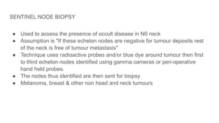
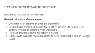
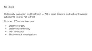
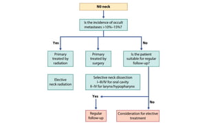
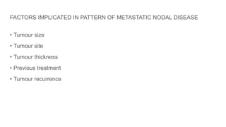
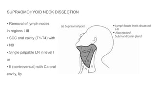
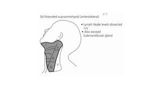
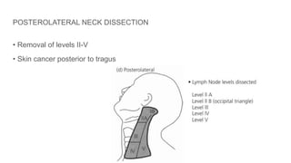
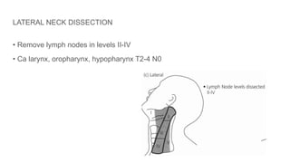
METASTATIC NECK
DISEASE
DR SHARADH CHANDRA
PGY2
KOTI ENT
INTRODUCTION
● When cancer cells spread to neck lymph nodes its called Neck Metastasis
● The degree of lymph node involvement should be regarded as an indirect index of the
systemic tumour burden
● Cure rates drop in half when there is regional lymph node involvement
● Elective removal of regional lymph nodes serves as a staging procedure for treatment-
surgical and non surgical
TUMOUR BIOLOGY - METASTASIS
● Understanding the molecular processes triggering
metastatic cascades in cancers is Advancing.
● Cancer cells access the lymphatic system through pre-
existing vessels
● Solid tumors can induce lymphangiogenesis.
● Vascular endothelial growth factors (VEGF)-C and VEGF-
D produced by cancer cells play an active role in
lymphangiogenesis by binding to VEGF receptor-3 on
lymphatic endothelial cells
Metastatic primary tumours have been shown to express distinct signature genes that set them apart from non-
metastatic tumours
This metastatic profile includes genes related to the extracellular matrix, adhesion, motility and protease inhibition.
This is facilitated by three molecular events:
(1) Cellular adhesion molecules - E cadherin downregulation - making the cancer cells free to migrate.
(2) Overexpression of integrins + production of matrix metalloproteinases (MMP 1, 2, 3, 9 and 14) that act upon
the matrix to increased cellular motility.
(3) autocrine and paracrine cytokines, mediated by integrin receptors
genotypic (e.g. p53 and MET oncogene mutation), phenotypic (e.g. E cadherin downregulation)
microenvironmental (e.g. expression of VEGF-C) processes combine to facilitate Metastasis
Behaviour Of Disease Within The Cervical Lymph Nodes
Toker described four distinct growth patterns of squamous cell carcinoma
(SCC) within cervical lymph nodes
1.Cancerous deposits in the subcapsular sinus, growth within the affected
node takes place, replacing the architecture of the node before extracapsular
spread occurs.
2.Metastasis infiltrate the lymphatic sinuses (Germinal centres,trabeculae -
intact) -
Ultimately, extracapsular spread occurs by the direct penetration and
destruction of the capsule, or by the arrest of further underlying capsular or
juxtacapsular lymphatics.
3.(less common pattern) - deposition of a malignant embolus within the subcapsular sinus + arrest
of tumour within capsular/juxtacapsular lymphatics. =>cancer growth both within and outside the
node
4.(uncommon metastatic pattern) is where capsular/juxtacapsular emboli grow with no intranodal
cancer.
extracapsular spread - much earlier.
Metastatic involvement of various lymph node regions usually progresses from superior to inferior
but lymph node groups can be bypassed, leading to ‘skip metastases’.
Early contralateral spread is present in tumors which are at midline or near midline
Neck Lymph Node Levels
● There are approximately 150
lymph nodes on either side of
the neck
● Size is from 3mm to 3cm but
most nodes are less than
1 centimetre
REGION SPECIFIC LYMPHATIC DRAINAGE
REGION LYMPHATIC DRAINAGE
NASOPHARYNX, NASAL CAVITIES &
SINUSES
Retropharyngeal or submandibular LNs →
LEVEL II & III
OROPHARYNX Retropharyngeal nodes → LEVEL II, III, IV
ORAL CAVITY Posterior: IB LEVEL II & III
Anterior: IA LEVEL II, III & IV
Skip metastases(Tongue): LEVEL IV
LARYNX Supraglottis: B/L LEVEL II & III
Subglottis: Prelaryngeal, Pretracheal Or
Paratracheal
Nodes LEVEL III & IV & VI
Echelon Nodes
SITE ECHELON LN'S
Oral cavity Levels I, II & III
Larynx & pharynx Levels II, III, IV
Thyroid Levels IV, VI & VII
Parotid Gland Pre-auricular, Periparotid & Intraparotid
LNs
Levels II, III & Va LN
Submandibular & sublingual gland Levels I, II & III
METASTATIC BEHAVIOUR IN THE PREVIOUSLY TREATED NECK
Surgery →Collateral lymphatic channels Diverted lymph flow →Contralateral or
Retrograde spread
Radiation therapy →↓Lymphocytes within LNs & Thickening of the walls →
Lymphatic obstruction
Shunting
Contralateral spread
Surgical scar may trap tumor cells which may lead to recurrence
METASTATIC NODAL DISEASE
• Level I-lip, anterior tongue, anterior floor of mouth, buccal mucosa
• Level II, III - tonsil, base of tongue (scalp, external auditory canal)
• Level IV - hypopharynx & larynx
• Level V - nasopharyngeal malignancy
• Level VI - thyroid, subglottic
● The nasopharynx, nasal cavities and sinuses- levels II, III & V
● The oropharynx similarly - levels II, III and IV
The oral cavity:
posterior parts of the oral cavity - level II/III or indirectly via the level Ib
More anterior parts of the oral cavity and tongue also drain to these
More anterior parts of the oral cavity and tongue - level II/III + level la or directly to
the jugular nodal chain (levels II to IV).
The larynx:
The supraglottis - levels II/III, with a greater tendency for bilateral nodal drainage.
The lower system - levels III/IV through lymphatics which pass through or behind
the cricothyroid membrane and also into the prelaryngeal, pretracheal or
paratracheal nodes (level VI) before reaching the deep cervical nodes.
Vocal cords are relatively avascular, they have an extremely sparse lymphatic
drainage and, as such, lymph node metastases from small carcinomas at this site
are uncommon
The hypopharynx -Drainage is to levels III, IV, VI and VII
OCCULT NODAL DISEASE
• Presence of neck metastasis in the neck nodes that cannot be clinically or
radiologically identified-
• 2 categories
1) Occult metastasis that can be identified by the light microscopy
2) Micrometastases less than 2mm that need special histological techniques
(immunohistochemistry, step serial sectioning and molecular analysis) for
identication.
Occult metastasis can only be identified only when it is removed
• Lymph node negative in histology means
CANCER NEVER REACHED THE NODE
Or else
IT REACHED BUT WAS DESTROYED IN THE NODE
• This make several controversies in the Management of occult neck disease
• Observational data suggests conversion of N0 to N+ is similar to the incidence of
pathological positive nodes In END specimens (30%)
CYSTIC NECK METASTASIS
● usually of oropharyngeal origin,
● most common sites being the tonsil or tongue base.
● Human papillomavirus (HPV) related tumours are more often associated with
cystic metastases than HPV negative tumours.
● These lymph node metastases tend to have a better prognosis than their non-
cystic counterparts.
● reported as branchiogenic carcinoma in the past.
● Given the exponential rise in HPV positive oropharyngeal carcinoma
throughout the world, any cystic level II neck mass in an adult should be
considered as a potential metastasis and should be investigated
EXTRANODAL EXTENSION(ENE)
• Associated with a poor prognosis (½ survival rate)
Presence of skin involvement/soft tissue involvement
with deep fixation or tethering to underline muscle/
structures or clinical sign of nerve Involvement
• Increased risk of local recurrence, distant
metastases and early recurrence
• If ENE is detected following surgery add post-
operative radiotherapy or chemoradiation
PROGNOSTIC NODAL FEATURES
• Site Size and number
• Low neck nodes
• Extracapsular spread
• Morphology
• Bilateral or contralateral spread
FACTORS IMPLICATED IN PATTERN OF METASTATIC NODAL DISEASE
• Tumour size
• Tumour site
• Tumour thickness
• Previous treatment
• Tumour recurrence
CLINICAL STAGING (UICC)
• Nx - Regional lymph nodes cannot be assessed
• N0 - No regional lymph node metastasis
• N1-Single ipsilateral node <3cm, ENE -Negative
• N2a-single ipsilateral node 3-6cm
• N2b - Multiple ipsilateral nodes 3-6cm
• N2c-Bilateral or contralateral nodes 3-6cm
• N3a-Node/ Nodes >6cm, ENE -Negative
• N3b-Node/ Nodes >6cm, ENE -positive
They now take into account ENE status and HPV (P16) status, and split the staging into distinct clinical and
pathological classication
Joint UICC AJCC classification (extra model extension has poor prognosis; HPV has better prognosis)
NECK DISSECTION TERMINOLOGY
A Surgery which removes the lymph node bearing areas of the neck with/without the non-lymphatic
structures - SCM, IJV & accessory nerves
Standardized neck dissection terminology by the American Academy of Otolaryngology and Head and
Neck Surgery in 1991
Updated by the Committee for Neck Dissection Classification of the American Head and Neck Society
in 2002
Neck dissections divided into two broad types with subdivisions:
1. comprehensive (removal of levels I–V)
2. selective (SND) (less than five levels).
Elective neck dissection (END) is any type of neck dissection that is performed on the clinically and
radiologically negative neck (cN0)
Classification of neck dissection techniques
TYPE STRUCTURES REMOVED
Radical neck dissection Level (I-V), IJV, SCM, spinal accessory
nerve
Modified radical neck dissection Level I-V
One of non lymphatic is preserved
Type 1: accessory spinal
Type 2: accessory spinal + IJV
Type 3: accessory spinal + IJV + SCM
Selective neck dissection One or more LN level preserved
Extended radical neck dissection Additional LN groups or non lymphatic
Selective Neck Dissection (SND)
Supraomohyoid I-III
Extended supraomohyoid (anterolateral) I-IV
Lateral II-IV
Posterolateral II-V
Anterior or central VI (Perithyroid, Delphian, Tracheo-
oesophageal & anteriosuperior
mediastinum)
Classification of Neck Dissections
Based on 4 concepts
1. Radical neck dissection RND is the standard basic procedure for cervical
lymphadenectomy against which all other modifications
2. Modifications of the RND which include preservation of any nonlymphatic
structures are referred to as modified radical neck dissection (MRND)
3. Any neck dissection that preserves one or more groups or levels of lymph
nodes is referred to as a selective neck dissection (SND)
4. An extended neck dissection refers to the removal of additional lymph node
groups or non-lymphatic structures relative to the RND
SKIN INCISION
• Vascularisation of flaps
• Exposure
• Protection of major vessels
• Localization of primary tumour
• Consider previous radiotherapy & reconstruction
• Cosmesis
• Previous surgical field
Blood Supply Of Cervical Neck Skin
Blood enters from above, below and either side with a
resultant watershed in the middle of the neck.
• Incisions can be planned to utilize this so as to maximize
blood supply to each of the neck flaps
RADICAL NECK DISSECTION
• Lymph nodes level I - V
• Non-lymphatic structures
• Accessory nerve
• Internal jugular vein
• Sternocleidomastoid muscle
Indication: extensive cervical involvement or matted lymph nodes with gross
extracapsular spread and invasion into SAN, IJV, SCM.
Contraindication
• Untreatable primary tumour or unresectable neck disease
• Patient unfit
• Distant metastasis
• Simultaneous bilateral dissection
MODIFIED NECK DISSECTION
Medina classification (1989)
• Modified radical neck dissection
- Type I (XI preserved)
- Type II (XI, IJV preserved)
- Type III (XI, IJV, and SCM preserved) (FND)
SELECTIVE NECK DISSECTION
• Any type of cervical lymphadenectomy with preservation of one or more lymph
node groups
• Four subtype:
Supraomohyoid neck dissection
Posterolateral neck dissection
Lateral neck dissection
Anterior neck dissection
SUPRAOMOHYOID NECK DISSECTION
• Removal of lymph nodes
in regions I-III
• SCC oral cavity (T1-T4) with
• N0
• Single palpable LN in level I
or
• II (controversial) with Ca oral
cavity, lip
POSTEROLATERAL NECK DISSECTION
• Removal of levels II-V
• Skin cancer posterior to tragus
LATERAL NECK DISSECTION
• Remove lymph nodes in levels II-IV
• Ca larynx, oropharynx, hypopharynx T2-4 N0
ANTERIOR NECK DISSECTION
• Removal of LN surrounding the visceral
structure in the anterior aspect of the
neck, level VI
• Differentiated and medullary Ca of
thyroid with thyroidectomy
EXTENDED RADICAL NECK DISSECTION
• Removal of additional lymphatic structure & Non lymphatic structure other than
RND
• Retropharyngeal LN
• Level VII LN
• Hypoglossal nerve
• Carotid artery
• Skin of neck
GENERAL COMPLICATIONS
• Anaesthetic complications
• Post operative atelectasis with basal collapse
• Pneumonia
• Ischaemic heart disease
• Urinary retention
• Deep vein thrombosis
LOCAL COMPLICATIONS
• Hemorrhage
• Wound infection
• Carotid artery rupture
• Nerve injuries
• Chylous fistula
• Pneumothorax
• Cerebral edema
ASSESSMENT OF CERVICAL LYMPHADENOPATHY
● Clinical assessment
● Fine needle aspiration cytology
● Ultrasound scan
● Computed Tomography
● Magnetic Resonance Imaging
● Positron Emission Tomography
● Open Biopsy
CLINICAL EXAMINATION
● Remains the most important initial method of assessment
● Variable in accuracy (Clinicians experience)
● Sensitivity of 74% specificity of 81% overall accuracy of 77% (Merrit et al)
● Difficult in short stocky necks
● Drawback is nodes may go unnoticed until they reach a considerable size
FINE NEEDLE ASPIRATION CYTOLOGY
● Technique is particularly useful in assessment of a palpable node when
searching for a primary
● In the presence of a primary less useful (evidence of HPV or EBV
transcripsts) (HPV oropharynx EBV nasopharynx)
● Easy to perform and quick in reporting
● Overall accuracy >90% (depends on pathologists experience)
ULTRASOUND SCAN
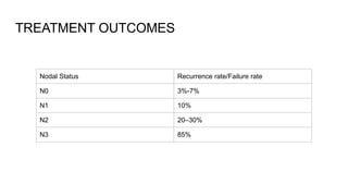
● Can detect the presence malignant cervical lymph nodes with sensitivity of
70% to 90%
● When combined with FNAC it increases to 90%
● No absolute criteria to differentiate between malignant vs benign nodes
[absent hilar echoes and increases in short axis length are generally
considered to be features of metastatic neck nodes.]
COMPUTED TOMOGRAPHY (C.T.)
• Detection of malignant neck nodes is higher than clinical examination
• Criteria to categorize as metastatic node
1) short axis diameter of 1cm
2) cluster of three or more nodes >0.8cm
3) nodal necrosis or patchy enhancement within nodes
Central Necrosis+Enlarged Nodes+Thin Rim of Inflammation (RIM enhancement)
MAGNETIC RESONANCE IMAGING (M.R.I.)
• Meta analysis suggests MRI can detect metastatic neck disease similar to CT
• MRI is better in assessing N0 neck in the presence of deep invasion
POSITRON EMISSION TOMOGRAPHY (Ρ.Ε.Τ.)
● Uses 18F-2-fluoro-2-deoxy-D-glucose (FDG) as radioactive tracer
● Can identify the areas of higher glucose uptake
● Anatomical localization difficult
● Coregistered anatomic and functional imaging systems (PET-CT) lead to
accurate image fusion by harvesting advantages of both techniques
● Role in occult primary, recurrent, residual disease & distant metastasis
● Sensitivity - 95 to 98%, Specificity - 95 to 98%
● False positive results are common first few weeks after chemo radiation.Scan
should be deferred for 10 to 12 weeks after completion of treatment
OPEN BIOPSY
● In general best avoided as initial diagnostic modality
● When FNAC not available, non diagnostic, results suggesting a lymphoma, or
anaplastic Carcinoma or primary site is occult may be necessary
● Prognosis will not be different if the definitive treatment is done within 6 weeks
● Any incision should made to facilitate to remove the scar via subsequent neck
dissection and vital structures like sternocleidomastoid may have to be
removed due to scar tissue
SENTINEL NODE BIOPSY
● Used to assess the presence of occult disease in N0 neck
● Assumption is "If these echelon nodes are negative for tumour deposits rest
of the neck is free of tumour metastasis"
● Technique uses radioactive probes and/or blue dye around tumour then first
to third echelon nodes identified using gamma cameras or peri-operative
hand held probes.
● The nodes thus identified are then sent for biopsy
● Melanoma, breast & other non head and neck tumours
TREATMENT OF METASTATIC NECK DISEASE
Decided by the stage of neck disease
General principles of neck spread
● Untreated neck patterns of spread is predictable
● In occult neck metastasis usually found at first echelon LN Region. This
permits principle of Selective Neck Dissection.
● Previous Treatment alters the pattern of spread
● Patients with palpable neck nodes likely to have non palpable spread in other
levels
N0 NECK
Historically evaluation and treatment for N0 is great dilemma and still controversial
Whether to treat or not to treat
Number of Treatment options
● Elective surgery
● Elective radiotherapy
● Wait and watch
● Elective neck investigations
INDICATIONS FOR ELECTIVE NECK TREATMENT
• 15% to 20% chance of subclinical neck disease
• Vigilant follow up not possible
• Clinical evaluation of the neck is difficult
• Access to the neck for reconstruction
CHOICE OF TREATMENT MODALITY FOR N0
• Elective neck dissection and irradiation are equally effective
• Choice dictated by many factors
Patient and surgeons choices
quality of life issues and how the primary is treated
THE CONTRALATERAL N0 NECK
• Incidence of occult metastases: 30-70%
• Supraglottic, Hypopharyngeal & Oropharyngeal Tumors
• Contralateral neck treatment is warranted
ARGUMENTS FOR ELECTIVE NECK SURGERY
• High incidence of occult metastatic disease
•Selective Neck Dissection got low morbidity and mortality
• Routine follow up will not detect the earliest conversion from No to N+
• Untreated neck mets can induce distant metastasis
• Elective neck surgery can be performed at primary surgery
• Patient compliance for regular follow ups cannot be assured
• Presence of multiple occult metastasis will dictate adjuvant irradiation
ARGUMENTS AGAINST ELECTIVE NECK SURGERY
• Careful clinical follow up combined with imaging has the potential to detect early
conversion
• Control rates no lower than when wait and watch policy is followed
• Elective surgery result in inevitable morbidity
• Elective Neck Dissection removes the natural barrier to the spread
• Elective Neck Dissection creates a scarred hypoxic field that can reduce the
radiation kill
THE N+ NECK (N1 NECK)
● Treatment modality is usually dictates by treatment to primary site
● Primary treated with Surgery → MRND (1)
● Primary treated with Radiation Neck—>Irradiation
● Upstaging after pathological assessment—>Adjuvant Radiotherapy
● Chemoradiation followed by Neck Dissection (Residual disease)
● In palpable neck disease all 5 LN levels should be dissected
● Majority of the cases accessory nerve can be preserved (MRND)
INDICATIONS OF POSTOPERATIVE RADIOTHERAPY
• Extracapsular spread
• Two or more positive nodes
• N2 N3 Stages
• Residual disease
N2A AND N2B NECK
• MRND sparing spinal accessory nerve where possible
• PORT(Totally implantable venous access device) is indicated
Primary treated with Surgery—>Comprehensive neck dissection
Adjuvant RT (success rate ↑10%)
• Adjuvant chemo-radiation in ENE
Primary treated with RT→ Residual—>Salvage surgery
N2C NECK
• Common primary site is tongue base, supraglottic larynx and hypopharynx
• Earlier it was considered as grave prognosis
• Now evidence shows the size number and the ECS determines the prognosis
rather than the laterality
• If operable (Radical neck dissection + conservative neck dissection on less
involved site)+ adjuvant radiotherapy
• Chemo-radiation
N3 NECK (MASSIVE NODES)
● Commonly fixed to skin or underlying structures
● High risk for distant metastasis -----PET CT may be warranted
● Mostly incurable
● Decision depends on stage of disease, presence or absence of fixation and
structure to which node is fixed, surgeon experience
● Long term control is poor and the benefit of treatment should be carefully
weighed against morbidity
● Radical treatment is warranted in patients who have less advanced primary
● Fixation to mandible, SCM, midline muscles -------treatable
● Fixation to brachial plexus or skull base: contraindication to surgery
● Fixation to the skin may be treated by wide resection and flap repair
● Arterial invasion -------careful assessment
● RND OR ERND + chemoradiation
● MANY PATIENTS PALLIATION WILL BE THE BEST OPTION
NECK METASTASIS FROM UNKNOWN PRIMARY
● Unknown or occult primary carcinoma
● Presentation of metastatic neck lymphadenopathy without the development of
a primary lesion within a subsequent five year period.
● Failure to identify occult primary-
Spontaneous regression of primary tumour
Autoimmune destruction
Accelerated tumour progression
Metastatic carcinoma with no evidence of primary site after history, physical
examination and radiological imaging.
Secondary of neck<10% of all unknown primary
Most likely head and neck primary site: tonsil (45%), base of tongue (40%) and
piriform fossa (10%).
Unknown Primary treatment
T0N1M0
Single modality treatment (neck dissection or radiotherapy)
T0N2M0 AND T0N3M0
Neck dissection + adjuvant chemo-radiation
TREATMENT OUTCOMES
Nodal Status Recurrence rate/Failure rate
N0 3%-7%
N1 10%
N2 20–30%
N3 85%
Management of recurrence
Unresectable Re-irradiation +/- Chemotherapy
Resectable Wide Resection + Post op RT
Ipsilateral untreated neck Neck Dissection +/- Post op RT
RT+ Salvage surgery
Contralateral untreated neck Salvage surgery
Previously treated neck Radical radiotherapy (if primary was
surgery)
Salvage Surgery (if primary was radiation)
Recurrence after combination treatment Wide excision of the tumor & the overlying
skin + Flap reconstruction + Brachytherapy
Reference
Scott-Brown’s Otorhinolaryngology Head and Neck Surgery VOLUME 3
Stell and Maran’s Textbook of Head and Neck Surgery and Oncology

Metastatic Neck Disease: Evaluation & Treatment

  • 1.
  • 2.
    INTRODUCTION ● When cancercells spread to neck lymph nodes its called Neck Metastasis ● The degree of lymph node involvement should be regarded as an indirect index of the systemic tumour burden ● Cure rates drop in half when there is regional lymph node involvement ● Elective removal of regional lymph nodes serves as a staging procedure for treatment- surgical and non surgical
  • 3.
    TUMOUR BIOLOGY -METASTASIS ● Understanding the molecular processes triggering metastatic cascades in cancers is Advancing. ● Cancer cells access the lymphatic system through pre- existing vessels ● Solid tumors can induce lymphangiogenesis. ● Vascular endothelial growth factors (VEGF)-C and VEGF- D produced by cancer cells play an active role in lymphangiogenesis by binding to VEGF receptor-3 on lymphatic endothelial cells
  • 4.
    Metastatic primary tumourshave been shown to express distinct signature genes that set them apart from non- metastatic tumours This metastatic profile includes genes related to the extracellular matrix, adhesion, motility and protease inhibition. This is facilitated by three molecular events: (1) Cellular adhesion molecules - E cadherin downregulation - making the cancer cells free to migrate. (2) Overexpression of integrins + production of matrix metalloproteinases (MMP 1, 2, 3, 9 and 14) that act upon the matrix to increased cellular motility. (3) autocrine and paracrine cytokines, mediated by integrin receptors genotypic (e.g. p53 and MET oncogene mutation), phenotypic (e.g. E cadherin downregulation) microenvironmental (e.g. expression of VEGF-C) processes combine to facilitate Metastasis
  • 5.
    Behaviour Of DiseaseWithin The Cervical Lymph Nodes Toker described four distinct growth patterns of squamous cell carcinoma (SCC) within cervical lymph nodes 1.Cancerous deposits in the subcapsular sinus, growth within the affected node takes place, replacing the architecture of the node before extracapsular spread occurs. 2.Metastasis infiltrate the lymphatic sinuses (Germinal centres,trabeculae - intact) - Ultimately, extracapsular spread occurs by the direct penetration and destruction of the capsule, or by the arrest of further underlying capsular or juxtacapsular lymphatics.
  • 6.
    3.(less common pattern)- deposition of a malignant embolus within the subcapsular sinus + arrest of tumour within capsular/juxtacapsular lymphatics. =>cancer growth both within and outside the node 4.(uncommon metastatic pattern) is where capsular/juxtacapsular emboli grow with no intranodal cancer. extracapsular spread - much earlier. Metastatic involvement of various lymph node regions usually progresses from superior to inferior but lymph node groups can be bypassed, leading to ‘skip metastases’. Early contralateral spread is present in tumors which are at midline or near midline
  • 7.
    Neck Lymph NodeLevels ● There are approximately 150 lymph nodes on either side of the neck ● Size is from 3mm to 3cm but most nodes are less than 1 centimetre
  • 10.
    REGION SPECIFIC LYMPHATICDRAINAGE REGION LYMPHATIC DRAINAGE NASOPHARYNX, NASAL CAVITIES & SINUSES Retropharyngeal or submandibular LNs → LEVEL II & III OROPHARYNX Retropharyngeal nodes → LEVEL II, III, IV ORAL CAVITY Posterior: IB LEVEL II & III Anterior: IA LEVEL II, III & IV Skip metastases(Tongue): LEVEL IV LARYNX Supraglottis: B/L LEVEL II & III Subglottis: Prelaryngeal, Pretracheal Or Paratracheal Nodes LEVEL III & IV & VI
  • 11.
    Echelon Nodes SITE ECHELONLN'S Oral cavity Levels I, II & III Larynx & pharynx Levels II, III, IV Thyroid Levels IV, VI & VII Parotid Gland Pre-auricular, Periparotid & Intraparotid LNs Levels II, III & Va LN Submandibular & sublingual gland Levels I, II & III
  • 12.
    METASTATIC BEHAVIOUR INTHE PREVIOUSLY TREATED NECK Surgery →Collateral lymphatic channels Diverted lymph flow →Contralateral or Retrograde spread Radiation therapy →↓Lymphocytes within LNs & Thickening of the walls → Lymphatic obstruction Shunting Contralateral spread Surgical scar may trap tumor cells which may lead to recurrence
  • 13.
    METASTATIC NODAL DISEASE •Level I-lip, anterior tongue, anterior floor of mouth, buccal mucosa • Level II, III - tonsil, base of tongue (scalp, external auditory canal) • Level IV - hypopharynx & larynx • Level V - nasopharyngeal malignancy • Level VI - thyroid, subglottic
  • 14.
    ● The nasopharynx,nasal cavities and sinuses- levels II, III & V ● The oropharynx similarly - levels II, III and IV The oral cavity: posterior parts of the oral cavity - level II/III or indirectly via the level Ib More anterior parts of the oral cavity and tongue also drain to these More anterior parts of the oral cavity and tongue - level II/III + level la or directly to the jugular nodal chain (levels II to IV).
  • 15.
    The larynx: The supraglottis- levels II/III, with a greater tendency for bilateral nodal drainage. The lower system - levels III/IV through lymphatics which pass through or behind the cricothyroid membrane and also into the prelaryngeal, pretracheal or paratracheal nodes (level VI) before reaching the deep cervical nodes. Vocal cords are relatively avascular, they have an extremely sparse lymphatic drainage and, as such, lymph node metastases from small carcinomas at this site are uncommon The hypopharynx -Drainage is to levels III, IV, VI and VII
  • 16.
    OCCULT NODAL DISEASE •Presence of neck metastasis in the neck nodes that cannot be clinically or radiologically identified- • 2 categories 1) Occult metastasis that can be identified by the light microscopy 2) Micrometastases less than 2mm that need special histological techniques (immunohistochemistry, step serial sectioning and molecular analysis) for identication. Occult metastasis can only be identified only when it is removed
  • 17.
    • Lymph nodenegative in histology means CANCER NEVER REACHED THE NODE Or else IT REACHED BUT WAS DESTROYED IN THE NODE • This make several controversies in the Management of occult neck disease • Observational data suggests conversion of N0 to N+ is similar to the incidence of pathological positive nodes In END specimens (30%)
  • 18.
    CYSTIC NECK METASTASIS ●usually of oropharyngeal origin, ● most common sites being the tonsil or tongue base. ● Human papillomavirus (HPV) related tumours are more often associated with cystic metastases than HPV negative tumours. ● These lymph node metastases tend to have a better prognosis than their non- cystic counterparts. ● reported as branchiogenic carcinoma in the past. ● Given the exponential rise in HPV positive oropharyngeal carcinoma throughout the world, any cystic level II neck mass in an adult should be considered as a potential metastasis and should be investigated
  • 19.
    EXTRANODAL EXTENSION(ENE) • Associatedwith a poor prognosis (½ survival rate) Presence of skin involvement/soft tissue involvement with deep fixation or tethering to underline muscle/ structures or clinical sign of nerve Involvement • Increased risk of local recurrence, distant metastases and early recurrence • If ENE is detected following surgery add post- operative radiotherapy or chemoradiation
  • 20.
    PROGNOSTIC NODAL FEATURES •Site Size and number • Low neck nodes • Extracapsular spread • Morphology • Bilateral or contralateral spread
  • 21.
    FACTORS IMPLICATED INPATTERN OF METASTATIC NODAL DISEASE • Tumour size • Tumour site • Tumour thickness • Previous treatment • Tumour recurrence
  • 22.
    CLINICAL STAGING (UICC) •Nx - Regional lymph nodes cannot be assessed • N0 - No regional lymph node metastasis • N1-Single ipsilateral node <3cm, ENE -Negative • N2a-single ipsilateral node 3-6cm • N2b - Multiple ipsilateral nodes 3-6cm • N2c-Bilateral or contralateral nodes 3-6cm • N3a-Node/ Nodes >6cm, ENE -Negative • N3b-Node/ Nodes >6cm, ENE -positive They now take into account ENE status and HPV (P16) status, and split the staging into distinct clinical and pathological classication
  • 23.
    Joint UICC AJCCclassification (extra model extension has poor prognosis; HPV has better prognosis)
  • 26.
    NECK DISSECTION TERMINOLOGY ASurgery which removes the lymph node bearing areas of the neck with/without the non-lymphatic structures - SCM, IJV & accessory nerves Standardized neck dissection terminology by the American Academy of Otolaryngology and Head and Neck Surgery in 1991 Updated by the Committee for Neck Dissection Classification of the American Head and Neck Society in 2002 Neck dissections divided into two broad types with subdivisions: 1. comprehensive (removal of levels I–V) 2. selective (SND) (less than five levels). Elective neck dissection (END) is any type of neck dissection that is performed on the clinically and radiologically negative neck (cN0)
  • 27.
    Classification of neckdissection techniques TYPE STRUCTURES REMOVED Radical neck dissection Level (I-V), IJV, SCM, spinal accessory nerve Modified radical neck dissection Level I-V One of non lymphatic is preserved Type 1: accessory spinal Type 2: accessory spinal + IJV Type 3: accessory spinal + IJV + SCM Selective neck dissection One or more LN level preserved Extended radical neck dissection Additional LN groups or non lymphatic
  • 28.
    Selective Neck Dissection(SND) Supraomohyoid I-III Extended supraomohyoid (anterolateral) I-IV Lateral II-IV Posterolateral II-V Anterior or central VI (Perithyroid, Delphian, Tracheo- oesophageal & anteriosuperior mediastinum)
  • 29.
    Classification of NeckDissections Based on 4 concepts 1. Radical neck dissection RND is the standard basic procedure for cervical lymphadenectomy against which all other modifications 2. Modifications of the RND which include preservation of any nonlymphatic structures are referred to as modified radical neck dissection (MRND) 3. Any neck dissection that preserves one or more groups or levels of lymph nodes is referred to as a selective neck dissection (SND) 4. An extended neck dissection refers to the removal of additional lymph node groups or non-lymphatic structures relative to the RND
  • 30.
    SKIN INCISION • Vascularisationof flaps • Exposure • Protection of major vessels • Localization of primary tumour • Consider previous radiotherapy & reconstruction • Cosmesis • Previous surgical field
  • 31.
    Blood Supply OfCervical Neck Skin Blood enters from above, below and either side with a resultant watershed in the middle of the neck. • Incisions can be planned to utilize this so as to maximize blood supply to each of the neck flaps
  • 33.
    RADICAL NECK DISSECTION •Lymph nodes level I - V • Non-lymphatic structures • Accessory nerve • Internal jugular vein • Sternocleidomastoid muscle
  • 34.
    Indication: extensive cervicalinvolvement or matted lymph nodes with gross extracapsular spread and invasion into SAN, IJV, SCM. Contraindication • Untreatable primary tumour or unresectable neck disease • Patient unfit • Distant metastasis • Simultaneous bilateral dissection
  • 35.
    MODIFIED NECK DISSECTION Medinaclassification (1989) • Modified radical neck dissection - Type I (XI preserved) - Type II (XI, IJV preserved) - Type III (XI, IJV, and SCM preserved) (FND)
  • 36.
    SELECTIVE NECK DISSECTION •Any type of cervical lymphadenectomy with preservation of one or more lymph node groups • Four subtype: Supraomohyoid neck dissection Posterolateral neck dissection Lateral neck dissection Anterior neck dissection
  • 37.
    SUPRAOMOHYOID NECK DISSECTION •Removal of lymph nodes in regions I-III • SCC oral cavity (T1-T4) with • N0 • Single palpable LN in level I or • II (controversial) with Ca oral cavity, lip
  • 39.
    POSTEROLATERAL NECK DISSECTION •Removal of levels II-V • Skin cancer posterior to tragus
  • 40.
    LATERAL NECK DISSECTION •Remove lymph nodes in levels II-IV • Ca larynx, oropharynx, hypopharynx T2-4 N0
  • 41.
    ANTERIOR NECK DISSECTION •Removal of LN surrounding the visceral structure in the anterior aspect of the neck, level VI • Differentiated and medullary Ca of thyroid with thyroidectomy
  • 42.
    EXTENDED RADICAL NECKDISSECTION • Removal of additional lymphatic structure & Non lymphatic structure other than RND • Retropharyngeal LN • Level VII LN • Hypoglossal nerve • Carotid artery • Skin of neck
  • 43.
    GENERAL COMPLICATIONS • Anaestheticcomplications • Post operative atelectasis with basal collapse • Pneumonia • Ischaemic heart disease • Urinary retention • Deep vein thrombosis
  • 44.
    LOCAL COMPLICATIONS • Hemorrhage •Wound infection • Carotid artery rupture • Nerve injuries • Chylous fistula • Pneumothorax • Cerebral edema
  • 45.
    ASSESSMENT OF CERVICALLYMPHADENOPATHY ● Clinical assessment ● Fine needle aspiration cytology ● Ultrasound scan ● Computed Tomography ● Magnetic Resonance Imaging ● Positron Emission Tomography ● Open Biopsy
  • 46.
    CLINICAL EXAMINATION ● Remainsthe most important initial method of assessment ● Variable in accuracy (Clinicians experience) ● Sensitivity of 74% specificity of 81% overall accuracy of 77% (Merrit et al) ● Difficult in short stocky necks ● Drawback is nodes may go unnoticed until they reach a considerable size
  • 47.
    FINE NEEDLE ASPIRATIONCYTOLOGY ● Technique is particularly useful in assessment of a palpable node when searching for a primary ● In the presence of a primary less useful (evidence of HPV or EBV transcripsts) (HPV oropharynx EBV nasopharynx) ● Easy to perform and quick in reporting ● Overall accuracy >90% (depends on pathologists experience)
  • 48.
    ULTRASOUND SCAN ● Candetect the presence malignant cervical lymph nodes with sensitivity of 70% to 90% ● When combined with FNAC it increases to 90% ● No absolute criteria to differentiate between malignant vs benign nodes [absent hilar echoes and increases in short axis length are generally considered to be features of metastatic neck nodes.]
  • 49.
    COMPUTED TOMOGRAPHY (C.T.) •Detection of malignant neck nodes is higher than clinical examination • Criteria to categorize as metastatic node 1) short axis diameter of 1cm 2) cluster of three or more nodes >0.8cm 3) nodal necrosis or patchy enhancement within nodes Central Necrosis+Enlarged Nodes+Thin Rim of Inflammation (RIM enhancement)
  • 50.
    MAGNETIC RESONANCE IMAGING(M.R.I.) • Meta analysis suggests MRI can detect metastatic neck disease similar to CT • MRI is better in assessing N0 neck in the presence of deep invasion
  • 51.
    POSITRON EMISSION TOMOGRAPHY(Ρ.Ε.Τ.) ● Uses 18F-2-fluoro-2-deoxy-D-glucose (FDG) as radioactive tracer ● Can identify the areas of higher glucose uptake ● Anatomical localization difficult ● Coregistered anatomic and functional imaging systems (PET-CT) lead to accurate image fusion by harvesting advantages of both techniques ● Role in occult primary, recurrent, residual disease & distant metastasis ● Sensitivity - 95 to 98%, Specificity - 95 to 98% ● False positive results are common first few weeks after chemo radiation.Scan should be deferred for 10 to 12 weeks after completion of treatment
  • 52.
    OPEN BIOPSY ● Ingeneral best avoided as initial diagnostic modality ● When FNAC not available, non diagnostic, results suggesting a lymphoma, or anaplastic Carcinoma or primary site is occult may be necessary ● Prognosis will not be different if the definitive treatment is done within 6 weeks ● Any incision should made to facilitate to remove the scar via subsequent neck dissection and vital structures like sternocleidomastoid may have to be removed due to scar tissue
  • 53.
    SENTINEL NODE BIOPSY ●Used to assess the presence of occult disease in N0 neck ● Assumption is "If these echelon nodes are negative for tumour deposits rest of the neck is free of tumour metastasis" ● Technique uses radioactive probes and/or blue dye around tumour then first to third echelon nodes identified using gamma cameras or peri-operative hand held probes. ● The nodes thus identified are then sent for biopsy ● Melanoma, breast & other non head and neck tumours
  • 54.
    TREATMENT OF METASTATICNECK DISEASE Decided by the stage of neck disease General principles of neck spread ● Untreated neck patterns of spread is predictable ● In occult neck metastasis usually found at first echelon LN Region. This permits principle of Selective Neck Dissection. ● Previous Treatment alters the pattern of spread ● Patients with palpable neck nodes likely to have non palpable spread in other levels
  • 55.
    N0 NECK Historically evaluationand treatment for N0 is great dilemma and still controversial Whether to treat or not to treat Number of Treatment options ● Elective surgery ● Elective radiotherapy ● Wait and watch ● Elective neck investigations
  • 57.
    INDICATIONS FOR ELECTIVENECK TREATMENT • 15% to 20% chance of subclinical neck disease • Vigilant follow up not possible • Clinical evaluation of the neck is difficult • Access to the neck for reconstruction
  • 58.
    CHOICE OF TREATMENTMODALITY FOR N0 • Elective neck dissection and irradiation are equally effective • Choice dictated by many factors Patient and surgeons choices quality of life issues and how the primary is treated
  • 59.
    THE CONTRALATERAL N0NECK • Incidence of occult metastases: 30-70% • Supraglottic, Hypopharyngeal & Oropharyngeal Tumors • Contralateral neck treatment is warranted
  • 60.
    ARGUMENTS FOR ELECTIVENECK SURGERY • High incidence of occult metastatic disease •Selective Neck Dissection got low morbidity and mortality • Routine follow up will not detect the earliest conversion from No to N+ • Untreated neck mets can induce distant metastasis • Elective neck surgery can be performed at primary surgery • Patient compliance for regular follow ups cannot be assured • Presence of multiple occult metastasis will dictate adjuvant irradiation
  • 61.
    ARGUMENTS AGAINST ELECTIVENECK SURGERY • Careful clinical follow up combined with imaging has the potential to detect early conversion • Control rates no lower than when wait and watch policy is followed • Elective surgery result in inevitable morbidity • Elective Neck Dissection removes the natural barrier to the spread • Elective Neck Dissection creates a scarred hypoxic field that can reduce the radiation kill
  • 62.
    THE N+ NECK(N1 NECK) ● Treatment modality is usually dictates by treatment to primary site ● Primary treated with Surgery → MRND (1) ● Primary treated with Radiation Neck—>Irradiation ● Upstaging after pathological assessment—>Adjuvant Radiotherapy ● Chemoradiation followed by Neck Dissection (Residual disease) ● In palpable neck disease all 5 LN levels should be dissected ● Majority of the cases accessory nerve can be preserved (MRND)
  • 65.
    INDICATIONS OF POSTOPERATIVERADIOTHERAPY • Extracapsular spread • Two or more positive nodes • N2 N3 Stages • Residual disease
  • 66.
    N2A AND N2BNECK • MRND sparing spinal accessory nerve where possible • PORT(Totally implantable venous access device) is indicated Primary treated with Surgery—>Comprehensive neck dissection Adjuvant RT (success rate ↑10%) • Adjuvant chemo-radiation in ENE Primary treated with RT→ Residual—>Salvage surgery
  • 67.
    N2C NECK • Commonprimary site is tongue base, supraglottic larynx and hypopharynx • Earlier it was considered as grave prognosis • Now evidence shows the size number and the ECS determines the prognosis rather than the laterality • If operable (Radical neck dissection + conservative neck dissection on less involved site)+ adjuvant radiotherapy • Chemo-radiation
  • 68.
    N3 NECK (MASSIVENODES) ● Commonly fixed to skin or underlying structures ● High risk for distant metastasis -----PET CT may be warranted ● Mostly incurable ● Decision depends on stage of disease, presence or absence of fixation and structure to which node is fixed, surgeon experience
  • 69.
    ● Long termcontrol is poor and the benefit of treatment should be carefully weighed against morbidity ● Radical treatment is warranted in patients who have less advanced primary ● Fixation to mandible, SCM, midline muscles -------treatable ● Fixation to brachial plexus or skull base: contraindication to surgery ● Fixation to the skin may be treated by wide resection and flap repair ● Arterial invasion -------careful assessment ● RND OR ERND + chemoradiation ● MANY PATIENTS PALLIATION WILL BE THE BEST OPTION
  • 70.
    NECK METASTASIS FROMUNKNOWN PRIMARY ● Unknown or occult primary carcinoma ● Presentation of metastatic neck lymphadenopathy without the development of a primary lesion within a subsequent five year period. ● Failure to identify occult primary- Spontaneous regression of primary tumour Autoimmune destruction Accelerated tumour progression
  • 71.
    Metastatic carcinoma withno evidence of primary site after history, physical examination and radiological imaging. Secondary of neck<10% of all unknown primary Most likely head and neck primary site: tonsil (45%), base of tongue (40%) and piriform fossa (10%).
  • 73.
    Unknown Primary treatment T0N1M0 Singlemodality treatment (neck dissection or radiotherapy) T0N2M0 AND T0N3M0 Neck dissection + adjuvant chemo-radiation
  • 74.
    TREATMENT OUTCOMES Nodal StatusRecurrence rate/Failure rate N0 3%-7% N1 10% N2 20–30% N3 85%
  • 75.
    Management of recurrence UnresectableRe-irradiation +/- Chemotherapy Resectable Wide Resection + Post op RT Ipsilateral untreated neck Neck Dissection +/- Post op RT RT+ Salvage surgery Contralateral untreated neck Salvage surgery Previously treated neck Radical radiotherapy (if primary was surgery) Salvage Surgery (if primary was radiation) Recurrence after combination treatment Wide excision of the tumor & the overlying skin + Flap reconstruction + Brachytherapy
  • 76.
    Reference Scott-Brown’s Otorhinolaryngology Headand Neck Surgery VOLUME 3 Stell and Maran’s Textbook of Head and Neck Surgery and Oncology