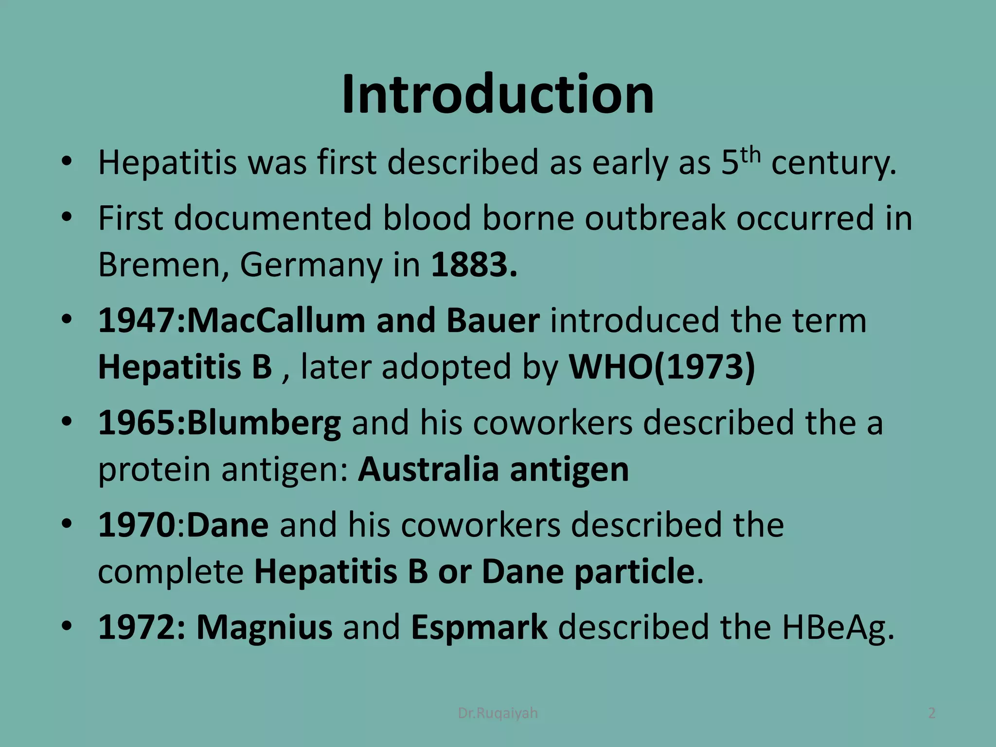
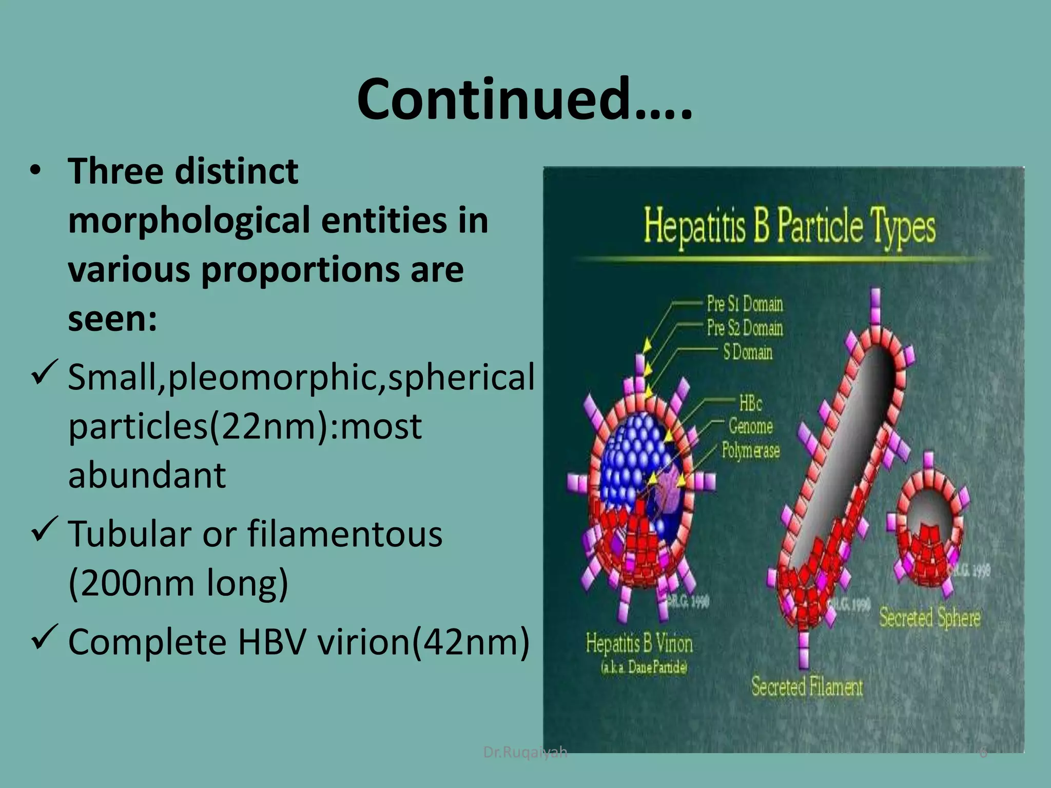
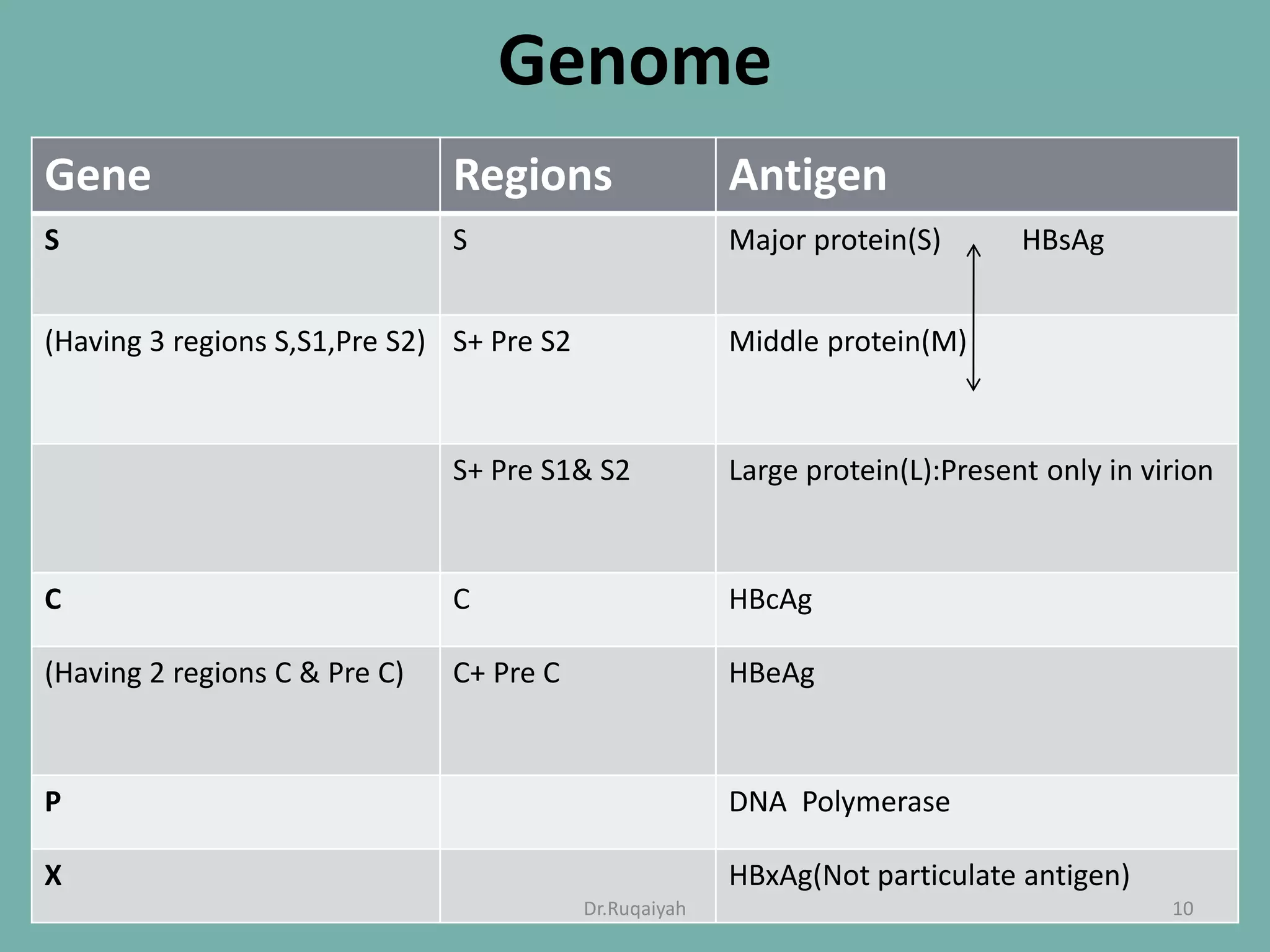
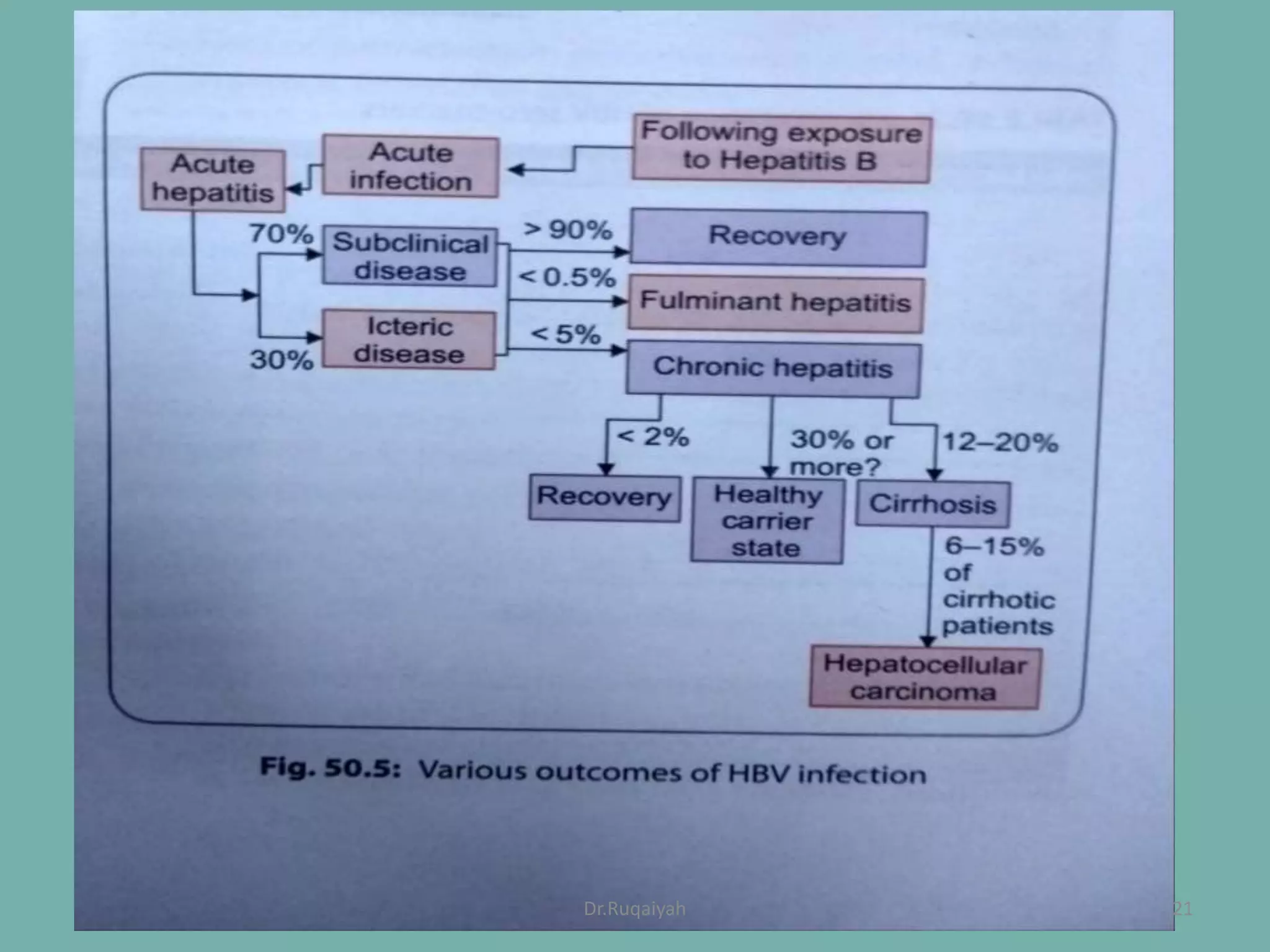
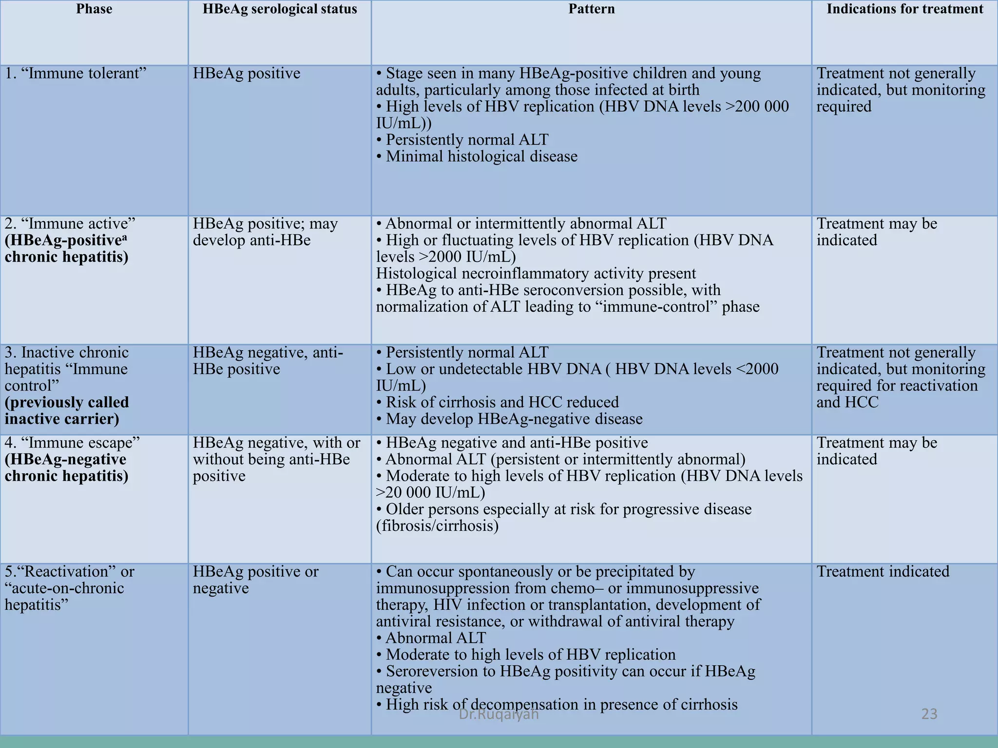
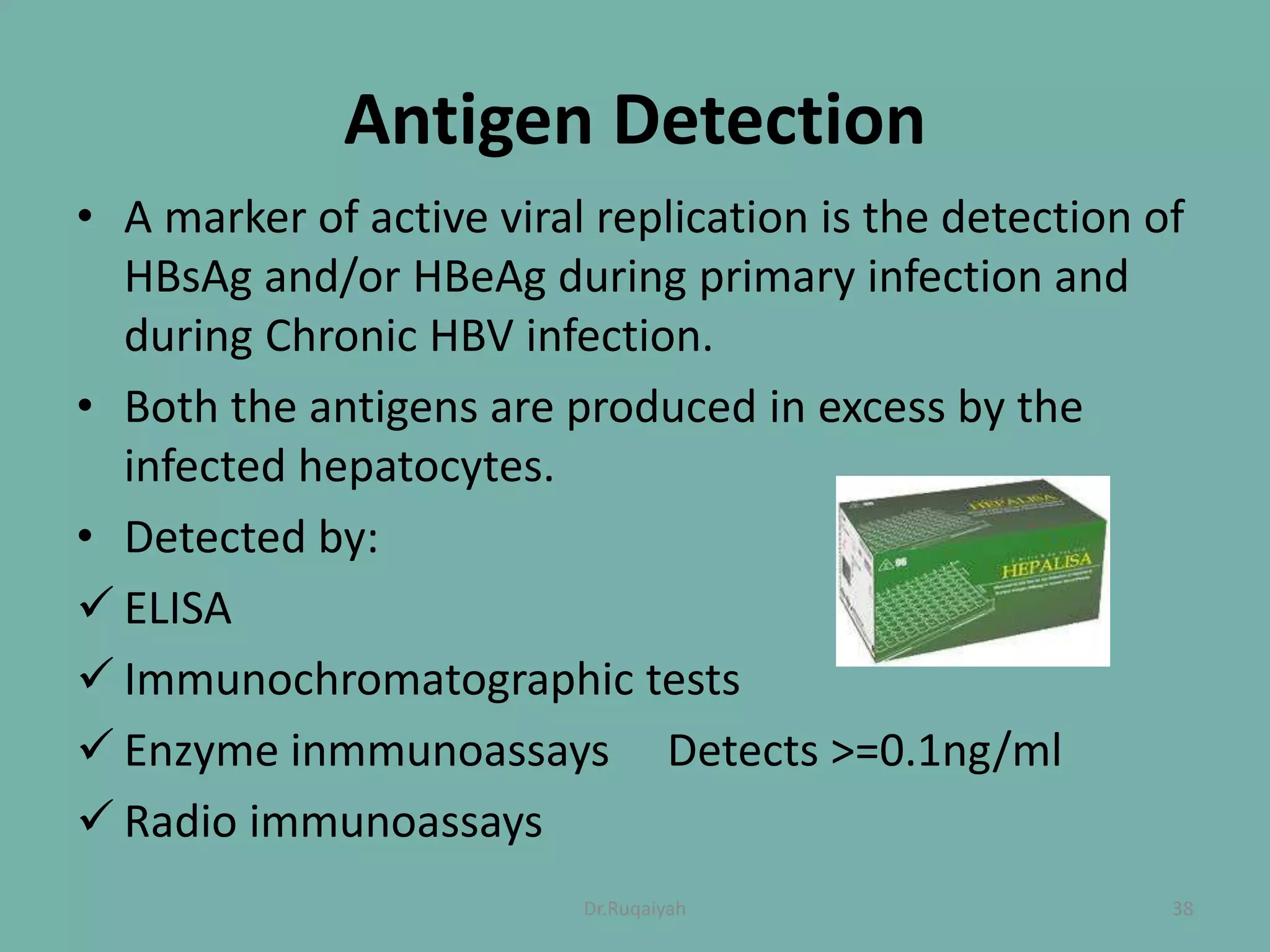
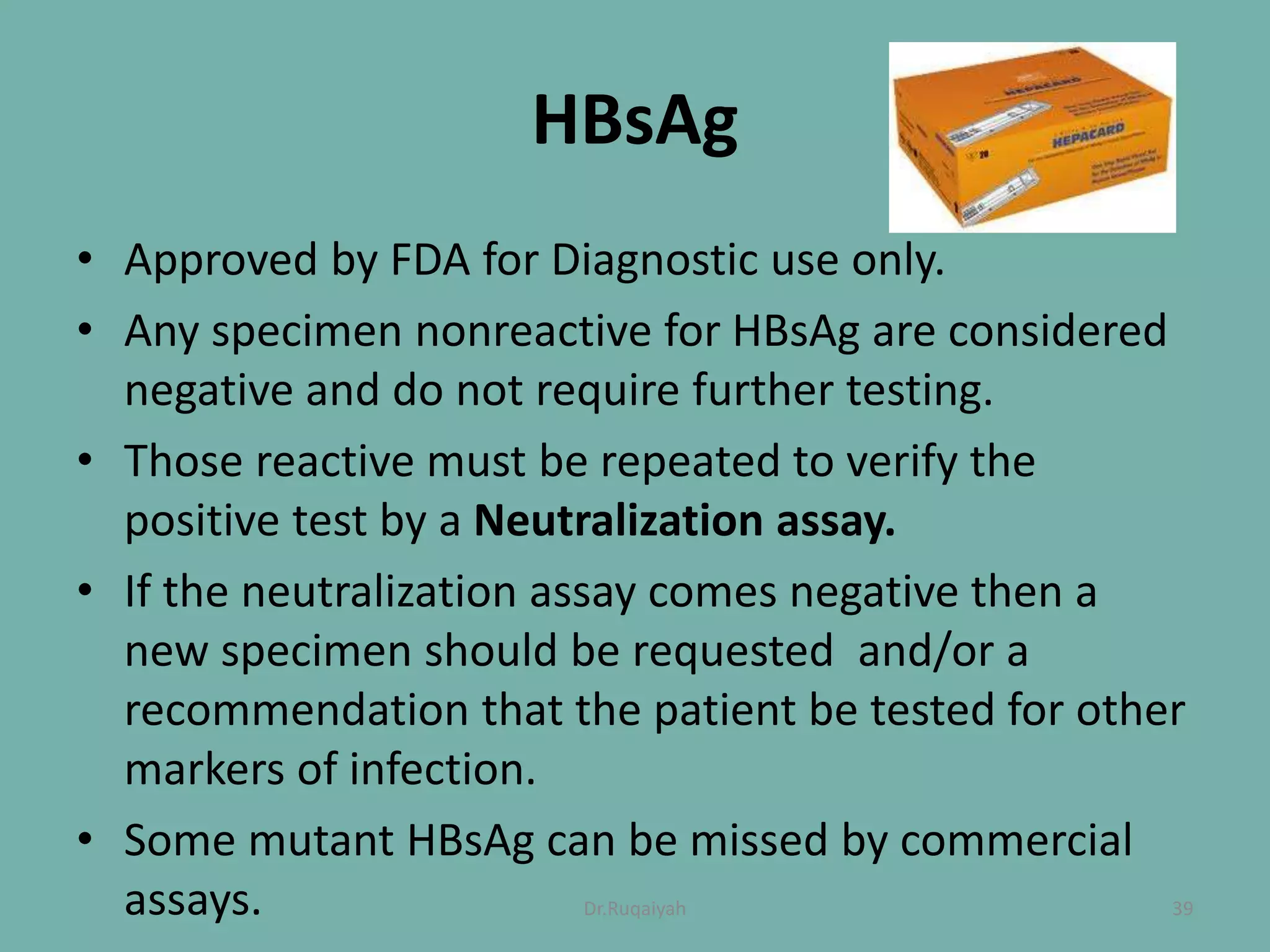
HEPATITIS B
Dr. Ruqaiyah Nadeem
1Dr.Ruqaiyah
Introduction
• Hepatitis was first described as early as 5th century.
• First documented blood borne outbreak occurred in
Bremen, Germany in 1883.
• 1947:MacCallum and Bauer introduced the term
Hepatitis B , later adopted by WHO(1973)
• 1965:Blumberg and his coworkers described the a
protein antigen: Australia antigen
• 1970:Dane and his coworkers described the
complete Hepatitis B or Dane particle.
• 1972: Magnius and Espmark described the HBeAg.
2Dr.Ruqaiyah
Taxonomy
• Group: Group VII(ds DNA RT)
• Order: Unassigned
• Family:
Hepadnaviridae Avihepadnaviridae Not assingned
• Genus: Orthohepadnavirus
• Species:
Hepatitis B Ground Squirrel Wood Chuck Wolly Monkey
Hepa. Hepa. Hepa.
3Dr.Ruqaiyah
Morphology
4Dr.Ruqaiyah
5Dr.Ruqaiyah
Continued….
• Three distinct
morphological entities in
various proportions are
seen:
 Small,pleomorphic,spherical
particles(22nm):most
abundant
 Tubular or filamentous
(200nm long)
 Complete HBV virion(42nm)
6Dr.Ruqaiyah
Viral antigens
• HbsAg
• HbcAg
• HbeAg
• Anti- HBs
• Anti- HBc
• Anti- Hbe
7Dr.Ruqaiyah
• HBsAg is antigenically complex
• Group reactive antigen- a
• Type specific antigen- d/y and w/r
8Dr.Ruqaiyah
Typing of HBV
Serotypes
• Its divided into 4 serotypes adr, adw, ayr, ayw
Genotypes
• Eight genotypes A- H
9Dr.Ruqaiyah
Genome
Gene Regions Antigen
S S Major protein(S) HBsAg
(Having 3 regions S,S1,Pre S2) S+ Pre S2 Middle protein(M)
S+ Pre S1& S2 Large protein(L):Present only in virion
C C HBcAg
(Having 2 regions C & Pre C) C+ Pre C HBeAg
P DNA Polymerase
X HBxAg(Not particulate antigen)
10Dr.Ruqaiyah
Genome
• Genome is approx. 3200 base long and contains
overlapping genes.
11Dr.Ruqaiyah
Virus Replication
13Dr.Ruqaiyah
14Dr.Ruqaiyah
Epidemiology
• Reservoir of infection
– Cases
– Carriers
• Carriers
– Simple carriers
– Super carriers
• Prevalence
– Low endemicity- <2 %. Nepal, SriLanka
– Intermediate endemicity- 2-8%. India, bhutan,
Indonesia, Maldives
– High endemicity- >8 %. Bangladesh, korea
15Dr.Ruqaiyah
• 2 billion people have been infected (1 out of 3
people).
• 350 million people are chronically infected.
• An estimated 6 lakh people die each year from
hepatitis B and its complications.
16Dr.Ruqaiyah
In India
• The average estimated carrier rate of hepatitis B
virus (HBV) is 3.7%.
• An approximate total of 40 million HBV carriers
• South indians have high carrier rates
• Second most common cause of acute hepatitis after
HEV.
17Dr.Ruqaiyah
Age
• Chance of developing acute hepatitis is directly
related to age
• 1% (perinatal)
• 10 % (early childhood)
• 30 % ( late childhood)
• Chance of developing chronic hepatitis is inversely
proportional to age
• 80-90 % (perinatal)
• 30% (early childhood)
• 5% (late childhood)
20Dr.Ruqaiyah
21Dr.Ruqaiyah
Clinical manifestations
• Incubation period 30-180 days
• It is indistinguishable from other hepatitis viruses
• Pre-icteric phase- nausea, vomiting
• Icteric phase- jaundice
• Hepatic complications- fulminant hepatitis, cirrhosis,
HCC
22Dr.Ruqaiyah
Continued…
• 4 phases
 Immuno tolerance
 Immuno clearance
 Inactive HBsAg carrier
 HBeAg – CHB
 Occult Hepatitis B
Phase HBeAg serological status Pattern Indications for treatment
1. “Immune tolerant” HBeAg positive • Stage seen in many HBeAg-positive children and young
adults, particularly among those infected at birth
• High levels of HBV replication (HBV DNA levels >200 000
IU/mL))
• Persistently normal ALT
• Minimal histological disease
Treatment not generally
indicated, but monitoring
required
2. “Immune active”
(HBeAg-positivea
chronic hepatitis)
HBeAg positive; may
develop anti-HBe
• Abnormal or intermittently abnormal ALT
• High or fluctuating levels of HBV replication (HBV DNA
levels >2000 IU/mL)
Histological necroinflammatory activity present
• HBeAg to anti-HBe seroconversion possible, with
normalization of ALT leading to “immune-control” phase
Treatment may be
indicated
3. Inactive chronic
hepatitis “Immune
control”
(previously called
inactive carrier)
HBeAg negative, anti-
HBe positive
• Persistently normal ALT
• Low or undetectable HBV DNA ( HBV DNA levels <2000
IU/mL)
• Risk of cirrhosis and HCC reduced
• May develop HBeAg-negative disease
Treatment not generally
indicated, but monitoring
required for reactivation
and HCC
4. “Immune escape”
(HBeAg-negative
chronic hepatitis)
HBeAg negative, with or
without being anti-HBe
positive
• HBeAg negative and anti-HBe positive
• Abnormal ALT (persistent or intermittently abnormal)
• Moderate to high levels of HBV replication (HBV DNA levels
>20 000 IU/mL)
• Older persons especially at risk for progressive disease
(fibrosis/cirrhosis)
Treatment may be
indicated
5.“Reactivation” or
“acute-on-chronic
hepatitis”
HBeAg positive or
negative
• Can occur spontaneously or be precipitated by
immunosuppression from chemo– or immunosuppressive
therapy, HIV infection or transplantation, development of
antiviral resistance, or withdrawal of antiviral therapy
• Abnormal ALT
• Moderate to high levels of HBV replication
• Seroreversion to HBeAg positivity can occur if HBeAg
negative
• High risk of decompensation in presence of cirrhosis
Treatment indicated
23Dr.Ruqaiyah
Collection, Transport and Storage
of Specimens
• HBV infection is diagnosed by serological and
molecular methods using serum and plasma.
• In general HBV antigens and antibodies are stable at
room temperature for several hours to days,can be
stored at 4ᴼC for months and can be frozen at -20ᴼC
to -70ᴼC for many years.
24Dr.Ruqaiyah
Lab diagnosis
25Dr.Ruqaiyah
26Dr.Ruqaiyah
HBsAg
• Appears in 1-12 weeks of infection (8-12 wks)
• Presence indicates onset of infectivity
• Becomes undetectable 1-2 months after
jaundice
• Used to calculate prevalence
27Dr.Ruqaiyah
HBeAg and HBV DNA
• Appear shortly after appearance of HBsAg
• They are markers of-
– Active viral replication
– High viral infectivity
• They cannot differentiate stages
28Dr.Ruqaiyah
HBcAg
• Non-secretory
• Hepatocytes by immunofluorescence
29Dr.Ruqaiyah
Anti-HBc IgM
• Appears 1-2 wks after HBsAg and lasts for 3-6
month
• Indicates acute infection
30Dr.Ruqaiyah
Anti- HBc IgG
• Appears in late stage and remains positive
indefinitely
• Chronic stage
• Carrier stage
• Recovery
31Dr.Ruqaiyah
Total Anti HBc
• Negative indicates that a person has not been
infected with HBV.
• Indicate Acute: (HBsAg +ve; IgM Anti HBc +ve)
Resolved(HBsAg –ve)
Chronic(HBsAg +ve)
32Dr.Ruqaiyah
Anti- HBe
• Appear after clearance of HBeAg
• Indicates diminished replication and
decreased infectivity
33Dr.Ruqaiyah
Anti -HBs
• Appears after clearance of HBsAg remains
indifinetly
• Indicates recovery, immunity, non infectivity
• Marker of hepatitis B vaccination
34Dr.Ruqaiyah
Detection Methods
35Dr.Ruqaiyah
Detection Methods:
Antigenic
Molecular
Serological
Others: Biochem,Microscopy,Culture
36Dr.Ruqaiyah
Microscopy
• Microscopic detection of HBV does not play a specific
role in the Diagnosis of disease.
• However liver Biopsy is typically used to assess the
extent of Histologic involvement and damage as well
as a response to therapy.
Culture
• Athough HBV can infect hepatocytes in vitro,culture
of HBV is not used for the diagnosis of infection.
37Dr.Ruqaiyah
Antigen Detection
• A marker of active viral replication is the detection of
HBsAg and/or HBeAg during primary infection and
during Chronic HBV infection.
• Both the antigens are produced in excess by the
infected hepatocytes.
• Detected by:
 ELISA
 Immunochromatographic tests
 Enzyme inmmunoassays Detects >=0.1ng/ml
 Radio immunoassays
38Dr.Ruqaiyah
HBsAg
• Approved by FDA for Diagnostic use only.
• Any specimen nonreactive for HBsAg are considered
negative and do not require further testing.
• Those reactive must be repeated to verify the
positive test by a Neutralization assay.
• If the neutralization assay comes negative then a
new specimen should be requested and/or a
recommendation that the patient be tested for other
markers of infection.
• Some mutant HBsAg can be missed by commercial
assays. 39Dr.Ruqaiyah
40Dr.Ruqaiyah
Nucleic Acid Detection:Quantitative
• Recommended for initial evaluation of Chronic
Hepatitis B and during management, particularly in
the decision making to initiate treatment and in
therapeutic monitoring.
 Criteria for Chronic HB:>=20,000IU/ml
45Dr.Ruqaiyah
47Dr.Ruqaiyah
HBV genotyping
• HBV genotype potential influence the outcome of
Chronic Hepatitis B and the success of antiviral therapy.
• Genotype B has more favorable outcome than C.
• Genotyping is more important for IFN therapy as A
have more changes of seroconversion than D.
• Methods:
 Genome Sequencing
 RFLP
 Genotype specific Amplification techniques
 Hybridization techniques
48Dr.Ruqaiyah
Detecting Core promoter/Pre core
mutation
• PCR plus hybridisation format detects
 Basal core promoter nucleotide 1762
 Basal core promoter nucleotide 1964
 Precore codon 28
• Hybridisation/direct sequencing
 Basal core promoter nucleotide 1964
 Precore codon 28
50Dr.Ruqaiyah
Biochemistry
• Serum Transaminases
 ALT> AST but this reverses once Cirrhosis sets in.
• Direct and Total Bilirubin
 0.3- 1 mg/100ml
• Albumin and Total protein
 Albumin 4-5.5 mg/100ml
• Coagulation tests
 Prothrombin time(11-12.5 sec) increases
• Alpha feto protein
52Dr.Ruqaiyah
Evaluation, Interpretation and
Reporting of Results
57Dr.Ruqaiyah
HBV Markers in different Stages:
Stage of
infection
HBV
DNA
HBsAg HBeAg Anti-
HBc
Ig M
Anti-
HBc
Total
Anti-
HBe
Anti-
HBs
Susceptible _ _ _ _ _ _ _
Early
incubation
+ _ _ _ _ _ _
Late
incubation
+ + +/_ _ _ _ _
Acute
infection
+ + + + _ _ _
Recent
infection
_/+ _ _ + + + +++
60Dr.Ruqaiyah
Stage of
infection
HBV DNA HBsAg HBeAg Anti-HBc
Ig M
Anti-
HBc
Total
Anti-
HBe
Anti-
HBs
Remote
infection
_ or very
low
_ _ _ + +/_ +
HBsAg-ve
Acute infect
_ _ _ + + _ _
HBsAg
variant
infect.
_/+ _ _/+ +/_ + _ _
Immune
active
carrier
++ + _/+ _/+ +++ _ _
Healthy
HBsAg
carrier
_ + _ _ + + _
Vaccination
response
_ _ _ _ _ _ +
61Dr.Ruqaiyah
Treatment
• 99% previously healthy person recover and
treatment is not required
• Indicated in fulminant hepatitis or severe
chronic hepatitis
– Interferon
– Nucleoside/ nucleotide analogues- lamuvidine,
adefovir, entecavir, telbivudine, tenofovir
62Dr.Ruqaiyah
Prophylaxis
• Recombinant subunit vaccine
• HBsAg is used as a vaccine candidate in
baker’s yeast by DNA recombinant technology
• Route- I/M
• Dosage- 10-20 μg/kg
• Schedule 0,1,6 and 6,10, 14
• Marker of protection- anti- HBs titer more
than 10 IU/ml
63Dr.Ruqaiyah
• Booster after 5 yr for high risk group or
antibody titer falls below 10 IU/ml
64Dr.Ruqaiyah
Passive immunisation
• HBIG should be given within 6 hrs but not
later than 48 hrs
• Dose- 0.05-.07 ml/kg
• Two doses 30 days apart
65Dr.Ruqaiyah
Thank you….
66Dr.Ruqaiyah

Hepatitis B

  • 1.
    HEPATITIS B Dr. RuqaiyahNadeem 1Dr.Ruqaiyah
  • 2.
    Introduction • Hepatitis wasfirst described as early as 5th century. • First documented blood borne outbreak occurred in Bremen, Germany in 1883. • 1947:MacCallum and Bauer introduced the term Hepatitis B , later adopted by WHO(1973) • 1965:Blumberg and his coworkers described the a protein antigen: Australia antigen • 1970:Dane and his coworkers described the complete Hepatitis B or Dane particle. • 1972: Magnius and Espmark described the HBeAg. 2Dr.Ruqaiyah
  • 3.
    Taxonomy • Group: GroupVII(ds DNA RT) • Order: Unassigned • Family: Hepadnaviridae Avihepadnaviridae Not assingned • Genus: Orthohepadnavirus • Species: Hepatitis B Ground Squirrel Wood Chuck Wolly Monkey Hepa. Hepa. Hepa. 3Dr.Ruqaiyah
  • 4.
  • 5.
  • 6.
    Continued…. • Three distinct morphologicalentities in various proportions are seen:  Small,pleomorphic,spherical particles(22nm):most abundant  Tubular or filamentous (200nm long)  Complete HBV virion(42nm) 6Dr.Ruqaiyah
  • 7.
    Viral antigens • HbsAg •HbcAg • HbeAg • Anti- HBs • Anti- HBc • Anti- Hbe 7Dr.Ruqaiyah
  • 8.
    • HBsAg isantigenically complex • Group reactive antigen- a • Type specific antigen- d/y and w/r 8Dr.Ruqaiyah
  • 9.
    Typing of HBV Serotypes •Its divided into 4 serotypes adr, adw, ayr, ayw Genotypes • Eight genotypes A- H 9Dr.Ruqaiyah
  • 10.
    Genome Gene Regions Antigen SS Major protein(S) HBsAg (Having 3 regions S,S1,Pre S2) S+ Pre S2 Middle protein(M) S+ Pre S1& S2 Large protein(L):Present only in virion C C HBcAg (Having 2 regions C & Pre C) C+ Pre C HBeAg P DNA Polymerase X HBxAg(Not particulate antigen) 10Dr.Ruqaiyah
  • 11.
    Genome • Genome isapprox. 3200 base long and contains overlapping genes. 11Dr.Ruqaiyah
  • 12.
  • 13.
  • 14.
    Epidemiology • Reservoir ofinfection – Cases – Carriers • Carriers – Simple carriers – Super carriers • Prevalence – Low endemicity- <2 %. Nepal, SriLanka – Intermediate endemicity- 2-8%. India, bhutan, Indonesia, Maldives – High endemicity- >8 %. Bangladesh, korea 15Dr.Ruqaiyah
  • 15.
    • 2 billionpeople have been infected (1 out of 3 people). • 350 million people are chronically infected. • An estimated 6 lakh people die each year from hepatitis B and its complications. 16Dr.Ruqaiyah
  • 16.
    In India • Theaverage estimated carrier rate of hepatitis B virus (HBV) is 3.7%. • An approximate total of 40 million HBV carriers • South indians have high carrier rates • Second most common cause of acute hepatitis after HEV. 17Dr.Ruqaiyah
  • 17.
    Age • Chance ofdeveloping acute hepatitis is directly related to age • 1% (perinatal) • 10 % (early childhood) • 30 % ( late childhood) • Chance of developing chronic hepatitis is inversely proportional to age • 80-90 % (perinatal) • 30% (early childhood) • 5% (late childhood) 20Dr.Ruqaiyah
  • 18.
  • 19.
    Clinical manifestations • Incubationperiod 30-180 days • It is indistinguishable from other hepatitis viruses • Pre-icteric phase- nausea, vomiting • Icteric phase- jaundice • Hepatic complications- fulminant hepatitis, cirrhosis, HCC 22Dr.Ruqaiyah
  • 20.
    Continued… • 4 phases Immuno tolerance  Immuno clearance  Inactive HBsAg carrier  HBeAg – CHB  Occult Hepatitis B Phase HBeAg serological status Pattern Indications for treatment 1. “Immune tolerant” HBeAg positive • Stage seen in many HBeAg-positive children and young adults, particularly among those infected at birth • High levels of HBV replication (HBV DNA levels >200 000 IU/mL)) • Persistently normal ALT • Minimal histological disease Treatment not generally indicated, but monitoring required 2. “Immune active” (HBeAg-positivea chronic hepatitis) HBeAg positive; may develop anti-HBe • Abnormal or intermittently abnormal ALT • High or fluctuating levels of HBV replication (HBV DNA levels >2000 IU/mL) Histological necroinflammatory activity present • HBeAg to anti-HBe seroconversion possible, with normalization of ALT leading to “immune-control” phase Treatment may be indicated 3. Inactive chronic hepatitis “Immune control” (previously called inactive carrier) HBeAg negative, anti- HBe positive • Persistently normal ALT • Low or undetectable HBV DNA ( HBV DNA levels <2000 IU/mL) • Risk of cirrhosis and HCC reduced • May develop HBeAg-negative disease Treatment not generally indicated, but monitoring required for reactivation and HCC 4. “Immune escape” (HBeAg-negative chronic hepatitis) HBeAg negative, with or without being anti-HBe positive • HBeAg negative and anti-HBe positive • Abnormal ALT (persistent or intermittently abnormal) • Moderate to high levels of HBV replication (HBV DNA levels >20 000 IU/mL) • Older persons especially at risk for progressive disease (fibrosis/cirrhosis) Treatment may be indicated 5.“Reactivation” or “acute-on-chronic hepatitis” HBeAg positive or negative • Can occur spontaneously or be precipitated by immunosuppression from chemo– or immunosuppressive therapy, HIV infection or transplantation, development of antiviral resistance, or withdrawal of antiviral therapy • Abnormal ALT • Moderate to high levels of HBV replication • Seroreversion to HBeAg positivity can occur if HBeAg negative • High risk of decompensation in presence of cirrhosis Treatment indicated 23Dr.Ruqaiyah
  • 21.
    Collection, Transport andStorage of Specimens • HBV infection is diagnosed by serological and molecular methods using serum and plasma. • In general HBV antigens and antibodies are stable at room temperature for several hours to days,can be stored at 4ᴼC for months and can be frozen at -20ᴼC to -70ᴼC for many years. 24Dr.Ruqaiyah
  • 22.
  • 23.
  • 24.
    HBsAg • Appears in1-12 weeks of infection (8-12 wks) • Presence indicates onset of infectivity • Becomes undetectable 1-2 months after jaundice • Used to calculate prevalence 27Dr.Ruqaiyah
  • 25.
    HBeAg and HBVDNA • Appear shortly after appearance of HBsAg • They are markers of- – Active viral replication – High viral infectivity • They cannot differentiate stages 28Dr.Ruqaiyah
  • 26.
    HBcAg • Non-secretory • Hepatocytesby immunofluorescence 29Dr.Ruqaiyah
  • 27.
    Anti-HBc IgM • Appears1-2 wks after HBsAg and lasts for 3-6 month • Indicates acute infection 30Dr.Ruqaiyah
  • 28.
    Anti- HBc IgG •Appears in late stage and remains positive indefinitely • Chronic stage • Carrier stage • Recovery 31Dr.Ruqaiyah
  • 29.
    Total Anti HBc •Negative indicates that a person has not been infected with HBV. • Indicate Acute: (HBsAg +ve; IgM Anti HBc +ve) Resolved(HBsAg –ve) Chronic(HBsAg +ve) 32Dr.Ruqaiyah
  • 30.
    Anti- HBe • Appearafter clearance of HBeAg • Indicates diminished replication and decreased infectivity 33Dr.Ruqaiyah
  • 31.
    Anti -HBs • Appearsafter clearance of HBsAg remains indifinetly • Indicates recovery, immunity, non infectivity • Marker of hepatitis B vaccination 34Dr.Ruqaiyah
  • 32.
  • 33.
  • 34.
    Microscopy • Microscopic detectionof HBV does not play a specific role in the Diagnosis of disease. • However liver Biopsy is typically used to assess the extent of Histologic involvement and damage as well as a response to therapy. Culture • Athough HBV can infect hepatocytes in vitro,culture of HBV is not used for the diagnosis of infection. 37Dr.Ruqaiyah
  • 35.
    Antigen Detection • Amarker of active viral replication is the detection of HBsAg and/or HBeAg during primary infection and during Chronic HBV infection. • Both the antigens are produced in excess by the infected hepatocytes. • Detected by:  ELISA  Immunochromatographic tests  Enzyme inmmunoassays Detects >=0.1ng/ml  Radio immunoassays 38Dr.Ruqaiyah
  • 36.
    HBsAg • Approved byFDA for Diagnostic use only. • Any specimen nonreactive for HBsAg are considered negative and do not require further testing. • Those reactive must be repeated to verify the positive test by a Neutralization assay. • If the neutralization assay comes negative then a new specimen should be requested and/or a recommendation that the patient be tested for other markers of infection. • Some mutant HBsAg can be missed by commercial assays. 39Dr.Ruqaiyah
  • 37.
  • 38.
    Nucleic Acid Detection:Quantitative •Recommended for initial evaluation of Chronic Hepatitis B and during management, particularly in the decision making to initiate treatment and in therapeutic monitoring.  Criteria for Chronic HB:>=20,000IU/ml 45Dr.Ruqaiyah
  • 39.
  • 40.
    HBV genotyping • HBVgenotype potential influence the outcome of Chronic Hepatitis B and the success of antiviral therapy. • Genotype B has more favorable outcome than C. • Genotyping is more important for IFN therapy as A have more changes of seroconversion than D. • Methods:  Genome Sequencing  RFLP  Genotype specific Amplification techniques  Hybridization techniques 48Dr.Ruqaiyah
  • 41.
    Detecting Core promoter/Precore mutation • PCR plus hybridisation format detects  Basal core promoter nucleotide 1762  Basal core promoter nucleotide 1964  Precore codon 28 • Hybridisation/direct sequencing  Basal core promoter nucleotide 1964  Precore codon 28 50Dr.Ruqaiyah
  • 42.
    Biochemistry • Serum Transaminases ALT> AST but this reverses once Cirrhosis sets in. • Direct and Total Bilirubin  0.3- 1 mg/100ml • Albumin and Total protein  Albumin 4-5.5 mg/100ml • Coagulation tests  Prothrombin time(11-12.5 sec) increases • Alpha feto protein 52Dr.Ruqaiyah
  • 43.
  • 44.
    HBV Markers indifferent Stages: Stage of infection HBV DNA HBsAg HBeAg Anti- HBc Ig M Anti- HBc Total Anti- HBe Anti- HBs Susceptible _ _ _ _ _ _ _ Early incubation + _ _ _ _ _ _ Late incubation + + +/_ _ _ _ _ Acute infection + + + + _ _ _ Recent infection _/+ _ _ + + + +++ 60Dr.Ruqaiyah
  • 45.
    Stage of infection HBV DNAHBsAg HBeAg Anti-HBc Ig M Anti- HBc Total Anti- HBe Anti- HBs Remote infection _ or very low _ _ _ + +/_ + HBsAg-ve Acute infect _ _ _ + + _ _ HBsAg variant infect. _/+ _ _/+ +/_ + _ _ Immune active carrier ++ + _/+ _/+ +++ _ _ Healthy HBsAg carrier _ + _ _ + + _ Vaccination response _ _ _ _ _ _ + 61Dr.Ruqaiyah
  • 46.
    Treatment • 99% previouslyhealthy person recover and treatment is not required • Indicated in fulminant hepatitis or severe chronic hepatitis – Interferon – Nucleoside/ nucleotide analogues- lamuvidine, adefovir, entecavir, telbivudine, tenofovir 62Dr.Ruqaiyah
  • 47.
    Prophylaxis • Recombinant subunitvaccine • HBsAg is used as a vaccine candidate in baker’s yeast by DNA recombinant technology • Route- I/M • Dosage- 10-20 μg/kg • Schedule 0,1,6 and 6,10, 14 • Marker of protection- anti- HBs titer more than 10 IU/ml 63Dr.Ruqaiyah
  • 48.
    • Booster after5 yr for high risk group or antibody titer falls below 10 IU/ml 64Dr.Ruqaiyah
  • 49.
    Passive immunisation • HBIGshould be given within 6 hrs but not later than 48 hrs • Dose- 0.05-.07 ml/kg • Two doses 30 days apart 65Dr.Ruqaiyah
  • 50.

Editor's Notes

  • #25 For nucleic acid analysis: EDTA or Citrated Plasma should be used. Plasma should be separated from RBCs within 6 hrs and stored at 4ᴼC.