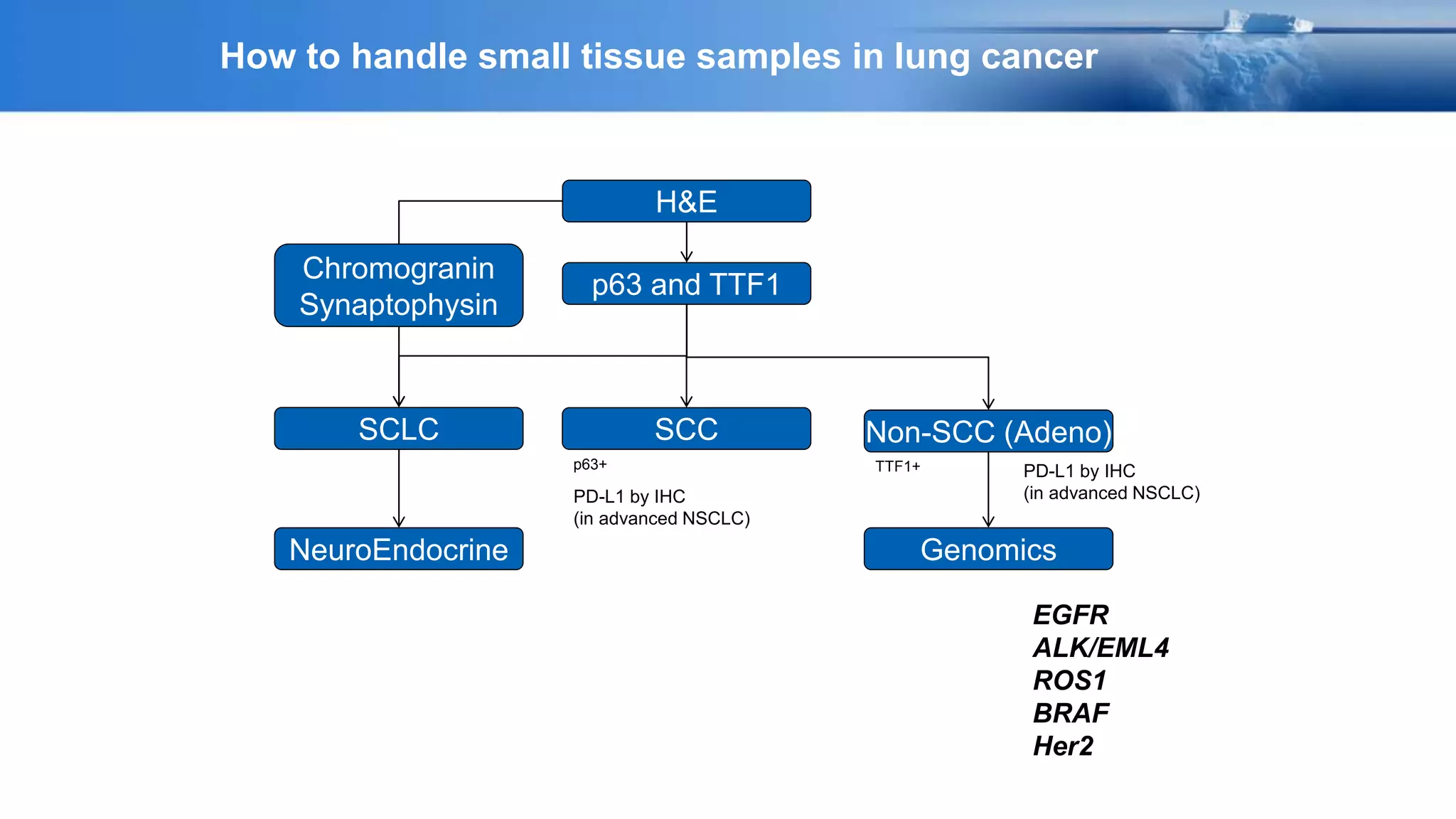
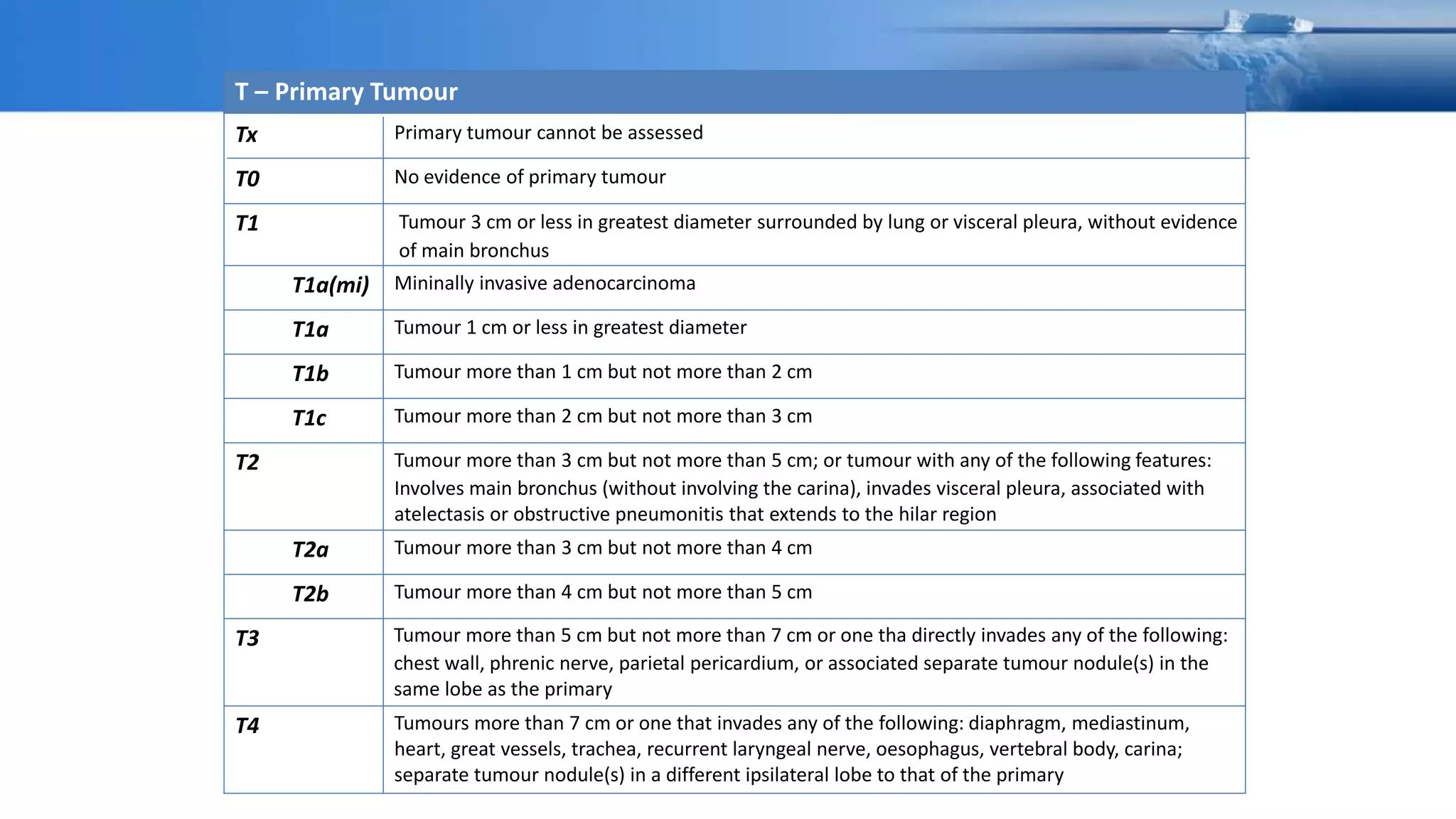
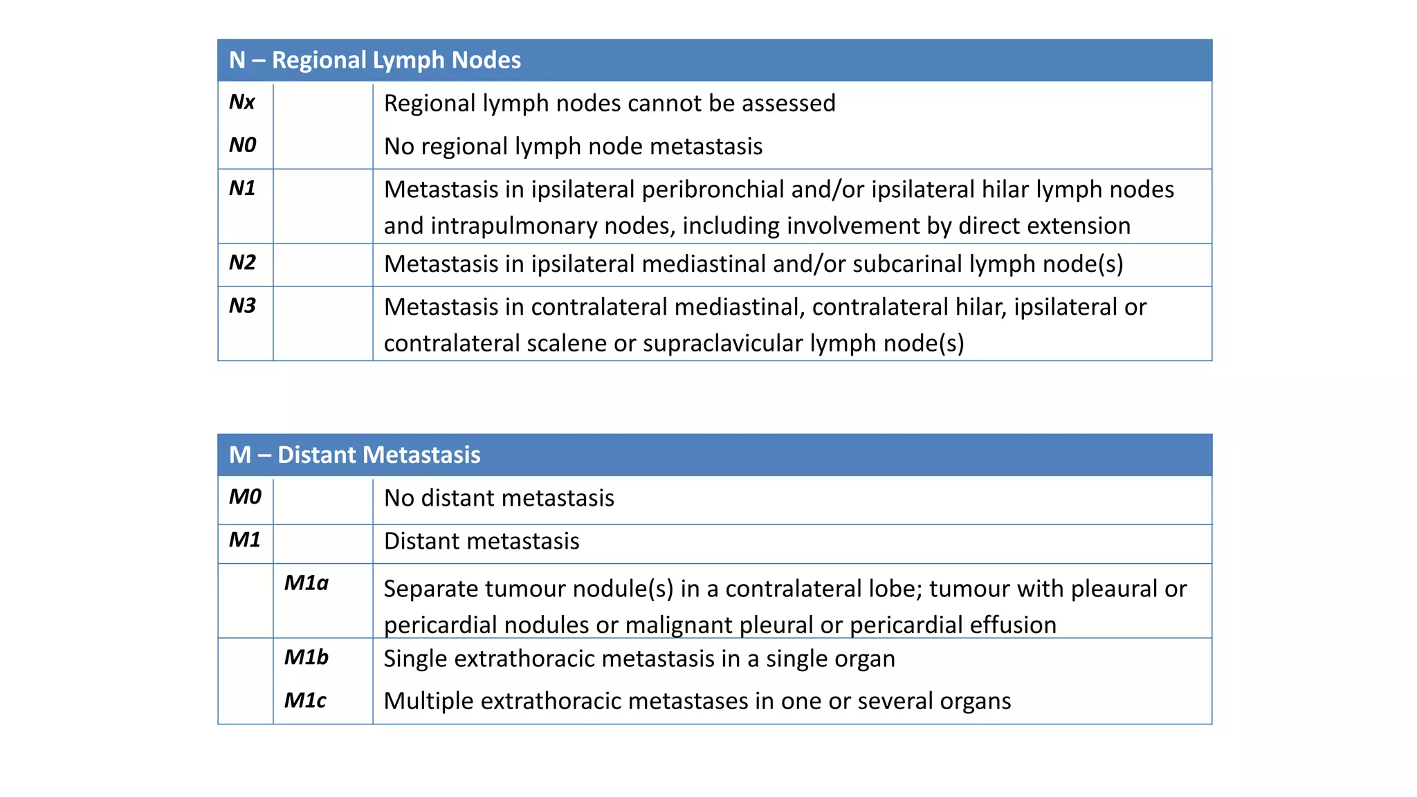
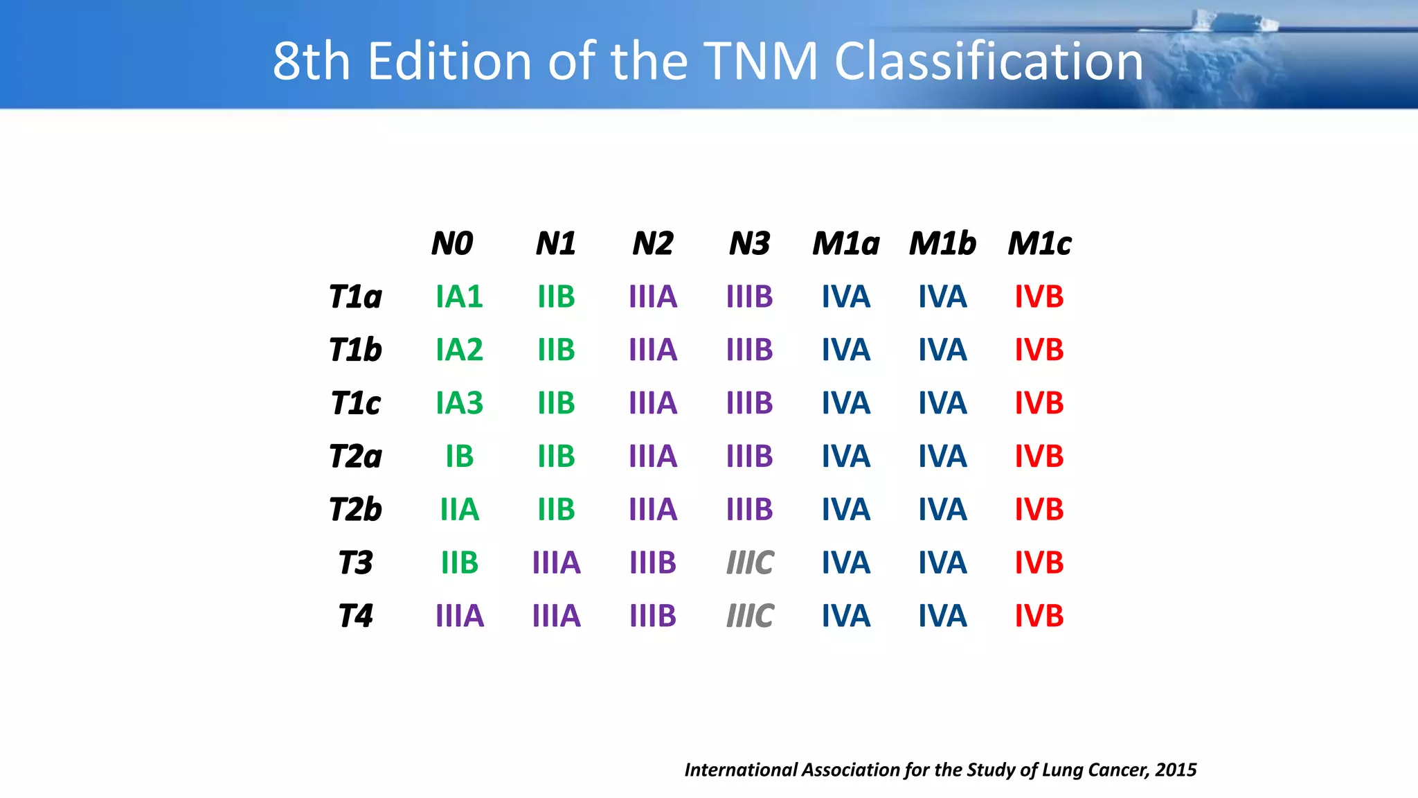
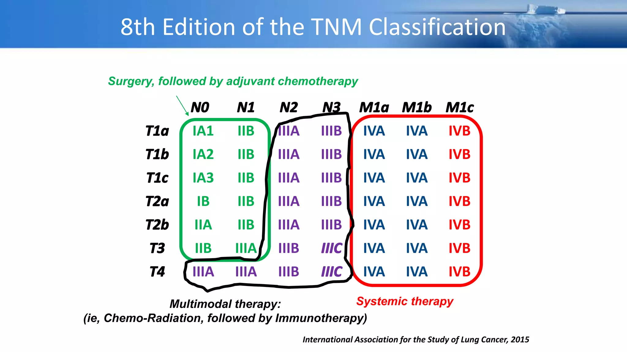
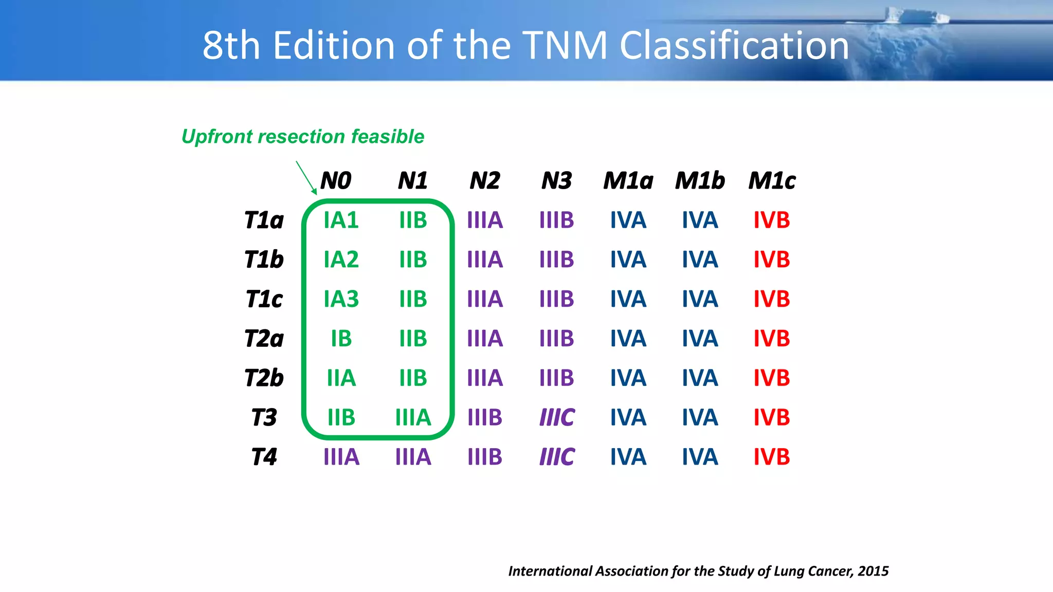
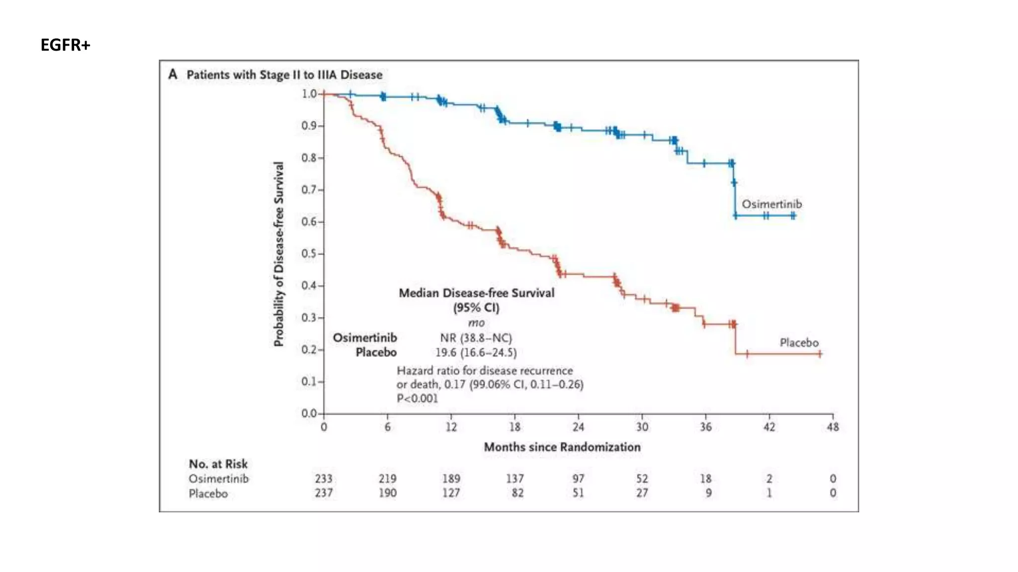
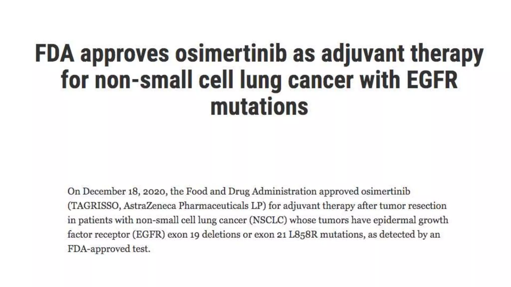
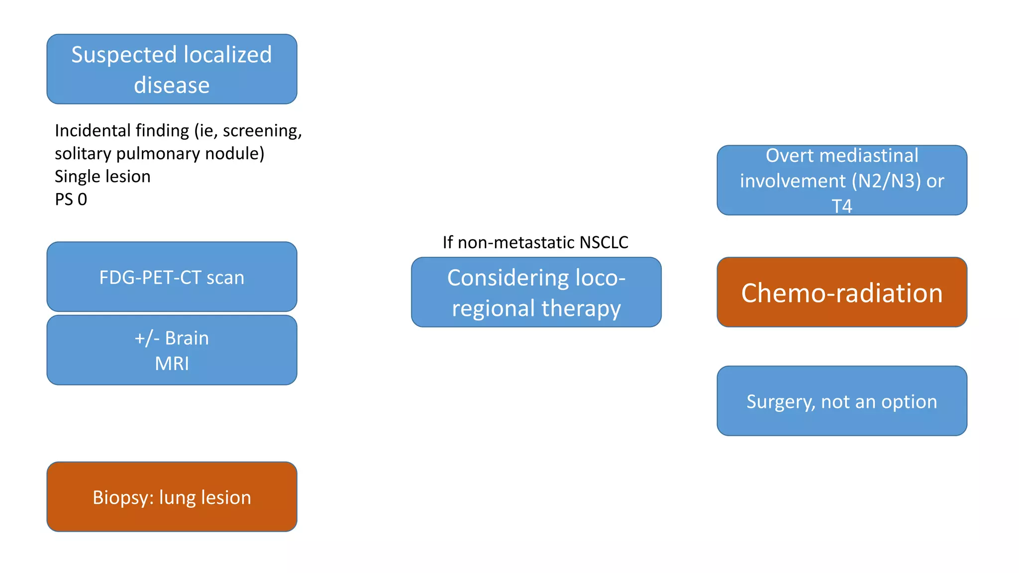
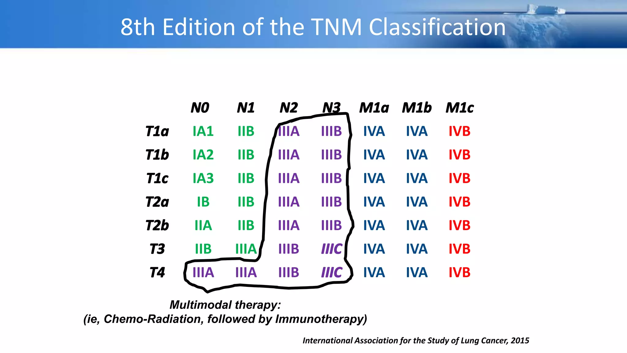
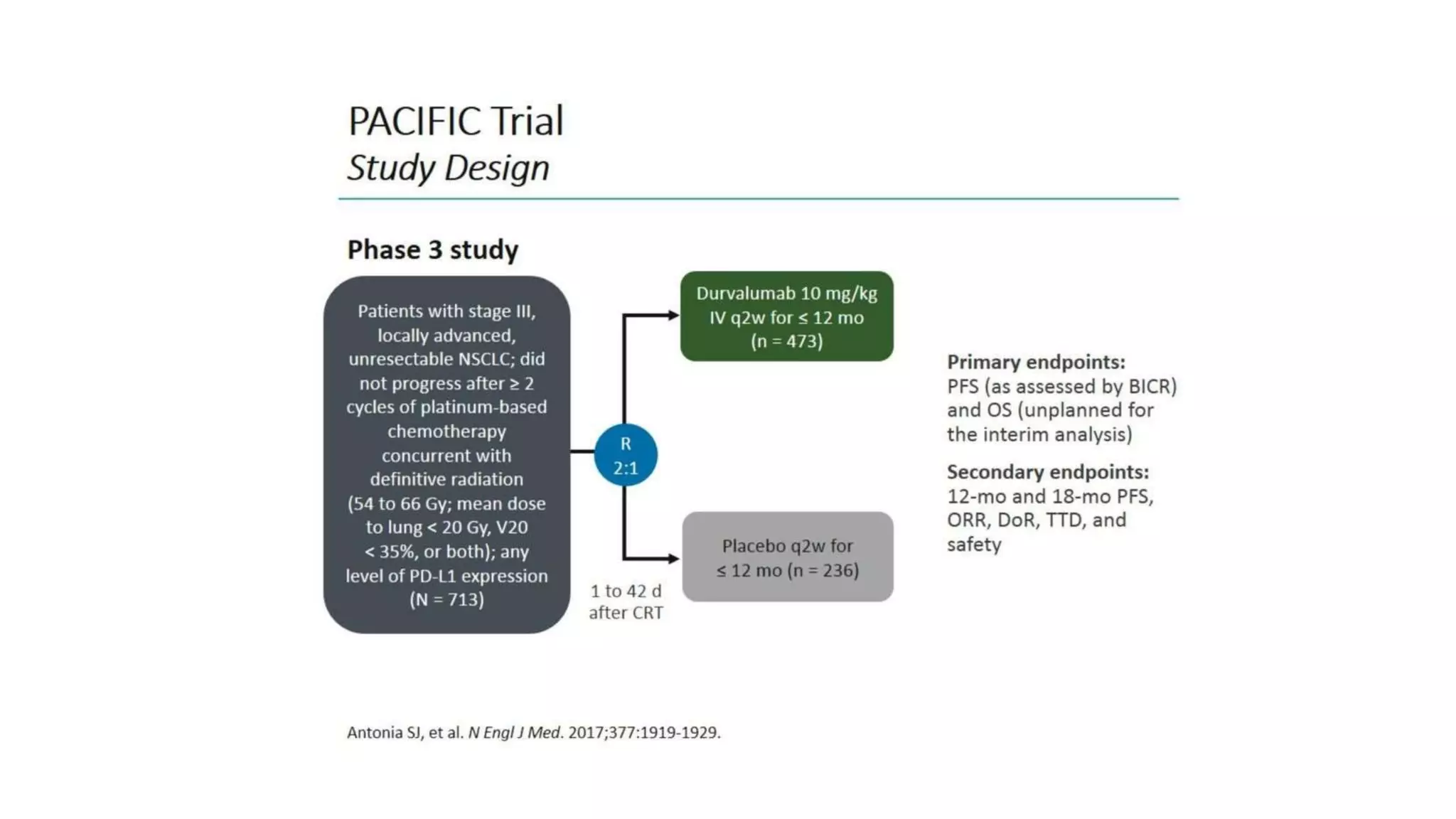
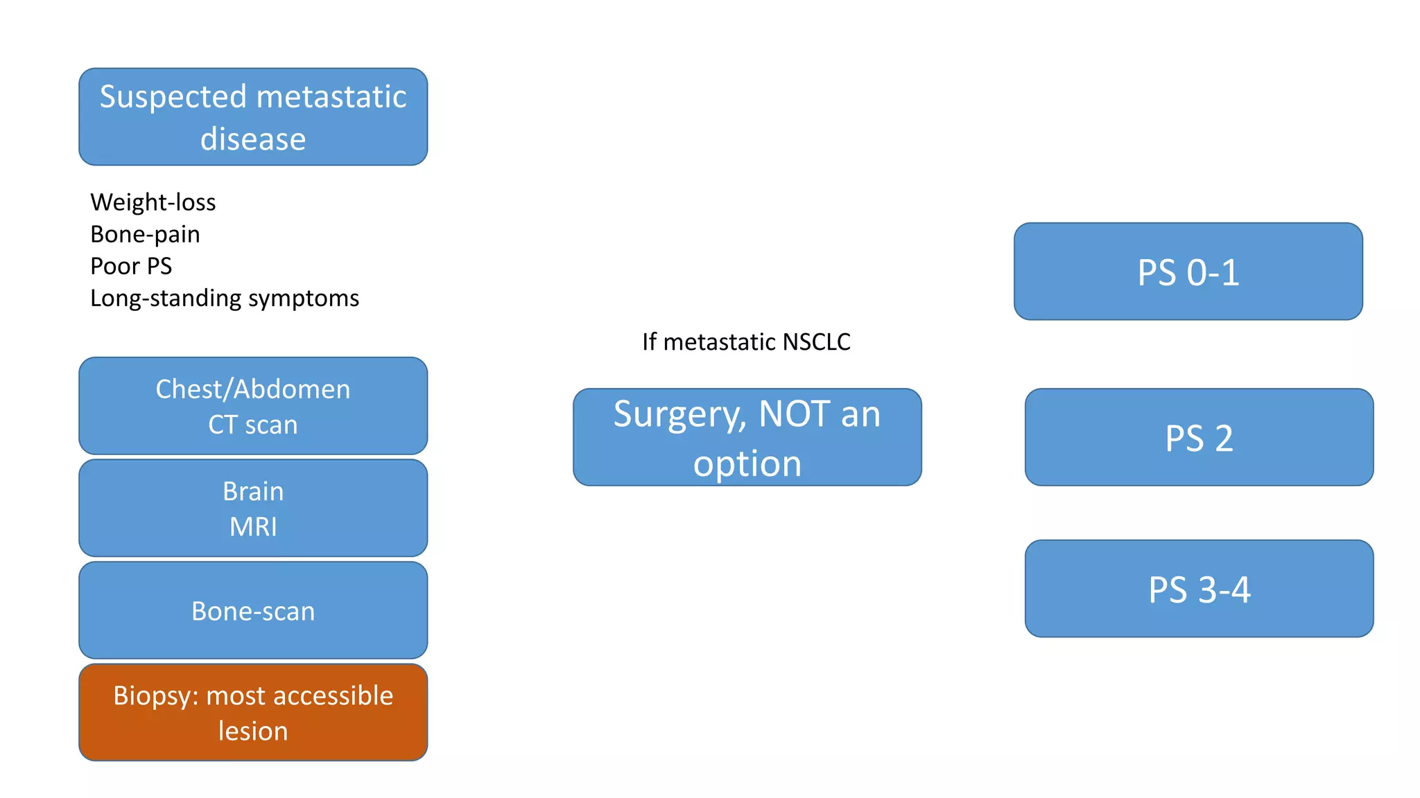
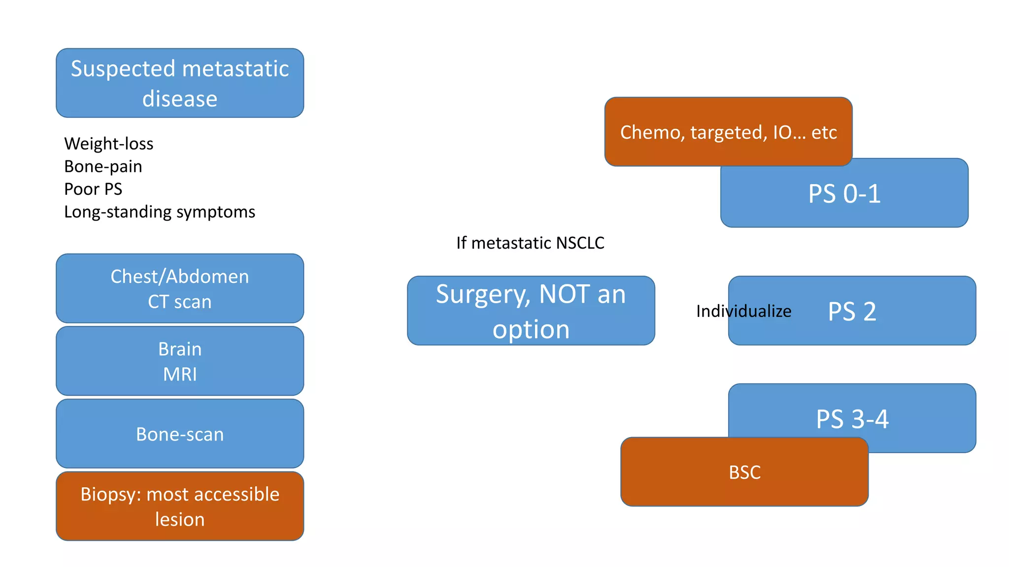
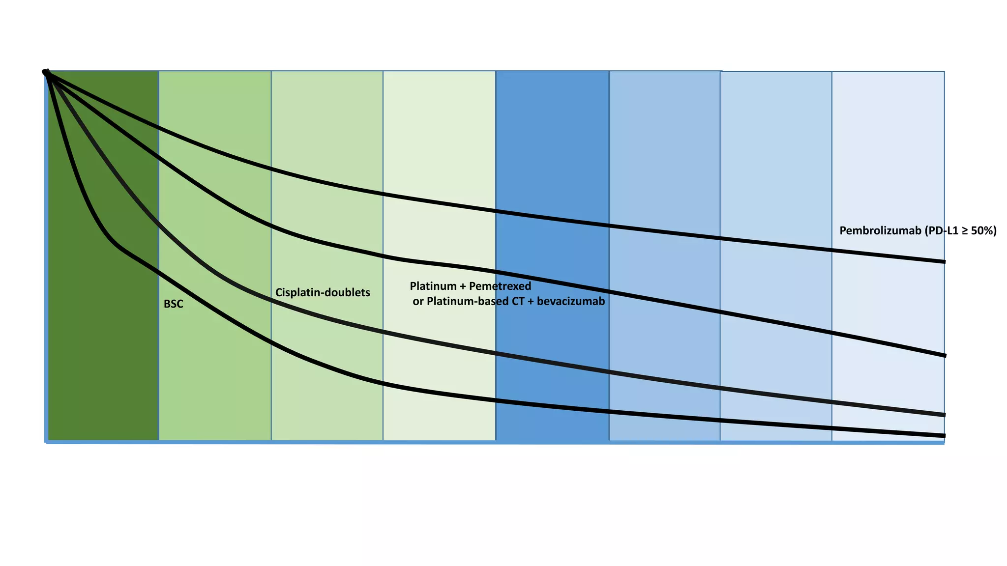
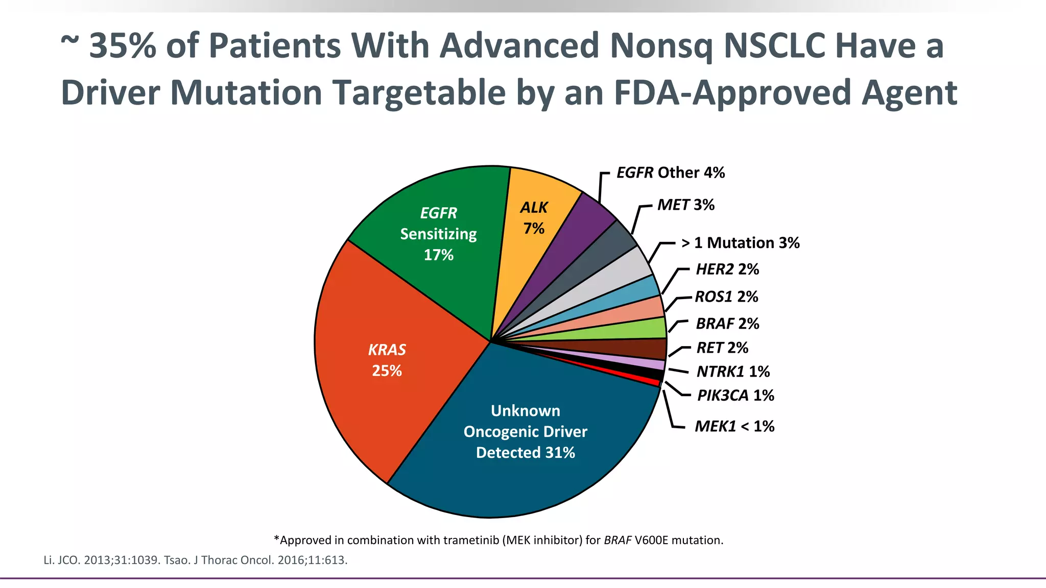
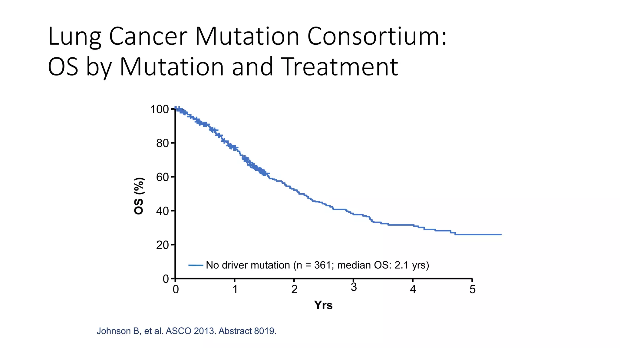
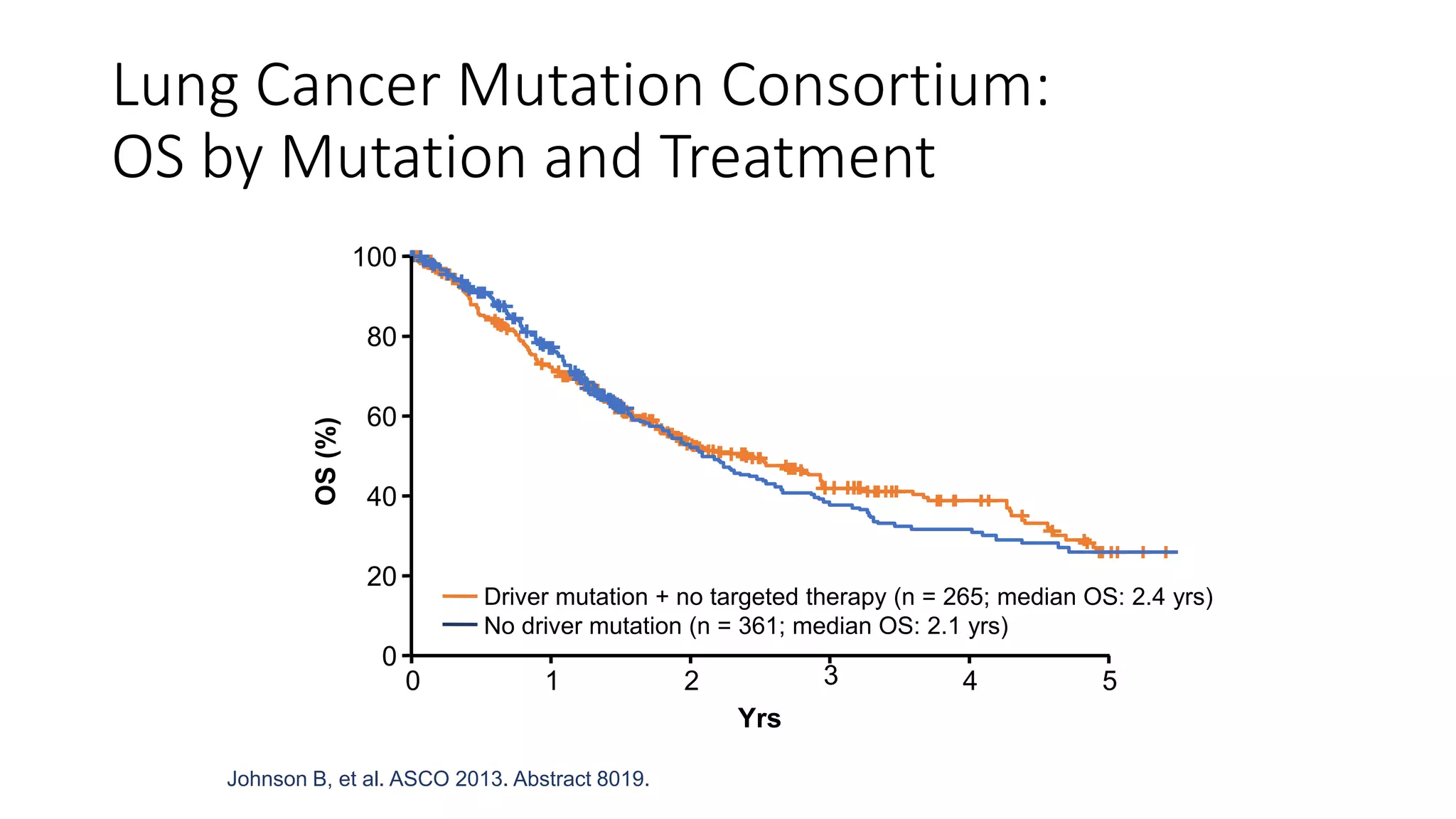
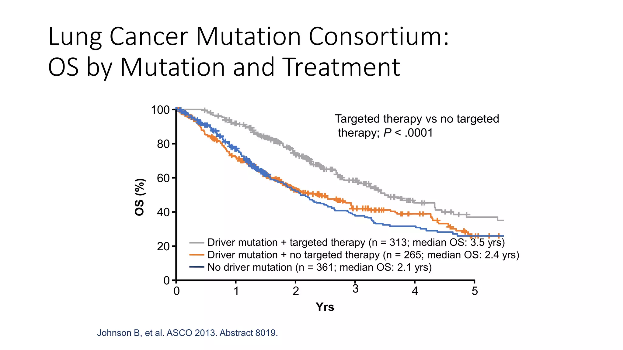
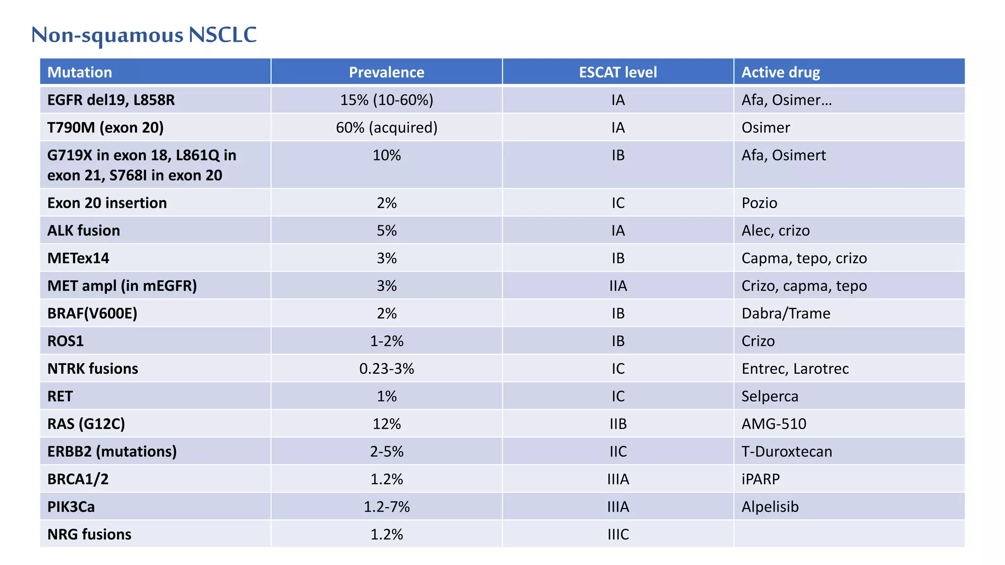
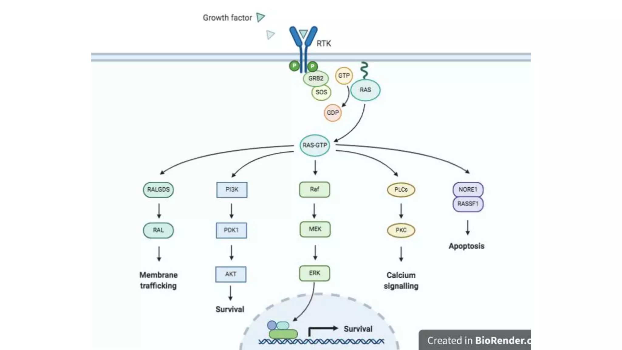
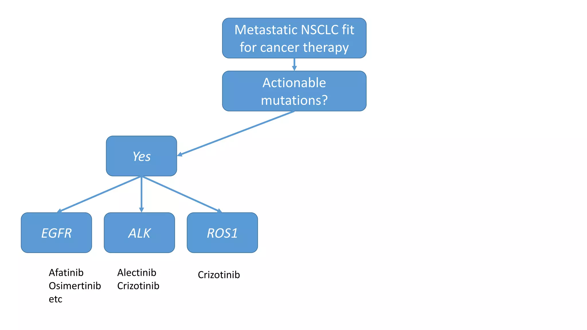
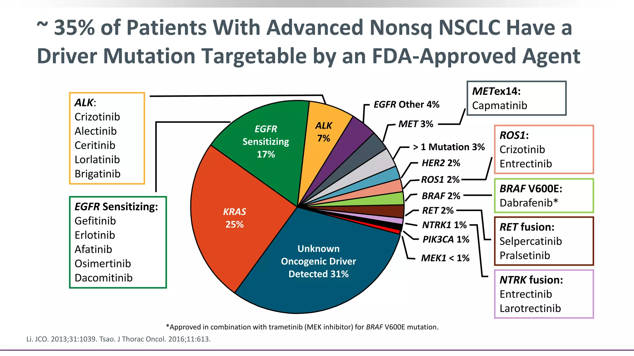
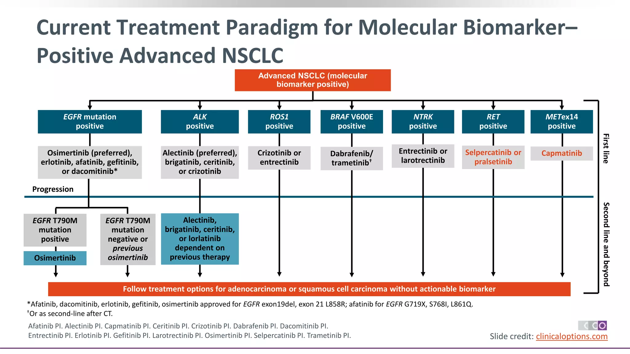
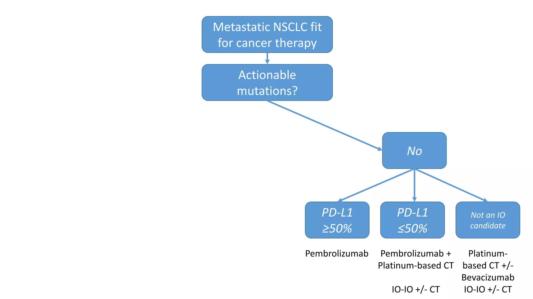
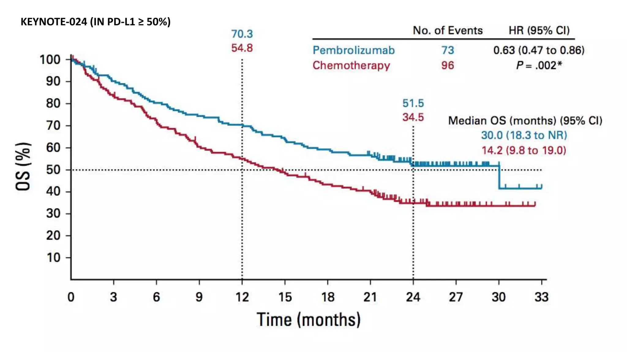
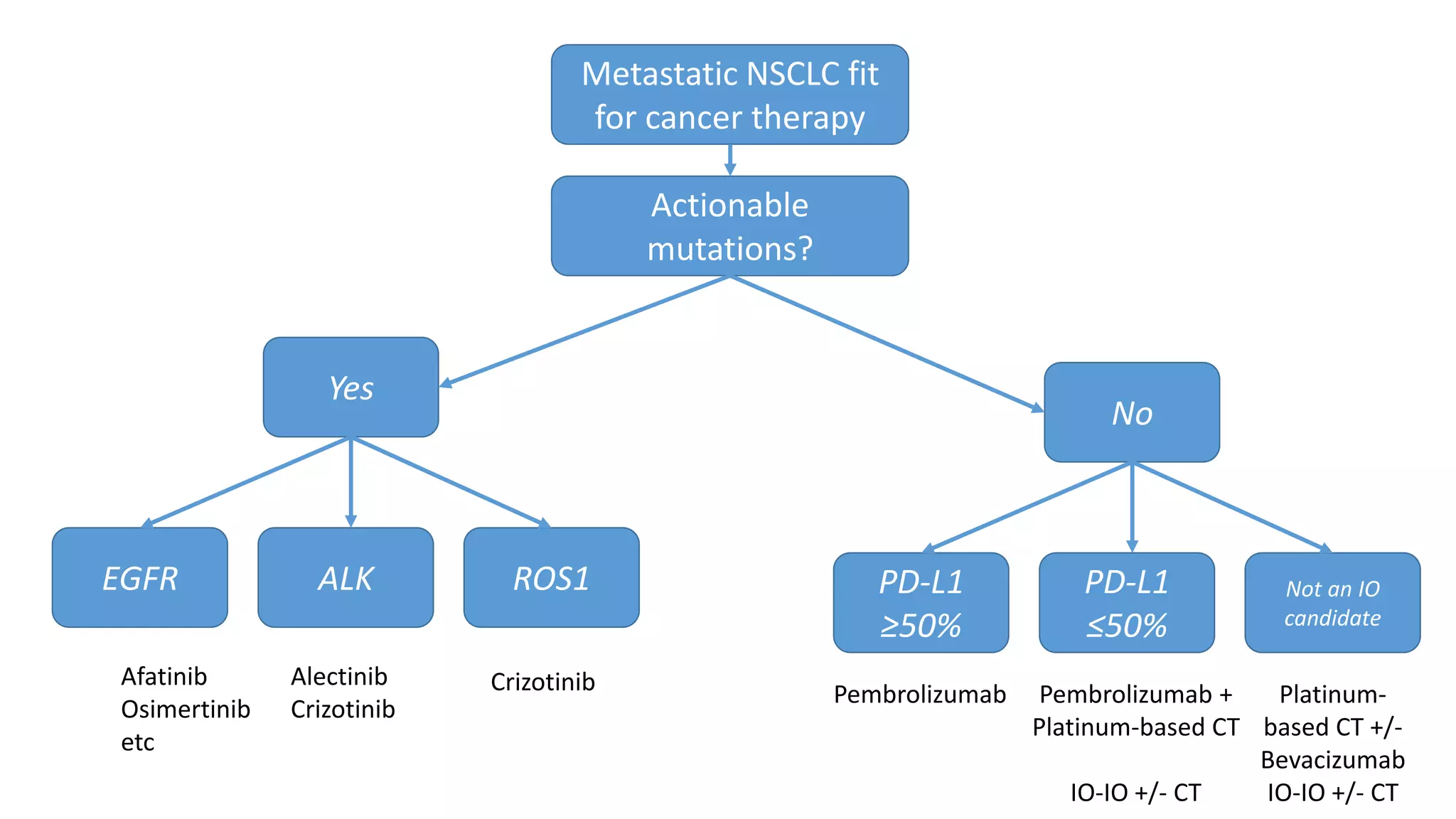
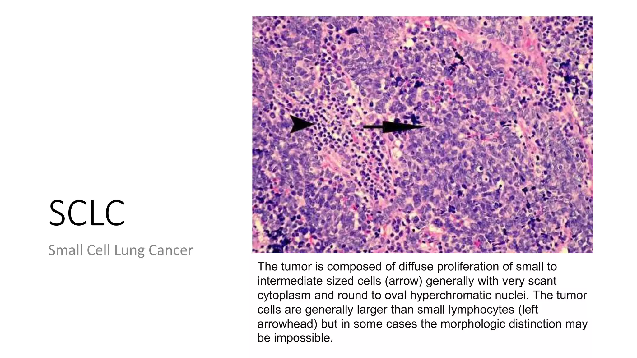
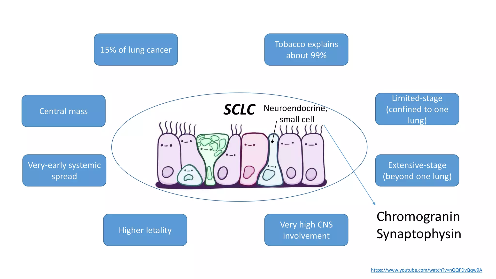
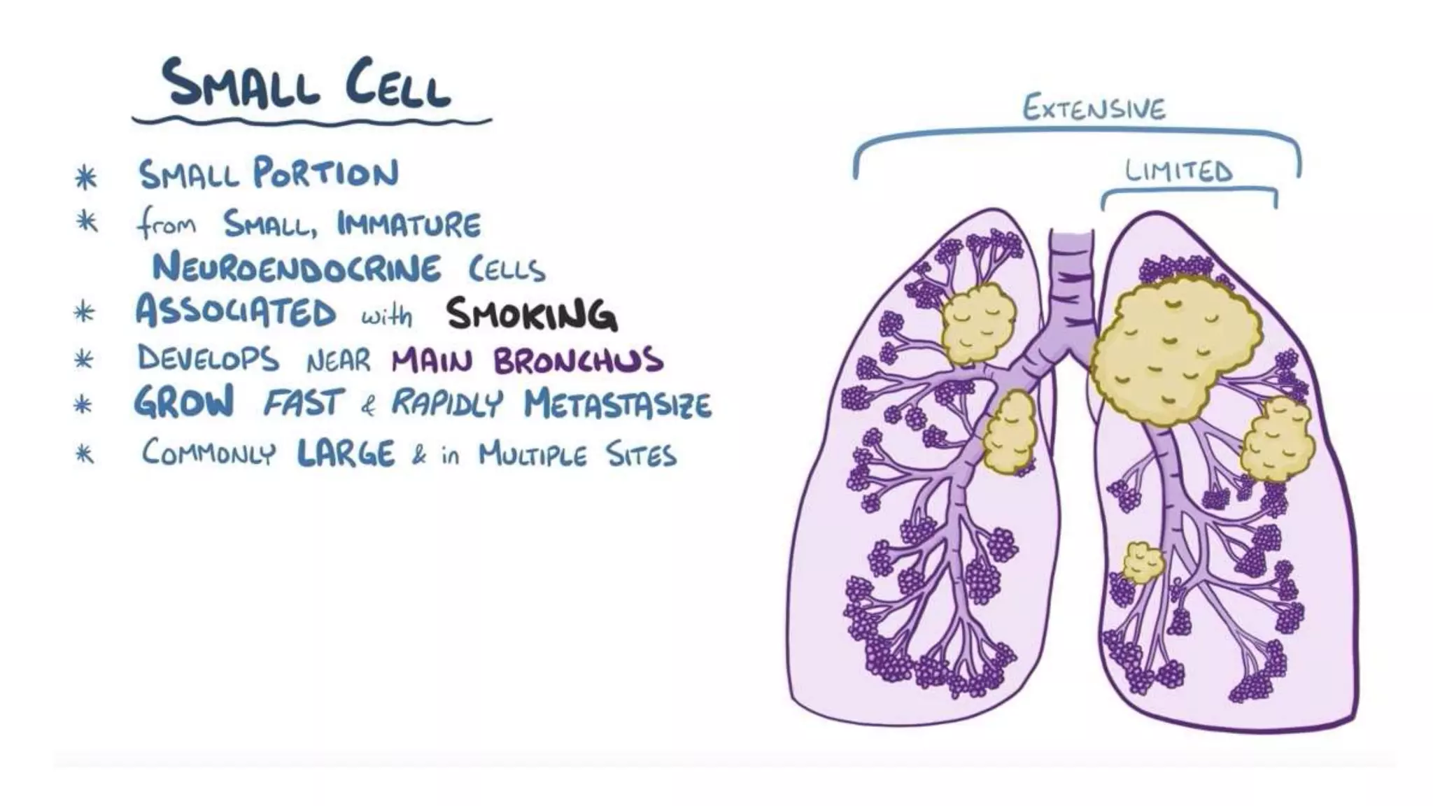
Cáncer de pulmón (lung cancer)
Parte 2/2
How to handle small tissue samples in lung cancer
p63 and TTF1
H&E
SCC Non-SCC (Adeno)
Genomics
SCLC
NeuroEndocrine
EGFR
ALK/EML4
ROS1
BRAF
Her2
p63+ TTF1+
PD-L1 by IHC
(in advanced NSCLC)
PD-L1 by IHC
(in advanced NSCLC)
Chromogranin
Synaptophysin
Glosario…
• Ki-67, cromogranina A, sinaptofisina, TTF-1, p63, p40, SCLC, NSCLC,
NET, neuroendocrino, mutación, mutación conductora (driver),
mutación pasajera (passenger), PD-1, PD-L1, nivolumab,
pembrolizumab, durvalumab, secuenciación de ADN, NGS,
anormalidades genéticas recurrentes, oncogén, terapia estándar, IO,
IO-IO, mutaciones accionables (actionable), medicina de precisión,
terapia dirigida (targeted therapy), inhibidor de tirosina kinasa (TKI),
bevacizumab, cisplatino, carboplatino, vinorelbina, osimertinib,
supervivencia general (OS), PET, PET-CT, FDG
TNM staging system
T – Primary Tumour
Tx Primary tumour cannot be assessed
T0 No evidence of primary tumour
T1 Tumour 3 cm or less in greatest diameter surrounded by lung or visceral pleura, without evidence
of main bronchus
T1a(mi) Mininally invasive adenocarcinoma
T1a Tumour 1 cm or less in greatest diameter
T1b Tumour more than 1 cm but not more than 2 cm
T1c Tumour more than 2 cm but not more than 3 cm
T2 Tumour more than 3 cm but not more than 5 cm; or tumour with any of the following features:
Involves main bronchus (without involving the carina), invades visceral pleura, associated with
atelectasis or obstructive pneumonitis that extends to the hilar region
T2a Tumour more than 3 cm but not more than 4 cm
T2b Tumour more than 4 cm but not more than 5 cm
T3 Tumour more than 5 cm but not more than 7 cm or one tha directly invades any of the following:
chest wall, phrenic nerve, parietal pericardium, or associated separate tumour nodule(s) in the
same lobe as the primary
T4 Tumours more than 7 cm or one that invades any of the following: diaphragm, mediastinum,
heart, great vessels, trachea, recurrent laryngeal nerve, oesophagus, vertebral body, carina;
separate tumour nodule(s) in a different ipsilateral lobe to that of the primary
N – Regional Lymph Nodes
Regional lymph nodes cannot be assessed
Nx
No regional lymph node metastasis
N0
Metastasis in ipsilateral peribronchial and/or ipsilateral hilar lymph nodes
and intrapulmonary nodes, including involvement by direct extension
N1
Metastasis in ipsilateral mediastinal and/or subcarinal lymph node(s)
N2
Metastasis in contralateral mediastinal, contralateral hilar, ipsilateral or
contralateral scalene or supraclavicular lymph node(s)
N3
M – Distant Metastasis
No distant metastasis
M0
Distant metastasis
M1
Separate tumour nodule(s) in a contralateral lobe; tumour with pleaural or
pericardial nodules or malignant pleural or pericardial effusion
M1a
Single extrathoracic metastasis in a single organ
M1b
Multiple extrathoracic metastases in one or several organs
M1c
International Association for the Study of Lung Cancer, 2015
8th Edition of the TNM Classification
for Lung Cancer
N0 N1 N2 N3 M1a M1b M1c
T1a IA1 IIB IIIA IIIB IVA IVA IVB
T1b IA2 IIB IIIA IIIB IVA IVA IVB
T1c IA3 IIB IIIA IIIB IVA IVA IVB
T2a IB IIB IIIA IIIB IVA IVA IVB
T2b IIA IIB IIIA IIIB IVA IVA IVB
T3 IIB IIIA IIIB IIIC IVA IVA IVB
T4 IIIA IIIA IIIB IIIC IVA IVA IVB
International Association for the Study of Lung Cancer, 2015
Treatment of NSCLC
8th Edition of the TNM Classification
for Lung Cancer
N0 N1 N2 N3 M1a M1b M1c
T1a IA1 IIB IIIA IIIB IVA IVA IVB
T1b IA2 IIB IIIA IIIB IVA IVA IVB
T1c IA3 IIB IIIA IIIB IVA IVA IVB
T2a IB IIB IIIA IIIB IVA IVA IVB
T2b IIA IIB IIIA IIIB IVA IVA IVB
T3 IIB IIIA IIIB IIIC IVA IVA IVB
T4 IIIA IIIA IIIB IIIC IVA IVA IVB
International Association for the Study of Lung Cancer, 2015
Surgery, followed by adjuvant chemotherapy
Systemic therapy
Multimodal therapy:
(ie, Chemo-Radiation, followed by Immunotherapy)
Suspected localized
disease
Incidental finding (ie, screening,
solitary pulmonary nodule)
Single lesion
PS 0
FDG-PET-CT scan
+/- Brain
MRI
Biopsy: lung lesion
Considering loco-
regional therapy
If non-metastatic NSCLC
No evidence of mediastinal
disease (PET-CT/MRI…)
Mediastinoscopy
Negative (no tumor in
mediastinal LNs)
Patient appropriate for
resection
Lobectomy/Pneumonectomy
+ LN dissection
Preoperative physiologic assessment
Dyspnea Spirometry (FEV1)
Cardiovascular evaluation
Enfermedad pulmonar
difusa
+/- DLCO
FEV1 or DLCO ≥ 80% FEV1 or DLCO < 80%
Post-Op pulmonary reserve estimation
Estimated Post-Op FEV1 or DLCO < 40%
Cardiopulmonary Exercise Testing
Estimated Post-Op FEV1 or DLCO ≥ 40%
Vo(2)max of > 15 mL/kg/min
Surgery
Colice, CHEST, 2007
8th Edition of the TNM Classification
for Lung Cancer
N0 N1 N2 N3 M1a M1b M1c
T1a IA1 IIB IIIA IIIB IVA IVA IVB
T1b IA2 IIB IIIA IIIB IVA IVA IVB
T1c IA3 IIB IIIA IIIB IVA IVA IVB
T2a IB IIB IIIA IIIB IVA IVA IVB
T2b IIA IIB IIIA IIIB IVA IVA IVB
T3 IIB IIIA IIIB IIIC IVA IVA IVB
T4 IIIA IIIA IIIB IIIC IVA IVA IVB
International Association for the Study of Lung Cancer, 2015
Upfront resection feasible
Lobectomy/pneumonectomy
+ LN dissection
T1/T2a (≤ 4 cm) N0 M0
(Stages IA, IB)
No adjuvant therapy
required
≥4 cm or N+
(IIA-III)
Platin-based adjuvant
therapy
Consider RT if: + Margins or +LNs
Oversimplification… I know.
EGFR+
Suspected localized
disease
Incidental finding (ie, screening,
solitary pulmonary nodule)
Single lesion
PS 0
FDG-PET-CT scan
+/- Brain
MRI
Biopsy: lung lesion
Considering loco-
regional therapy
If non-metastatic NSCLC
Overt mediastinal
involvement (N2/N3) or
T4
Chemo-radiation
Surgery, not an option
8th Edition of the TNM Classification
for Lung Cancer
N0 N1 N2 N3 M1a M1b M1c
T1a IA1 IIB IIIA IIIB IVA IVA IVB
T1b IA2 IIB IIIA IIIB IVA IVA IVB
T1c IA3 IIB IIIA IIIB IVA IVA IVB
T2a IB IIB IIIA IIIB IVA IVA IVB
T2b IIA IIB IIIA IIIB IVA IVA IVB
T3 IIB IIIA IIIB IIIC IVA IVA IVB
T4 IIIA IIIA IIIB IIIC IVA IVA IVB
International Association for the Study of Lung Cancer, 2015
Multimodal therapy:
(ie, Chemo-Radiation, followed by Immunotherapy)
Immune response: ON
Immune response: OFF
PD-L1
PD-1
Anti PD1 (Pembrolizumab / Nivolumab)
Anti PD-L1 (Durvalumab/Atezolizumab/Avelumab)
Tumor Cell
Immune response: ON
Suspected metastatic
disease
Weight-loss
Bone-pain
Poor PS
Long-standing symptoms
Chest/Abdomen
CT scan
Brain
MRI
Bone-scan
Biopsy: most accessible
lesion
Surgery, NOT an
option
PS 0-1
PS 2
PS 3-4
If metastatic NSCLC
8th Edition of the TNM Classification
for Lung Cancer
N0 N1 N2 N3 M1a M1b M1c
T1a IA1 IIB IIIA IIIB IVA IVA IVB
T1b IA2 IIB IIIA IIIB IVA IVA IVB
T1c IA3 IIB IIIA IIIB IVA IVA IVB
T2a IB IIB IIIA IIIB IVA IVA IVB
T2b IIA IIB IIIA IIIB IVA IVA IVB
T3 IIB IIIA IIIB IIIC IVA IVA IVB
T4 IIIA IIIA IIIB IIIC IVA IVA IVB
International Association for the Study of Lung Cancer, 2015
Systemic therapy
Suspected metastatic
disease
Weight-loss
Bone-pain
Poor PS
Long-standing symptoms
Chest/Abdomen
CT scan
Brain
MRI
Bone-scan
Biopsy: most accessible
lesion
Surgery, NOT an
option
PS 0-1
PS 2
PS 3-4
If metastatic NSCLC
BSC
Chemo, targeted, IO… etc
Individualize
BSC
Cisplatin-doublets
Platinum + Pemetrexed
or Platinum-based CT + bevacizumab
Pembrolizumab (PD-L1 ≥ 50%)
Recurrent genetic abnormalities in
lung cancer
~ 35% of Patients With Advanced Nonsq NSCLC Have a
Driver Mutation Targetable by an FDA-Approved Agent
Li. JCO. 2013;31:1039. Tsao. J Thorac Oncol. 2016;11:613.
ALK
7%
EGFR Other 4%
MET 3%
> 1 Mutation 3%
HER2 2%
ROS1 2%
RET 2%
NTRK1 1%
PIK3CA 1%
MEK1 < 1%
BRAF 2%
Unknown
Oncogenic Driver
Detected 31%
KRAS
25%
EGFR
Sensitizing
17%
ALK
7%
*Approved in combination with trametinib (MEK inhibitor) for BRAF V600E mutation.
Impacto de la terapia de
precisión en cáncer de pulmón
metastásico
Johnson B, et al. ASCO 2013. Abstract 8019.
Lung Cancer Mutation Consortium:
OS by Mutation and Treatment
No driver mutation (n = 361; median OS: 2.1 yrs)
100
80
60
40
20
0
OS
(%)
0 1 2 3 4 5
Yrs
Johnson B, et al. ASCO 2013. Abstract 8019.
Lung Cancer Mutation Consortium:
OS by Mutation and Treatment
Driver mutation + no targeted therapy (n = 265; median OS: 2.4 yrs)
No driver mutation (n = 361; median OS: 2.1 yrs)
100
80
60
40
20
0
OS
(%)
0 1 2 3 4 5
Yrs
Johnson B, et al. ASCO 2013. Abstract 8019.
Lung Cancer Mutation Consortium:
OS by Mutation and Treatment
Driver mutation + targeted therapy (n = 313; median OS: 3.5 yrs)
Driver mutation + no targeted therapy (n = 265; median OS: 2.4 yrs)
No driver mutation (n = 361; median OS: 2.1 yrs)
100
80
60
40
20
0
OS
(%)
0 1 2 3 4 5
Yrs
Targeted therapy vs no targeted
therapy; P < .0001
Non-squamousNSCLC
2020
Mutation Prevalence ESCAT level Active drug
EGFR del19, L858R 15% (10-60%) IA Afa, Osimer…
T790M (exon 20) 60% (acquired) IA Osimer
G719X in exon 18, L861Q in
exon 21, S768I in exon 20
10% IB Afa, Osimert
Exon 20 insertion 2% IC Pozio
ALK fusion 5% IA Alec, crizo
METex14 3% IB Capma, tepo, crizo
MET ampl (in mEGFR) 3% IIA Crizo, capma, tepo
BRAF(V600E) 2% IB Dabra/Trame
ROS1 1-2% IB Crizo
NTRK fusions 0.23-3% IC Entrec, Larotrec
RET 1% IC Selperca
RAS (G12C) 12% IIB AMG-510
ERBB2 (mutations) 2-5% IIC T-Duroxtecan
BRCA1/2 1.2% IIIA iPARP
PIK3Ca 1.2-7% IIIA Alpelisib
NRG fusions 1.2% IIIC
Metastatic NSCLC fit
for cancer therapy
Actionable
mutations?
EGFR ALK ROS1
Afatinib
Osimertinib
etc
Alectinib
Crizotinib
Crizotinib
Yes
~ 35% of Patients With Advanced Nonsq NSCLC Have a
Driver Mutation Targetable by an FDA-Approved Agent
Li. JCO. 2013;31:1039. Tsao. J Thorac Oncol. 2016;11:613.
EGFR Sensitizing:
Gefitinib
Erlotinib
Afatinib
Osimertinib
Dacomitinib
ALK:
Crizotinib
Alectinib
Ceritinib
Lorlatinib
Brigatinib
ROS1:
Crizotinib
Entrectinib
BRAF V600E:
Dabrafenib*
NTRK fusion:
Entrectinib
Larotrectinib
ALK
7%
EGFR Other 4%
MET 3%
> 1 Mutation 3%
HER2 2%
ROS1 2%
RET 2%
NTRK1 1%
PIK3CA 1%
MEK1 < 1%
BRAF 2%
Unknown
Oncogenic Driver
Detected 31%
KRAS
25%
EGFR
Sensitizing
17%
ALK
7%
*Approved in combination with trametinib (MEK inhibitor) for BRAF V600E mutation.
METex14:
Capmatinib
RET fusion:
Selpercatinib
Pralsetinib
Current Treatment Paradigm for Molecular Biomarker–
Positive Advanced NSCLC
Advanced NSCLC (molecular
biomarker positive)
Slide credit: clinicaloptions.com
*Afatinib, dacomitinib, erlotinib, gefitinib, osimertinib approved for EGFR exon19del, exon 21 L858R; afatinib for EGFR G719X, S768I, L861Q.
†Or as second-line after CT.
Afatinib PI. Alectinib PI. Capmatinib PI. Ceritinib PI. Crizotinib PI. Dabrafenib PI. Dacomitinib PI.
Entrectinib PI. Erlotinib PI. Gefitinib PI. Larotrectinib PI. Osimertinib PI. Selpercatinib PI. Trametinib PI.
ALK
positive
Progression
EGFR mutation
positive
ROS1
positive
Crizotinib or
entrectinib
Follow treatment options for adenocarcinoma or squamous cell carcinoma without actionable biomarker
Osimertinib
EGFR T790M
mutation
negative or
previous
osimertinib
Alectinib,
brigatinib, ceritinib,
or lorlatinib
dependent on
previous therapy
Alectinib (preferred),
brigatinib, ceritinib,
or crizotinib
Osimertinib (preferred),
erlotinib, afatinib, gefitinib,
or dacomitinib*
EGFR T790M
mutation
positive
BRAF V600E
positive
Dabrafenib/
trametinib†
First
line
Second
line
and
beyond
Entrectinib or
larotrectinib
NTRK
positive
Selpercatinib or
pralsetinib
RET
positive
Capmatinib
METex14
positive
Metastatic NSCLC fit
for cancer therapy
Actionable
mutations?
PD-L1
≥50%
PD-L1
≤50%
Not an IO
candidate
No
Pembrolizumab Pembrolizumab +
Platinum-based CT
IO-IO +/- CT
Platinum-
based CT +/-
Bevacizumab
IO-IO +/- CT
KEYNOTE-024 (IN PD-L1 ≥ 50%)
Metastatic NSCLC fit
for cancer therapy
Actionable
mutations?
EGFR ALK ROS1
Afatinib
Osimertinib
etc
Alectinib
Crizotinib
Crizotinib
Yes
PD-L1
≥50%
PD-L1
≤50%
Not an IO
candidate
No
Pembrolizumab +
Platinum-based CT
IO-IO +/- CT
Platinum-
based CT +/-
Bevacizumab
IO-IO +/- CT
Pembrolizumab
SCLC
Small Cell Lung Cancer
SCLC
Small Cell Lung Cancer
The tumor is composed of diffuse proliferation of small to
intermediate sized cells (arrow) generally with very scant
cytoplasm and round to oval hyperchromatic nuclei. The tumor
cells are generally larger than small lymphocytes (left
arrowhead) but in some cases the morphologic distinction may
be impossible.
15% of lung cancer
Central mass
Very-early systemic
spread
Higher letality
Tobacco explains
about 99%
Limited-stage
(confined to one
lung)
Extensive-stage
(beyond one lung)
Very high CNS
involvement
SCLC
https://www.youtube.com/watch?v=nQQFDvQqw9A
Neuroendocrine,
small cell
Chromogranin
Synaptophysin
Small-Cell Lung Cancer: work-up and management
CT-Chest/Abdomen + Brain MRI +/- Bone Scan
SCLC
Stage I All others
PET-CT + Brain MRI
Confirmed Stage I
Surgery + EP
Limited-Stage Extended-stage
EP + RT + PCI Atezolizumab
+
Carboplatin
+
Etoposide +/- PCI
EP: Etoposide + Cisplatin x4 months
70% LT survival Median OS: 20 months
Median OS: 12.3 months
IMpower133

CES2021 - Clase 13 - Cáncer de pulmón (2/2)

  • 1.
    Cáncer de pulmón(lung cancer) Parte 2/2
  • 2.
    How to handlesmall tissue samples in lung cancer p63 and TTF1 H&E SCC Non-SCC (Adeno) Genomics SCLC NeuroEndocrine EGFR ALK/EML4 ROS1 BRAF Her2 p63+ TTF1+ PD-L1 by IHC (in advanced NSCLC) PD-L1 by IHC (in advanced NSCLC) Chromogranin Synaptophysin
  • 3.
    Glosario… • Ki-67, cromograninaA, sinaptofisina, TTF-1, p63, p40, SCLC, NSCLC, NET, neuroendocrino, mutación, mutación conductora (driver), mutación pasajera (passenger), PD-1, PD-L1, nivolumab, pembrolizumab, durvalumab, secuenciación de ADN, NGS, anormalidades genéticas recurrentes, oncogén, terapia estándar, IO, IO-IO, mutaciones accionables (actionable), medicina de precisión, terapia dirigida (targeted therapy), inhibidor de tirosina kinasa (TKI), bevacizumab, cisplatino, carboplatino, vinorelbina, osimertinib, supervivencia general (OS), PET, PET-CT, FDG
  • 4.
  • 5.
    T – PrimaryTumour Tx Primary tumour cannot be assessed T0 No evidence of primary tumour T1 Tumour 3 cm or less in greatest diameter surrounded by lung or visceral pleura, without evidence of main bronchus T1a(mi) Mininally invasive adenocarcinoma T1a Tumour 1 cm or less in greatest diameter T1b Tumour more than 1 cm but not more than 2 cm T1c Tumour more than 2 cm but not more than 3 cm T2 Tumour more than 3 cm but not more than 5 cm; or tumour with any of the following features: Involves main bronchus (without involving the carina), invades visceral pleura, associated with atelectasis or obstructive pneumonitis that extends to the hilar region T2a Tumour more than 3 cm but not more than 4 cm T2b Tumour more than 4 cm but not more than 5 cm T3 Tumour more than 5 cm but not more than 7 cm or one tha directly invades any of the following: chest wall, phrenic nerve, parietal pericardium, or associated separate tumour nodule(s) in the same lobe as the primary T4 Tumours more than 7 cm or one that invades any of the following: diaphragm, mediastinum, heart, great vessels, trachea, recurrent laryngeal nerve, oesophagus, vertebral body, carina; separate tumour nodule(s) in a different ipsilateral lobe to that of the primary
  • 6.
    N – RegionalLymph Nodes Regional lymph nodes cannot be assessed Nx No regional lymph node metastasis N0 Metastasis in ipsilateral peribronchial and/or ipsilateral hilar lymph nodes and intrapulmonary nodes, including involvement by direct extension N1 Metastasis in ipsilateral mediastinal and/or subcarinal lymph node(s) N2 Metastasis in contralateral mediastinal, contralateral hilar, ipsilateral or contralateral scalene or supraclavicular lymph node(s) N3 M – Distant Metastasis No distant metastasis M0 Distant metastasis M1 Separate tumour nodule(s) in a contralateral lobe; tumour with pleaural or pericardial nodules or malignant pleural or pericardial effusion M1a Single extrathoracic metastasis in a single organ M1b Multiple extrathoracic metastases in one or several organs M1c International Association for the Study of Lung Cancer, 2015
  • 7.
    8th Edition ofthe TNM Classification for Lung Cancer N0 N1 N2 N3 M1a M1b M1c T1a IA1 IIB IIIA IIIB IVA IVA IVB T1b IA2 IIB IIIA IIIB IVA IVA IVB T1c IA3 IIB IIIA IIIB IVA IVA IVB T2a IB IIB IIIA IIIB IVA IVA IVB T2b IIA IIB IIIA IIIB IVA IVA IVB T3 IIB IIIA IIIB IIIC IVA IVA IVB T4 IIIA IIIA IIIB IIIC IVA IVA IVB International Association for the Study of Lung Cancer, 2015
  • 9.
  • 10.
    8th Edition ofthe TNM Classification for Lung Cancer N0 N1 N2 N3 M1a M1b M1c T1a IA1 IIB IIIA IIIB IVA IVA IVB T1b IA2 IIB IIIA IIIB IVA IVA IVB T1c IA3 IIB IIIA IIIB IVA IVA IVB T2a IB IIB IIIA IIIB IVA IVA IVB T2b IIA IIB IIIA IIIB IVA IVA IVB T3 IIB IIIA IIIB IIIC IVA IVA IVB T4 IIIA IIIA IIIB IIIC IVA IVA IVB International Association for the Study of Lung Cancer, 2015 Surgery, followed by adjuvant chemotherapy Systemic therapy Multimodal therapy: (ie, Chemo-Radiation, followed by Immunotherapy)
  • 11.
    Suspected localized disease Incidental finding(ie, screening, solitary pulmonary nodule) Single lesion PS 0 FDG-PET-CT scan +/- Brain MRI Biopsy: lung lesion Considering loco- regional therapy If non-metastatic NSCLC No evidence of mediastinal disease (PET-CT/MRI…) Mediastinoscopy Negative (no tumor in mediastinal LNs) Patient appropriate for resection Lobectomy/Pneumonectomy + LN dissection
  • 12.
    Preoperative physiologic assessment DyspneaSpirometry (FEV1) Cardiovascular evaluation Enfermedad pulmonar difusa +/- DLCO FEV1 or DLCO ≥ 80% FEV1 or DLCO < 80% Post-Op pulmonary reserve estimation Estimated Post-Op FEV1 or DLCO < 40% Cardiopulmonary Exercise Testing Estimated Post-Op FEV1 or DLCO ≥ 40% Vo(2)max of > 15 mL/kg/min Surgery Colice, CHEST, 2007
  • 13.
    8th Edition ofthe TNM Classification for Lung Cancer N0 N1 N2 N3 M1a M1b M1c T1a IA1 IIB IIIA IIIB IVA IVA IVB T1b IA2 IIB IIIA IIIB IVA IVA IVB T1c IA3 IIB IIIA IIIB IVA IVA IVB T2a IB IIB IIIA IIIB IVA IVA IVB T2b IIA IIB IIIA IIIB IVA IVA IVB T3 IIB IIIA IIIB IIIC IVA IVA IVB T4 IIIA IIIA IIIB IIIC IVA IVA IVB International Association for the Study of Lung Cancer, 2015 Upfront resection feasible
  • 14.
    Lobectomy/pneumonectomy + LN dissection T1/T2a(≤ 4 cm) N0 M0 (Stages IA, IB) No adjuvant therapy required ≥4 cm or N+ (IIA-III) Platin-based adjuvant therapy Consider RT if: + Margins or +LNs Oversimplification… I know.
  • 15.
  • 17.
    Suspected localized disease Incidental finding(ie, screening, solitary pulmonary nodule) Single lesion PS 0 FDG-PET-CT scan +/- Brain MRI Biopsy: lung lesion Considering loco- regional therapy If non-metastatic NSCLC Overt mediastinal involvement (N2/N3) or T4 Chemo-radiation Surgery, not an option
  • 18.
    8th Edition ofthe TNM Classification for Lung Cancer N0 N1 N2 N3 M1a M1b M1c T1a IA1 IIB IIIA IIIB IVA IVA IVB T1b IA2 IIB IIIA IIIB IVA IVA IVB T1c IA3 IIB IIIA IIIB IVA IVA IVB T2a IB IIB IIIA IIIB IVA IVA IVB T2b IIA IIB IIIA IIIB IVA IVA IVB T3 IIB IIIA IIIB IIIC IVA IVA IVB T4 IIIA IIIA IIIB IIIC IVA IVA IVB International Association for the Study of Lung Cancer, 2015 Multimodal therapy: (ie, Chemo-Radiation, followed by Immunotherapy)
  • 20.
    Immune response: ON Immuneresponse: OFF PD-L1 PD-1
  • 21.
    Anti PD1 (Pembrolizumab/ Nivolumab) Anti PD-L1 (Durvalumab/Atezolizumab/Avelumab) Tumor Cell Immune response: ON
  • 23.
    Suspected metastatic disease Weight-loss Bone-pain Poor PS Long-standingsymptoms Chest/Abdomen CT scan Brain MRI Bone-scan Biopsy: most accessible lesion Surgery, NOT an option PS 0-1 PS 2 PS 3-4 If metastatic NSCLC
  • 24.
    8th Edition ofthe TNM Classification for Lung Cancer N0 N1 N2 N3 M1a M1b M1c T1a IA1 IIB IIIA IIIB IVA IVA IVB T1b IA2 IIB IIIA IIIB IVA IVA IVB T1c IA3 IIB IIIA IIIB IVA IVA IVB T2a IB IIB IIIA IIIB IVA IVA IVB T2b IIA IIB IIIA IIIB IVA IVA IVB T3 IIB IIIA IIIB IIIC IVA IVA IVB T4 IIIA IIIA IIIB IIIC IVA IVA IVB International Association for the Study of Lung Cancer, 2015 Systemic therapy
  • 25.
    Suspected metastatic disease Weight-loss Bone-pain Poor PS Long-standingsymptoms Chest/Abdomen CT scan Brain MRI Bone-scan Biopsy: most accessible lesion Surgery, NOT an option PS 0-1 PS 2 PS 3-4 If metastatic NSCLC BSC Chemo, targeted, IO… etc Individualize
  • 26.
    BSC Cisplatin-doublets Platinum + Pemetrexed orPlatinum-based CT + bevacizumab Pembrolizumab (PD-L1 ≥ 50%)
  • 27.
  • 28.
    ~ 35% ofPatients With Advanced Nonsq NSCLC Have a Driver Mutation Targetable by an FDA-Approved Agent Li. JCO. 2013;31:1039. Tsao. J Thorac Oncol. 2016;11:613. ALK 7% EGFR Other 4% MET 3% > 1 Mutation 3% HER2 2% ROS1 2% RET 2% NTRK1 1% PIK3CA 1% MEK1 < 1% BRAF 2% Unknown Oncogenic Driver Detected 31% KRAS 25% EGFR Sensitizing 17% ALK 7% *Approved in combination with trametinib (MEK inhibitor) for BRAF V600E mutation.
  • 29.
    Impacto de laterapia de precisión en cáncer de pulmón metastásico
  • 30.
    Johnson B, etal. ASCO 2013. Abstract 8019. Lung Cancer Mutation Consortium: OS by Mutation and Treatment No driver mutation (n = 361; median OS: 2.1 yrs) 100 80 60 40 20 0 OS (%) 0 1 2 3 4 5 Yrs
  • 31.
    Johnson B, etal. ASCO 2013. Abstract 8019. Lung Cancer Mutation Consortium: OS by Mutation and Treatment Driver mutation + no targeted therapy (n = 265; median OS: 2.4 yrs) No driver mutation (n = 361; median OS: 2.1 yrs) 100 80 60 40 20 0 OS (%) 0 1 2 3 4 5 Yrs
  • 32.
    Johnson B, etal. ASCO 2013. Abstract 8019. Lung Cancer Mutation Consortium: OS by Mutation and Treatment Driver mutation + targeted therapy (n = 313; median OS: 3.5 yrs) Driver mutation + no targeted therapy (n = 265; median OS: 2.4 yrs) No driver mutation (n = 361; median OS: 2.1 yrs) 100 80 60 40 20 0 OS (%) 0 1 2 3 4 5 Yrs Targeted therapy vs no targeted therapy; P < .0001
  • 33.
    Non-squamousNSCLC 2020 Mutation Prevalence ESCATlevel Active drug EGFR del19, L858R 15% (10-60%) IA Afa, Osimer… T790M (exon 20) 60% (acquired) IA Osimer G719X in exon 18, L861Q in exon 21, S768I in exon 20 10% IB Afa, Osimert Exon 20 insertion 2% IC Pozio ALK fusion 5% IA Alec, crizo METex14 3% IB Capma, tepo, crizo MET ampl (in mEGFR) 3% IIA Crizo, capma, tepo BRAF(V600E) 2% IB Dabra/Trame ROS1 1-2% IB Crizo NTRK fusions 0.23-3% IC Entrec, Larotrec RET 1% IC Selperca RAS (G12C) 12% IIB AMG-510 ERBB2 (mutations) 2-5% IIC T-Duroxtecan BRCA1/2 1.2% IIIA iPARP PIK3Ca 1.2-7% IIIA Alpelisib NRG fusions 1.2% IIIC
  • 35.
    Metastatic NSCLC fit forcancer therapy Actionable mutations? EGFR ALK ROS1 Afatinib Osimertinib etc Alectinib Crizotinib Crizotinib Yes
  • 36.
    ~ 35% ofPatients With Advanced Nonsq NSCLC Have a Driver Mutation Targetable by an FDA-Approved Agent Li. JCO. 2013;31:1039. Tsao. J Thorac Oncol. 2016;11:613. EGFR Sensitizing: Gefitinib Erlotinib Afatinib Osimertinib Dacomitinib ALK: Crizotinib Alectinib Ceritinib Lorlatinib Brigatinib ROS1: Crizotinib Entrectinib BRAF V600E: Dabrafenib* NTRK fusion: Entrectinib Larotrectinib ALK 7% EGFR Other 4% MET 3% > 1 Mutation 3% HER2 2% ROS1 2% RET 2% NTRK1 1% PIK3CA 1% MEK1 < 1% BRAF 2% Unknown Oncogenic Driver Detected 31% KRAS 25% EGFR Sensitizing 17% ALK 7% *Approved in combination with trametinib (MEK inhibitor) for BRAF V600E mutation. METex14: Capmatinib RET fusion: Selpercatinib Pralsetinib
  • 37.
    Current Treatment Paradigmfor Molecular Biomarker– Positive Advanced NSCLC Advanced NSCLC (molecular biomarker positive) Slide credit: clinicaloptions.com *Afatinib, dacomitinib, erlotinib, gefitinib, osimertinib approved for EGFR exon19del, exon 21 L858R; afatinib for EGFR G719X, S768I, L861Q. †Or as second-line after CT. Afatinib PI. Alectinib PI. Capmatinib PI. Ceritinib PI. Crizotinib PI. Dabrafenib PI. Dacomitinib PI. Entrectinib PI. Erlotinib PI. Gefitinib PI. Larotrectinib PI. Osimertinib PI. Selpercatinib PI. Trametinib PI. ALK positive Progression EGFR mutation positive ROS1 positive Crizotinib or entrectinib Follow treatment options for adenocarcinoma or squamous cell carcinoma without actionable biomarker Osimertinib EGFR T790M mutation negative or previous osimertinib Alectinib, brigatinib, ceritinib, or lorlatinib dependent on previous therapy Alectinib (preferred), brigatinib, ceritinib, or crizotinib Osimertinib (preferred), erlotinib, afatinib, gefitinib, or dacomitinib* EGFR T790M mutation positive BRAF V600E positive Dabrafenib/ trametinib† First line Second line and beyond Entrectinib or larotrectinib NTRK positive Selpercatinib or pralsetinib RET positive Capmatinib METex14 positive
  • 38.
    Metastatic NSCLC fit forcancer therapy Actionable mutations? PD-L1 ≥50% PD-L1 ≤50% Not an IO candidate No Pembrolizumab Pembrolizumab + Platinum-based CT IO-IO +/- CT Platinum- based CT +/- Bevacizumab IO-IO +/- CT
  • 39.
  • 40.
    Metastatic NSCLC fit forcancer therapy Actionable mutations? EGFR ALK ROS1 Afatinib Osimertinib etc Alectinib Crizotinib Crizotinib Yes PD-L1 ≥50% PD-L1 ≤50% Not an IO candidate No Pembrolizumab + Platinum-based CT IO-IO +/- CT Platinum- based CT +/- Bevacizumab IO-IO +/- CT Pembrolizumab
  • 41.
  • 42.
    SCLC Small Cell LungCancer The tumor is composed of diffuse proliferation of small to intermediate sized cells (arrow) generally with very scant cytoplasm and round to oval hyperchromatic nuclei. The tumor cells are generally larger than small lymphocytes (left arrowhead) but in some cases the morphologic distinction may be impossible.
  • 43.
    15% of lungcancer Central mass Very-early systemic spread Higher letality Tobacco explains about 99% Limited-stage (confined to one lung) Extensive-stage (beyond one lung) Very high CNS involvement SCLC https://www.youtube.com/watch?v=nQQFDvQqw9A Neuroendocrine, small cell Chromogranin Synaptophysin
  • 45.
    Small-Cell Lung Cancer:work-up and management CT-Chest/Abdomen + Brain MRI +/- Bone Scan SCLC Stage I All others PET-CT + Brain MRI Confirmed Stage I Surgery + EP Limited-Stage Extended-stage EP + RT + PCI Atezolizumab + Carboplatin + Etoposide +/- PCI EP: Etoposide + Cisplatin x4 months 70% LT survival Median OS: 20 months Median OS: 12.3 months IMpower133

Editor's Notes

  • #29 Adenoca, adenocarcinoma; Nonsq, nonsquamous; NSCLC, non-small-cell lung cancer.
  • #31 OS, overall survival.
  • #32 OS, overall survival.
  • #33 OS, overall survival.
  • #37 Adenoca, adenocarcinoma; Nonsq, nonsquamous; NSCLC, non-small-cell lung cancer.
  • #38 CT, chemotherapy; NSCLC, non-small-cell lung cancer.