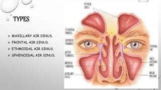
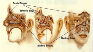
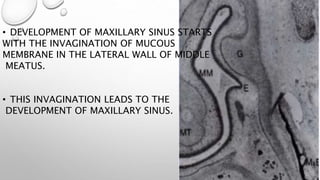
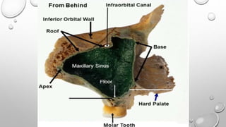
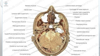
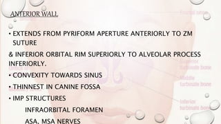
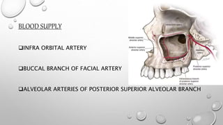
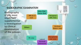
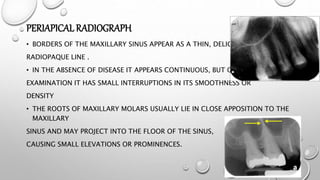
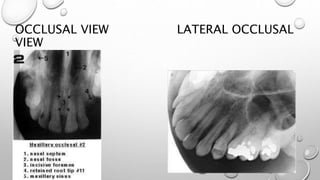
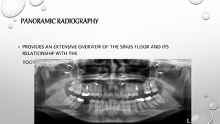
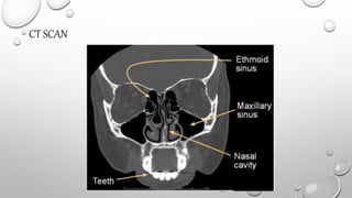
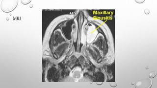
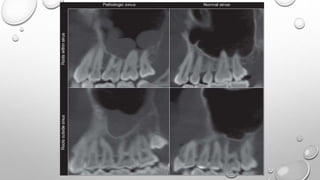
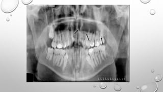
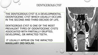
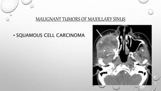
The document provides information about the maxillary sinus, including its development, anatomy, blood and nerve supply, functions, diagnostic evaluation, developmental anomalies, and pathologic conditions. It discusses the maxillary sinus's development beginning in the 4th month of gestation. Anatomically, it is the largest paranasal sinus and has superior, inferior, lateral, medial, and anterior walls. Common pathologies include maxillary sinusitis, odontogenic cysts impacting the sinus, and tumors. Radiographic imaging plays an important role in evaluating sinus disease.