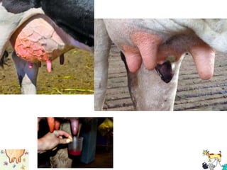
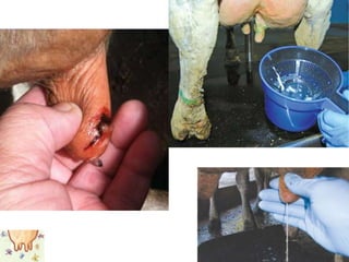
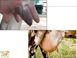
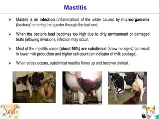
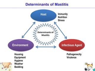
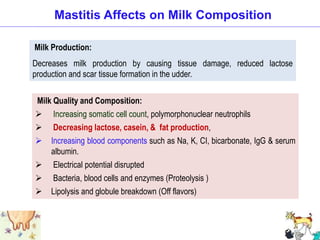
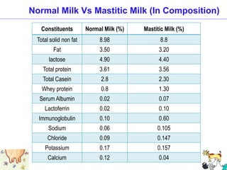
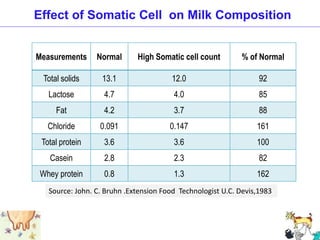
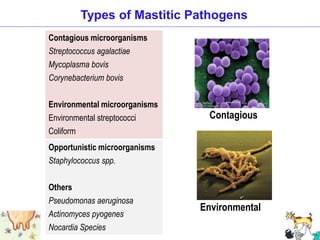
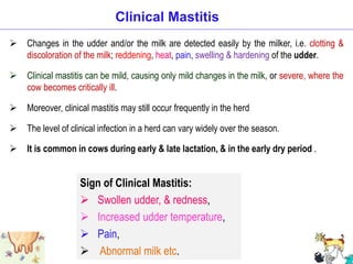
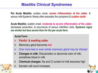
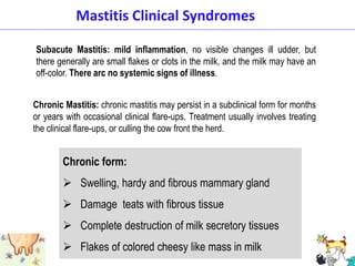
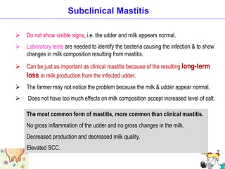
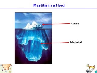
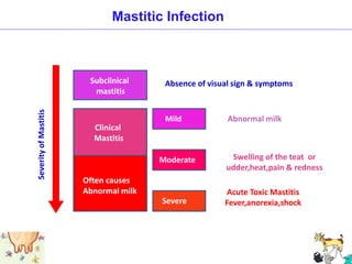
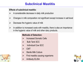
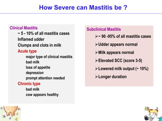
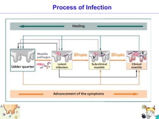
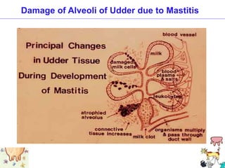
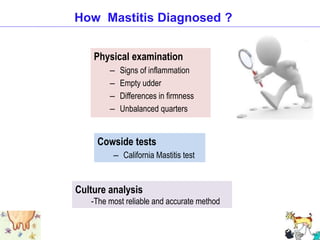
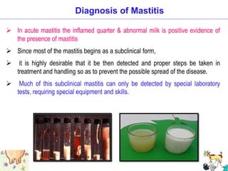
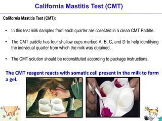
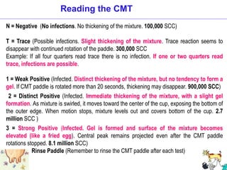
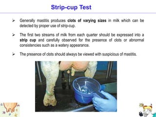
5. 5
Outlines
Introduction
Mastitis
Effects of Mastitis
Mastitic Pathogens
Form of Mastitis
Process of Mastitic Infection
Prevention of Mastitis
6. Introduction
Mastitis is Characterized by Physical, Chemical & usually Bacteriological
changes in milk and by Pathological changes in the Udder.
Mastitis is the inflammation of the mammary gland & udder
tissue, and is a major endemic disease of dairy cattle.
The most costly disease of dairy cattle.
Olanpaka, Olanfula, Thunko in BD
7. • The incidence is high in 3rd lactation in cattle & 4th lactation in buffalo
• Large pendulous udder with external injury to teats make animal
susceptible to mastitis
• Mastitis is more prevalent in animals that houses in unhygienic
condition & hot & humid environmental condition
• Prevalence of mastitis very high in winter & low in autumn in Bd
8. Mastitis
An inflammation of the milk secreting tissues of the udder, caused by microbial
infections in one or more quarters.
“Mastitis” derived from the Geek word
“Mastos” meaning “Breast” & “Itis” denotes “Inflammation”
Mastitis: Inflammation of the mammary gland regardless of causes. It
is characterized by the physical, chemical and bacteriological changes
in milk and pathological changes in the mammary gland tissues.
Affects 25 to 30 percent of all quarters
9. Mastitis is an infection (inflammation) of the udder caused by microorganisms
(bacteria) entering the quarter through the teat end.
When the bacteria load becomes too high due to dirty environment or damaged
teats (allowing invasion), infection may occur.
Most of the mastitis cases (about 95%) are subclinical (show no signs) but result
in lower milk production and higher cell count (an indicator of milk spoilage).
When stress occurs, subclinical mastitis flares up and become clinical.
Mastitis
10. Effects of Mastitis
Direct losses due to mastitis.
Treatment cost.
Discarded milk.
Fatality.
Repeated cases of mastitis.
Decrease in milk yield.
Milk quality changes.
Decreased hygienic quality of milk and public
health considerations.
11. Loss of the affected quarter when it becomes chronic
Loss of daily milk production by about 60-70%
Deterioration in milk quality: reduces use & sell of that milk
It is very fast spreading disease
It destroy the milk synthesizing cells & tissues and
recovery of that tissues are not possible
Treatment of mastitis is costly & production after
treatment is not satisfactory
Loss of the animal or replacements are expensive
Treatment cost of animal increases
Economic Losses Caused by Mastitis
12. Miscellaneous = 1%
Components of Total Economic Losses by Mastitis
Milk production loss (Reduced milk production ) = 60-70%
Milk discarded or downgraded = 7%
Cows lost by pre-mature culling = 14%
Treatment and veterinary expenses = 8%
14. Mastitis Affects on Milk Composition
Milk Production:
Decreases milk production by causing tissue damage, reduced lactose
production and scar tissue formation in the udder.
Milk Quality and Composition:
Increasing somatic cell count, polymorphonuclear neutrophils
Decreasing lactose, casein, & fat production,
Increasing blood components such as Na, K, CI, bicarbonate, IgG & serum
albumin.
Electrical potential disrupted
Bacteria, blood cells and enzymes (Proteolysis )
Lipolysis and globule breakdown (Off flavors)
15. Constituents Normal Milk (%) Mastitic Milk (%)
Total solid non fat 8.98 8.8
Fat 3.50 3.20
lactose 4.90 4.40
Total protein 3.61 3.56
Total Casein 2.8 2.30
Whey protein 0.8 1.30
Serum Albumin 0.02 0.07
Lactoferrin 0.02 0.10
Immunoglobulin 0.10 0.60
Sodium 0.06 0.105
Chloride 0.09 0.147
Potassium 0.17 0.157
Calcium 0.12 0.04
Normal Milk Vs Mastitic Milk (In Composition)
16. Measurements Normal High Somatic cell count % of Normal
Total solids 13.1 12.0 92
Lactose 4.7 4.0 85
Fat 4.2 3.7 88
Chloride 0.091 0.147 161
Total protein 3.6 3.6 100
Casein 2.8 2.3 82
Whey protein 0.8 1.3 162
Source: John. C. Bruhn .Extension Food Technologist U.C. Devis,1983
Effect of Somatic Cell on Milk Composition
17. What are the Health Concerns of Mastitis ?
• Animal health
– Loss of functional quarter
– Lowered milk production
– Death of cow
• Human health
– Poor quality milk
– antibiotic residues in milk
22. Contagious Mastitis
Primary habitat: bacteria live on/in the udder and teat lesions
Poor survival of bacteria in the environment
Spread from cow to cow, primarily during milking by milk contaminated
fomites at milking, sponge, milker's hands, milking machine
Staphylococcus aureus, Streptococcus agalactia, Mycoplasma bovis
& sometimes Streptococcus uberis are contagious mastitis causing
organisms.
Usually chronic, subclinical mastitis
23. Environment Mastitis
Environment to cow
Incidence increases as the incidence of contagious mastitis decreases
Primary habitat of bacterial is in the environment (feces, soil, bedding, water)
Environmental contact at milking time or between milking
Organisms from the bedding, stalls etc. gain entrance through fatigued teat
canals after or during milking to cause infection.
Streptococcus dysgalactia, Streptococcus uberus, & Coliform
(E. coli, Klebsiella) are a few the organisms that live in the environment
24. Forms of Mastitis
This disease is seen in various forms or degrees of severity, depending on
type of bacteria causing the infection,
degree of tissue damage and
amount of inflammation of the mammary gland.
Mastitis is usually classified according to its visibility to the dairy farmer, i.e. clinical
or subclinical mastitis.
Clinical mastitis can become subclinical if treatment is ineffective,
Subclinical mastitis can become clinical at any time.
The level at which a subclinical infection becomes clinical can depend on an
individual cow or how frequently and closely the milk or the cow is examined for
clinical signs.
25. Per acute: very severe and of very short duration, generally proving quickly
fatal
Acute: present or experienced to a severe or intense degree
Sub-acute: between acute and chronic
Chronic: persisting for a long time or constantly recurring
Subclinical: relating to or denoting a disease which is not severe enough to
present definite or readily observable symptoms.
Forms of Mastitis
26. Changes in the udder and/or the milk are detected easily by the milker, i.e. clotting &
discoloration of the milk; reddening, heat, pain, swelling & hardening of the udder.
Clinical mastitis can be mild, causing only mild changes in the milk, or severe, where the
cow becomes critically ill.
Moreover, clinical mastitis may still occur frequently in the herd
The level of clinical infection in a herd can vary widely over the season.
It is common in cows during early & late lactation, & in the early dry period .
Clinical Mastitis
Sign of Clinical Mastitis:
Swollen udder, & redness,
Increased udder temperature,
Pain,
Abnormal milk etc.
27. Mastitis Clinical Syndromes
Per Acute Mastitis: sudden onset, severe inflammation of the udder, &
serous milk-Systemic illness often precedes the symptoms & sudden death
Acute Mastitis: sudden onset, moderate to severe inflammation of the udder,
decreased production, & occurrence of serous milk/fibrin clots, Systemic signs
are similar but less severe than for the per acute form.
Acute Form:
Painful & swelling udder
Mammary gland becomes hot
One/ more teat or even whole mammary gland may be infected
Changes in milk: Discoloration or abnormal color of milk,
sometimes blood in milk
Chemical changes: Na and Cl content of milk becomes high
Somatic cell count increases
28. Mastitis Clinical Syndromes
Subacute Mastitis: mild inflammation, no visible changes ill udder, but
there generally are small flakes or clots in the milk, and the milk may have an
off-color. There arc no systemic signs of illness.
Chronic form:
Swelling, hardy and fibrous mammary gland
Damage teats with fibrous tissue
Complete destruction of milk secretory tissues
Flakes of colored cheesy like mass in milk
Chronic Mastitis: chronic mastitis may persist in a subclinical form for months
or years with occasional clinical flare-ups. Treatment usually involves treating
the clinical flare-ups, or culling the cow front the herd.
29. Do not show visible signs, i.e. the udder and milk appears normal.
Laboratory tests are needed to identify the bacteria causing the infection & to show
changes in milk composition resulting from mastitis.
Can be just as important as clinical mastitis because of the resulting long-term
loss in milk production from the infected udder.
The farmer may not notice the problem because the milk & udder appear normal.
Does not have too much effects on milk composition accept increased level of salt.
Subclinical Mastitis
The most common form of mastitis, more common than clinical mastitis.
No gross inflammation of the udder and no gross changes in the milk.
Decreased production and decreased milk quality.
Elevated SCC.
31. Absence of visual sign & symptoms
Abnormal milk
Swelling of the teat or
udder,heat,pain & redness
Acute Toxic Mastitis
Fever,anorexia,shock
Subclinical
mastitis
Clinical
Mastitis
Often causes
Abnormal milk
Mild
Moderate
Severe
Severity
of
Mastitis
Mastitic Infection
32. Effects of subclinical mastitis:
A considerable decrease in daily milk production
Changes in milk composition not significant except increase in salt level
Decrease the hygienic value of milk
In addition to increased costs with mastitis, there is also an importance
in the hygienic value of milk and other dairy products.
Subclinical Mastitis
Methods of Detection:
Increased Somatic Cells
Bulk Tank SCC
Individual Cow SCC
CMT
Sterile Milk Culture
Find mastitis causing organisms
Antibody ELISA
33. How Severe can Mastitis be ?
Subclinical Mastitis
~ 90 -95% of all mastitis cases
Udder appears normal
Milk appears normal
Elevated SCC (score 3-5)
Lowered milk output (~ 10%)
Longer duration
Clinical Mastitis
~ 5 - 10% of all mastitis cases
Inflamed udder
Clumps and clots in milk
Acute type
major type of clinical mastitis
bad milk
loss of appetite
depression
prompt attention needed
Chronic type
bad milk
cow appears healthy
34. Infected udder
Environment
bedding
soil
water
manure
Replacement animals
Where do These organisms come from ?
35. Infected udder
Unhygienic environment (bedding, soil, water, grazing
land, housing, manure etc.)
Introducing new cows without clinical examinations
Unhygienic milking conditions (dirty floor, unclean
milking pail, unclean hand of milk man, milking without
washing and drying of cow’s udder etc.)
Milking with un-experienced milkman
Hard and unhygienic floor of farm
Possible Reasons for Spreading Mastitis
36. Process of infection
Colonized organisms produce toxic substances
harmful to the milk producing cells
Migrate up the teat canal and colonize the
secretory cells
Organisms invade the udder through
teat canal
37. Teat canal of milking cows remain open up to 25 minutes of milking
Organisms invade the udder through the open teat canal immediately
after milking
Migration of organisms into the teat canal & colonization of milk
secretion cells
Colonized organisms produce toxic substances that affect milk
secretion cells
The immune system send somatic cells to fight against bacteria
Hot ,swollen, red & painful udder when defense system fails
Simultaneously cows affected by fever
Process of Infection
40. How Mastitis Diagnosed ?
Physical examination
– Signs of inflammation
– Empty udder
– Differences in firmness
– Unbalanced quarters
Cowside tests
– California Mastitis test
Culture analysis
-The most reliable and accurate method
41. Diagnosis of Mastitis
In acute mastitis the inflamed quarter & abnormal milk is positive evidence of
the presence of mastitis
Since most of the mastitis begins as a subclinical form,
it is highly desirable that it be then detected and proper steps be taken in
treatment and handling so as to prevent the possible spread of the disease.
Much of this subclinical mastitis can only be detected by special laboratory
tests, requiring special equipment and skills.
42. California Mastitis Test (CMT)
• In this test milk samples from each quarter are collected in a clean CMT Paddle.
• The CMT paddle has four shallow cups marked A, B, C, and D to help identifying
the individual quarter from which the milk was obtained.
• The CMT solution should be reconstituted according to package instructions.
The CMT reagent reacts with somatic cell present in the milk to form
a gel.
California Mastitis Test (CMT):
43. Step 1: Take about 1 teaspoon (2 cc) milk from each quarter. This is the amount of
milk that would be left in the cups if the CMT Paddle were held nearly vertical
Step 2: Add an equal amount of CMT solution to each cup in the paddle.
Step 3: Rotate the CMT Paddle in a circular motion to thoroughly mix the contents.
Do not mix more than 10 seconds.
Step 4: Read the test quickly. Visible reaction disintegrates after about 20 seconds. The
reaction is scored visually. The more gel formation, the higher the score
The CMT reaction must be scored within 15 to 20 seconds of mixing because weak
reactions will disappear after that time.
How to Use CMT
44. N = Negative (No infections. No thickening of the mixture. 100,000 SCC)
T = Trace (Possible infections. Slight thickening of the mixture. Trace reaction seems to
disappear with continued rotation of the paddle. 300,000 SCC
Example: If all four quarters read trace there is no infection. If one or two quarters read
trace, infections are possible.
1 = Weak Positive (Infected. Distinct thickening of the mixture, but no tendency to form a
gel. If CMT paddle is rotated more than 20 seconds, thickening may disappear. 900,000 SCC)
2 = Distinct Positive (Infected. Immediate thickening of the mixture, with a slight gel
formation. As mixture is swirled, it moves toward the center of the cup, exposing the bottom of
the outer edge. When motion stops, mixture levels out and covers bottom of the cup. 2.7
million SCC )
3 = Strong Positive (Infected. Gel is formed and surface of the mixture becomes
elevated (like a fried egg). Central peak remains projected even after the CMT paddle
rotations stopped. 8.1 million SCC)
Rinse Paddle (Remember to rinse the CMT paddle after each test)
Reading the CMT
45. Interpretation of CMT Scores
CMT Score Somatic Cell Range Interpretation
N (Negative) 0-200,000 Healthy Quarter
T (Trace) 200,000-400,000 Subclinical Mastitis
1 400,000-1,200,000 Subclinical Mastitis
2 1,200,000-5,000,000 Serious Mastitis Infection
3 Over 5,000,000 Serious Mastitis Infection
Any reaction of T (Trace) or Higher indicates that the quarter has subclinical mastitis.
CMT scores are directly related to average somatic cell counts. The following table
shows how they are related.
46. Generally mastitis produces clots of varying sizes in milk which can be
detected by proper use of strip-cup.
The first two streams of milk from each quarter should be expressed into a
strip cup and carefully observed for the presence of clots or abnormal
consistencies such as a watery appearance.
The presence of clots should always be viewed with suspicious of mastitis.
Strip-cup Test
47. Whiteside test is roughly correlated with the leukocyte content of the milk.
It is, however, simply conducted, is inexpensive, and of value in detecting early
mastitis when good judgment is used in the interpretation of the results.
In this test 5 drops of milk are placed upon a glass plate, one drop of 4%
NaOH solution is added by means of a medium dropper,
The mixture is stirred for about 20 seconds with a small tooth pick or stirring rod.
Normal milk will give a homogenous mixture following stirring.
Clumps of varying sizes will indicate subclinical mastitis.
Whiteside Test
48. Skill milkman are able to detect any changes in udder by simple palpation.
If the udder or a quarter of a udder become slightly congested or nodules are
felt inside the udder then it can be suspected as mastitis infection.
This field tests don’t give any indication about the type of organism that may be
involved.
More accurate laboratory tests together with skilled bacteriological examinations
are necessary for a complete diagnosis of mastitis.
Palpation Test
49. Reduction of the duration of the infection
Treating all the quarter of the cow during dry-off season
Treatment of the clinical cases
Culling of the chronic cases
Reduction of future causes of the infection
Maintaining of hygienic condition during milking period
Washing of cows udder with warm water
Disinfection of milk utensils and milking machine
Disinfection of the teat cup specially
Should be economically advantageous to the farm
Has to simple and technical skill and understanding
Basic Principles in Prevention Program
50. Manage the pre-disposing factors of mastitis properly
Don’t milk cows in unhygienic condition
Before milking, clean the udder well with clean water
and wipe dry with clean towel.
Should use separate cloth, towel for each animal
Disposable paper towel is also an option. Repeated
use of unclean towel may itself predispose to mastitis
Milking should be quick, complete & hygienic
Milk animals with chronic mastitis at the end
Prevention of Mastitis
51. Don’t mix milk of affected cows with others
Carry out teat dipping or spray immediately after milking
Prevent the animal from sitting for at least 30-45 minutes after milking.
Periodically check and treat for sub-clinical mastitis
Keep the floor of the cattle shed without holes and as dry as possible
Continue teat dipping/spray 2 weeks after drying off and start the
practice two weeks before calving
Carry out proper fly control
Manure pit should not be close to milking shed
Prevention of Mastitis
53. The general line of treatment is as follows:
Mild cases of clinical mastitis may not require treatment
Milk may be cultured for microbial sensitivity of antibiotics and treatment given
accordingly
Intramammary infusion containing ampicillin, amoxicillin, cloxacillin, penicillin,
tetracycline, erythromycin, tylosin, gentamycin, neomycin or nitrofurazones etc.
For better result, antibiotics by parental route may be used
Acute and per-acute mastitis may be treated with antimicrobials and always
require fluid and electrolytes with non-steroidal anti-inflammatory agents
Antihistaminics
Mastitis Treatment
54. Mastitis Treatment
IMM Therapy
-Injection of antibiotics into udder
Systemic Therapy
-Antibiotics IV or IM
Supportive Therapy
-Remove toxins - Frequent milkout
-Treat dehydration, swelling and pain
-Know bug
Lactational Therapy
-Likelihood of success?
Dry Cow Therapy
-Larger dose, longer acting product
55. Masticare (powder) 30gm twice daily/3-5 days
Mastikort, Mastijet, Mastinil, Mastilex (Infusion) – Infuse 1 syringe
after complete milking for 4 days
Diadin, Pronapen Vet, Amcox vet, Keto-A-Vet,- Injection for 3-5 days
56. Two vaccine named Startvac & Topvac has given in mastitis
First vaccine: at 45 days before the expected parturition date
Second vaccine: 35 days thereafter (corresponding to 10 days
before the expected parturition date)
Third vaccine:62 days after the second vaccine (equivalent to 52
days after post-parturition)
Vaccination for Mastitis
Administration route and dose:
Route: Intramuscular into the neck muscle (the injections should be
preferably administered on the alternative sides of the neck)
Dose: 2 ml per cattle (Both heifers and cows).
57. Dipal is a conventional iodine-based teat dip solution,
Offering a fast kill of bacteria, spores & other mastitis-causing microbes.
Dipal is available in ready-to-use solutions, concentrates & formulations with increased
emollient content, for even better teat skin conditioning.
Washing of udder with dipal solution generally done after milking
Take 50 ml of dipal solution & 250 ml of water in a dip cup & mix properly
Then use it after milking & it makes a thin layer around the whole udder that prevents
the entrance of microbes through teat canal up to 45 min.
Use of Dipal Solution
58. Through & complete milking which is followed by dry off teats after each
milking
The udder & perineal region of the cow should be washed properly with
warm water before milking
Milkers hand & cloths should be washed with soap & water before each
milking
Detection in infection and culling of the infected animal should be practiced
Young cow should be milked before than older one, then the newly introduced
one should be milked separately and suspected and known cases should be
milked at the last
For the reduction of infection. frequent testing of the fluid milk from the
udder teat of all cows should be practiced
Maintenance of Sanitary Condition during Milking
60. 1. Milking Hygiene
Prepare the udder to be milked. The appropriate use of pre-
dipping is very important
Single use of towels
Use of gloves
Dry teats before milking or inserting the milking machine
2. Good Milking System
Milking equipment should be adequate in size, functioning
properly, and regularly cleaned and maintained
3.Good use of Machinery
Apply and remove machine carefully
Remove machine when cow is milked out, shut off vacuum at
claw before removal
The 10 Steps to Mastitis Control
61. 4. Pre-dipping
Effective teat disinfection reduces somatic cell count (SCC).
5. Monitor Mastitis Trough SCC
Monitor your mastitis score of CMT regularly
6. Treat clinical Mastitis
Treat clinical cows with a qualified Veterinarian. Withhold treated
cows' milk from milk supply chain until recovery.
The 10 Steps to Mastitis Control
62. 7. Eliminate Chronic Cows from the Herd or Milk Them Last
Segregate chronic mastitis cows, milk them last, cull when necessary
Cows with chronic mastitis serve as reservoirs of organisms and could
infect susceptible cows
8. Dry Cow Therapy
Dry treat each quarter using partial insertion techniques with an approved
dry cow treatment at drying off.
Cure rate is twice high as that during lactation Lowers the risk of clinical
and subclinical mastitis during subsequent lactation
The 10 Steps to Mastitis Control
63. 9. Keep Environment Clean as Much as Possible
Keep cow’s clean, udders free from soil and manure.
Fence off wet, swampy areas.
Keep free stalls and stanchions bedded properly.
Keep calving areas clean, properly bedded (straw preferred)
10. Good Nutrition
Properly feed and care for cows.
It has been shown by different studies that there is an association between
mastitis and supplementation with vitamins and minerals
Mastitis is a management problem and can be controlled. Mastitis control
and quality milk production depend on keeping excellent hygiene
standards. Prevention programs work if followed correctly.
The 10 Steps to Mastitis Control
64. Summary
Mastitis is primarily a management problem
Mastitis can be controlled
Prevention programs work best when correctly followed
“Clean Environment & Proper Management
Protects the Animal from Mastitis”
“Better management practices will reduce the incidence of mastitis”