Boutonniere deformity- extensionof MCP and DIP joints and flexion of PIP joint.
occurs due to rupture of extensor
tendon.
Seen in Rheumatoid arthritis patients.
Also called as buttonhole deformity
Stages : 1-passively correctable deformity
Stage 2:fixed contracture
Stage 3; fixed contracture with fibrosis
Stage 4: stage 3+ PIP arthritis
3.
Jersey finger
• Oppositeof mallet finger
• Pt is unable to flex distal IP joint
• Jersey finger (rugby finger) is an
avulsion of the flexor digitorum
profundus tendon (FDP) from its
distal insertion on the distal
phalanx (zone I).
4.
Claw hand
• Appearslike claw of animal.
• Occurs due to injury to ulnar nerve.
• Involves lumbricals of hand.
• Hyperextension at MCP and flexion at IP
joint resembles claw hand.
• Seen in leprosy and RA
• Wasting of muscles can be seen.
• Can be treated conservative or surgical.
• Difficulty in grips and grasps.
5.
Swan neck deformity
•PIP joint hyper extension and DIP joint
flexion.
• Occurs due to damage to extensor
tendon and results in loss of ROM.
• Seen in RA.
• Treated by splint, surgical procedures.
• PT- waxbath, flexibility ex
• Fig of 8 ring splint/anti swan neck
orthosis
6.
Mallet finger
• Injuryto tendon that extends DIP joint.
• Occurs due to injury such as hit by ball.
• Also called as hammer finger.
• Can cause avulsion fracture.
• Treated by splint or surgical procedures.
• Might limit ROM.
• Also called as baseball finger or drop
finger
7.
Classification of malletfinger-doyle
classification
• Type 1- closed injury with or without avulsion fracture
• Type 2- open injury(only lacerations)
• Type 3- open injury( deep, involving skin and tendon)
• Type 4- mallet fracture
8.
Zig zag deformityof thumb
• Occurs due to loss of
stabilizing muscle and
rupture of ligaments.
• Seen in RA
9.
Ulnar drift
• Occursdue to MCP joint synovitis and loosening of collateral
ligaments
• Interosseous muscle contracture occurs
• Can be mild,moderate,severe
10.
Opera glass hand
•Shortening of fingers due to destruction of phalanges
• Excess skin gets folded
11.
Hammer toe
• Ahammer toe is a deformity of the second,
third, or fourth toes. In this condition, the
toe is bent at the middle joint, so that it
resembles a hammer. Initially, hammer toes
are flexible and can be corrected with
simple measures; however, if left untreated,
they can become fixed and require surgery.
12.
Hallux valgus orbunions
• A bony bump that forms on the joint at the base
of the big toe.
• A bunion is formed when the big toe pushes
against the next toe and becomes red and
painful. Tight shoes, foot stress, and arthritis are
the causes.
• The main symptoms are bone deformity, pain
and stiffness.
• Treatments include changing shoes, padding the
foot and pain medication. Painful bunions can
be removed surgically.
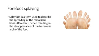
Forefoot splaying
• Splayfootis a term used to describe
the spreading of the metatarsal
bones (forefoot), hence resulting in
the disappearance of the transverse
arch of the foot.