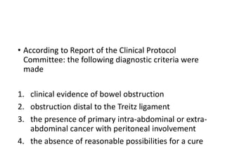
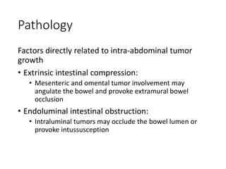
60 year old male with malignant
bowel obstruction.
Discuss the pathology and
management of malignant bowel
obstruction.
By
Dr Olofin. K. E
Senior registrar
General surgery unit
National hospital
Outline
• Introduction
• Definition
• Epidemiology
• Etiology
• Classification
• Pathology
• Management
• History
• Physical examination
• Investigation
• Treatment
• Conclusion
• References
Introduction
Definition
• Malignant bowel obstruction is defined as luminal
narrowing of small or large bowel with clinical
evidence of bowel obstruction in the setting of
metastatic intra-abdominal cancer.
• According to Report of the Clinical Protocol
Committee: the following diagnostic criteria were
made
1. clinical evidence of bowel obstruction
2. obstruction distal to the Treitz ligament
3. the presence of primary intra-abdominal or extra-
abdominal cancer with peritoneal involvement
4. the absence of reasonable possibilities for a cure
Introduction
Epidemiology
• The global prevalence rate is estimated to range from 3% to 15%
of cancer patients
• Primary cancers of abdominal origin that most frequently
produce MBO are those of the
• colon 25%–40%
• ovary 16%–29%
• stomach 6%–19%
• pancreas 6%–13%
• bladder 3%–10%
• endometrium 3%–11%
• Those of extra-abdominal origin due to peritoneal infiltration are
those of the
• breast 2%–3%
• melanoma 3%
• The mean age at presentation is 61 years (from 58–65 years)
Introduction
Aetiology
• Intra-abdominal
• Colorectal cancer
• Ovarian
• Extra-abdominal
• Breast cancer
• Melanoma
Introduction
Classification
A. Mechanical
• Extrinsic
• Intrinsic
• Adynamic (caused by tumour infiltrating bowel
wall, nerve and plexus)
B. Partial or complete
C. Proximal or distal
Pathology
Factors directly related to intra-abdominal tumor
growth
• Extrinsic intestinal compression:
• Mesenteric and omental tumor involvement may
angulate the bowel and provoke extramural bowel
occlusion
• Endoluminal intestinal obstruction:
• Intraluminal tumors may occlude the bowel lumen or
provoke intussusception
• Intramural intestinal infiltration:
• Intramural infiltration through the mucosa may obstruct
the lumen or impair peristaltic movements.
• Infiltration of the mesenteries and plexus:
• Infiltration of the enteric or celiac plexus may cause
severe impairment in peristalsis and consequent
obstruction due to dysmotility
• Factors not directly related to intra-abdominal
tumor growth
• Paraneoplastic neuropathy
• Opioid-induced intestinal dysfunction: Chronic
constipation
• Adynamic ileum Inflammatory intestinal disease
• Renal insufficiency/dehydratation
• Mesenteric thrombosis
• Postsurgical adherences
• Radiogenic fibrosis
Management
History
• nausea 100%
• vomiting 87%–100%
• colic pain 72%–80%
• pain due to distension 56%–90%
• Absolute constipation in the previous 72 hours in
85%–93%
• Overflow diarrhea (bacteria overgrowth)
Management
Physical examination
Anthropometry, vitals, Pallor, Jaundice, hydration
status
Abdomen:
Size, tenderness, masses, ascites organomegaly,
bowel sounds
DRE: masses
Investigations
• Plain abdominal x-ray;
• Dilated loop of bowel
• Multiple air fluid level
• Gasless pelvis
• Ground glass appearance
• In some cases, Multiple air/fluid level may be unremarkable
because tumour encasement of the bowel wall may prevent
the classical sign of bowel dilatation seen in non-malignant
bowel obstruction.
• Small bowel contrast
• Using either barium or gastrograffin opinions are divided on
this. But a failure of contrast to reach the caecum in 24 hours
suggests high grade or complete obstruction.
• Barium enema.
• If this shows obstruction in addition with small bowel
blockage this suggests multiple levels of obstruction
consistent with carcinomatosis
• CT-Scan:
• This is essential in all cases of MBO if surgical treatment
is being considered.
• It is now the gold standard in diagnosing malignant
bowel obstruction
• Sensitivity of CT-Scan in the diagnosis of malignant
bowel obstruction = (78-100%)
• Specificity = (> 90%)
• These will show the sites of obstruction, possible bowel
strangulation or ischaemia.
• MRI
• FBC
• E/U
• LFT
• Clotting profile
•
Management
Treatment
• The decision making process in advanced oncologic
patients requires individualized evaluation based
on
• the extension of the neoplasm
• the global prognosis
• the possibility of specific cancer treatments
• associated comorbidities
• the general status
• the particular options available to the duly informed
patient
Management
Treatment
• Realize this is end of life management, hence
treatment is palliative to improve the quality of life.
No cure is expected
• proper counseling of patients and relatives.
Increase in length of survival is bonus.
• About 15% of patients are terminally ill
Management
• Primary goal of treatment
• Alleviate nausea, vomiting and pain
• Make patient able to eat
• Return patient home or a nursing facility
• Possible treatments include
• Surgery
• endoscopic palliation
• digestive aspiration
• symptomatic palliative pharmacologic therapy
Surgical treatment
• Operative mortality = (5-32%)
• Operative morbidity = (42%)
• Re-obstruction = (10-50%)
• Therefore proper consideration must be given
before performing surgery.
• No rush to surgery.
• LESS LIKELY TO BENEFIT FROM SURGERY.
• Those with
• Ascites
• Carcinomatosis
• Abdominal mass that is palpable,
• Multiple obstruction,
• Very advanced carcinoma
• Those with very poor clinical status.
The Krebs and Goplerud prognostic index
• Palliation is regarded as successful if survival is at least 2
MONTHS
• This depends also on age, nutritional status, tumour
status, ascites, previous chemotherapy, and radiation
treatment.
Surgical options
• The quickest and the safest is preferred
• RESECTION with or without anastomosis
• INTESTINAL BY-PASS especially for radiation-
induced obstruction
• INTESTINAL STOMA, enterostomy, entero-
colostomy, entero- gastrostomy
• GASTROSTOMY is essentially for drainage to relieve
nausea and vomiting which are really very
troublesome symptoms.
Endoscopic treatment
• Usually for a single site obstruction
• Patients NOT fit for operation
• Extensive disease
• Patients refusing operation
Endoluminal wall stents
• Successful in 64 -100% in rectal carcinoma either
complete or partial.
• In 70% of cases of upper intestinal obstruction,
gastric outlet obstruction, duodenal and jejunal
obstructions.
• Expertise and necessary equipment are needed for
this procedure
• The procedure
• Canalize bowel using laser or balloon dilatation.
• insert a guide wire under fluoroscopy (seldinger's technique
to canalize the bowel. The neodymium-doped yttrium
aluminium garnet (nd:yag) laser can be used at the time of
stenting for initial canalization of bowel for tow rectal
carcinoma
• not ideal for long term palliation
• Laser therapy requires repeated treatments to maintain
luminal patency.
• But balloon dilatation can be a short term measure at the
time of stenting or use of nd:yag laser. If stenting is possible
it is probably the optimal endoscopic technique.
• SEMS show success of about 90%.
• Show to maintain patency longer.
• Complications;
• Perforation
• Stent migration
• Stent obstruction
• PERCUTANEOUS ENDOSCOPIC GASTROSTOMY
(PEG)
• Usually well tolerated
• Alleviate nausea and vomiting
• Allows intermittent oral intake.
• Patients with ascites are poor candidates for
Percutaneous Endoscopic Gastrostomy. (PEG)
Non-operative treatment
• Naso-gastric tube drainage
• It is very discomforting.
• Used only on a time-limited basis for decompression.
• IV fluid; rehydrate.
• Nutrition; parenteral
• Pharmacological; the goals are:
• Alleviate pain
• Check nausea
• Check vomiting
• Intestinal inflammation and oedema
Pharmacological
• Octreotide
• One of the most effective drugs for the relief of symptoms of
MBO.
• It is a synthetic analog of somatostatin.
• It reduces G. I. Secretions, increases small bowel transit time,
delays onset of oedema and ischaemia in anti-mesenteric
border of intestines.
• Effect can be dramatic. Within a few hours! Response is 75-
100%
• Dose: 0.3-0.6 mg/day subcutaneous route.
• Response is control of nausea and vomiting.
• Duration of treatment (median 9.4-17.5 days).
• Relief period is for life of the patient
• Opioids
• Morphine and hydromorphine
• Alleviate pain, produces adynamic ileus
• Methadone very effective when used with
metoclopramide
• Metoclopramide
• some feel it is contraindicated in bowel obstruction because it
promotes gastric motility, but it is efficacious in partial bowel
obstruction.
• Antiemetics
• oral medications should be avoided because of
vomiting.
• Prochloperazine given rectally
• Promethazine given rectally
• Hydroxyzine given rectally
• Ondansetron given subcutaneously
• Methotrimprazine given intramuscularly
• Haloperidol given subcutaneously.
• Haloperidol is believed to be the drug of choice, for it controls
nausea, vomiting and agitated delirium.
• With anti-emetics, complete relief of emesis is achieved
in only 30% of patients.
• Anticholinergics
• They decrease peristalsis, secretions, vomiting and
intestinal colic
• Scopolamine:
• might be more cost effective than octreotide.
• It is given subcutaneously or as a transdermal patch
Corticosteroids
• This reduces peri-tumoral oedema,
• Activate central and peripheral anti-emetic effect
• It is co-analgesic in intestinal obstruction related pain.
Dexamethasone dose is 2-60mg per day.
• Usually prescribed for terminal patients.
Intra-peritoneum chemotherapy
• Can be used for recurrent Intra-abdominal carcinoma
Management
• Radiotherapy
• This is to produce local palliation to pelvis, duodenal
area, and to intestinal stoma blockages by tumor.
• Combination with 5-FU is beneficial
• Generally, complication of radiation will not occur before
patient dies. This is END OF LIFE (EOL)
management/palliation.
Conclusion
• MBO is a common and difficult problem.
• Objectives are to relief pain, nausea, vomiting,
early removal of N/G tube, keep patient out of the
hospital as much as possible and to restore ability
to eat.
• Non-surgical interventions should be considered in
all patients.
• The decision to pursue surgical vs non-surgical
treatment hinge on variety of factors; general
patient condition and the extent of the malignancy.
References
• Albert Tuca, Ernest Guell, Emilio Martinez-Losada,
Nuria Codorniu. Malignant bowel obstruction in
advanced cancer patients: epidemiology, management,
and factors influencing spontaneous resolution: Cancer
Management and Research 2012:4 159–169
• Prof. O. G. Ajao, Dept of Surgery, U. C. H. Ibadan,
Nigeria. Update material. Wacs.
• Sarah FH et ael. Malignant bowel obstruction. Expert
analysis. 2015
• Eric R, Charles F V. Current concept in malignant bowel
obstruction management. Current Oncology. 2009;
11(4):293-303.
•Thank you

MALIGNANT BOWEL-WPS Office.pptx

  • 1.
    60 year oldmale with malignant bowel obstruction. Discuss the pathology and management of malignant bowel obstruction. By Dr Olofin. K. E Senior registrar General surgery unit National hospital
  • 2.
    Outline • Introduction • Definition •Epidemiology • Etiology • Classification • Pathology • Management • History • Physical examination • Investigation • Treatment • Conclusion • References
  • 3.
    Introduction Definition • Malignant bowelobstruction is defined as luminal narrowing of small or large bowel with clinical evidence of bowel obstruction in the setting of metastatic intra-abdominal cancer.
  • 4.
    • According toReport of the Clinical Protocol Committee: the following diagnostic criteria were made 1. clinical evidence of bowel obstruction 2. obstruction distal to the Treitz ligament 3. the presence of primary intra-abdominal or extra- abdominal cancer with peritoneal involvement 4. the absence of reasonable possibilities for a cure
  • 5.
    Introduction Epidemiology • The globalprevalence rate is estimated to range from 3% to 15% of cancer patients • Primary cancers of abdominal origin that most frequently produce MBO are those of the • colon 25%–40% • ovary 16%–29% • stomach 6%–19% • pancreas 6%–13% • bladder 3%–10% • endometrium 3%–11% • Those of extra-abdominal origin due to peritoneal infiltration are those of the • breast 2%–3% • melanoma 3% • The mean age at presentation is 61 years (from 58–65 years)
  • 6.
    Introduction Aetiology • Intra-abdominal • Colorectalcancer • Ovarian • Extra-abdominal • Breast cancer • Melanoma
  • 7.
    Introduction Classification A. Mechanical • Extrinsic •Intrinsic • Adynamic (caused by tumour infiltrating bowel wall, nerve and plexus) B. Partial or complete C. Proximal or distal
  • 8.
    Pathology Factors directly relatedto intra-abdominal tumor growth • Extrinsic intestinal compression: • Mesenteric and omental tumor involvement may angulate the bowel and provoke extramural bowel occlusion • Endoluminal intestinal obstruction: • Intraluminal tumors may occlude the bowel lumen or provoke intussusception
  • 9.
    • Intramural intestinalinfiltration: • Intramural infiltration through the mucosa may obstruct the lumen or impair peristaltic movements. • Infiltration of the mesenteries and plexus: • Infiltration of the enteric or celiac plexus may cause severe impairment in peristalsis and consequent obstruction due to dysmotility
  • 10.
    • Factors notdirectly related to intra-abdominal tumor growth • Paraneoplastic neuropathy • Opioid-induced intestinal dysfunction: Chronic constipation • Adynamic ileum Inflammatory intestinal disease • Renal insufficiency/dehydratation • Mesenteric thrombosis • Postsurgical adherences • Radiogenic fibrosis
  • 11.
    Management History • nausea 100% •vomiting 87%–100% • colic pain 72%–80% • pain due to distension 56%–90% • Absolute constipation in the previous 72 hours in 85%–93% • Overflow diarrhea (bacteria overgrowth)
  • 12.
    Management Physical examination Anthropometry, vitals,Pallor, Jaundice, hydration status Abdomen: Size, tenderness, masses, ascites organomegaly, bowel sounds DRE: masses
  • 13.
    Investigations • Plain abdominalx-ray; • Dilated loop of bowel • Multiple air fluid level • Gasless pelvis • Ground glass appearance • In some cases, Multiple air/fluid level may be unremarkable because tumour encasement of the bowel wall may prevent the classical sign of bowel dilatation seen in non-malignant bowel obstruction. • Small bowel contrast • Using either barium or gastrograffin opinions are divided on this. But a failure of contrast to reach the caecum in 24 hours suggests high grade or complete obstruction.
  • 14.
    • Barium enema. •If this shows obstruction in addition with small bowel blockage this suggests multiple levels of obstruction consistent with carcinomatosis • CT-Scan: • This is essential in all cases of MBO if surgical treatment is being considered. • It is now the gold standard in diagnosing malignant bowel obstruction
  • 15.
    • Sensitivity ofCT-Scan in the diagnosis of malignant bowel obstruction = (78-100%) • Specificity = (> 90%) • These will show the sites of obstruction, possible bowel strangulation or ischaemia. • MRI
  • 16.
    • FBC • E/U •LFT • Clotting profile •
  • 17.
    Management Treatment • The decisionmaking process in advanced oncologic patients requires individualized evaluation based on • the extension of the neoplasm • the global prognosis • the possibility of specific cancer treatments • associated comorbidities • the general status • the particular options available to the duly informed patient
  • 18.
    Management Treatment • Realize thisis end of life management, hence treatment is palliative to improve the quality of life. No cure is expected • proper counseling of patients and relatives. Increase in length of survival is bonus. • About 15% of patients are terminally ill
  • 19.
    Management • Primary goalof treatment • Alleviate nausea, vomiting and pain • Make patient able to eat • Return patient home or a nursing facility
  • 20.
    • Possible treatmentsinclude • Surgery • endoscopic palliation • digestive aspiration • symptomatic palliative pharmacologic therapy
  • 21.
    Surgical treatment • Operativemortality = (5-32%) • Operative morbidity = (42%) • Re-obstruction = (10-50%) • Therefore proper consideration must be given before performing surgery. • No rush to surgery.
  • 22.
    • LESS LIKELYTO BENEFIT FROM SURGERY. • Those with • Ascites • Carcinomatosis • Abdominal mass that is palpable, • Multiple obstruction, • Very advanced carcinoma • Those with very poor clinical status.
  • 23.
    The Krebs andGoplerud prognostic index • Palliation is regarded as successful if survival is at least 2 MONTHS • This depends also on age, nutritional status, tumour status, ascites, previous chemotherapy, and radiation treatment.
  • 24.
    Surgical options • Thequickest and the safest is preferred • RESECTION with or without anastomosis • INTESTINAL BY-PASS especially for radiation- induced obstruction • INTESTINAL STOMA, enterostomy, entero- colostomy, entero- gastrostomy • GASTROSTOMY is essentially for drainage to relieve nausea and vomiting which are really very troublesome symptoms.
  • 25.
    Endoscopic treatment • Usuallyfor a single site obstruction • Patients NOT fit for operation • Extensive disease • Patients refusing operation
  • 26.
    Endoluminal wall stents •Successful in 64 -100% in rectal carcinoma either complete or partial. • In 70% of cases of upper intestinal obstruction, gastric outlet obstruction, duodenal and jejunal obstructions. • Expertise and necessary equipment are needed for this procedure
  • 27.
    • The procedure •Canalize bowel using laser or balloon dilatation. • insert a guide wire under fluoroscopy (seldinger's technique to canalize the bowel. The neodymium-doped yttrium aluminium garnet (nd:yag) laser can be used at the time of stenting for initial canalization of bowel for tow rectal carcinoma • not ideal for long term palliation • Laser therapy requires repeated treatments to maintain luminal patency. • But balloon dilatation can be a short term measure at the time of stenting or use of nd:yag laser. If stenting is possible it is probably the optimal endoscopic technique.
  • 28.
    • SEMS showsuccess of about 90%. • Show to maintain patency longer. • Complications; • Perforation • Stent migration • Stent obstruction
  • 29.
    • PERCUTANEOUS ENDOSCOPICGASTROSTOMY (PEG) • Usually well tolerated • Alleviate nausea and vomiting • Allows intermittent oral intake. • Patients with ascites are poor candidates for Percutaneous Endoscopic Gastrostomy. (PEG)
  • 30.
    Non-operative treatment • Naso-gastrictube drainage • It is very discomforting. • Used only on a time-limited basis for decompression. • IV fluid; rehydrate. • Nutrition; parenteral • Pharmacological; the goals are: • Alleviate pain • Check nausea • Check vomiting • Intestinal inflammation and oedema
  • 31.
    Pharmacological • Octreotide • Oneof the most effective drugs for the relief of symptoms of MBO. • It is a synthetic analog of somatostatin. • It reduces G. I. Secretions, increases small bowel transit time, delays onset of oedema and ischaemia in anti-mesenteric border of intestines. • Effect can be dramatic. Within a few hours! Response is 75- 100% • Dose: 0.3-0.6 mg/day subcutaneous route. • Response is control of nausea and vomiting. • Duration of treatment (median 9.4-17.5 days). • Relief period is for life of the patient
  • 32.
    • Opioids • Morphineand hydromorphine • Alleviate pain, produces adynamic ileus • Methadone very effective when used with metoclopramide • Metoclopramide • some feel it is contraindicated in bowel obstruction because it promotes gastric motility, but it is efficacious in partial bowel obstruction.
  • 33.
    • Antiemetics • oralmedications should be avoided because of vomiting. • Prochloperazine given rectally • Promethazine given rectally • Hydroxyzine given rectally • Ondansetron given subcutaneously • Methotrimprazine given intramuscularly • Haloperidol given subcutaneously. • Haloperidol is believed to be the drug of choice, for it controls nausea, vomiting and agitated delirium. • With anti-emetics, complete relief of emesis is achieved in only 30% of patients.
  • 34.
    • Anticholinergics • Theydecrease peristalsis, secretions, vomiting and intestinal colic • Scopolamine: • might be more cost effective than octreotide. • It is given subcutaneously or as a transdermal patch
  • 35.
    Corticosteroids • This reducesperi-tumoral oedema, • Activate central and peripheral anti-emetic effect • It is co-analgesic in intestinal obstruction related pain. Dexamethasone dose is 2-60mg per day. • Usually prescribed for terminal patients.
  • 36.
    Intra-peritoneum chemotherapy • Canbe used for recurrent Intra-abdominal carcinoma
  • 37.
    Management • Radiotherapy • Thisis to produce local palliation to pelvis, duodenal area, and to intestinal stoma blockages by tumor. • Combination with 5-FU is beneficial • Generally, complication of radiation will not occur before patient dies. This is END OF LIFE (EOL) management/palliation.
  • 38.
    Conclusion • MBO isa common and difficult problem. • Objectives are to relief pain, nausea, vomiting, early removal of N/G tube, keep patient out of the hospital as much as possible and to restore ability to eat. • Non-surgical interventions should be considered in all patients. • The decision to pursue surgical vs non-surgical treatment hinge on variety of factors; general patient condition and the extent of the malignancy.
  • 39.
    References • Albert Tuca,Ernest Guell, Emilio Martinez-Losada, Nuria Codorniu. Malignant bowel obstruction in advanced cancer patients: epidemiology, management, and factors influencing spontaneous resolution: Cancer Management and Research 2012:4 159–169 • Prof. O. G. Ajao, Dept of Surgery, U. C. H. Ibadan, Nigeria. Update material. Wacs. • Sarah FH et ael. Malignant bowel obstruction. Expert analysis. 2015 • Eric R, Charles F V. Current concept in malignant bowel obstruction management. Current Oncology. 2009; 11(4):293-303.
  • 40.

Editor's Notes

  • #11 Paraneoplastic syndromes are rare disorders that are triggered by an altered immune system response to a neoplasm