
Recommended
More Related Content
What's hot
What's hot (20)
Similar to RADIOLOGY EMERGENCY RESPONSE
Similar to RADIOLOGY EMERGENCY RESPONSE (20)
More from Anupam Niraula
Recently uploaded
Recently uploaded (20)
RADIOLOGY EMERGENCY RESPONSE
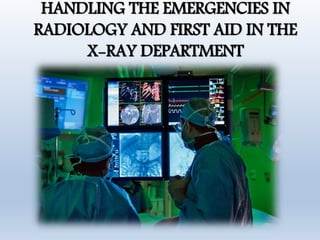
- 1. HANDLING THE EMERGENCIES IN RADIOLOGY AND FIRST AID IN THE X-RAY DEPARTMENT
- 2. Introduction • Emergency means serious, unexpected event that demands immediate attention. • Serious problem need active response to the doctors • We must be prepared to minimize the possibility of further injury or complication.
- 3. Emergency department • Also known as an Accident and emergency department or causality department. • The emergency departments (EDs) of most hospitals serve a variety of clients. • It is medical treatment facility specialized in emergency medicine. • Acute care of patient without prior appointment.
- 4. Trauma units • Trauma units are designed to cope with life- threatening injuries. • Trauma units are usually staffed with one or more trauma physicians who receive highly specialized training in the diagnosis and treatment of traumatic injuries. • There are three designated levels of trauma facilities: Level 1 Level 2 Level 3
- 5. Emergency code response • When working alone, or when qualified assistance is not immediately available. • You can obtain help by using the emergency call system. • Blue code – medical emergency (24444) • Red code – fire (22589)
- 6. Emergency response Team • Hospitals have a designated group of health care workers who respond to this type of code. • Usually consists of one or more physicians, several nurses, a respiratory therapist, and an electro-cardiographer.
- 7. Patient assessment • Assess patients and observe changes in their clinical signs and conditions is very important • Patients with a history of chronic cardiac or pulmonary disease are at greater risk. • Before any patient is injected with a contrast medium or subjected to an invasive procedure, a thorough history of previous cardiac events, allergies, chronic diseases, and medications should be taken. • Baseline vital signs must also be taken and recorded.
- 8. Cont... • Patients in the ED are classified as non-urgent, urgent or acute (life-threatening). • The most acute cases are seen first.
- 9. Management of Severely Injured Patients • The acute trauma setting is not the place for disagreement about the patient. Immediate management decisions must be made by the head physician. • The trauma team leader/head physician is an overall charge in acute care. • Trauma team leader must be an experienced consultant, there must be a consultant in Radiology in charge of trauma.
- 10. Location and Facilities • Triaging (decide the order of treatment)of patients is very important. • Imaging technique of choice is the one which is definitive in trauma setting, most often head to thigh CE-MDCT. • The MDCT should be adjacent to emergency room. • Radiography must also be present near the emergency room. • The imaging environment requires all the life support facilities available in the emergency room. This will include monitoring and gases.
- 11. Radiography • CXR-Chest radiograph must be obtained to document the position of tubes and to evaluate for pneumothorax or hemothorax and mediastinal abnormalities • AXR or pelvic X Ray are usually irrelevant if patient is going for CT. • Cervical spinal injury precautions and pelvic binders should remain in place until the MDCT has been fully assessed
- 12. C6 #
- 13. Focused Abdominal Sonography in Trauma (FAST) • FAST is used to demonstrate - intra-abdominal hemorrhage - Solid organ injuries- spleen, liver, kidney - Pericardial effusion
- 14. Focused Abdominal Sonography in Trauma (FAST)
- 15. Focused Abdominal Sonography in Trauma (FAST)
- 16. MDCT • Clear of the need for protocols must exist for notifying the CT department urgent imaging and how the department will respond to ensure that the scanner is clear to receive the incoming injured patient. • IV assess right ante-cubital assess is preferred for contrast administration. • Radiation dose should be considered.
- 17. Poly-trauma protocol MDCT is indicated when: • There is hemodynamic instability • The mechanism of injury suggests that there may be severe injuries that cannot be excluded by clinical examination or plain films.
- 18. Interventional Radiology(IR) • The role of IR in the emergency dept. is to stop hemorrhage as quickly as possible • The decision on whether a patient with traumatic hemorrhage undergoes endovascular treatment, open surgery, a combination of the two is typically a decision made by both the trauma team leader and interventional radiologist after consultation. • Interventional treatment modalities include Balloon occlusion, or transarterial embolization to stop hemorrhage.
- 20. MRI • MRI is not indicated in the setting of acute trauma care. However availability of clear protocols for the transfer of emergency to MRI facilities after stabilizing the patient is recommended.
- 21. Upcoming Slide contains sensitive image
- 22. No Imaging ! • There may be circumstances where imaging is inappropriate; for example, where a emergency patient is admitted with profound shock, is not responding to intravenous fluids and the site of bleeding is clear from the mechanism of injury. • Such patients may be best taken straight to theatre.
- 23. Contrast Media Reactions : Management and Preventions • The cause of reactions to iodinated contrast agents has been studied at length but is still unknown. • An appropriate history is helpful. • Who had no adverse reaction to an iodine contrast agent at one time might experience a reaction on a subsequent occasion. • Contrast medium sensitivity test is appropriate to minimize this reaction
- 24. Categories of reactions Mild: nausea, vomiting, cough, sneezing, warmth, headache, itching, chills, sweats, rash, anxiety. Moderate: tachycardia / bradycardia, hypertension / hypotension, swelling – eyes, face. Severe: convulsions, hypotension, arrhythmias, unresponsiveness, cardiopulmonary arrest.
- 25. Nausea and vomiting • Breathing suggestions. • "Breathe through your mouth, taking short, rapid, panting breaths," or "Take some long, slow, deep breaths through your nose," are both effective instructions. • On the other hand: if a patient expresses a need for a basin. Offer it immediately. Provide tissues and water to rinse the mouth. support the patient in a sitting or lateral recumbent position to avoid aspiration of vomitus. The lateral recumbent position is safest for the patient with nausea who is unable to sit up.
- 26. Urticaria • TRANSIENT(lasting for short time) • Supportive Treatment Including Observation • PROTRACTED(lasting for longer time) • Diphenhydramine (AVIL-50 mg) Intramuscular Or Intravenous • PROFOUND(intense emergency) • H1 and H2 Anti-histamine (CIMETIDINE-300 mg IN 200ml) • Adrenaline 1:1000- 0.1-0.3 ml In Adults (Max- 3mg) • 0.1 mg/Kg Up to 0.3 Max Children
- 27. Respiratory reaction • LARYNGEAL EDEMA(excess ofwatery fluid collection in the cavitiesor tissues in the body) – Oxygen- 6- 10 l/min – EPINEPHRINE-Intramuscular/nebulization (1:1000) 0.5ml(0.5mg)-adult dose – Intubation • BRONCHOSPASM(Narrowing of bronchi) • PULMONARY EDEMA: – Elevate Head End Of Bed – O2- 10 L/Min – Furosemide/Laxis- 40 Mg Iv Slowly – Morphine – 1-3mg – Shift To Icu
- 28. Hypotension WITH BRADYCARDIA • Elevate The Feet • O2-- 6-10 l/min • Normal Saline Rapidly To Be Given • Atropine 0.6-1.0 mg Iv Repeat After 3-5min Max 3mg Total • Pediatric 0.02mg ( Max 0.6mg) WITH TACHYCARDIA • Elevate The Feet • O2-- 6-10 l/min • Normal Saline Rapidly To Be Given • Adrenaline 0.5 ml
- 29. Convulsion • Mild: – Turn patient around to avoid aspiration – Airway Should Be Clean And Open – O2- 10l/Min • Severe: – Diazepam- 5mg Iv Slowly (anti- convulsion) • Hypertensive Crisis: – O2 10 l/Min – Nitroglycerine- 0.4 mg Tab Sublingually – If No Response: – Nifedipine- 10mg Capsule Sublingually – Monitor Bp Closely
- 30. Extravasation policy Initial • Elevate extremity • Icepack - 15 -60 min t.d.s x 3 days Observe for 2-4 hrs if volume > 5ml Call referring physician Surgical consultation if : • Increasing pain after 2-4 Hrs • Change in sensation distal to site of Extravasation • Volume – Ionic : >30ml Nonionic : > 100ml
- 31. Note • Severe reactions may begin as mild / moderate reactions • Ensure resolution prior to discharge (E.g.) in vagal reactions, bradycardia, and hypotension should resolve. • Otherwise, call code or transfer to emergency department
- 33. Reference • Ehrlich - Patient Care in Radiography - With an Introduction to Medical Imaging, 7th ed. • https://radiology.ucsf.edu/patient-care/patient- safety/contrast/iodinated/contrast-extravasation.
Editor's Notes
- Hemodynamic instability is a term used to indicate abnormal or unstable blood pressure and can suggest inadequate arterial blood flow
- https://radiology.ucsf.edu/patient-care/patient-safety/contrast/iodinated/contrast-extravasation