Mediasternum.pptx
•Download as PPTX, PDF•
1 like•596 views

Definition: A median septum or median partition between the two pleural cavities. It includes all the structures which lie in the intermediate compartments of the thoracic cavity It is a partition between the right & left pleural sacs. It includes all the structures which lie in the intermediate compartments of the thoracic cavity (all thoracic viscera EXCEPT lungs) Superior boundary: Superior thoracic aperture Inferior boundary: Diaphragm Anterior boundary: Sternum Posterior boundary: Bodies of vertebrae T1 to T12 Lateral boundaries: Mediastinal parietal pleura (left and right).

Recommended
More Related Content
What's hot
What's hot (20)
Similar to Mediasternum.pptx
Similar to Mediasternum.pptx (20)
More from Dr Ndayisaba Corneille
More from Dr Ndayisaba Corneille (20)
Recently uploaded
Recently uploaded (20)
Mediasternum.pptx
- 1. Dr. NDAYISABA CORNEILLE CEO of CHG MBChB,DCM,BCSIT,CCNA Supported BY
- 3. Mediastinum Definition: A median septum or median partition between the two pleural cavities. It includes all the structures which lie in the intermediate compartments of the thoracic cavity Dr Ndayisaba Corneille
- 4. DEFINITION OF MEDIASTINUM • It is a partition between the right & left pleural sacs. It includes all the structures which lie in the intermediate compartments of the thoracic cavity (all thoracic viscera EXCEPT lungs) Dr Ndayisaba Corneille
- 5. Superior boundary: Superior thoracic aperture Inferior boundary: Diaphragm Anterior boundary: Sternum Posterior boundary: Bodies of vertebrae T1 to T12 Lateral boundaries: Mediastinal parietal pleura (left and right). Boundaries of the Mediastinum Dr Ndayisaba Corneille
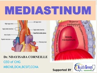
- 6. DIVISIONS OF MEDIASTINUM • It is divided by a horizontal plane extending from sternal angle to lower border of 4th thoracic vertebra into: 1. Superior mediastinum: above the plane 2. Inferior mediastinum: Dr Ndayisaba Corneille
- 7. DIVISIONS OF MEDIASTINUM Dr Ndayisaba Corneille
- 9. Divisions of Mediastinum 1. Superior mediastinum: Above the imaginary plane between the sternal angle and the lower border of the 4th thoracic vertebra. Dr Ndayisaba Corneille
- 10. Divisions of Mediastinum 2. Inferior mediastinum: Below the imaginary plane and it is further subdivided into: a. Anterior mediastinum: Behind the body and xiphoid process of the sternum and in front of the middle mediastinum (pericardium). b. Middle mediastinum: Contains pericardium, heart and the roots of the great vessels. c. Posterior mediastinum: lies behind the middle mediastinum (the part that lies posterior to the pericardium and anterior to the bodies of vertebrae T5 to T12). Dr Ndayisaba Corneille
- 12. Divisions of Mediastinum Dr Ndayisaba Corneille
- 13. SUPERIOR MEDIASTINUM BOUNDARIES: • Anterior: manubrium sterni • Posterior: Upper 4 thoracic vertebrae • Superior: Plane of thoracic inlet at the of T1 • Inferior: Horizontal plane • On each side: Pleura of level the lung Dr Ndayisaba Corneille
- 14. SUPERIOR MEDIASTINUM CONTENTS: • FROM BEHIND FORWARD: 1.Esophagus 2.Trachea 3.Arch of aorta & its 3 branches: brachiocephalic, left common carotid & left subclavian arteries 4.Right & left brachiocephalic veins & superior vena cava 5.Thymus gland Dr Ndayisaba Corneille
- 15. SUPERIOR MEDIASTINUM OTHER CONTENTS: • Nerves: 1. Right & left vagus 2. Right & left phrenic 3. Right & left sympathetic trunks 4. Left recurrent laryngeal • Lymphatic structures: 1. Thoracic duct 2. Lymph nodes Dr Ndayisaba Corneille
- 17. The Superior Mediastinum Dr Ndayisaba Corneille
- 18. SECTION at T-4 Dr Ndayisaba Corneille
- 19. INFERIOR MEDIASTINUM 1. It is subdivided into: • Anterior mediastinum: in front of pericardium • Middle mediastinum: contains heart & pericardium • Posterior mediastinum: behind pericardium Dr Ndayisaba Corneille
- 20. Anterior Mediastinum Smallest subdivision of the Inferior Mediastinum Boundaries: Anterior : body of sternum & trans thoracis muscles Posterior : pericardium Contents: Loose CT (Sternopericardial Ligament) Adipose tissue Thymus Lymphatic Vessels & lymph nodes Branches of Internal Thoracic Vessels Dr Ndayisaba Corneille
- 21. POSTERIOR MEDIASTINUM BOUNDARIES: • Anterior: Pericardium & diaphragm • Posterior: Lower 8 thoracic vertebrae • Superior: Horizontal plane • Inferior: Diaphragm • On each side: Pleura Dr Ndayisaba Corneille
- 22. POSTERIOR MEDIASTINUM Dr Ndayisaba Corneille
- 23. POSTERIOR MEDIASTINUM • CONTENTS: 1. Esophagus (most anterior structure) 2. Thoracic duct 3. Right & left vagus 4. Descending aorta 5. Azygos & hemiazygos veins 6. Right & left sympathetic trunks & their branches (splanchnic nerves) 7. Lymph nodes Dr Ndayisaba Corneille
- 24. MIDDLE MEDIASTINUM • CONTENTS: 1. Pericardium & heart 2. Arteries: ascending aorta, pulmonary trunk 3. Veins: lower half of superior vena cava, terminations of inferior vena cava & pulmonary veins 4. Nerves: phrenic 5. Lymph nodes Dr Ndayisaba Corneille
- 25. VEINS BRACHIOCEPHALIC: (Superior mediastinum) • FORMATION: by union of internal jugular & subclavian vein (behind medial end of clavicle) • END: Both veins unite to form S.V.C. • RIGHT VEIN: shorter & has a vertical course, related laterally to right phrenic nerve & right pleura & lung, its tributaries in thorax: right 1st posterior intercostal vein, right internal thoracic vein, right lymphatic duct • LEFT VEIN: longer & has an oblique course, related anteriorly to manubrium & thymus gland, & posteriorly to branches of arch of aorta, its tributaries in thorax: left 1st posterior intercostal vein, left superior intercostal vein, left internal thoracic vein, thoracic duct Dr Ndayisaba Corneille
- 26. VEINS SUPERIOR VENA CAVA: (Superior & middle mediastinum) • FORMATION: by union of brachiocephalic veins, behind lower border of right 1st costal cartilage • END: opens into right atrium behind right 3rd costal cartilage • TRIBUTARIES: azygos vein Dr Ndayisaba Corneille
- 27. ARTERIES AORTA: • ASCENDING AORTA: (Middle mediastinum) 1. ORIGIN: at the base of left ventricle opposite lower border of left 3rd costal cartilage 2. END: ascends upward, forward & to the right & continues as arch of aorta 3. BRANCHES: right & left coronary arteries Dr Ndayisaba Corneille
- 28. • ARCH OF AORTA: (Superior mediastinum) 1. ORIGIN: continuation of ascending aorta, opposite upper border of right 2nd costal cartilage 2. COURSE & RELATIONS: ascends upward backward & to the left (behind manubrium & in front of trachea) then curves backward (to the left of trachea) then finally curves downward 3. TERMINATION: continues as descending aorta, opposite lower border of T4 ARTERIES Dr Ndayisaba Corneille
- 29. ARTERIES • BRANCHES OF ARCH OF AORTA: (Superior mediastinum) 1. BRACHIOCEPHALIC: ascends upward & to the right (behind left brachiocephalic vein & in front of trachea) & divides into right common carotid & right subclavian arteries (behind right sternoclavicular joint) 2. LEFT COMMON CAROTID: ascends upward & to the left (to the left side of brachiocephalic artery) & enters the neck (behind left sternoclavicular joint) 3. LEFT SUBCLAVIAN: ascends upward (behind left common carotid artery, in front of esophagus, to the left side of trachea), arches over apex of left lung to enter neck Dr Ndayisaba Corneille
- 30. ARTERIES DESCENDING AORTA: (Posterior mediastinum) • ORIGIN: continuation of arch of aorta • TERMINATION: passes through aortic opening of diaphragm (opposite T12) & continues as abdominal aorta • RELATIONS: 1. Anterior: esophagus 2. Posterior: thoracic vertebrae 3. Right: thoracic duct 4. Left: left pleura & lung • BRANCHES: posterior intercostal (from 3rd to 11th), subcostal, bronchial, esophageal, pericardial arteries Dr Ndayisaba Corneille
- 31. ARTERIES PULMONARY TRUNK (Middle mediastinum) • ORIGIN: from upper part of right ventricle, behind sternal end of left 3rd costal cartilage • COURSE: ascends upward & to the left & divides (at lower border of T4) into: 1. Right pulmonary: runs behind ascending aorta & S.V.C to enter root of right lung 2. Left pulmonary: runs in front of desending aorta to enter root of left lung Dr Ndayisaba Corneille
- 32. The Thymus gland • DEVELOPMENT- bilateral 3rd pharyngeal pouches • EVOLUTION- largest at birth or during infancy increases slightly during 1st decade of life and decrease thereafter the site of T-cell production, Secrets Thomasine hormone which promotes the maturation of T cells • Roughly a bi-lobed structure • Normal weight- 5 – 50 gm Dr Ndayisaba Corneille
- 34. Thymus • Description: It is a lymphoid organ located in the inferior part of the neck and anterior part of superior mediastinum. • Shape: It has a flask shaped lobes • Morphogenesis: It undergoes involution after puberty and replaced by fat. Dr Ndayisaba Corneille
- 35. TRACHEA • BEGINNING: continuation of larynx, opposite C6 • TERMINATION: bifurcates into 2 bronchi, opposite lower border of T4 • RELATIONS: (in superior mediastinum) 1. Anterior: arch of aorta, brachiocephalic & left common carotid arteries 2. Posterior: left recurrent laryngeal nerve, esophagus 3. Right: right vagus nerve 4. Left: arch of aorta, left subclavian artery • NERVE SUPPLY: sympathetic trunks & vagus • BLOOD SUPPLY: inferior thyroid vessels • LYMPHATIC DRAINAGE: pretracheal & paratracheal Dr Ndayisaba Corneille
- 36. ESOPHAGUS • BEGINNING: continuation of pharynx, opposite C6 • TERMINATION: passes through esophageal opening of diaphragm (opposite T10) & joins stomach • RELATIONS: (in superior mediastinum) 1. Anterior: left recurrent laryngeal nerve, trachea, left subclavian artery 2. Posterior: thoracic vertebrae 3. Right: right pleura & lung 4. Left: thoracic duct, left pleura & lung Dr Ndayisaba Corneille
- 37. ESOPHAGUS • RELATIONS: (in posterior mediastinum) 1. Anterior: pericardium, separating it from left atrium 2. Posterior: thoracic duct, descending aorta, azygos vein 3. Right: right pleura & lung 4. Left: descending aorta, left pleura & lung • NERVE SUPPLY: as trachea • ARTERIAL SUPPLY: descending aorta • VENOUS DRAINAGE: azygos & hemiazygos • LYMPHATIC DRAINAGE: posterior mediastinal lymph nodes Dr Ndayisaba Corneille
- 38. NERVES PHRENIC NERVES: (Superior & middle mediastinum) • ORIGIN: anterior rami of C3,4,5 • COURSE & RELATIONS IN THORAX: 1. RIGHT: descends to the right side of: right brachiocephalic vein, S.V.C., pericardium, I.V.C. 2. LEFT: descends to the left side of: arch aorta, pericardium • BRANCHES: 1. Motor branches to: diaphragm 2. Sensory branches from: • Mediastinal & central part of diaphragmatic pleura • Fibrous pericardium & parietal layer of serous pericardium • Peritoneum covering central part of undersurface of diaphragm Dr Ndayisaba Corneille
- 39. NERVES • VAGUS NERVES: (Superior & posterior mediastinum) • ORIGIN: 10th cranial nerve • COURSE & RELATIONS IN THORAX: 1. RIGHT: descends to the right side of: trachea, behind root of right lung (pulmonary plexus), behind esophagus (esophageal plexus), passes through esophageal opening of diaphragm to reach posterior surface of stomach 2. LEFT: descends to the left side of: arch aorta, behind root of left lung (pulmonary plexus), in front of esophagus (esophageal plexus), passes through esophageal opening of diaphragm to reach anterior surface of stomach Dr Ndayisaba Corneille
- 40. NERVES BRANCHES IN THORAX: • BOTH VAGI: to lungs & esophagus • RIGHT VAGUS: to heart • LEFT VAGUS: left recurrent laryngeal nerve: curves below arch of aorta, behind ligamentum arteriosum, ascends in groove between trachea & esophagus to reach the neck. It supplies: heart, trachea, esophagus (in thorax) & larynx (in neck) Dr Ndayisaba Corneille
- 41. NERVES THORACIC PART OF SYMPATHETIC TRUNKS: (Superior & posterior mediastinum) • BEGINNING: the cervical part continues as thoracic part by passing in front of neck of first rib • TERMINATION: the thoracic part continues as lumbar part by passing behind medial arcuate ligament • COURSE: 1. In upper part of thorax: descend in front of heads of ribs 2. In lower part of thorax: descend on the sides of bodies of vertebrae • GANGLIA: usually 11 (1st thoracic ganglion fuses with inferior cervical ganglion forming stellate ganglion) Dr Ndayisaba Corneille
- 42. NERVES • BRANCHES: 1. Rami communicantes: each ganglion receives a white ramus (preganglionic) & gives a grey ramus (postganglionic) to corresponding thoracic spinal nerve 2. Visceral branches (postganglionic) to thoracic organs (from upper 5 ganglia): to heart, lungs, esophagus, descending aorta 3. Visceral branches (preganglionic) to abdominal organs: • Greater splanchnic nerve (from 5th to 9th ganglia) • Lesser splanchnic nerve (from 10th 7 11th ganglia) • Lowest splanchnic nerve (from 12th ganglion) Dr Ndayisaba Corneille
- 43. END Dr Ndayisaba Corneille THANKS FOR LISTENING By DR NDAYISABA CORNEILLE MBChB,DCM,BCSIT,CCNA Contact us: amentalhealths@gmail.com/ ndayicoll@gmail.com whatsaps :+256772497591 /+250788958241