The document provides an overview of lower respiratory tract infections, including pneumonia and pulmonary tuberculosis. It discusses various types of pneumonia such as community-acquired pneumonia, hospital-acquired pneumonia, and pneumonia in immunocompromised patients. For community-acquired pneumonia, it describes common causative agents and their characteristics. It also covers treatments for different types of pneumonia. The document additionally discusses pneumonia seen in immunocompromised individuals, including Pneumocystis pneumonia and fungal pneumonia.
For health students and medical sciences students
How to make plan
For medical science students
How to upload the care plan
Curriculum development
For medical science students
To evaluate students
Palestine last event orientationfvgnh .pptxRaedMohamed3
An EFL lesson about the current events in Palestine. It is intended to be for intermediate students who wish to increase their listening skills through a short lesson in power point.
Embracing GenAI - A Strategic ImperativePeter Windle
Artificial Intelligence (AI) technologies such as Generative AI, Image Generators and Large Language Models have had a dramatic impact on teaching, learning and assessment over the past 18 months. The most immediate threat AI posed was to Academic Integrity with Higher Education Institutes (HEIs) focusing their efforts on combating the use of GenAI in assessment. Guidelines were developed for staff and students, policies put in place too. Innovative educators have forged paths in the use of Generative AI for teaching, learning and assessments leading to pockets of transformation springing up across HEIs, often with little or no top-down guidance, support or direction.
This Gasta posits a strategic approach to integrating AI into HEIs to prepare staff, students and the curriculum for an evolving world and workplace. We will highlight the advantages of working with these technologies beyond the realm of teaching, learning and assessment by considering prompt engineering skills, industry impact, curriculum changes, and the need for staff upskilling. In contrast, not engaging strategically with Generative AI poses risks, including falling behind peers, missed opportunities and failing to ensure our graduates remain employable. The rapid evolution of AI technologies necessitates a proactive and strategic approach if we are to remain relevant.
Unit 8 - Information and Communication Technology (Paper I).pdfThiyagu K
This slides describes the basic concepts of ICT, basics of Email, Emerging Technology and Digital Initiatives in Education. This presentations aligns with the UGC Paper I syllabus.
Operation “Blue Star” is the only event in the history of Independent India where the state went into war with its own people. Even after about 40 years it is not clear if it was culmination of states anger over people of the region, a political game of power or start of dictatorial chapter in the democratic setup.
The people of Punjab felt alienated from main stream due to denial of their just demands during a long democratic struggle since independence. As it happen all over the word, it led to militant struggle with great loss of lives of military, police and civilian personnel. Killing of Indira Gandhi and massacre of innocent Sikhs in Delhi and other India cities was also associated with this movement.
Model Attribute Check Company Auto PropertyCeline George
In Odoo, the multi-company feature allows you to manage multiple companies within a single Odoo database instance. Each company can have its own configurations while still sharing common resources such as products, customers, and suppliers.
2. Overview of RS Anatomy & Physiology
7/27/2022 by Amanuel.O 2
3. Overview of Anatomy & Physiology RS
The respiratory system is composed of the upper and
lower respiratory tracts.
The Upper airway structures consist of the:-
nose and nasal passages
sinuses,
pharynx,
tonsils and adenoids
larynx and
trachea.
URT warms and filters inspired air
BY A.O 3
7/27/2022
4. Overview of Anatomy & Physiology of RS
• The lower respiratory tract consists of the
Lungs, which contain the bronchial and alveolar
structures.
• The LRT (the lungs) can accomplish gas exchange.
• Both URT & LRT are responsible for ventilation .
The RS works in concert with the cardiovascular system.
The RS is responsible for ventilation and diffusion &
The CVS is responsible for perfusion.
BY A.O 4
7/27/2022
5. Overview of Anatomy & Physiology RS…
The lungs and wall of the thorax are lined with a serous
membrane called the pleura.
The pleural fluid is small amount of fluid found between
these two membranes serve as a lubricant.
Lobes:-
The left lung consists of an upper and lower lobes
The right lung has an upper, middle, and lower lobe.
BY A.O 5
7/27/2022
6. Overview of Anatomy & Physiology RS…
Bronchi and Bronchioles:
There are several divisions of the bronchi within each
lobe of the lung.
First are the lobar bronchi (3 in the right lung and 2 in
the left lung).
The bronchioles then branch into terminal bronchioles.
Terminal bronchioles then become respiratory
bronchioles
The respiratory bronchioles then lead into alveolar
ducts and alveolar sacs and then alveoli.
Oxygen and carbon dioxide exchange takes place in the
alveoli.
BY A.O 6
7/27/2022
7. Overview of Anatomy & Physiology of RS
Alveoli: About 300 million alveoli are made the lung.
Three different types of cells are found in the alvioli:-
Type I alveolar cells are epithelial cells that form
the alveolar walls.
Type II alveolar cells secrete surfactant that lines
the inner surface and prevents alveolar collapse.
Type III alveolar cells are large phagocytic cells
that ingest foreign matter (e.g, mucus, bacteria) and
act as an important defense mechanism.
7/27/2022 by Amanuel.O 7
9. I. PNEUMONIA
Pneumonia is an inflammation of the lung parenchyma
caused by various microorganisms, including bacteria,
fungi, parasites, and viruses.
Pneumonitis is a more general term that describes an
inflammatory process in the lung tissue that may
predispose or place the patient at risk for microbial
invasion.
by Amanuel.O 9
7/27/2022
10. Classification of Pneumonia
• Classically, there are four categories of pneumonia:
Bacterial(typical= if s.pneumonie, H. influenzae,S. aureus)
Atypical( if other bacterias and other causitive agents)
Anaerobic/ cavitary, and
Opportunistic.
• However, The more widely used classification are as
follow:
Community-acquired pneumonia (CAP),
Hospital-acquired (nosocomial)pneumonia (HAP),
Pneumonia in the Immunocompromised host, and
Aspiration pneumonia
by Amanuel.O 10
7/27/2022
11. 1. Community-Acquired Pneumonia
CAP occurs either in the community setting or with the
normal social contact.
Hospitalization for CAP depends on its severity.
Causative agents are:
S. pneumoniae, H. influenzae, Legionella,
Pseudomonas aeruginosa, and other gram-negative rods.
It is most prevalent during the winter and spring, when
URTIs are most frequent.
by Amanuel.O 11
7/27/2022
12. CAP
Streptococcal pneumonia (pneumococcal)
Highest occurrence in winter months
Incidence greatest in the:
Elderly and
Patients with COPD,
Heart failure,
Alcoholism, and after influenza.
Death occurs in 14% of hospitalized adults with invasive
disease.
Abrupt onset, toxic appearance, pleuritic chest pain
Bacteremia in 15% to 25% of all patients
by Amanuel.O 12
7/27/2022
13. CAP…
The organism colonizes the URT and can cause the
disseminated invasive infections:
URTIs-otitis media and sinusitis
Pneumonia and other LRTIs, and
It may occur as a:
Lobar or
Broncho pneumonic form and may follow a recent
respiratory illness.
by Amanuel.O
13
7/27/2022
14. CAP
Treatment
Penicillins
Alternative antibiotic therapy, such as
Cefotaxime or
Ceftriaxone;
Antipseudomonal fluoroquinolones
levofloxacin, gatifloxacin, moxifloxacin
by Amanuel.O 14
7/27/2022
15. CAP…
Mycoplasma pneumonia
It is CAP, w/c is caused by M. pneumoniae
It occurs most often in:
Older children and
Young adults
It is spread by infected respiratory droplets through person-to-
person contact.
It affects entire respiratory tract and has the characteristics of a
bronchopneumonia.
by Amanuel.O 15
7/27/2022
16. CAP
Increase in fall and winter
Responsible for epidemics of respiratory illness
Most common type of atypical pneumonia
Accounts for 20% of CAP
Mortality rate:<0.1%
by Amanuel.O 16
7/27/2022
17. CAP
Onset is usually insidious
Patients not usually as ill as in other pneumonias
Sore throat, nasal congestion, ear pain,
headache, low-grade fever, pleuritic pain,
Myalgias, diarrhea, Erythematous rash,
Pharyngitis
Interstitial infiltrates on chest x-ray.
by Amanuel.O 17
7/27/2022
18. CAP
Treatment of Mycoplasma pneumonia caused CAP
Doxycycline,
Macrolide : ERT,clarithromycin,azithromycin
Fluoroquinolone
by Amanuel.O 18
7/27/2022
19. CAP
H. influenzae .
Incidence greatest in:-
Alcoholics,
Elderly,
Pts with DM or COPD, and
children <5 years of age
Accounts for 5–20% of CAP
Mortality rate:30%
by Amanuel.O 19
7/27/2022
20. CAP
Frequently insidious onset associated with URTI 2 to 6
weeks before onset of illness
low-grade fever, chills, productive cough
Usually involves one or more lobes
Bacteremia is common
Chest x-rays may reveal:
Multi lobar,
Patchy bronchopneumonia or
Consolidation (alveoli tissue is solidified)
by Amanuel.O 20
7/27/2022
21. CAP
Treatment of H. influenzae caused CAP
Ampicillin,
Third Cephalosporin,
Macrolides (Azithromycin, Clarithromycin),
Fluoroquinolones
by Amanuel.O 21
7/27/2022
22. CAP
Viral pneumonia is another cause of CAP
Influenza viruses types A, B
Adenovirus, parainfluenza, CMV, Corona virus
Incidence greatest in winter months.
Epidemics occur every 2 to 3 years.
Accounts for 20% of CAP
Viruses are the most common cause of pneumonia in
infants and children.
by Amanuel.O 22
7/27/2022
23. CAP
The chief causes of viral pneumonia In immunocompetent
adults: -
Influenza viruses types A and B,
Parainfluenza virus,
Adenovirus, corona virus, and
Varicella-zoster virus.
The chief causes of viral pneumonia, In immuno
compromised adults:
Cytomegalovirus is the leading
HSV,
Adenovirus, and respiratory syncytial virus
by Amanuel.O 23
7/27/2022
24. CAP
Chest X-ray
Patchy in filtrate,
small pleural effusion
Begins as an acute URTIs- in most patients
bronchitis,
pleurisy
by Amanuel.O 24
7/27/2022
25. CAP
Treatment of viral CAP
Type A: Amantadine and Rimantadine
Type A/B: zanamivir, oseltamivir phosphate
Treated symptomatically
Does not respond to treatment with currently available
antimicrobials
by Amanuel.O 25
7/27/2022
26. 2. Hospital-Acquired Pneumonia
HAP, also known as nosocomial pneumonia, is defined as
the onset of pneumonia symptoms more than 48 hours
after admission in patients with no evidence of infection
at the time of admission.
HAP accounts for 15% of hospital-acquired infections but
is the most lethal nosocomial infection.
by Amanuel.O 26
7/27/2022
27. HAP
Ventilator-associated pneumonia is considered as a type of
nosocomial pneumonia
It is bacterial pneumonia that develops in patients with
acute respiratory failure who have been receiving :-
Mechanical ventilation for at least 48 hours or
Endotracheal intubation
Hands of health care personnel is also another potential
source of nosocomial infection.
by Amanuel.O 27
7/27/2022
28. HAP…
HAP occurs when at least one of three conditions exists:
Inoculum of organisms reaches the LRT and
overwhelms the host's defenses, or
Presence of highly virulent organism
Impaired host defenses
Immunocompromised patients are at particular risk.
by Amanuel.O 28
7/27/2022
29. HAP
Predisposing factors to HAP includes:
Impaired host defenses (acute or chronic illness),
A variety of co-morbid conditions
Supine positioning and aspiration
Coma
Malnutrition
Prolonged hospitalization
Hypotension, and
Metabolic disorders.
by Amanuel.O 29
7/27/2022
30. HAP
HAP is associated with a high mortality rate because of:
The virulence of the organisms,
Their resistance to antibiotics, and
The patient's underlying disorder
The common organisms responsible for HAP include:
Enterobacter species, E. coli, H. influenzae, Klebsiella
species, Proteus, Serratia marcescens, P. aeruginosa,
Methicillin-sensitive/resistant-S.aureus & S.
pneumoniae
by Amanuel.O 30
7/27/2022
31. HAP
Staphylococcal pneumonia accounts for >30%cases of HAP
but <10% of cases of CAP.
• It can occur through inhalation or hematogenous route.
• It is often accompanied by bacteremia and positive
blood cultures.
• Its mortality rate is high
• Specific strains of staphylococci are resistant to all
available antimicrobial agents except vancomycin.
by Amanuel.O 31
7/27/2022
32. HAP
Pseudomonal pneumonia accounts for 15% cases of HAP
and mortality rate:40–60%.
It occurs:-
In debilitated patients ,
Altered mental status, and
Prolonged intubation or with tracheotomy
by Amanuel.O 32
7/27/2022
33. Clinical manifestations of HAP…
Cough and sputum production
General malaise
Fever, chills, productive cough, relative bradycardia,
Leukocytosis
Pleural effusion,
Diffuse consolidation on chest x-ray.
• Even with treatment, the mortality rate remains high(40-60%).
by Amanuel.O 33
7/27/2022
35. 3. Pneumonia in Immune compromised Host
Pneumonia in Immunocompromised hosts includes:
PCP, and other Fungal pneumonias
Mycobacterium tuberculosis.
The organism that causes PCP is now known as
Pneumocystis jiroveci instead of Pneumocystis carinii.
by Amanuel.O 35
7/27/2022
36. Pneumonia in the immunocompromised host occurs
with:
Immunosuppressive agents
Use of corticosteroids or
Chemotherapy
Nutritional depletion,
Use of broad-spectrum antimicrobial agents,
HIV/AIDS
long-term advanced life-support technology
(mechanical ventilation).
by Amanuel.O 36
7/27/2022
37. Pneumonia in an Immunocompromised…
Immune compromised Patients commonly develop
pneumonia from organisms of low virulence.
Patients with impaired defenses develop HAP from gram-
negative bacilli (Klebsiella, Pseudomonas, E. coli,
Enterobacteriaceae, Proteus, Serratia).
Whether patients are immunocompromised or immuno
competent, the clinical presentation of pneumonia is
similar.
by Amanuel.O 37
7/27/2022
38. Pneumonia in an Immunocompromised…
PCP/Pneumocystis jiroveci
Incidence greatest in patients with:
AIDS and
Immunosuppressive therapy for cancer, organ
transplantation
Frequently seen with CMV infection
Mortality rate 15–20% in hospitalized patients and fatal if
not treated.
by Amanuel.O 38
7/27/2022
39. Pneumonia in an Immunocompromised…
Pulmonary infiltrates on chest x-ray
Non-productive cough, fever, dyspnea
Treatment
Trimethoprim/sulfamethoxazole (TMP-SMZ),
Primequine plus clindamycin
by Amanuel.O 39
7/27/2022
40. Pneumonia in an Immunocompromised…
Fungal Pneumonia
Incidence greatest in
Immunocompromised and
Neutropenic patients
Mortality rate:15–20%
Cough, hemoptysis,
On chest x-ray-infiltrates and fungus ball
by Amanuel.O 40
7/27/2022
41. Pneumonia in an Immunocompromised…
Treatment
Flucytosine with amphotericin B in non-neutropenic
patients,
Amphotericin B, Itraconazole, ketoconazole
Lobectomy for fungus ball
by Amanuel.O 41
7/27/2022
42. 4. Aspiration Pneumonia
Aspiration pneumonia refers to pneumonia resulting from
entry of endogenous or exogenous substances into the
lower airway.
The most common form of aspiration pneumonia is
bacterial infection from aspiration of bacteria that
normally reside in the upper airways.
Common pathogens are S. pneumoniae, H. influenza,
and S. aureus.
by Amanuel.O 42
7/27/2022
43. Aspiration Pneumonia…
Substances other than bacteria may be aspirated into the
lung, such as gastric contents, exogenous chemical
contents, or irritating gases.
This type of aspiration or ingestion may:-
Impair the lung defenses,
Cause inflammatory changes, and
Lead to bacterial growth and a resulting pneumonia.
by Amanuel.O 43
7/27/2022
44. Other Classification of Pneumonia
lobar pneumonia- If a substantial portion of one or more
lobes is involved.
Bronchopneumonia- is a pneumonia that is distributed in a
patchy fashion, and originated in one or more localized
areas within the bronchi and extending to the adjacent
surrounding lung parenchyma.
Bronchopneumonia is more common than lobar
pneumonia.
by Amanuel.O 44
7/27/2022
45. by Amanuel.O 45
.
Distribution of lung involvement in bronchial and lobar
pneumonia.
In bronchopneumonia patchy areas of consolidation occur.
In lobar pneumonia, an entire lobe is consolidated
7/27/2022
46. Clinical manifestation of pneumonia
Rapidly rising fever (38.5° to 40.5°C) and
pleuritic chest pain that is aggravated by deep breathing
and coughing.
Marked tachypnea (25 to 45 breaths/min),
shortness of breath,
use of accessory muscles in respiration
sudden onset of shaking chill in pneumococcal .
by Amanuel.O 46
7/27/2022
47. Assessment and Diagnostic Findings
consolidation of lung tissue, including increased tactile
fremitus.
crackles
percussion dullness
Egophony (secondary to consolidation)
by Amanuel.O 47
7/27/2022
48. Assessment and Diagnostic Findings
History (recent RTI),
Physical examination,
Chest x-ray studies,
Blood culture (bacteremia)
Sputum examination.
by Amanuel.O 48
7/27/2022
49. Medical Management
Administration of the appropriate antibiotic as
determined by the results of the Gram stain.
However, an etiologic agent is not identified in 50% of
CAP cases and empiric therapy must be initiated.
In suspected HAP pneumonia, empirical treatment is
usually initiated with a broad-spectrum IV antibiotic and
May be mono therapy or combination therapy
Cephalosporin groups or
Anti staphylococcal penicillin could be used.
If hypoxemia develops, oxygen is administered.
Pulse oximetry-to determine the need for oxygen and
evaluate the effectiveness of the therapy.
by Amanuel.O 49
7/27/2022
51. II. TUBERCULOSIS (TB)
Introduction :-
TB is an infectious disease that primarily affects the lung
parenchyma
It also may be transmitted to other parts of the body, including
the meninges, kidneys, bones, and lymph nodes
TB is caused by mycobacterium tuberculosis, a rod-shaped
‘acid fast’ bacillus
Occasionally ,the disease can also be caused by mycobacterium
bovis and africanum
7/27/2022 by Amanuel.O 51
52. Introduction…
If Properly treated, tuberculosis caused by drug-
susceptible strains is curable in virtually all cases.
If Untreated, the disease may be fatal
Transmission: airborne spread of droplet nuclei produced
by patients with infectious pulmonary tuberculosis.
52
BY: A.O
7/27/2022
53. Routes of transmission
.
53
1) By inhalation of infected droplet nuclei. This is most
common MOT.
3000 droplet nuclei can be produced during a single
cough
Droplet nuclei are so small that they pass the defenses
of the bronchi and
multiplication and infection begin in to the terminal
alveoli of the lungs
BY: A.O
7/27/2022
54. Routes of transmission…
54
2) Consumption of raw milk containing M .bovine
It is much less frequent
The risk of infection is high with close, prolonged,
indoor, exposure to a person with sputum smear-positive
pulmonary TB.
BY: A.O
7/27/2022
55. Risk Factors
55
Household contact with a newly diagnosed smear
positive case
Age less than 5 years and elders
Immunosuppressive therapy
HIV infection ,Malnutrition ,Over crowding
Poor living condition
Alcohol abuse & drug use
Co morbid condition(DM, Chronic Renal Failure, Ca).
BY: A.O
7/27/2022
56. Pathophysiology
Primary infection: occurs in people who have not had any
previous exposure to tubercle bacilli.
Infection begins when person inhales droplet nuclei containing
tubercle bacilli that reach the alveoli(lungs).
They are ingested by alveoli macrophages and the majority of
bacilli are inhibited.
A small number of bacilli may multiply intracelulary &
released when macrophages die.
A localized granulomatous inflammatory process occurs in the
lung & this is called the primary (Ghon) focus.
56
BY: A.O
7/27/2022
57. Pathophysiology…
From the Ghon focus, bacilli drain via lymphatic to the
regional lymph nodes.
The Ghon focus associated with regional lymphadenopathy
form the primary Ghon Complex.
If alive, the bacilli may spread by lymphatic channel or via
blood stream from the primary complex to distant tissue or
organs.
Then the future course depends on the dynamic balance b/n
the host immunity & the pathogen.
At this level most of patients are asymptomatic.
57
BY: A.O
7/27/2022
58. Latent TB Infection (LTBI)
The process of LTBI begins when extra cellular bacilli are
ingested by microphages & presented to other WBCs.
This triggers the immune response in w/c WBCs kill or
encapsulate most of bacilli, leading to formation of a
granuloma.
At this point, LTBI may be detected by using the tuberculin
skin test(TST) or Interferon gamma release assay(IGRA).
It can take 2-8 wks after initial infection for body’s immune
system to be able to react to tuberculin & for the infection to
detected by TST & IGRA.
58
BY: A.O
7/27/2022
59. Signs and symptoms…
The commonest symptoms of pulmonary Tb are:
Cough with or with out sputum production ,
Chest pain, hemoptysis and mild dyspnea
Fever, night sweats, anorexia, and decreased activity
Some infants and young children with bronchial obstruction have:
localized wheezing or
decreased breath sounds that may be accompanied by tachypnea
or,
rarely, respiratory distress
59
BY: A.O
7/27/2022
60. Signs and symptoms…
60
These pulmonary symptoms and signs are occasionally
alleviated by antibiotics, suggesting bacterial super
infection.
Symptom of EPTB
•Bone TB; localized pain, swelling, muscle weakness, paralyzing
and stiffness of joint .
•Intestinal; loss appetite ,loss of weight, abdominal pain, diarrhea
and ascites.
•TB meningitis; headache , fever, neck stiffness ,and vomiting.
BY: A.O
7/27/2022
61. Classification of TB
1. Anatomical site of disease
2. Bacteriological results (including drug resistance)
3. History of previous treatment
4. HIV status of the patient
61
BY: A.O
7/27/2022
62. 1. Anatomical site of TB diseases
In general recommended Rx regimens are similar,
irrespective of site.
A. Pulmonary Tuberculosis(PTB)
Refer to a case of TB involving the lung parenchyma.
62
BY: A.O
7/27/2022
63. 1. Anatomical site of TB diseases…
B. Extra pulmonary tuberculosis(EPTB)
Refer to a case of TB involving organ other than lung
such as:
Lymph nodes, Pleura, GUT, Bones and Joints, Meninges,
Peritoneum, and Pericardium.
Virtually all organ systems may be affected.
EPTB is seen more commonly in HIV-infected today than
in the past.
63
BY: A.O
7/27/2022
64. 2. Bacteriological classification
Refer to the smear status of pulmonary case and the
identification of MTB by culture or newer methods.
A. Smear PTB+ If a pt With :
A t least two initial sputum smear +ve for AFB or
One initial smear +ve for AFB and culture +ve or
one initial smear +ve for AFB and radiographic
abnormalities
Consistent with active TB as determined by a clinician.
64
BY: A.O
7/27/2022
65. 2. Bacteriological classification …
B. Smear- Negative PTB/PTB-ve
1. A suggestive symptoms of TB with at least 3 initial-ve
for AFB and No response to a course of broad spectrum
antibiotic
2. Again three smear AFB–ve and Radiological abnormality
consistent with pulmonary TB.
65
BY: A.O
7/27/2022
66. 2. Bacteriological classification …
C. Extra pulmonary TB(EPTB)
TB in organ other than the lung , proven by one culture
+ve specimen from an extra-pulmonary site or histo-
pathological evidence from a biopsy or
TB based on strong clinical evidence with active EPTB
and the decision by a physician to treat with a full course
of anti TB therapy.
66
BY: A.O
7/27/2022
67. 3. Hx of previous Rx pt registration group
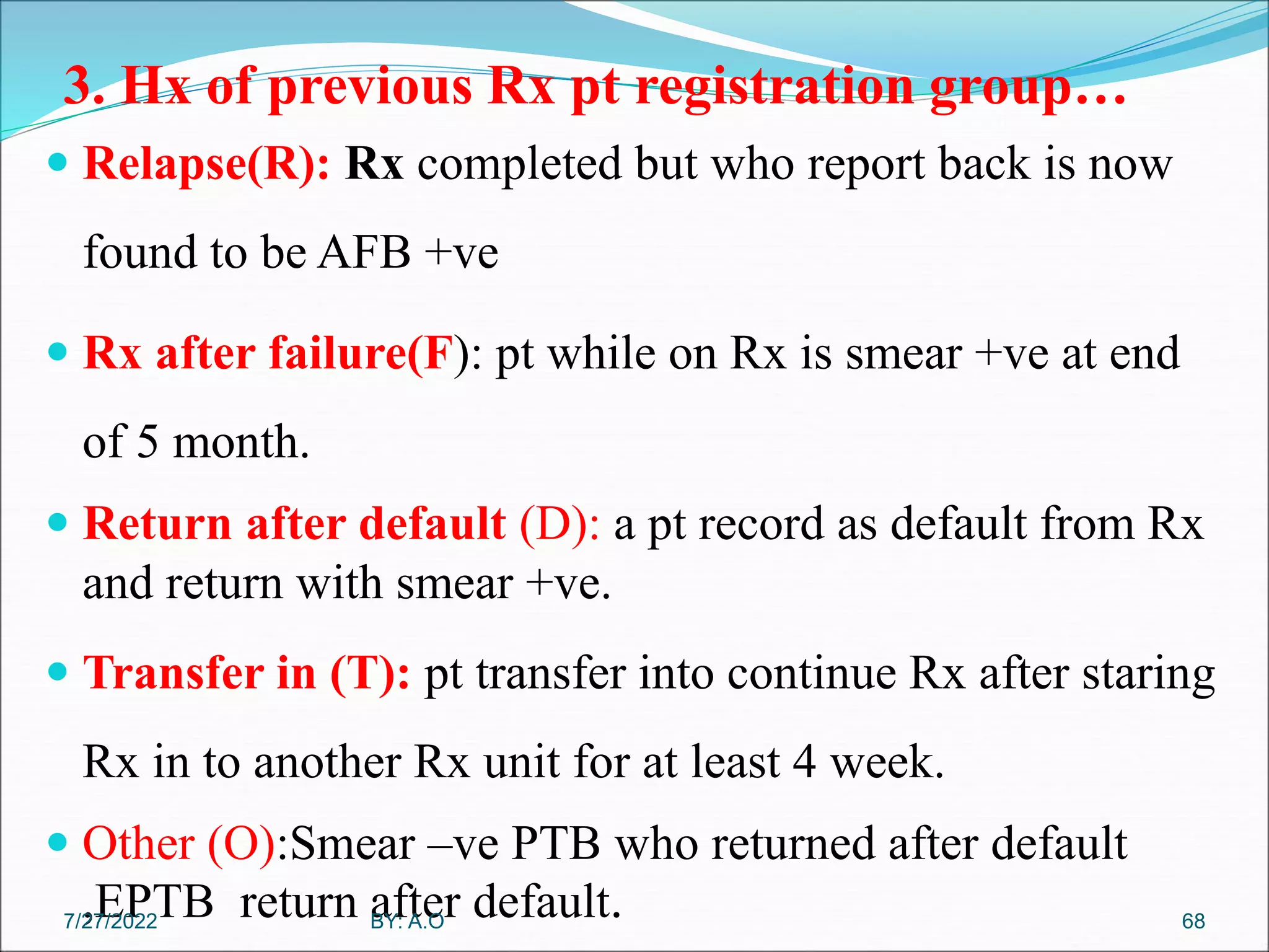
It is important to identify previously treated pt . B/c they
are high risk for drug resistance including MDR-TB.
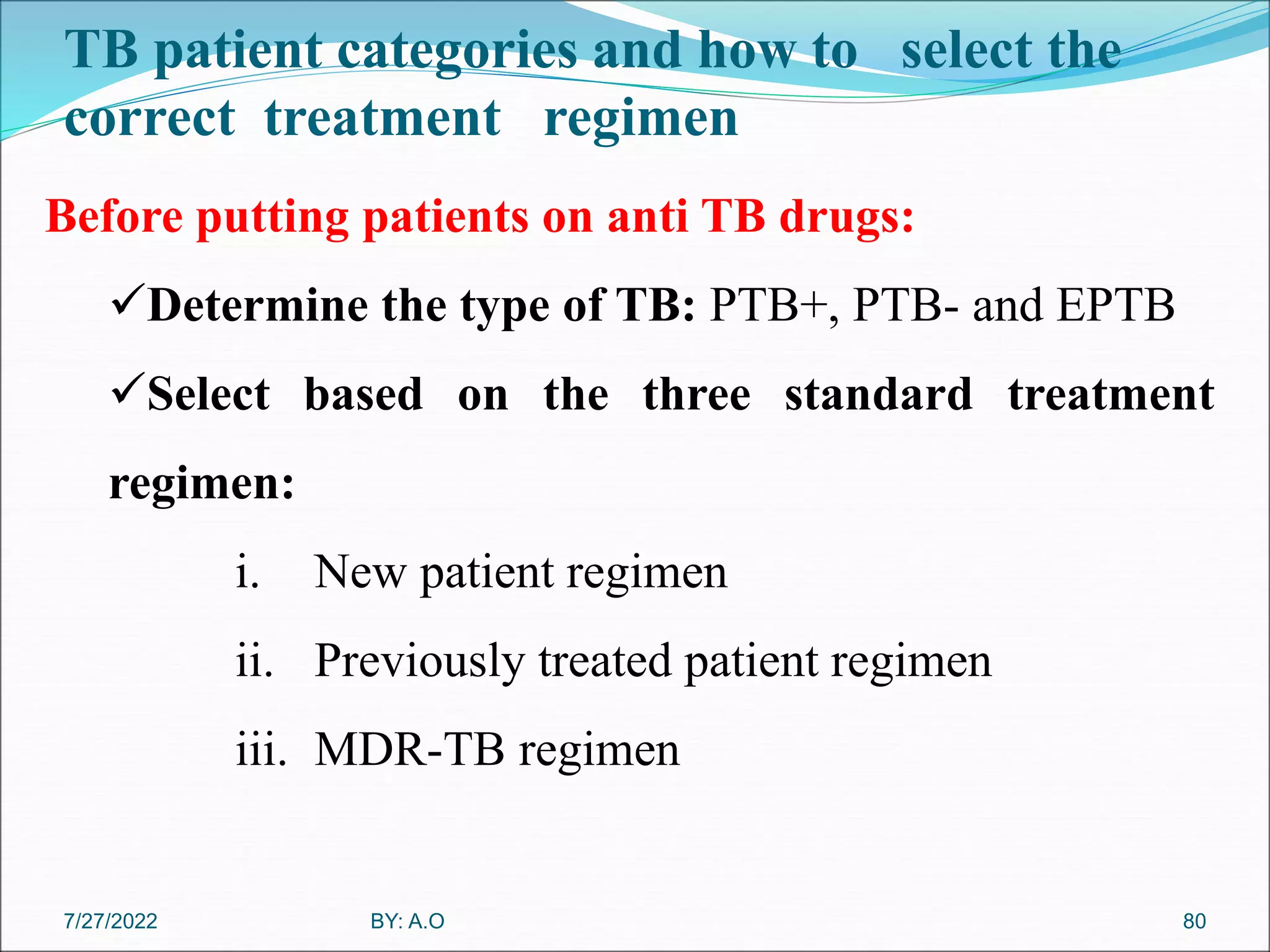
New patient: A who never had Rx or have taken anti TB
for less than 1 month [New case (N)].
Previously Treated patient: A patient who have received
1 month or more of anti TB drug in the past& may have
+ve or –ve bacterlogical and may be any diseases at an
anatomical site .
67
BY: A.O
7/27/2022
68. 3. Hx of previous Rx pt registration group…
Relapse(R): Rx completed but who report back is now
found to be AFB +ve
Rx after failure(F): pt while on Rx is smear +ve at end
of 5 month.
Return after default (D): a pt record as default from Rx
and return with smear +ve.
Transfer in (T): pt transfer into continue Rx after staring
Rx in to another Rx unit for at least 4 week.
Other (O):Smear –ve PTB who returned after default
,EPTB return after default. 68
BY: A.O
7/27/2022
69. 4. HIV Status in HIV+ve individuals
Smear +ve PTB :
One sputum smear +ve & HIV +ve/strong clinical evidence of HIV
infection.
Smear -ve PTB :
Three septum smear-ve & radiological abnormality, or
HIV +ve /strong clinical evidence of HIV infection & Decision by
clinician to Rx with Anti TB or
A pt with AFB –ve & culture +ve.
EPTB:
The definition is the same as HIV-ve TB cases
69
BY: A.O
7/27/2022
70. Diagnosis of Tuberculosis
The key to the diagnosis of tuberculosis is a high index of
suspicion.
Diagnosis is not difficult with a high-risk patient e.g., a
Homeless, alcoholic who presents with typical
symptoms and
A classic chest radiograph showing upper-lobe
infiltrates with cavities .
70
BY: A.O
7/27/2022
72. Diagnostic Method…
B. Molecular test for TB Dx
1. Line probe Assay (LPA): show Rifampicin & INH drug
sensitivity & used for smear +ve only to check presence or
absence of a specific mutation.
2. Gene Xpert MTB/RIF :Shows Rifampicin resistance
only.
C. Histo-pathological examination
D. Radiological examination
72
BY: A.O
7/27/2022
73. Standard TB Case Definition
73
•Tuberculosis suspect
cough of 2weeks or more duration with SOB, chest
pain, hemoptysis & constitutional symptoms is TB
suspect.
•Case of tuberculosis
A definite case of TB or one a health worker has
diagnosed TB and has decided to Rx with a full course
of TB Rx .
•A definite/proven case of tuberculosis
A pt with two sputum smears +ve (one sputum +ve is
enough for HIV +ve pt )or
culture +ve for mycobacterium tuberculosis .
BY: A.O
7/27/2022
74. Treatment of TB
The aim of TB treatment is:-
To cure the TB patient and restore QOL and productivity.
To prevent death from active TB or its late effects.
To Prevent relapse of TB.
To prevent the development and transmission of drug
resistance.
To decrease TB transmission to others.
To reduces the number of actively multiplying bacteria.
74
BY: A.O
7/27/2022
75. Drugs-used For TB
The drugs used for the TB treatment are safe and effective if
properly used:
First line drugs for the treatment of TB in Ethiopia include:
Rifampicin (R)
Ethambutol (E)
Isoniazid (H)
Pyrazinamide (Z)
Streptomycin(S)
75
BY: A.O
7/27/2022
76. Drugs-used For TB…
The fixed dose combination(FDC) drugs available for
adult and adolescent:
RHZE 150/75/400/275mg
RHZ 150/75/400mg
RH 150/75mg
EH 400/150mg
76
BY: A.O
7/27/2022
77. Chemotherapy of TB
TB drugs available as loose form:
Ethambutol 400mg
Isoniazid 300mg
Streptomycin sulphate vials 1 gm
NB: streptomycin is administered by injection and the
other anti TB drugs are to be taken orally
All drugs should be taken together as a single ,daily
dose, preferably on an empty stomach.
77
BY: A.O
7/27/2022
78. Phases of medical therapy
.
78
1. Intensive phase:
For new cases; a phase consists of combination of
four drugs for the first 8 weeks followed by two
drugs, to be taken for 4 months
For re-treatment cases: with combination of five drugs
for the first 8 weeks followed by four drugs for the
next four weeks.
BY: A.O
7/27/2022
79. Phases of medical therapy …
79
2.Continuation phase
This phase immediately follows the intensive phase and
is important to ensure cure or completion of treatment.
Necessary to avoid relapse after completion of treatment.
For new cases: treatment with a combination of two
drugs, to be taken for 4 months and
For re-treatment cases : treatment with a combination
of four drugs for 4 months or three drugs for 5 months.
BY: A.O
7/27/2022
80. TB patient categories and how to select the
correct treatment regimen
80
Before putting patients on anti TB drugs:
Determine the type of TB: PTB+, PTB- and EPTB
Select based on the three standard treatment
regimen:
i. New patient regimen
ii. Previously treated patient regimen
iii. MDR-TB regimen
BY: A.O
7/27/2022
81. Conti…
.
81
TB patient type Recommended regimen
New Treatment as new
2RHZE/4RH
Previously
treated
Treatment after failure Treat as retreatment
2RHZES/RHZE/5RHE
Treatment after defaulter or relapse
after one course of Rx
Treat as retreatment
2RHZES/RHZE/5RHE
Transfer in Continue same Rx regimen
Others
Previously successfully Rx pt coming
with PTB-ve or EPTB
Treatment as new
2RHZE/4RH
Defaulted pt coming with smear –ve
TB ,EPTB,or previously Rxed pt with
unknown RX outcome
Treat as retreatment
2RHZES/RHZE/5RHE
7/27/2022
82. Recommended Dose of First-Line Anti-TB
Drugs for Adults
Drugs Recommended dose
Dose and range
(mg/kg Bwt)
Maximum
(mg)
Isoniazid 5(4-6) 300
Rifampicin 10(8-12) 600
Pyrazinamide 25(20-30) 2,000
Ethambutol 15(15-20) 1600
Streptomycin 15(12-18) 1000
82
BY: A.O
7/27/2022
83. Anti TB Drugs Dosage of New TB cases
83
Patient’s Weight in
Kgs
Treatment regimen and dose
Intensive phase
2RHZE
Continuation phase
4RH
20-29 1½ 1½
30-39 2 2
40-54 3 3
≥55 4 4
BY: A.O
7/27/2022
85. Standard code for TB treatment regimen
There is a standard code for writing out TB treatment
regimens.
Each antituberculosis drug has an abbreviation.
a M(X)DR-TB regimen consists of two phases:
1) The first phase is the period in which the injectable
agent is used and the second is after it has been stopped.
For instance in Ethiopia standard treatment for
MDR-TB is 6E-Z-KM(AM)-LFX-Eto-Cs/12/E-Z-Lfx-Eto-Cs
85
BY: A.O
7/27/2022
86. 86
Grouping Drugs
Group 1:- first line oral agents Isoniazid (H) ; Rifampicin (R); Ethambutol
(E); pyrazinamide (Z); Rifabutin (Rfb)n
Group 2:- Inject able agents Kanamycin (Km); Amikacin (Am);
Capreomycine (Cm); Streptomycin (S)
Group 3:- Fluoroquinolones Moxifloxacilin (Mfx); Levofloxacilin (Lfx)
Group 4:- Oral bacteriostatic second line
agents
Ethionamide (Eto); Cycloserine (Cs); para-
aminosalicylic acid (PAS)
Group 5:- Agents with unclear role in DR-
TB treatment (not recommended by the
WHO for routine use in DR-TB patients)
Clofazimine (Cfz); Linezolid (Lzd);
Amoxicillin /clavulanate (Amx/Clv);
Thioacetazone (Thz); Imipenem/ciliastain
(Imp/Cln); High-dose isoniazid (High-dose
H)b; clarithromycin (Clr)
BY: A.O
7/27/2022
87. Standard MDR-TB Regimen
87
MDR-TB patients susceptible to both Kanamycin and
Quinolone
Regimen: E-Z-KM (AM)-Lfx-Eto-Cs
MDR-TB patients susceptible to both Kanamycin, but
resistant to Quinolone
Regimen: E-Z-KM (AM)-Mfx-Eto-Cs-PAS
MDR-TB patients susceptible to Quinolone, but
resistant to Kanamycin
Regimen: E-Z-Cm-Lfx-Eto-Cs
XDR-TB cases (I.e. MDR-TB and resistance to
Quinolone
Regimen: E-Z-Cm -Mfx-Eto-Cs-PAS
BY: A.O
7/27/2022
88. 88
Promote air way clearance
Increase fluid intake promote systemic rehydration.
Pt understand that TB is communicable diseases and
taking the drag regular for prescribed and duration.
Instruct about important hygiene
Mouth care
Covering mouth and nose when cough and sneezing
Proper disposal of tissue property
Hand wash
Promote adduct nutrition
BY: A.O
7/27/2022
89. Prevention
89
General preventive measures (e.g. staying at home,
avoiding visitors, covering mouth during coughs with
hand ,opening window .
Vaccination:
The BCG vaccine, made from an attenuated strain of M.
bovis is given to > 80% of the world's children, primarily
in high-burden countries.
BY: A.O
7/27/2022
91. Introduction to Pleural disorder
Pleural disorder is a disorder that involves:
The membranes covering the lungs (visceral pleura)
The surface of the chest wall (parietal pleura) or
The disorders affecting the pleural space.
7/27/2022 by Amanuel.O 91
93. 1. Pleurisy
Pleurisy (pleuritis) refers to inflammation of both parietal
and visceral layers of the pleurae.
Pathophysiology-Pleurisy may develop :
• In pneumonia, TB, or collagen disease
• After trauma to the chest,
• Pulmonary infarction, or pulmonary embolism;
• Primary and metastatic cancer; and
• After thoracotomy.
by Amanuel.O 93
7/27/2022
94. Pathophysiology…
The parietal pleura has nerve endings; the visceral pleura
does not.
When the inflamed pleural membranes rub together
during inspiration, the result is severe, sharp, knife like
pain.
by Amanuel.O 94
7/27/2022
95. Clinical Manifestations
Taking a deep breath, coughing, or sneezing worsens the
pain, i.e a key characteristic of pleuritic pain.
Pleuritic pain is restricted in distribution; usually occurs
only on one side.
Pain may be localized or radiate to the shoulder or
abdomen.
Latter, as pleural fluid develops , the pain decreases.
by Amanuel.O 95
7/27/2022
96. Assessment and Diagnostic Findings
Pleural friction rub sound in the early period, but it
disappears later as little fluid accumulates.
Chest x-rays
Sputum examinations,
Pleural fluid for examination via thoracentesis
Pleural biopsy - less commonly done.
by Amanuel.O 96
7/27/2022
97. Management
The objectives are:
To treat the underlying condition causing the pleurisy
(pneumonia, infection)
To relieve the pain and
To monitor signs and symptoms of pleural effusion such
as: - Shortness of breath.
Assumption of a position that decreases pain, and
decreased chest wall excursion.
by Amanuel.O 97
7/27/2022
98. Medical Management
NSAIDs: Indomethacin may provide pain relief during
deep breathing and coughing.
Topical heat or cold applications may provide
symptomatic relief.
Intercostals' nerve block may be required If the pain is
severe.
by Amanuel.O 98
7/27/2022
99. Nursing Management
Turning the patient frequently onto the affected side to
splint the chest wall and reduce the stretching of pleurae.
To enhance comfort and
To reduce pain during inspiration.
Teach about the use the hands or a pillow to splint the rib
cage while coughing.
by Amanuel.O 99
7/27/2022
100. 2. Pleural effusion
Pleural effusion is a collection of fluid in the pleural space,
in response to injury, inflammation or both.
It may represents a local response to disease or
manifestation of a systemic illness.
Normally, pleural space contains (5 to 15 ml) of pleural
fluid, which acts as a lubricant that allows the pleural
surfaces to move without friction.
by Amanuel.O 100
7/27/2022
101. Causes of plural effusion
Pleural effusion may be a complication of:
Heart failure,
TB,
Pneumonia,
Viral pulmonary infection
Nephrotic syndrome,
Connective tissue disease,
Pulmonary embolism, and
Bronchogenic carcinoma
by Amanuel.O 101
7/27/2022
102. Pathophysiology
In certain disorders, fluid may accumulate in the pleural
space to a point where it becomes clinically evident.
This always has pathologic significance.
The effusion can be:-
Clear fluid
Bloody
Transudates
Exudates(may be purulent)
by Amanuel.O 102
7/27/2022
103. Pathophysiology…
A transudate occurs when the formation and reabsorption
of pleural fluid are altered.
Transudative effusion implies that the pleural membranes
are not diseased.
The most common cause of a transudative effusion is
heart failure.
An exudate usually results from inflammation by
bacterial products/tumors involving the pleural surfaces.
by Amanuel.O 103
7/27/2022
104. Clinical Manifestations
If effusion is from Pneumonia :
fever, chills, and pleuritic chest pain,
Malignant effusion :
dyspnea and coughing
A large pleural effusion
shortness of breath
When a small to moderate pleural effusion is present,
minimal or absence of dyspnea.
by Amanuel.O 104
7/27/2022
105. Assessment and Diagnostic Findings
Physical examination
Decreased or absent breath sounds in auscultation
Decreased fremitus, and
Dull, flat sound when percussed
Acute respiratory distress- if extreme pleural effusion.
Tracheal deviation away from the affected side
Chest x-ray
Chest CT scan
by Amanuel.O 105
7/27/2022
106. Assessment and Diagnostic Findings…
Thoracentesis confirm the presence of fluid
Pleural fluid analysis:- bacterial culture, Gram stain,
AFB stain, RBC and WBC counts,
Chemistry studies(glucose, protein),
Cytological analysis for malignant cells, and
PH
Pleural biopsy
by Amanuel.O 106
7/27/2022
107. Management
The objectives are:
To treat the underlying cause (e.g, heart failure,
pneumonia, lung cancer, cirrhosis)
To prevent re-accumulation of fluid, and
To relieve discomfort, dyspnea, and respiratory
compromise.
If the pleural fluid is an exudate, more extensive
diagnostic procedures are performed to determine the
cause. by Amanuel.O 107
7/27/2022
108. Management…
Thoracentesis is performed:
To remove fluid, and to relieve dyspnea and respiratory
compromise.
To obtain a specimen for analysis,
Depending on the size of the pleural effusion, the patient
may be treated by:
Removing the fluid by thoracentesis
Inserting a chest tube connected to a water-seal drainage system or
Suctioning to remove fluid and re-expand the lung.
by Amanuel.O 108
7/27/2022
109. Management
Repeated thoracentesis result in:
pain,
depletion of protein and electrolytes, and
pneumothorax.
If the underlying cause is a malignancy, however, the
effusion tends to recur within a few days or weeks.
Therefore surgical:-
pleurectomy
Pleurodesis may be performed.
by Amanuel.O 109
7/27/2022
110. Management…
Pleurodesis is a medical procedure in which the pleural
space is artificially obliterated.
It involves the adhesion of the two pleurae using
chemically irritating agents (e.g., bleomycin or talc) are
instilled in the pleural space.
by Amanuel.O 110
7/27/2022
111. Nursing Management
Preparing and positioning for thoracentesis and offers
support throughout the procedure.
Pain management is a priority, and in assuming positions
that are the least painful.
Frequent turning and ambulation facilitate drainage.
Analgesics as prescribed and as needed.
Care of chest tube.
by Amanuel.O 111
7/27/2022
112. 3. Empyema
An empyema refers to collection of pus inside the
pleural space (dead cells and infected fluid).
Causes;-
Penetrating chest trauma,
Hematogenous infection of the pleural space,
Non-bacterial infections, or
Iatrogenic causes (after thoracic surgery or thoracentesis)
by Amanuel.O 112
7/27/2022
113. Causes….
Complications of bacterial pneumonia or lung abscess are
the two commonest ways that bacteria get into pleural
space.
In order for empyema to occur:
Bacteria,
Fungi, or
Chemicals must get into the pleural space and cause
inflammation, leading to the production of pus.
Bacteria can also get into the pleural space from medical
instruments that are used to do tests or operate the chest.
by Amanuel.O 113
7/27/2022
114. Risk for Empyema
The greatest risk factors for empyema are:
Pneumonia,
Medical procedures done in the lung and surrounding structures,
Chest trauma and
Pre-existing lung diseases (COPD and lung cancer).
People who have pre-existing lung diseases who develop
empyema are more likely to die than those who don’t.
by Amanuel.O 114
7/27/2022
115. Clinical Manifestations
Fever, night sweats, pleural pain, cough, dyspnea.
SOB-E.g. Pneumonia
Fatigue, loss of appetite, and weight loss
Empyema associated with sepsis is the most severe.
High fever, chills, tachypnea, tachycardia, and low B/P.
Sepsis is life-threatening and requires emergency treatment.
by Amanuel.O 115
7/27/2022
116. Diagnostic Findings
Blood cultures
WBCs count
X-ray (pneumonia, lung abscess)
CT scan of the chest
Thoracentesis for microscopic examination
Thoracic ultrasound
Chest auscultation: Decreased or absent breath sounds &
Dullness on chest percussion & decreased fremitus.
by Amanuel.O 116
7/27/2022
117. Management
The objectives of treatment are to drain the pleural cavity
and to achieve full expansion of the lung.
Draining the fluid
large doses of appropriate antibiotics based on the causative
organism.
Cephalosporins ,Metronidazole, and
Penicillins with Beta lactamase (Ampicillin/
sulbactam).
Sterilization of empyema requires 4 to 6 wks of antibiotics.
by Amanuel.O 117
7/27/2022
118. Management…
Drainage of the pleural fluid is accomplished by the ff:
Needle aspiration (Thoracentesis) or
Tube thoracostomy (Chest drainage using a large-
diameter inter costal tube attached to water-seal
drainage.
by Amanuel.O 118
7/27/2022
119. Nursing Management
Deep breathing exercises to restore normal respiratorion
Provide care(e.g, needle aspiration, closed chest drainage).
When a patient is discharged to home with a drainage tube
or system in place:
instructs the patient and family on care of the drainage
system and drain site, measurement and
observation of drainage, signs and symptoms of infection
by Amanuel.O 119
7/27/2022
120. CHRONIC OBSTRUCTIVE PULMONARY
DISEASE(COPD)
COPD is a disease state characterized by airflow limitation
that is not fully reversible.
COPD is the diseases that cause airflow obstruction (e.g,
emphysema, chronic bronchitis).
COPD can coexist with asthma & both diseases have the
same major symptoms.
Other diseases (bronchiectasis, and asthma) are now
classified as chronic pulmonary disorders.
by Amanuel.O 120
7/27/2022
121. Pathophysiology
In COPD, the airflow limitation is associated with an
abnormal inflammatory response of the lungs to noxious
particles or gases.
The inflammatory response occurs throughout the airways,
parenchyma, and pulmonary vasculature.
Because of the chronic inflammation and the body’s
attempts to repair it, narrowing occurs in the small
peripheral airways.
by Amanuel.O 121
7/27/2022
122. Pathophysiology…
Over time, this injury and repair process causes scar tissue
formation and narrowing of the airway lumen.
Airflow obstruction may also be due to parenchymal
destruction as seen with emphysema, a disease of the
alveoli or gas exchange units.
by Amanuel.O 122
7/27/2022
123. Pathophysiology…
Early in the course of COPD, the inflammatory response
causes pulmonary vasculature (thickening of vessel wall).
These changes may result from:
Exposure to cigarette smoke,
Use of tobacco products, or
Release of inflammatory mediators
by Amanuel.O 123
7/27/2022
124. Chronic Bronchitis
Chronic bronchitis, a disease of the airways, is defined as
the presence of cough and sputum production for at least 3
months in each of two consecutive years.
The causes are:
Cigarette smoking or
Other environmental pollutants irritate the airways.
This results in inflammation & hyper secretion of mucus.
by Amanuel.O 124
7/27/2022
125. Pathophysiology
This constant irritation causes the mucus-secreting glands
and goblet cells to increase in number.
Ciliary function is reduced, and more mucus is produced.
The bronchial walls become thickened, the lumen narrows,
and mucus may plug the airway.
Alveoli adjacent to the bronchioles may become damaged
and fibrosed,
Resulting in altered function of the alveolar macrophages
that play role in destroying foreign particles (bacteria).
by Amanuel.O 125
7/27/2022
126. Pathophysiology…
As a result, the patient becomes more susceptible to
respiratory infection.
A wide range of viral, and bacterial infections can produce
acute episodes of bronchitis.
Exacerbations of chronic bronchitis are most likely to
occur during the winter.
by Amanuel.O 126
7/27/2022
127. Risk Factors for COPD
Exposure to tobacco smoke accounts 80% to 90% of
COPD cases
Passive smoking
Occupational exposure
Deficiency of alpha1- antitrypsin.
by Amanuel.O 127
7/27/2022
128. Clinical Manifestations
COPD is characterized by three primary symptoms:
Cough,
Sputum production, and
Dyspnea on exertion is severe and often interferes with the
patient's activities.
Chronic cough and sputum production often precede the
development of airflow limitation by many years.
Weight loss is common, because dyspnea interferes with
eating and the work of breathing is energy-depleting.
by Amanuel.O 128
7/27/2022
129. Clinical Manifestations
Often patients cannot participate in even mild exercise because of
dyspnea
As COPD progresses, dyspnea occurs even at rest.
As the work of breathing increases over time, the accessory muscles
are recruited in an effort to breathe.
Patients with COPD are at risk for:
respiratory insufficiency,
respiratory infections, and
increase the risk of respiratory failure.
by Amanuel.O 129
7/27/2022
130. Diagnostic Findings
Exposure to risk factors
Past medical history: asthma, allergy, sinusitis, nasal polyps,
history of RTIs.
Family history of COPD
Pattern of symptom development
History of exacerbations or
History of previous hospitalizations for RTIs.
Presence of Co-morbidities
Barrel chest
by Amanuel.O 130
7/27/2022
131. Medical Management
Smoking cessation: is the single most effective
intervention to prevent COPD or slow its progression.
Bronchodilators: Relieve bronchospasm and increase
oxygen distribution throughout the lungs and improving
alveolar ventilation.
Corticosteroids: Inhaled and systemic may be used
Oxygen therapy: prevent acute dyspnea.
by Amanuel.O 131
7/27/2022
133. Emphysema
Emphysema is a destructive disease of the lung in which
the alveoli (small sacs) are destroyed.
Emphysema is a pathologic term that describes an
abnormal distention of the air spaces with destruction of
the walls of the alveoli.
In emphysema, impaired gas exchange results from
destruction of the walls of over distended alveoli.
by Amanuel.O 133
7/27/2022
134. Chronic Pulmonary Disorders
Bronchiectasis is a chronic, irreversible dilation of the
bronchi and bronchioles.
It may be caused by a variety of conditions:
Airway obstruction
Diffuse airway injury
Pulmonary infections and obstruction of the bronchus
Genetic disorders such as cystic fibrosis
Abnormal host defense (e.g, ciliary dyskinesia or
humoral immunodeficiency)
Idiopathic causes
by Amanuel.O 134
7/27/2022
135. Bronchiectasis….
A person may be predisposed to bronchiectasis as a result of
recurrent respiratory infections in early childhood:
Measles,
influenza,
tuberculosis, and
immunodeficiency disorders.
Bronchiectasis is usually localized, affecting a segment or
lobe of a lung, most frequently the lower lobes.
by Amanuel.O 135
7/27/2022
136. Bronchiectasis….
Cigarette smoking impairs bronchial drainage by:
Paralyzing ciliary action,
Hyperplasia of the mucous glands,
Increasing bronchial secretions, and
Causing inflammation of the mucous membranes
by Amanuel.O 136
7/27/2022
138. Pathophysiology
The inflammatory process associated with pulmonary
infections damages the bronchial wall and its supporting
structure
This results in thick sputum that ultimately obstructs the
bronchi.
The walls become permanently distended and distorted,
impairing muco ciliary clearance.
The inflammation and infection extend to the
peribronchial tissues.
by Amanuel.O 138
7/27/2022
139. Pathophysiology
The retention of secretions and subsequent obstruction
ultimately cause the alveoli distal to the obstruction to
collapse (atelectasis).
Inflammatory scarring or fibrosis replaces functioning
lung tissue.
There is ventilation–perfusion imbalance and hypoxemia.
by Amanuel.O 139
7/27/2022
140. Clinical Manifestations
Chronic cough & purulent sputum
Hemoptysis
Clubbing of the fingers =>b/c of respiratory insufficiency.
Repeated episodes of pulmonary infection
Even with modern treatment approaches, the mean age at
death is ~ 55 years.
by Amanuel.O 140
7/27/2022
141. Assessment and Diagnostic Findings
Bronchiectasis is not readily diagnosed because the
symptoms can mimic with chronic bronchitis.
A definite sign is prolonged Hx of productive cough,
with sputum consistently negative for AFB.
CT scan: Demonstrates bronchial dilation.
by Amanuel.O 141
7/27/2022
142. Medical Management
Treatment objectives are:
To promote bronchial drainage
To clear excessive secretions from the affected portion
of the lungs and
To prevent or control infection.
by Amanuel.O 142
7/27/2022
143. Medical Management
Chest physiotherapy-percussion and postural drainage, is
important in secretion management.
Smoking cessation
Antimicrobial therapy: based on the results of sputum
culture & sensitivity.
Surgical intervention: Diseased tissue is removed.
a segment of a lobe (segmental resection),
a lobe (lobectomy), or
rarely an entire lung (pneumonectomy).
by Amanuel.O 143
7/27/2022
144. Asthma
Asthma is a chronic inflammatory disease of the airways
that causes airway hyper responsiveness, mucosal edema,
and mucus production.
This inflammation ultimately leads to recurrent episodes
of asthma symptoms:
Cough
Chest tightness
Wheezing and
Dyspnea
by Amanuel.O 144
7/27/2022
145. Asthma…
Status asthmaticus is severe and persistent asthma that
does not respond to conventional therapy and is
considered life-threatening.
The attacks can last longer than 24 hours.
by Amanuel.O 145
7/27/2022
146. Asthma
Asthma differs from the other obstructive lung diseases in
that it is largely reversible, either spontaneously or with
treatment.
Patients with asthma may experience symptom-free
periods alternating with acute exacerbations.
Asthma can occur at any age.
by Amanuel.O 146
7/27/2022
147. Asthma…
Chronic exposure to airway irritants, or allergens also
increases the risk for developing asthma.
Allergy is the strongest predisposing factor for asthma.
Common allergens can be seasonal (e.g, grass, tree, and
weed pollens) or perennial ,mold, dust, or animal dander.
by Amanuel.O 147
7/27/2022
148. Common triggers
Common triggers of symptoms and exacerbations of
Asthma:
Air pollutants, cold, heat, weather changes, strong
odors or perfumes, smoke
Exercise
Stress or emotional upsets,
Medications, and
Gastro esophageal reflux
RTIs: viral RTI, sinusitis etc.
by Amanuel.O 148
7/27/2022
149. Pathophysiology
Asthma is a reversible and diffuse airway inflammation.
Inflammation causes:
Mucosal edema,
Increased mucus production which may entirely
plug to the bronchi &
Furtherly diminishes airway size or diameter.
This leads to alveoli hyper inflation
by Amanuel.O 149
7/27/2022
150. Pathophysiology…
The cells that play a key role in the inflammation of
asthma are :
Mast cells,
Neutrophil,
Eosinophils, and lymphocytes
Mast cells is activated, released several chemicals
mediators (Histamine, Bradykinin, Prostaglandins, and
Leukotrienes).
by Amanuel.O 150
7/27/2022
151. Pathophysiology…
These chemicals perpetuate the inflammatory response,
Vasodilatation – occurs after transient vasoconstriction
(lasting only for seconds),
Results in locally increased blood flow
Fluid leak from the vasculature,
Attraction of WBCs to the area, and
Broncho constriction.
by Amanuel.O 151
7/27/2022
152. Pathophysiology…
Further stimulation of alpha and beta2-adrenergic
receptors of the sympathetic NS are located in the bronchi.
When the alpha adrenergic receptors are stimulated,
broncho constriction occurs.
when the beta2 -adrenergic receptors are stimulated,
broncho dilation occurs.
by Amanuel.O 152
7/27/2022
153. Pathophysiology…
The balance between alpha and beta2 receptors is controlled
primarily by cyclic adenosine mono phosphate (cAMP).
Alpha-adrenergic receptor stimulation results in a decrease in
cAMP, which leads to an increase of chemical mediators released
by the mast cells and bronchoconstriction.
Beta2-receptor stimulation results in increased levels of cAMP,
which inhibits the release of chemical mediators and causes
bronchodilation.
by Amanuel.O 153
7/27/2022
155. Clinical manifestations
An asthma exacerbation may begin abruptly
The three most common symptoms of asthma are
Cough,
Dyspnea, and
Wheezing
Asthma attacks often occur at night or early in the
morning.
by Amanuel.O 155
7/27/2022
156. Clinical manifestations…
Generalized chest tightness
Expiration requires effort and becomes prolonged
As the exacerbation progresses, diaphoresis, tachycardia,
and a widened pulse pressure along with hypoxemia and
central cyanosis (a late sign).
The hypoxemia is 2 0 to a ventilation–perfusion mismatch
and readily responds to supplemental oxygenation.
by Amanuel.O 156
7/27/2022
157. Clinical manifestations….
Symptoms of exercise-induced asthma include:
Maximal symptoms during exercise,
Absence of nocturnal symptoms, and
Sometimes only a sign of a “choking” sensation during
exercise.
by Amanuel.O 157
7/27/2022
158. Diagnostic findings
A positive family Hx,
Occupational history
Environmental factors:
seasonal changes,
high pollen counts, mold,
Climate changes (particularly cold air), and
Air pollution are primarily associated with asthma.
by Amanuel.O 158
7/27/2022
159. Diagnostic findings…
Occupation-related chemicals and compounds:
metal salts, wood and vegetable dust, medications (e.g,
ASA, Antibiotics, Piperazine, Cimetidine),
Industrial chemicals and plastics, (e.g, laundry
detergents),
Animal and insect dusts, secretions.
by Amanuel.O 159
7/27/2022
160. Diagnostic findings…
Sputum and blood tests may disclose elevated levels of
eosinophils.
Serum levels of IgE may be elevated if allergy is present.
Arterial blood gas analysis and Pulse oximetry reveal
hypoxemia during acute attacks
by Amanuel.O 160
7/27/2022
161. MEDICAL MANAGEMENT
Goals of Asthma Treatment
Prevent chronic and troublesome symptoms
Maintain near-normal pulmonary function
Maintain normal activity levels (exercise and other
physical activity)
Prevent recurrent exacerbations of asthma and minimize
the need for emergency OPD visits or hospitalizations
by Amanuel.O 161
7/27/2022
164. Nursing management
Monitoring the severity of symptoms, breath sounds, peak
flow , pulse oximetry, and vital signs.
Obtain a history of allergic reactions to medications before
administering medications.
Administer medications as prescribed and monitor the
patient’s responses to those medications
Administer fluids if the patient is dehydrated.
by Amanuel.O 164
7/27/2022