Downloaded 16 times


![[1] Jack J kanski, Clinical Ophthalmology : a systemic approach. (Sixth edition ed.)Elsevier
limited; 2007. pg: 491-494
21
REFERENCES](https://image.slidesharecdn.com/lowvisioncase-231010072631-9dd685c4/85/Low-vision-case-Retinitis-Pigmentosa-pptx-21-320.jpg)


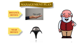
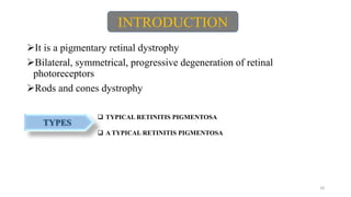
The document provides a detailed clinical overview of a 76-year-old female patient with a history of progressive vision loss and underlying retinal dystrophy. It includes examination findings, visual acuity measurements, management plans, and recommendations for rehabilitative services. The patient's condition is diagnosed as atypical retinitis pigmentosa, and multiple assistive device trials are discussed.


![Optics of contact lens and nomenclature copy [repaired] (1)](https://cdn.slidesharecdn.com/ss_thumbnails/opticsofcontactlensandnomenclature-copyrepaired1-170218054900-thumbnail.jpg?width=640&height=640&fit=bounds)


