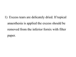
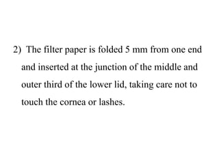
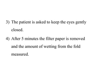
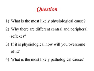
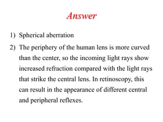
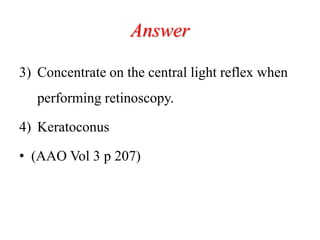
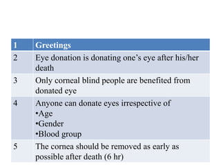
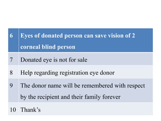
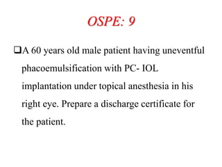
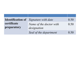
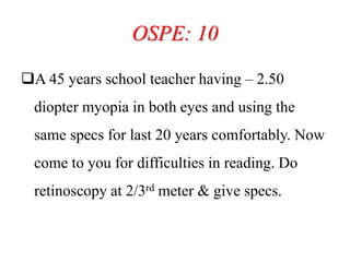
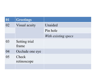
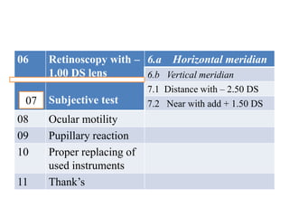
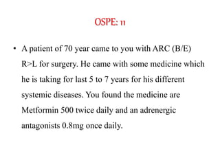
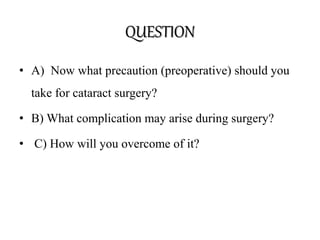
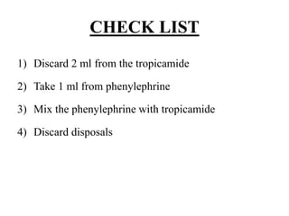
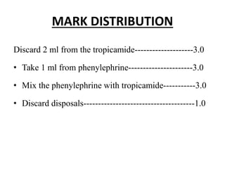
Objective Structured Practical
Examination
Dr Md Anisur Rahman (Anjum)
Professor & Head of the Department
Dhaka Medical College. Dhaka
OSPE: 1
Rapidly progressive unilateral proptosis is
usual, Average age of onset is 7 years. The
tumour is derived from undifferentiated
mesenchymal cells. Various genetic
predispositions have been identified, including
variants of the RB1 gene.
Question
1) What is the probable diagnosis?
2) What is the most common site in the orbit?
3) Which type is the worse prognosis?
4) Which is the most confirmatory diagnosis?
Answer
1) Rhabdomyosarcoma
2) Most commonly superonasal or superior orbit
3) Alveolar
4) Incisional biopsy followed by histopathology
• Kanski 8th edition p 109
OSPE: 2
Do the Schirmer test 2 & write down your
interpretation
1) Excess tears are delicately dried. If topical
anaesthesia is applied the excess should be
removed from the inferior fornix with filter
paper.
2) The filter paper is folded 5 mm from one end
and inserted at the junction of the middle and
outer third of the lower lid, taking care not to
touch the cornea or lashes.
3) The patient is asked to keep the eyes gently
closed.
4) After 5 minutes the filter paper is removed
and the amount of wetting from the fold
measured.
Interpretation: Less than 10 mm of wetting
after 5 minutes without anaesthesia or less than
6 mm with anaesthesia is considered abnormal.
• Kanski 8th edition p 124
OSPE 3
A patient came to you with corneal opacity.
What history should you take from that patient
mention with explanation for the relevant
histories?
Answer
• History of onset: Age of onset. If early onset
best possible treatment could not help
satisfactorily because of amblyopia
• History of trauma: If so, there may be cataract,
retain foreign body in presence trauma. And B-
Scan should be done before surgery
• H/O Associated pain, redness, watering,
discharge to exclude any corneal ulcer,
aphakic/pseudophakic bullous keratopathy
• H/O past surgeries
• H/O associated frequent change of glass
keratoconus
• H/O associated systemic problem
Hyperlipidaemia/Hypercalcemia
• Sankara Nethralaya 124
OSPE: 4. History taking R.P
1) Age of onset of symptoms.
2) Duration of night blindness.
3) Duration of progressive loss of visual field.
4) Duration of dimness of vision . Is it
progressive?
5) Family history of R.P.
Wednesday, February 05,
2014
14anjumk38dmc@gmail.com
6) H/O consanguinity.
7) H/O trauma.
8) H/O drug intake.
9) H/O hearing disorder, ataxia, nystagmus.
10)H/O mental retardation.
History taking R.P
11)H/O heart disease.
12)H/O hypogenitalism, obesity, polydactyly.
13)H/O diarrhea, skeletal deformity.
Wednesday, February 05,
2014
16anjumk38dmc@gmail.com
OSPE=5
Q: History taking of a patient suffering from
recurrent uveitis
A: Following points to be noted during history
taking:
19 FEB 2014 17anjumk38dmc@gmail.com
1) PATIENT DETAILS:
1) Age: Juvenile rheumatoid arthritis (JRA) is
common in patients less than 15 years.
2) Sex: JRA is common in females, HLA – B 27
associated uveitis in males. (but during
history taking you should not asked about
gender)
2) OCULAR HISTORY:
Is the disease unilateral or bilateral ?
When was the first attack?
When was the last/current attack?
What was the approximate frequency of the
attacks between the first and the last attack?
Details of prior ocular treatment.
Any previous history of rise IOP or use any
antiglaucoma agents.
19 FEB 2014 19anjumk38dmc@gmail.com
3) SYSTEMIC HISTORY:
H/O arthritis or low backache (JRA, HLA –B27
related uveitis).
H/O fever or respiratory symptoms, gastro-
intestinal, neurological symptoms, genital
lesions.
H/O DM, HTN, TB.
19 FEB 2014 20anjumk38dmc@gmail.com
H/O exposure/ IV drug abuse/ blood
transfusions.
H/O skin lesions (HZO, Psoriasis)
Details of prior systemic treatment.
OSPE: 6
When performing cycloplegic retinoscopy on an
anxious 7-year-old boy, you notice that the
central reflex shows with movement while the
peripheral reflex shows against movement.
Question
1) What is the most likely physiological cause?
2) Why there are different central and peripheral
reflexes?
3) If it is physiological how will you overcome
of it?
4) What is the most likely pathological cause?
Answer
1) Spherical aberration
2) The periphery of the human lens is more curved
than the center, so the incoming light rays show
increased refraction compared with the light rays
that strike the central lens. In retinoscopy, this
can result in the appearance of different central
and peripheral reflexes.
Answer
3) Concentrate on the central light reflex when
performing retinoscopy.
4) Keratoconus
• (AAO Vol 3 p 207)
OSPE: 7
• You are planning cataract surgery to achieve
emmetropia for a patient with the following
measurements:
• Refraction: -3.00 +2.00 x 120
• K: 42.50 D/42.75 D @ 120deg
Question
1) Which IOL will you prefer to achieve
emmetropia?
2) Give one reason in favour of your IOL choice
3) What are the options to correct astigmatism
during cataract surgery? Mention 2
Answer
1) Mono focal
2) The astigmatism is due to cataractous lens
3) .
a) toric lens implants
b) relaxing incisions
OSPE: 8
 Motivate a person for eye donation
1 Greetings
2 Eye donation is donating one’s eye after his/her
death
3 Only corneal blind people are benefited from
donated eye
4 Anyone can donate eyes irrespective of
•Age
•Gender
•Blood group
5 The cornea should be removed as early as
possible after death (6 hr)
6 Eyes of donated person can save vision of 2
corneal blind person
7 Donated eye is not for sale
8 Help regarding registration eye donor
9 The donor name will be remembered with respect
by the recipient and their family forever
10 Thank’s
OSPE: 9
A 60 years old male patient having uneventful
phacoemulsification with PC- IOL
implantation under topical anesthesia in his
right eye. Prepare a discharge certificate for
the patient.
Identificat
ion of the
patient
Name
Age 0.50
Gender 0.25
Address 0.50
Mobile no 0.25
Operation
Note
Date & time 0.50
Name of surgery 0.50
Name of anesthesia 0.50
Name of surgeon 0.50
Post operative
findings
Visual acuity 1.00
Anterior segment 1.00
Posterior segment 0.50
Post operative
treatment
Topical antibiotic 0.50
Topical steroid 0.50
Advice No water to eye 0.25
Use dark glass 0.25
Regular use of medicine 0.25
Any problem come to doctor 0.25
Follow up 0.25
Identification of
certificate
preparatory
Signature with date 0.50
Name of the doctor with
designation
0.50
Seal of the department 0.50
OSPE: 10
A 45 years school teacher having – 2.50
diopter myopia in both eyes and using the
same specs for last 20 years comfortably. Now
come to you for difficulties in reading. Do
retinoscopy at 2/3rd meter & give specs.
01 Greetings
02 Visual acuity Unaided
Pin hole
With existing specs
03 Setting trial
frame
04 Occlude one eye
05 Check
retinoscope
06 Retinoscopy with –
1.00 DS lens
Subjective test
6.a Horizontal meridian
6.b Vertical meridian
7.1 Distance with – 2.50 DS
7.2 Near with add + 1.50 DS
08 Ocular motility
09 Pupillary reaction
10 Proper replacing of
used instruments
11 Thank’s
07
OSPE: 11
• A patient of 70 year came to you with ARC (B/E)
R>L for surgery. He came with some medicine which
he is taking for last 5 to 7 years for his different
systemic diseases. You found the medicine are
Metformin 500 twice daily and an adrenergic
antagonists 0.8mg once daily.
QUESTION
• A) Now what precaution (preoperative) should you
take for cataract surgery?
• B) What complication may arise during surgery?
• C) How will you overcome of it?
A) What precaution (preoperative) should you take for
cataract surgery?
• a).Control DM
• b) Maximum dilatation of the pupil as far as can even
with atropine 1% eye drop
• B) What complication may arise during surgery?
a) Pupil may constrict during cataract surgery.
b) The iris may billow and prolapsed through the
incision.
c) The risk of capsule rupture and vitreous loss is
increased.
C) How will you overcome of it?
Strategies for management include the
a) Use of Healon 5,
b) preoperative pupillary dilatation with atropine,
c) intracameral epinephrine,
d) iris hooks, and
e) low aspiration flow rates.
OSPE: 12
What is the use of this
spectacle?
Ptosis props which are used to lift droopy
eyelid
OSPE: 13
A B C
QUESTION
a
i. How far should the Amsler's chart be placed in
front of the patients?
ii. How many degree(s) does each square subtend in
the macula when placed in the recommended
position?
• b.
i. What is the advantage of chart b over chart a?
ii. What is the advantage of chart c over chart a?
ANSWER
a)
i. The chart is placed 30 cm from the patient
ii. 1 degree
b)
• Chart b contains two diagonal lines, that cross at the
central black point. In patient who can not see the
black spot because of central scotoma, the lines help
to maintain fixation and allows the patient to outline
the limits of scotoma.
• The red lines on a black background in chart c is
useful for patient with neuro-logical disorder such as
optic nerve or chiasmal lesion or toxic amblyopia
OSPE: 14
 H/O DOUBLE VISION
1) Greetings & self introduction
2) Whether double vision is monocular or binocular
3) Direction of double vision: whether the diplopia is
horizontal,
vertical or
torsional.
4) Ask the patient in which direction of gaze the
diplopia is worse→ right, left, up, down, right and
up, right and down, left and up, left and down, or
distance or near.
5) Ask for diurnal variability and fatigability of
diplopia
6) Detailed history about :
 mode of onset,
 duration of onset,
 associated pain,
 history of strabismus in childhood,
 history of trauma,
 neurological symptoms such as dysphagia or
weakness,
7) Underlying systemic illness:
 hypertension,
 diabetes,
 cerebrovascular disease,
 cardiac atherosclerotic disease
 multiple sclerosis.
8) Thank’s to patient
OSPE: 15
For the detection of ROP you have to dilate the pupil
of pre mature infant with 2.5% Phenylephrine and
0.75% Tropicamide, but which is commercially not
available
Prepare above concentration with the supplied
materials
CHECK LIST
1) Discard 2 ml from the tropicamide
2) Take 1 ml from phenylephrine
3) Mix the phenylephrine with tropicamide
4) Discard disposals
MARK DISTRIBUTION
Discard 2 ml from the tropicamide--------------------3.0
• Take 1 ml from phenylephrine----------------------3.0
• Mix the phenylephrine with tropicamide-----------3.0
• Discard disposals--------------------------------------1.0

Ospe 25 march 2017

  • 1.
    Objective Structured Practical Examination DrMd Anisur Rahman (Anjum) Professor & Head of the Department Dhaka Medical College. Dhaka
  • 2.
    OSPE: 1 Rapidly progressiveunilateral proptosis is usual, Average age of onset is 7 years. The tumour is derived from undifferentiated mesenchymal cells. Various genetic predispositions have been identified, including variants of the RB1 gene.
  • 3.
    Question 1) What isthe probable diagnosis? 2) What is the most common site in the orbit? 3) Which type is the worse prognosis? 4) Which is the most confirmatory diagnosis?
  • 4.
    Answer 1) Rhabdomyosarcoma 2) Mostcommonly superonasal or superior orbit 3) Alveolar 4) Incisional biopsy followed by histopathology • Kanski 8th edition p 109
  • 5.
    OSPE: 2 Do theSchirmer test 2 & write down your interpretation
  • 6.
    1) Excess tearsare delicately dried. If topical anaesthesia is applied the excess should be removed from the inferior fornix with filter paper.
  • 7.
    2) The filterpaper is folded 5 mm from one end and inserted at the junction of the middle and outer third of the lower lid, taking care not to touch the cornea or lashes.
  • 8.
    3) The patientis asked to keep the eyes gently closed. 4) After 5 minutes the filter paper is removed and the amount of wetting from the fold measured.
  • 9.
    Interpretation: Less than10 mm of wetting after 5 minutes without anaesthesia or less than 6 mm with anaesthesia is considered abnormal. • Kanski 8th edition p 124
  • 10.
    OSPE 3 A patientcame to you with corneal opacity. What history should you take from that patient mention with explanation for the relevant histories?
  • 11.
    Answer • History ofonset: Age of onset. If early onset best possible treatment could not help satisfactorily because of amblyopia • History of trauma: If so, there may be cataract, retain foreign body in presence trauma. And B- Scan should be done before surgery
  • 12.
    • H/O Associatedpain, redness, watering, discharge to exclude any corneal ulcer, aphakic/pseudophakic bullous keratopathy • H/O past surgeries
  • 13.
    • H/O associatedfrequent change of glass keratoconus • H/O associated systemic problem Hyperlipidaemia/Hypercalcemia • Sankara Nethralaya 124
  • 14.
    OSPE: 4. Historytaking R.P 1) Age of onset of symptoms. 2) Duration of night blindness. 3) Duration of progressive loss of visual field. 4) Duration of dimness of vision . Is it progressive? 5) Family history of R.P. Wednesday, February 05, 2014 14anjumk38dmc@gmail.com
  • 15.
    6) H/O consanguinity. 7)H/O trauma. 8) H/O drug intake. 9) H/O hearing disorder, ataxia, nystagmus. 10)H/O mental retardation.
  • 16.
    History taking R.P 11)H/Oheart disease. 12)H/O hypogenitalism, obesity, polydactyly. 13)H/O diarrhea, skeletal deformity. Wednesday, February 05, 2014 16anjumk38dmc@gmail.com
  • 17.
    OSPE=5 Q: History takingof a patient suffering from recurrent uveitis A: Following points to be noted during history taking: 19 FEB 2014 17anjumk38dmc@gmail.com
  • 18.
    1) PATIENT DETAILS: 1)Age: Juvenile rheumatoid arthritis (JRA) is common in patients less than 15 years. 2) Sex: JRA is common in females, HLA – B 27 associated uveitis in males. (but during history taking you should not asked about gender)
  • 19.
    2) OCULAR HISTORY: Isthe disease unilateral or bilateral ? When was the first attack? When was the last/current attack? What was the approximate frequency of the attacks between the first and the last attack? Details of prior ocular treatment. Any previous history of rise IOP or use any antiglaucoma agents. 19 FEB 2014 19anjumk38dmc@gmail.com
  • 20.
    3) SYSTEMIC HISTORY: H/Oarthritis or low backache (JRA, HLA –B27 related uveitis). H/O fever or respiratory symptoms, gastro- intestinal, neurological symptoms, genital lesions. H/O DM, HTN, TB. 19 FEB 2014 20anjumk38dmc@gmail.com
  • 21.
    H/O exposure/ IVdrug abuse/ blood transfusions. H/O skin lesions (HZO, Psoriasis) Details of prior systemic treatment.
  • 22.
    OSPE: 6 When performingcycloplegic retinoscopy on an anxious 7-year-old boy, you notice that the central reflex shows with movement while the peripheral reflex shows against movement.
  • 23.
    Question 1) What isthe most likely physiological cause? 2) Why there are different central and peripheral reflexes? 3) If it is physiological how will you overcome of it? 4) What is the most likely pathological cause?
  • 24.
    Answer 1) Spherical aberration 2)The periphery of the human lens is more curved than the center, so the incoming light rays show increased refraction compared with the light rays that strike the central lens. In retinoscopy, this can result in the appearance of different central and peripheral reflexes.
  • 25.
    Answer 3) Concentrate onthe central light reflex when performing retinoscopy. 4) Keratoconus • (AAO Vol 3 p 207)
  • 26.
    OSPE: 7 • Youare planning cataract surgery to achieve emmetropia for a patient with the following measurements: • Refraction: -3.00 +2.00 x 120 • K: 42.50 D/42.75 D @ 120deg
  • 27.
    Question 1) Which IOLwill you prefer to achieve emmetropia? 2) Give one reason in favour of your IOL choice 3) What are the options to correct astigmatism during cataract surgery? Mention 2
  • 28.
    Answer 1) Mono focal 2)The astigmatism is due to cataractous lens 3) . a) toric lens implants b) relaxing incisions
  • 29.
    OSPE: 8  Motivatea person for eye donation
  • 30.
    1 Greetings 2 Eyedonation is donating one’s eye after his/her death 3 Only corneal blind people are benefited from donated eye 4 Anyone can donate eyes irrespective of •Age •Gender •Blood group 5 The cornea should be removed as early as possible after death (6 hr)
  • 31.
    6 Eyes ofdonated person can save vision of 2 corneal blind person 7 Donated eye is not for sale 8 Help regarding registration eye donor 9 The donor name will be remembered with respect by the recipient and their family forever 10 Thank’s
  • 32.
    OSPE: 9 A 60years old male patient having uneventful phacoemulsification with PC- IOL implantation under topical anesthesia in his right eye. Prepare a discharge certificate for the patient.
  • 33.
    Identificat ion of the patient Name Age0.50 Gender 0.25 Address 0.50 Mobile no 0.25 Operation Note Date & time 0.50 Name of surgery 0.50 Name of anesthesia 0.50 Name of surgeon 0.50
  • 34.
    Post operative findings Visual acuity1.00 Anterior segment 1.00 Posterior segment 0.50 Post operative treatment Topical antibiotic 0.50 Topical steroid 0.50 Advice No water to eye 0.25 Use dark glass 0.25 Regular use of medicine 0.25 Any problem come to doctor 0.25 Follow up 0.25
  • 35.
    Identification of certificate preparatory Signature withdate 0.50 Name of the doctor with designation 0.50 Seal of the department 0.50
  • 36.
    OSPE: 10 A 45years school teacher having – 2.50 diopter myopia in both eyes and using the same specs for last 20 years comfortably. Now come to you for difficulties in reading. Do retinoscopy at 2/3rd meter & give specs.
  • 37.
    01 Greetings 02 Visualacuity Unaided Pin hole With existing specs 03 Setting trial frame 04 Occlude one eye 05 Check retinoscope
  • 38.
    06 Retinoscopy with– 1.00 DS lens Subjective test 6.a Horizontal meridian 6.b Vertical meridian 7.1 Distance with – 2.50 DS 7.2 Near with add + 1.50 DS 08 Ocular motility 09 Pupillary reaction 10 Proper replacing of used instruments 11 Thank’s 07
  • 39.
    OSPE: 11 • Apatient of 70 year came to you with ARC (B/E) R>L for surgery. He came with some medicine which he is taking for last 5 to 7 years for his different systemic diseases. You found the medicine are Metformin 500 twice daily and an adrenergic antagonists 0.8mg once daily.
  • 40.
    QUESTION • A) Nowwhat precaution (preoperative) should you take for cataract surgery? • B) What complication may arise during surgery? • C) How will you overcome of it?
  • 41.
    A) What precaution(preoperative) should you take for cataract surgery? • a).Control DM • b) Maximum dilatation of the pupil as far as can even with atropine 1% eye drop
  • 42.
    • B) Whatcomplication may arise during surgery? a) Pupil may constrict during cataract surgery. b) The iris may billow and prolapsed through the incision. c) The risk of capsule rupture and vitreous loss is increased.
  • 43.
    C) How willyou overcome of it? Strategies for management include the a) Use of Healon 5, b) preoperative pupillary dilatation with atropine, c) intracameral epinephrine, d) iris hooks, and e) low aspiration flow rates.
  • 44.
    OSPE: 12 What isthe use of this spectacle?
  • 45.
    Ptosis props whichare used to lift droopy eyelid
  • 46.
  • 47.
    QUESTION a i. How farshould the Amsler's chart be placed in front of the patients? ii. How many degree(s) does each square subtend in the macula when placed in the recommended position?
  • 48.
    • b. i. Whatis the advantage of chart b over chart a? ii. What is the advantage of chart c over chart a?
  • 49.
    ANSWER a) i. The chartis placed 30 cm from the patient ii. 1 degree
  • 50.
    b) • Chart bcontains two diagonal lines, that cross at the central black point. In patient who can not see the black spot because of central scotoma, the lines help to maintain fixation and allows the patient to outline the limits of scotoma.
  • 51.
    • The redlines on a black background in chart c is useful for patient with neuro-logical disorder such as optic nerve or chiasmal lesion or toxic amblyopia
  • 52.
    OSPE: 14  H/ODOUBLE VISION
  • 53.
    1) Greetings &self introduction 2) Whether double vision is monocular or binocular 3) Direction of double vision: whether the diplopia is horizontal, vertical or torsional.
  • 54.
    4) Ask thepatient in which direction of gaze the diplopia is worse→ right, left, up, down, right and up, right and down, left and up, left and down, or distance or near. 5) Ask for diurnal variability and fatigability of diplopia
  • 55.
    6) Detailed historyabout :  mode of onset,  duration of onset,  associated pain,  history of strabismus in childhood,  history of trauma,  neurological symptoms such as dysphagia or weakness,
  • 56.
    7) Underlying systemicillness:  hypertension,  diabetes,  cerebrovascular disease,  cardiac atherosclerotic disease  multiple sclerosis. 8) Thank’s to patient
  • 57.
    OSPE: 15 For thedetection of ROP you have to dilate the pupil of pre mature infant with 2.5% Phenylephrine and 0.75% Tropicamide, but which is commercially not available Prepare above concentration with the supplied materials
  • 58.
    CHECK LIST 1) Discard2 ml from the tropicamide 2) Take 1 ml from phenylephrine 3) Mix the phenylephrine with tropicamide 4) Discard disposals
  • 59.
    MARK DISTRIBUTION Discard 2ml from the tropicamide--------------------3.0 • Take 1 ml from phenylephrine----------------------3.0 • Mix the phenylephrine with tropicamide-----------3.0 • Discard disposals--------------------------------------1.0