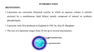
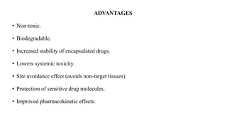
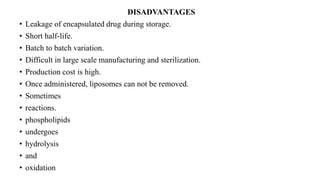
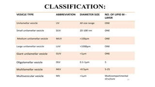
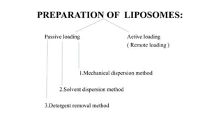
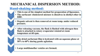
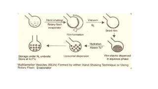
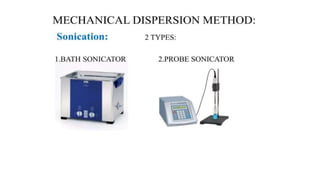
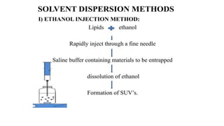
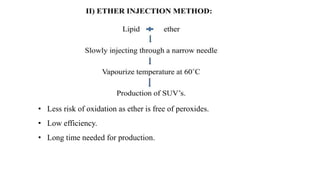
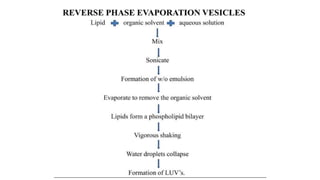
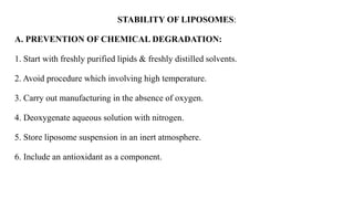
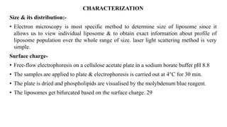
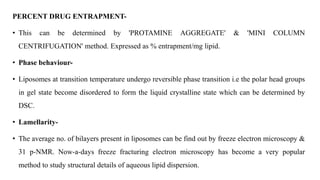
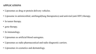
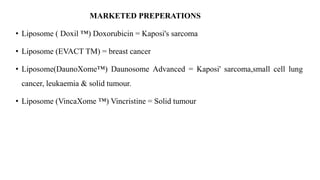
LIPOSOMES FORMULATION
AND EVALUATION
By
Sathish Kumar.P Mpharm 2nd Semester
Nandha College Of Pharmacy
CONTENTS
• Introduction
• Composition of liposome
• Advantages
• Disadvantages
• Classification
• Methods of preparation
• Stability
• Characterization / Evaluation
• Applications and marketed preparations
• References.
INTRODUCTION
DEFINITION:
• Liposomes are concentric bilayered vesicles in which an aqueous volume is entirely
enclosed by a membranous lipid bilayer mainly composed of natural or synthetic
phospholipids.
• Liposome were first produced in England in 1961 by Alec D. Bangham
• The size of a liposome ranges from 20 nm up to several micrometers.
PHOSPHOLIPIDS
• Phospholipids are the major structural components of biological membranes such
as the cell membrane.
PHOSPHOLIPIDS
• Phospholipids major structural are components of biological membranes in human
body, where 2 types of phospholipids exist phosphoglycerides & sphingolipids. i.e.
• Each phospholipid molecule has 3 major parts, 1 head & 2 tails. Head is made from 3
molecular components: choline, phosphate & glycerol which is hydrophilic. Each tail
with a long chain which are hydrophobic.
CHOLESTEROL
• Cholesterol stabilizes the membrane.
• It plays important role in bilayer formation.
• Cholesterol by itself does not form bilayer structure.
• Cholesterol act as fluidity buffer.
• Enhances the stability of the membrane.
• Enhances the rigidity of the phospholipid bilayer.
• Reduces the permeability of water soluble substance through the membrane.
ADVANTAGES
• Non-toxic.
• Biodegradable.
• Increased stability of encapsulated drugs.
• Lowers systemic toxicity.
• Site avoidance effect (avoids non-target tissues).
• Protection of sensitive drug molecules.
• Improved pharmacokinetic effects.
DISADVANTAGES
• Leakage of encapsulated drug during storage.
• Short half-life.
• Batch to batch variation.
• Difficult in large scale manufacturing and sterilization.
• Production cost is high.
• Once administered, liposomes can not be removed.
• Sometimes
• reactions.
• phospholipids
• undergoes
• hydrolysis
• and
• oxidation
MECHANICAL DISPERSION METHODS:
Hand-shaking method
• 2.Sonication
• 3.French pressure cell
• 4.Freeze-thaw technique
Solvent Dispersion Methods:
• 1.Ethanol injection
• 2.Ether injection
• 3.Reverse phase evaporation vesicles
Detergent Removal Methods:
• 1.Dialysis
• 2.Gel chromatography dilution
• At high energy levels, average size of vesicles is further reduced.
• Exposure of MLV's to ultrasonic irradiations is the most widely used
method for producing small vesicles.
• As chances of contamination are likely to occur in probe sonicator, bath
sonicator is widely used.
FRENCH PRESSURE CELL
• The french pressure cell is constructed from stainless steel and is capable of
withstanding very high pressures, even up to 20,000 - 40,000 psi.
• The body of the cell contains a pressure chamber, an outlet, a piston, bottom seal, etc.
both the piston and the bottom seal contain an O-ring each, which enables in tight
sealing the pressure cell.
WORKING:
• Initially the liposome suspension is added to the pressure cell and piston is pushed into
the body. Then the entire cell is turned upside down i.e., by an angle of 180°
• The liquid sample is then filled in the entire cavity till the outlet.
• After filling, the bottom seal is pressed down and the pressure cell is closed.
• The cell is brought back to upright position and the pressure is developed in the cell
using a hydraulic press.
• After sufficient pressure has been developed in the pressure cell, the valve is opened
very slowly and the product is allowed to exit in a drop-wise manner.
• Less risk of oxidation as ether is free of peroxides.
• Low efficiency.
• Long time needed for production.
STABILITY OF LIPOSOMES:
A. PREVENTION OF CHEMICAL DEGRADATION:
1. Start with freshly purified lipids & freshly distilled solvents.
2. Avoid procedure which involving high temperature.
3. Carry out manufacturing in the absence of oxygen.
4. Deoxygenate aqueous solution with nitrogen.
5. Store liposome suspension in an inert atmosphere.
6. Include an antioxidant as a component.
STABILITY OF LIPOSOMES:
B. PREVENTION OF PHYSICAL DEGRADATION:
1. ANNEALING' is best method to control physical degradation i.e incubating the liposomes
at a temperature high enough above the phase transition temperature.
2. The stability of liposomes may also be increased by cross linking membrane component
covalently using Gluteraldehyde fixation, osmification or polymerization of alkyne
containing phospholipids.
• These methods increases mechanical strength of the membrane & render them less
susceptible to disruption.
CHARACTERIZATION
Size & its distribution:-
• Electron microscopy is most specific method to determine size of liposome since it
allows us to view individual liposome & to obtain exact information about profile of
liposome population over the whole range of size. laser light scattering method is very
simple.
Surface charge-
• Free-flow electrophoresis on a cellulose acetate plate in a sodium borate buffer pH 8.8
• The samples are applied to plate & electrophoresis is carried out at 4°C for 30 min.
• The plate is dried and phospholipids are visualised by the molybdenum blue reagent.
• The liposomes get bifurcated based on the surface charge. 29
PERCENT DRUG ENTRAPMENT-
• This can be determined by 'PROTAMINE AGGREGATE' & 'MINI COLUMN
CENTRIFUGATION' method. Expressed as % entrapment/mg lipid.
• Phase behaviour-
• Liposomes at transition temperature undergo reversible phase transition i.e the polar head groups
in gel state become disordered to form the liquid crystalline state which can be determined by
DSC.
• Lamellarity-
• The average no. of bilayers present in liposomes can be find out by freeze electron microscopy &
31 p-NMR. Now-a-days freeze fracturing electron microscopy has become a very popular
method to study structural details of aqueous lipid dispersion.
APPLICATIONS
• Liposomes as drug or protein delivery vehicles.
• Liposome in antimicrobial, antifungal(lung therapeutics) and antiviral (anti HIV) therapy.
• In tumor therapy.
• gene therapy.
• In immunology.
• Liposomes as artificial blood surrogates.
• Liposomes as radio pharmaceutical and radio diagnostic carriers.
• Liposomes in cosmetics and dermatology.
MARKETED PREPERATIONS
• Liposome ( Doxil ™) Doxorubicin = Kaposi's sarcoma
• Liposome (EVACT TM) = breast cancer
• Liposome(DaunoXome™) Daunosome Advanced = Kaposi' sarcoma,small cell lung
cancer, leukaemia & solid tumour.
• Liposome (VincaXome ™) Vincristine = Solid tumour
REFERENCES:
• Baviskar D.T. and Jain D.K., Novel drug delivery systems, Nirali Prakashan, pg.no.-
14.15-14.27
• OY. Sultana., Liposomal Drug Delivery Systems: An Update Review., Current Drug
Delivery 2007, 4, 297-305.
• Sharma Shailesh, Sharma Neelam, Kumar Sandeep, Gupta GD., Liposomes: A
review., Journal of Pharmacy Research 2009, 2(7),1163-1167.

liposome

  • 1.
    LIPOSOMES FORMULATION AND EVALUATION By SathishKumar.P Mpharm 2nd Semester Nandha College Of Pharmacy
  • 2.
    CONTENTS • Introduction • Compositionof liposome • Advantages • Disadvantages • Classification • Methods of preparation • Stability • Characterization / Evaluation • Applications and marketed preparations • References.
  • 3.
    INTRODUCTION DEFINITION: • Liposomes areconcentric bilayered vesicles in which an aqueous volume is entirely enclosed by a membranous lipid bilayer mainly composed of natural or synthetic phospholipids. • Liposome were first produced in England in 1961 by Alec D. Bangham • The size of a liposome ranges from 20 nm up to several micrometers.
  • 4.
    PHOSPHOLIPIDS • Phospholipids arethe major structural components of biological membranes such as the cell membrane.
  • 5.
    PHOSPHOLIPIDS • Phospholipids majorstructural are components of biological membranes in human body, where 2 types of phospholipids exist phosphoglycerides & sphingolipids. i.e. • Each phospholipid molecule has 3 major parts, 1 head & 2 tails. Head is made from 3 molecular components: choline, phosphate & glycerol which is hydrophilic. Each tail with a long chain which are hydrophobic.
  • 7.
    CHOLESTEROL • Cholesterol stabilizesthe membrane. • It plays important role in bilayer formation. • Cholesterol by itself does not form bilayer structure. • Cholesterol act as fluidity buffer. • Enhances the stability of the membrane. • Enhances the rigidity of the phospholipid bilayer. • Reduces the permeability of water soluble substance through the membrane.
  • 8.
    ADVANTAGES • Non-toxic. • Biodegradable. •Increased stability of encapsulated drugs. • Lowers systemic toxicity. • Site avoidance effect (avoids non-target tissues). • Protection of sensitive drug molecules. • Improved pharmacokinetic effects.
  • 9.
    DISADVANTAGES • Leakage ofencapsulated drug during storage. • Short half-life. • Batch to batch variation. • Difficult in large scale manufacturing and sterilization. • Production cost is high. • Once administered, liposomes can not be removed. • Sometimes • reactions. • phospholipids • undergoes • hydrolysis • and • oxidation
  • 12.
    MECHANICAL DISPERSION METHODS: Hand-shakingmethod • 2.Sonication • 3.French pressure cell • 4.Freeze-thaw technique Solvent Dispersion Methods: • 1.Ethanol injection • 2.Ether injection • 3.Reverse phase evaporation vesicles Detergent Removal Methods: • 1.Dialysis • 2.Gel chromatography dilution
  • 16.
    • At highenergy levels, average size of vesicles is further reduced. • Exposure of MLV's to ultrasonic irradiations is the most widely used method for producing small vesicles. • As chances of contamination are likely to occur in probe sonicator, bath sonicator is widely used.
  • 17.
    FRENCH PRESSURE CELL •The french pressure cell is constructed from stainless steel and is capable of withstanding very high pressures, even up to 20,000 - 40,000 psi. • The body of the cell contains a pressure chamber, an outlet, a piston, bottom seal, etc. both the piston and the bottom seal contain an O-ring each, which enables in tight sealing the pressure cell.
  • 18.
    WORKING: • Initially theliposome suspension is added to the pressure cell and piston is pushed into the body. Then the entire cell is turned upside down i.e., by an angle of 180° • The liquid sample is then filled in the entire cavity till the outlet. • After filling, the bottom seal is pressed down and the pressure cell is closed. • The cell is brought back to upright position and the pressure is developed in the cell using a hydraulic press. • After sufficient pressure has been developed in the pressure cell, the valve is opened very slowly and the product is allowed to exit in a drop-wise manner.
  • 21.
    • Less riskof oxidation as ether is free of peroxides. • Low efficiency. • Long time needed for production.
  • 23.
    STABILITY OF LIPOSOMES: A.PREVENTION OF CHEMICAL DEGRADATION: 1. Start with freshly purified lipids & freshly distilled solvents. 2. Avoid procedure which involving high temperature. 3. Carry out manufacturing in the absence of oxygen. 4. Deoxygenate aqueous solution with nitrogen. 5. Store liposome suspension in an inert atmosphere. 6. Include an antioxidant as a component.
  • 24.
    STABILITY OF LIPOSOMES: B.PREVENTION OF PHYSICAL DEGRADATION: 1. ANNEALING' is best method to control physical degradation i.e incubating the liposomes at a temperature high enough above the phase transition temperature. 2. The stability of liposomes may also be increased by cross linking membrane component covalently using Gluteraldehyde fixation, osmification or polymerization of alkyne containing phospholipids. • These methods increases mechanical strength of the membrane & render them less susceptible to disruption.
  • 25.
    CHARACTERIZATION Size & itsdistribution:- • Electron microscopy is most specific method to determine size of liposome since it allows us to view individual liposome & to obtain exact information about profile of liposome population over the whole range of size. laser light scattering method is very simple. Surface charge- • Free-flow electrophoresis on a cellulose acetate plate in a sodium borate buffer pH 8.8 • The samples are applied to plate & electrophoresis is carried out at 4°C for 30 min. • The plate is dried and phospholipids are visualised by the molybdenum blue reagent. • The liposomes get bifurcated based on the surface charge. 29
  • 26.
    PERCENT DRUG ENTRAPMENT- •This can be determined by 'PROTAMINE AGGREGATE' & 'MINI COLUMN CENTRIFUGATION' method. Expressed as % entrapment/mg lipid. • Phase behaviour- • Liposomes at transition temperature undergo reversible phase transition i.e the polar head groups in gel state become disordered to form the liquid crystalline state which can be determined by DSC. • Lamellarity- • The average no. of bilayers present in liposomes can be find out by freeze electron microscopy & 31 p-NMR. Now-a-days freeze fracturing electron microscopy has become a very popular method to study structural details of aqueous lipid dispersion.
  • 27.
    APPLICATIONS • Liposomes asdrug or protein delivery vehicles. • Liposome in antimicrobial, antifungal(lung therapeutics) and antiviral (anti HIV) therapy. • In tumor therapy. • gene therapy. • In immunology. • Liposomes as artificial blood surrogates. • Liposomes as radio pharmaceutical and radio diagnostic carriers. • Liposomes in cosmetics and dermatology.
  • 28.
    MARKETED PREPERATIONS • Liposome( Doxil ™) Doxorubicin = Kaposi's sarcoma • Liposome (EVACT TM) = breast cancer • Liposome(DaunoXome™) Daunosome Advanced = Kaposi' sarcoma,small cell lung cancer, leukaemia & solid tumour. • Liposome (VincaXome ™) Vincristine = Solid tumour
  • 29.
    REFERENCES: • Baviskar D.T.and Jain D.K., Novel drug delivery systems, Nirali Prakashan, pg.no.- 14.15-14.27 • OY. Sultana., Liposomal Drug Delivery Systems: An Update Review., Current Drug Delivery 2007, 4, 297-305. • Sharma Shailesh, Sharma Neelam, Kumar Sandeep, Gupta GD., Liposomes: A review., Journal of Pharmacy Research 2009, 2(7),1163-1167.