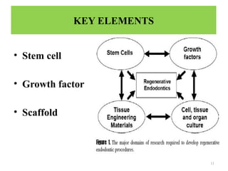
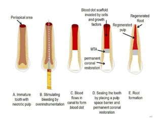
1
2
• Introduction
• Definitions
• Objective
• Key elements
Stem cells
Growth factors
Scaffolds
• POTENTIAL TECHNOLOGIES FOR REGENERATIVE
ENDODONTICS
• Root canal revascularization via blood clotting
• Postnatal stem cell therapy
• Scaffold implantation
• Injectable scaffold delivery
• Three-dimensional cell printing & gene delivery
• Measuring clinical outcomes
• Future application
• Conclusion
CONTENTS
3
INTRODUCTION
• Millions of teeth are saved each year by root canal
therapy. Although current treatment modalities offer
high levels of success for many conditions, an ideal
form of therapy might consist of regenerative
approaches in which diseased or necrotic pulp tissues
are removed and replaced with healthy pulp tissue to
revitalize teeth.
• Regenerative endodontics is the creation and delivery
of tissues to replace diseased, missing, and
traumatized pulp.
4
• Providing an overview of the methodological issues
is required to develop potential regenerative
endodontic therapies, and present a call for action to
develop these therapies for clinical use.
5
DEFINITION
• REGENERATIVE ENDODONTICS
• Regenerative endodontic procedures can be defined as
biologically based procedures designed to replace
damaged structures, including dentin and root
structures, as well as cells of the pulp-dentin complex.
• TISSUE ENGINEERING
• An interdisciplinary field that applies the principle of
engineering and life sciences towards the development
of biological substitutes that restore, maintain, or
improve tissue function.
6
REVASCULARIZATION
• Revascularization as defined by ‘andreasen’ is the
restoration of the vascularity to a tissue or organ.
REPAIR
• Repair is the restoration of tissue continuity without
the loss of original architecture and function.
7
HISTORY
• G. L. Feldman (1932) proposed that through
biological-aseptic principle of tooth therapy,
regeneration of pulp might be achieved and used
dentine fillings for stimulating pulp regeneration.
• In 1961, Nygaard ostby established a blood clot to
use as a scaffold to revascularize tissue within the
root canals of teeth.
• Concept of guided tissue and bone regenration were
first published by Meclcher in 1976.
8
• ƒIwaya, 2001:showed the revascularization potential
of an immature permanent tooth & this basically
renewed the interest in revascularization procedures.
• Trope in 2007 showed successful revascularization of
the necrotic , immature permanent max central incisor
induced in vivo by stimulation of blood clot from
periradicular tissues.
9
OBJECTIVES
• The objectives of regenerative endodontic procedure,
are to:
• Regenerate pulp like tissue, ideally. The pulp -dentin
complex
• Regenerate resorbed root, cervical or apical dentin.
10
GOALS
• Primary goal (essential): The elimination of
symptoms and the evidence of bony healing.
• Secondary goal (desirable): Increased root wall
thickness and/or increased root length.
• Tertiary goal: positive response to vitality
testing.
11
KEY ELEMENTS
• Stem cell
• Growth factor
• Scaffold
12
STEM CELLS
• A stem cell is defined as ‘‘
A cell that has the ability
to continuously divide and
produce progeny cells that
differentiate into various
other types of cells or
tissues.
13
All stem cells regardless of their source have three
general properties:
1. Capable of dividing and renewing themselves for
long periods
2. Unspecialized
3. May give rise to specialized cell types
14
CLASSIFICATION
Depending on the ability of the stem cells to produce the
different types of cells they are classified into pluripotent or
multipotent.
• Pluripotent stem cells are those which are capable of
differentiating into specialized cells of any three germ layers.
Truly pluripotent stem cells are found in the developing embryos.
• Stem cells, found in adults are restricted in their capacity to
differentiate and are thus termed multipotent. The mesenchymal
tissues (e.g. bone, dental pulp, periodontal ligament) appear to
have an enriched population of adult stem cells
15
• Stem cells are classified as embryonic/fetal and
postnatal/ adult stem cell.
• Embryonic stem cells derived from inner cell mass of
early embryo called blastocyst & are capable of dividing
and renewing themselves for long periods without
differentiating whereas adult stem cell cannot. Eg:
hematopoietic cell & hepatic cell etc.
• post natal cells taken from mature tissue. They are
lineage restricted & are referred to by their tissue of
origin. Its play an important role in local tissue repair
and regeneration. Eg: dental pulp stem cell
16
• Most stem cells in the oral region are of mesenchymal origin. It
is this multipotent capacity of the mesenchymal stem cells which
forms the basis of all regenerative endodontic procedures .
 Oral stems cells come under the post natal stem cell category
and includes
• Stem cells of the apical papilla (SCAP)
• Dental pulp stem cells (DPSCs)
• Inflamed periapical progenitor cells (iPAPCs)
• Periodontal ligament stem cells (PDLSCs)
• Bone marrow stem cells (BMSCs)
• Stem cells from human exfoliated deciduous teeth (SHED)
• Tooth germ progenitor cells (TGPCs)
• Dental follicle stem cells (DFSCs)
• Salivary gland stem cells (SGSCs)
J Istanbul Univ Fac Dent 2017;51(3 Suppl 1):S41-S51.
17
Schematic representation of the potential sources of postnatal stem cells in the oral
environment. Cell types include tooth germ progenitor cells (TGPCs), dental follicle
stem cells (DFSCs), salivary gland stem cells (SGSCs), stem cells of the apical papilla
(SCAP), dental pulp stem cells (DPSCs), stem cells from human exfoliated deciduous
teeth (SHED), periodontal ligament stem cells (PDLSCs), inflamed periapical
progenitor cells (iPAPCs) and bone marrow stem cells (BMSCs).
18
• Stem cells are often categorized by their source: The
most practical clinical application of a stem cell
therapy would be to use a patient’s own donor cells.
• Autologous stem cells are obtained from the same
individual to whom they will be implanted.
19
 Stem cells could be taken from the
• bone marrow
• peripheral blood
• fat removed by liposuction
• the periodontal ligament
• oral mucosa, or skin.
 An example of an autologous cell bank is one that stores
umbilical cord stem cells.
• It may be possible to use neuronal stem cells from adipose
fat as part of regenerative medicine instead of bone
marrow cells, possibly providing a less painful and less
threatening alternative collection method.
Seo BM, Miura M, Sonoyama W, Coppe C, Stanyon R, Shi S. Recovery of stem cells from
cryopreserved periodontal ligament. J Dent Res 2005;84:907–12.
20
• One potential disadvantage of harvesting cells from
patients is that surgical operations might lead to
postoperative sequelae, such as donor site infection.
• To accomplish endodontic regeneration, the most
promising cells are autologous postnatal stem cells,
because these appear to have the fewest
disadvantages that would prevent them from being
used clinically.
21
STEM CELL STORAGE
• CRYOPRESERVATION :
• It is the process of preserving cells or
whole tissues by cooling them to sub-
zero temperatures.
• Cells harvested near end of log phase
growth (approximately. 80–90%
confluent) are best for cryopreservation.
• Liquid nitrogen vapour is used to
preserve cells at a temperature of -196ºc
.
• In a vial 1.5 ml of freezing medium is
optimum for 1–2 ×10 6
cells
22
GROWTH FACTORS
• Growth factors are proteins that bind to receptors on
the cell and induce cellular proliferation and/or
differentiation.
• Many growth factors are quite versatile, stimulating
cellular division in numerous cell types, while others
are more cell specific.
• Growth factors play a role in signalling many
events in pulp dentine regeneration. Two
important families of growth factor that play a
vital role are transforming growth factor (TGF)
and bone morphogenetic protein (BMP).
23
GROWTH FACTORS CONTROL STEM CELLACTIVITY SUCH AS
24
25
26
• Fibroblast growth factors(FGF) are for general
growth-promoting effects on most fibroblastic cells:
it stimulates angiogenesis & cell migration in vivo.
• Platlet derived growth factor (PDGF) are derived
from platlets & endothelial cells: promotes
connective tissue cells.
27
SCAFFOLDS
• A scaffold is an artificial three-dimentional frame
structure that serves as a mimic of extracellular
matrix for cellular adhesion, migration, proliferation,
and tissue regeneration.
28
IDEAL REQUIREMENTS
29
CLASSIFICATION
30
BASED ON ORIGIN
 Platelet rich plasma
 Platelet rich fibrin
 Collagen
 Chitosan
 Glycosaminoglycans
 Hyaluronic acid
 Demineralized dentin
matrix
 Silk
Polymer
 Poly lactic acid
 Poly glycolic acid
 Polylacticcoglycolic
acid
Bioceramics
 Calcium
polyphosphate
 Bioactive glasses
 Glass ceramics
BIOLOGICAL/NATURAL
SCAFFOLDS
ARTIFICIAL/SYNTHETIC
SCAFFOLDS
gathani km, raghavendra ss. scaffolds in regenerative endodontics: a review. dent res j 2016;13:379-86
31
PLATLET RICH PLASMA(PRP)
• PRP is being established as a potentially ideal
scaffold for regenerative endodontic treatment
regimen.
• PRP contains growth factors, stimulates collagen
production, recruits other cells to the site of injury,
produces anti-inflammatory agents & intiates
vascular ingrowth.
32
• Blood sample from the patient’s arm(30 ml
approximately)
• Centrifuging the blood in the presence of an
anti-coagulant removal of erythrocytes from
the blood
• Then adding thrombin and calcium for
coagulation of prepared PRP then injected to
canal space
Preparation of PRP
#The first spin is at 2400 rpm for 10 minutes. With this spin we
separate the erythrocytes from platelet poor plasma. With the
second spin at 3600 rpm for 15 minutes we separate
the PRP from platelet poor plasma
33
34
PLATLET RICH FIBRIN
• Platelet-rich fibrin (PRF), introduced by Choukroun
in 2001, is one of the most commonly used
(autologous fibrin matrix) scaffolds in regenerative
endodontic procedures
• It is rich in pre-existing growth factors like PDGF,
TGF-ß etc., which helps in migration of fibroblasts
and endothelial cells.
35
Preparation of PRF
• PRF is prepared by collecting venous
blood from the patient’s arm without
anticoagulant.
• The blood is immediately centrifuged
at a speed of 3000 rpm for 10 mins.
• The resultant product consists of PRF
clot in the middle and RBC at the
bottom.
36
37
COLLAGEN
• It constitutes the extracellular matrix of all the tissues &
is responsible for the tensile strength of the tissues.
Advantages:
• Biocompatibility & biodegradability
• High alkaline phosphatase activity.
• Easy placement of cells & growth factors along with the
easy replacement with natural tissues after its
degradation.
• Collagen forms the hard and soft tissue which stimulates
the natural extracellular matrix of dentine
38
when used for REPs.
• Its available in various forms via
gels, sponges and sheets.
Disadvantage
• Rapid degradation and contraction
are the undesirable properties of
collagen
39
CHITOSAN
• Produced commercially by deacetylation of chitin,
which is a structural element in exoskeleton of
crustaceans (Crabs & Shrimps).
• Ability of chitosan to form porous scaffold makes it
is to be used in REPs.
• Advantages –
• Biocompatible, Easily absorbable,
• Antibacterial activity, increases alkaline phosphate
activity, shows fibroblast & odontoblastic
proliferation.
40
• Disadvantages –
• Low strength & inconsistency behavior with seeded
cells
41
GLYCOSAMINOGLYCANS
• Hyaluronic acid – One of the glycosaminoglycans in
ECM & plays an important role in maintaining
morphologic organization by preserving extracellular
spaces.
• It is bioactive and has properties of osteogenesis and
chondrogenesis. When used for REPs it can even
promote odontogenesis by forming pulp-dentin
complex.
42
• Advantages – Helps in differentiation of dental
mesenchymal cells to odontoblasts, contributes to
formation of dentin matrix & dental pulp,
• biocompatible, bioactive, non immunogenic
• Disadvantages –
• Water soluble, degrades rapidly by enzyme
hyaluronidase – Overcome by cross linking &
modification of HA.
43
DEMINERALIZED DENTIN MATRIX
• The organic matrix of dentin consist of 18% collagen,
2% noncollagenous proteins (NCPs), 70%
hydroxyapatite (HA), and 10% body fluid
(percentages indicating weight/volume). Their matrix
is a repository for growth factors, such as bone
morphogenetic proteins (BMPs), transforming growth
factor‑β, insulin like growth factor, and basic
‑
fibroblast growth factor.
44
Advantages:
• Non-immunogenic and mechanically superior
• It shows direct induction of differentiating
odontoblast like cells and indirect matrix synthesis
‑
leading to odontoblast differentiation
Disadvantage:
• Tooth demineralization is time consuming (usually 2–
6 days).Drawback of demineralization is that
prolonged acid exposure may negatively affect non-
collagenous proteins involved in new bone formation
45
ARTIFICIAL SCAFFOLDS
• Polymers with controlled physicochemical features
such as degradation rate, microstructure, and
mechanical strength. For eg:
• Polylactic acid , polyglycolic acid , and their
copolymers-poly lactic-co-glycolic acid etc.
• Synthetic hydrogels include polyethylene glycol based
polymers.
• Scaffolds modified with cell surface adhesion peptides
such as arginine, glycin & aspartic acid to improve cell
adhesion and matrix synthesis within the 3D network
46
• Scaffolds containing inorganic compounds such as
hydroxyapatite (HA), tricalcium phosphate (TCP) and
calcium polyphosphate (CPP), are used to enhance
bone conductivity.
47
DELIVERY SYSTEM
• Resultant mixture must be delivered in a spatially
appropriate fashion into the space of the root canal
system.
• two approaches:
• Inject a cell/scaffold/growth- factor mixture into the
root canal system. This approach is called cell
homing.
• Revascularization: inducing host stem cells from the
adjacent site to mobilize and inhabit the natural host
matrix.
48
AN OVERVIEW OF POTENTIAL
TECHNOLOGIES FOR
REGENERATIVE ENDODONTICS
49
REVASCULARIZATION
• Revascularization can be broadly defined as the
restoration of vascularity of a tissue or organ.
• The authors stated that the term revascularization
does not completely address the desired outcomes of
regenerative endodontic procedures, because the
desired outcome is regeneration of the pulp-dentin
complex.
50
MECHANISM
The process of revascularization takes place are as
follows:
• A few vital pulp cells remaining at the apical end
of the root canal might proliferate into the newly
formed matrix and differentiate into odontoblasts.
• Continued root development could be due to
multipotent dental pulp stem cells, which are
present abundantly in immature permanent teeth
51
• Stem cells in the periodontal ligament can proliferate
and grow into the apical end and within the root
canal.
• The fourth possible mechanism of root development
could be attributed to SCAP or to the bone marrow.
• Instrumentation beyond the confines of the root canal
to induce bleeding can also transplant mesenchymal
stem cells from the bone into the canal lumen.
• The blood clot is a rich source of growth factors such
as platelet-derived growth factor, vascular endothelial
growth factor, platelet derived epithelial growth
factor, and tissue growth factor.
52
CASE SELECTION
• Any tooth with necrotic pulp and an immaure apex is
a reasonable candidate for REPS, provided that the
pulp space is not needed for post/core and final
restoration.
• As with most endodontic procedures, compliance is
important due to the need for frequent follow-ups.
53
WHAT ARE THE CONSIDERATIONS FOR
CLINICAL REGENERATIVE ENDODONTIC
PROCEDURES
• Various regenerative endodontic treatment protocols have
been associated with a successful clinical outcome and
currently there is no single recommended protocol.
• Common features of cases with successful clinical outcomes
after REPs are:
1. Young patient
2. Necrotic pulp and immature apex
3. Minimal or no instrumentation of the dentinal walls
4. Placement of an intracanal medicament
5. Creation of a blood clot or protein scaffold in canal
6. Effective coronal seal
54
REVASCULARIZATION PROTOCOL
the ultimate goal of this approach is to develop a tissue engineering–
based method of pulpal regeneration in the fully developed
permanent tooth
Informed consent issues should include the number of appointments
(at least two), the possible adverse effects (primarily potential
minocycline staining of the crown), the potential lack of response to
treatment and alternative treatments, and possible post treatment
symptoms.
Clinical staining of the crown and any root structure above the
gingival margin appears to be due to the presence of minocycline.
This can be minimized by using a delivery system that restricts the
drug below the cementoenamel junction (CEJ).
Cohen 11th edition pg-467
55
First appointment
Following informed consent, the tooth is anesthetized, isolated, and
accessed
Minimal instrumentation should be accomplished, but the use of a
small file to “scout” the root canal system and determine working
length is important.
The root canal system is copiously and slowly irrigated with 20 ml of
NaOCl for 5 min followed by 20 ml of 0.12% to 2% chlorhexidine
(CHX).
It is important to place the needle into the apical third and irrigate
using needles with a closed end and side-port vents (e.g., Max-I-
Probe needles), together with a slow rate of infusion, to help to
reduce any irrigants passing through the open apex.
Cohen 11th edition pg-467
56
The root canal system is then dried with sterile paper points, and the
antimicrobial medicament is delivered into the root canal space.
Calcium hydroxide or an antibiotic paste or solution (1mg/ml) is
delivered to canal system. Access is temporarily restored
57
Second appointment
• The patient is evaluated for resolution of any signs or
symptoms of an acute infection (e.g., swelling, sinus
tract pain, etc.)
• The antimicrobial treatment is repeated if resolution has
not occurred.
• Since revascularization-induced bleeding will be evoked
at this appointment, the tooth should not be anesthetized
with a local anesthetic containing a vasoconstrictor.
• Instead, 3% mepivacaine can be used, which will
facilitate the ability to trigger bleeding into the root
canal system
58
• Following isolation and reestablishment of coronal
access, the intracanal medicament is removed by
irrigating with 17% EDTA(30ml/canal, 5 min)& then
final flush with saline(5ml/canal, 1 min).
• The canals are dried with paper points.
• Bleeding is induced by rotating a precurved k-file
size #25 at 2mm past the apical foramen with the goal
of having the whole canal filled with blood to the
level of the CEJ.
• Once a blood clot is formed, a premeasured piece of
collaplug is carefully placed on the top of the blood
clot to serve as an internal marix for the placement of
apporoximately 3mm white MTA or biodentin.
59
• A layer of GIC is flowed gently over the bioactive
coronal barrier and light cured for 40 sec. Then
composite resin restoration is placed over the GIC.
• A 12- to 18-month recall should be considered as the
earliest time point to conduct the clinical examination
and evaluate continued radiographic improvement in
root development
60
61
A COMPARISON BETWEEN REVASCULARIZATION
ENDODONTIC TREATMENT AND OTHER PULP
TREATMENT PROCEDURES
• There are several procedures designed to treat the
incompletely formed root that occur following
endodontic procedures.
• Apexification is defined as a method to induce a
calcified barrier in a root with an open apex or the
continued apical development of an incompletely
formed root in teeth with necrotic pulp tissue.
• This is distinct from revascularization, since
apexification does not attempt to regain vital tissue in
the canal space.
62
• The outcome of an apexification procedure is
establishment of an apical barrier against which an
obturating material may be placed.
• A second term, apexogenesis, is defined as a vital pulp
therapy procedure performed to encourage continued
physiologic development and formation of the root end.
• An important distinction is that apexogenesis is indicated
for teeth in which there has been no loss of vascularity,
thus no need to “revascularize” the canal space. (Cohen)
63
• In apexification with Ca(OH)2 :
1. Chances of root fracture and stem cell toxicity.
2. At least 6 months are required to create an apical
barrier, and multitple visits are needed to replenish
calcium hydroxide.
Andreasen JO, Farik B, Munksgaard EC. Long-term calcium hydroxide as a root canal dressing may increase risk of root
fracture. Dent Traumatol. 2002;18:134–137.
64
• MTA is used in the one or two step apexification
procedure, and therefore a fewer number of
appointments are needed.
• In spite of this, apexification with MTA neither
strengthens the root nor induces further root
development.
• As a result, the roots remain thin and fragile, and
hence another treatment approach is needed
Bose R, Nummikoski P, Hargreaves K. A retrospective evaluation of radiographic outcomes in immature teeth with necrotic root canal systems
treated with regenerative endodontic procedures. J Endod. 2009;35:1343–1349.
65
THE PERCENTAGE INCREASE IN ROOT WIDTH
AND ROOT LENGTH AFTER THE TREATMENT
PROCEDURE
Jeeruphan T, Jantarat J, Yanpiset K, Suwannapan L, Khewsawai P, Hargreaves KM. Mahidol study 1: comparison of radiographic
and survival outcomes of immature teeth treated with either regenerative endodontic or apexification methods: a retrospective
study. J Endod.2012;38:1330–1336.
66
MEDICAMENTS BEING USED IN
CASES OF REVASCULARIZATION
1.Triple antibiotic paste (1 : 1 : 1 mixture of
ciprofloxacin/metronidazole/minocycline)
2. Ca(OH)2 alone or with an antibiotic paste.
3. Formocresol
Bin-Na Lee, Jong-Wook Moon, ‘A review on regenerative endodontic treatment procedure’Restor Dent Endod. 2015 Aug; 40(3):
179–187
67
1.The triple antibiotic paste produced significantly greater
differences in dentinal wall thickness compared with either
the Ca(OH)2 or formocresol groups.
2.The formocresol group showed the smallest improvement
in root length and thickness.
3.Location of Ca(OH)2 placement appeared to be a strong
predictor of radiographic outcome.
4. When Ca(OH)2 placement was restricted to the coronal
half of the root canal, the increase in root wall thickness
was 55%, compared to a 3% increase when it was placed in
the apical half of the root canal system. This might be due
to residual Ca(OH)2 having a cytotoxic interaction with
stem cells
Bose R, Nummikoski P, Hargreaves K: A retrospective evaluation of radiographic outcomes in immature teeth with necrotic root
canal systems treated with regenerative endodontic procedures. J Endod 35:1343, 2009.
68
• ROLE OF ANTIBIOTIC PASTE
• The success of the regenerative endodontic procedure
depends on the effective disinfection of the canal.
• Antibiotic pastes are a combination of more than one
antibiotic mixed into a consistency of a paste.
• They are advocated as an effective alternative to
calcium hydroxide that has been traditionally used for
intracanal disinfection
69
Composition and mixing instructions
for the antibiotic paste (adapted from
Hoshino et al. 1996)
• 3- Mix- MP
• Antibiotics (3 Mix)
• Ciprofloxacin 200 mg
• Metronidazole 500 mg
• Minocycline 100 mg
• Carrier (MP)
• Macrogol ointment
• Propylene glycol
Hoshino E, Kurihara-Ando N, Sato I, Uematsu H, Sato M, Kota K. In-vitro antibacterialsusceptibility of bacteria taken from
infected root dentin to a mixture of ciprofloxacin,metronidazole and minocycline. Int Endod J 1996;29:125-30
70
• Protocol for preparation
• Antibiotics (3 Mix) –
• Remove sugar coating from tablets with surgical
blade, crush individually in separate mortars.
• Open capsules, crush in individually in separate
mortars. Grind each antibiotic to a fine powder.
• Combine equal amounts of antibiotics (1:1:1) on
mixing pad.
71
• Carrier (MP)
• Equal amounts of macrogol ointment and propylene glycol
(1:1).Using clean spatula, mix together on pad.
• MP: 3 Mix-1:7 (standard mix)
• If result is flaky or crumbly, then too much 3 mix has been
incorporated.
• Storage
• Antibiotics must be kept separately in moisture-tight porcelain
containers.
• Macrogol ointment and propylene glycol must be stored
separately.
• Discard if mixture is transparent (evidence of moisture
contamination)
72
• The triple antibiotic paste is the most commonly
advocated type and the following guidelines have to
be ensured when employing an antibiotic paste:
• It remains below CEJ (minimize crown staining).
• Concentration is adjusted to 0.1 mg/ml (100 μg of
each drug/ml).
• The pulp chamber is sealed with a dentin bonding
agent to avoid the risk of staining.
73
DISCOLORATION
• TAP is associated with severe discoloration due to the
presence of minocycline in it that binds with the
calcium of dentin forming insoluble complexes.
• To avoid staining while using TAP, the pulp chamber
should be sealed with dentine bonding agent and
ensure that TAP remains below the cementoenamel
junction (CEJ).
• The clinician should remove residual paste from the
pulp chamber and wipe clean it with cotton pellets
soaked in absolute alcohol.
Bansal, et al.: Challenges in regenerative endodontics Journal
74
• Modified TAP in which minocycline is substituted
with non-discoloring medicaments like
clarithromycin or fosfomycin or cefuroxime or
cefaclor have shown to be effective in eliminating
endodontic pathogens and were able to avoid the
permanent staining effect of the crown.
• Calcium hydroxide can also be used alternatively or
EndoVac apical negative-pressure irrigating system
along with sodium hypochlorite irrigation can be used
to avoid antibiotics completely as described in the
disinfection section.
75
• In addition, presence of gray mineral trioxide
aggregate (MTA) and white MTA might be another
source for discoloration which can be prevented by
using alternative tooth-colored bio-active materials
like calcium enriched mixture (CEM) cement over the
blood clot.
76
CLINICAL MEASURES OF
TREATMENT OUTCOME
• For revascularization not only radiographic evidence of
periradicular health but also radiographic and other
clinical evidence of functioning vital tissue in the canal
space.
• Radiographic evidence of functioning pulp (or pulp like)
tissue would include continued root growth, both in
length and wall thickness
• Other measures of the presence of vital, functioning
tissue in the canal space include laser Doppler blood
flowmetry, pulp testing involving heat, cold, and lack of
signs or symptoms.
77
• The ideal clinical outcome is an asymptomatic tooth
that does not require retreatment, but to validate that
regenerative endodontic techniques are truly
effective, non subjective vitality-assessment methods
are essential”
78
ADVANTAGES
• Achieving continued root development (root
lengthening) and strengthening due to reinforcement
of lateral dentinal walls with deposition of new
dentin/ hard tissue are the biggest advantages.
• Obturation of the canal is not required unlike in
calcium hydroxide–induced apexification (inherent
danger of splitting the root during lateral
condensation can be avoided).
• After control of infection, the procedure can be
completed in a single visit
79
DISADVANTAGES
• Discoloration due to use of minocycline in triple
antibiotic paste (revealed by Kim et al.)
• Prolonged treatment period and more appointments
(compared with one-visit MTA apical barrier
technique)
80
• POTENTIAL CAUSES OF FAILURE
• Poor root development (absence of increase in root
length, absence of increase in root wall thickness, or
lack of formation of tooth apex)
• Insufficient bleeding during the procedure
• Root canal calcification/obliteration
81
POSTNATAL STEM CELL THERAPY
• The simplest method to administer cells of appropriate
regenerative potential is to inject postnatal stem cells
into disinfected root canal systems after the apex is
opened.
• Postnatal stem cells can be derived from multiple tissues,
including skin, buccal mucosa, fat, and bone.
• Autologous or allogenic stem cells are delivered to teeth
via injectable matrix.
82
Advantages
• Quick, easy delivery, least painful and cells are easy o
harvest.
Disadvantages
• Low cell survival,
• Cells do not produce new functioning pulp & high
risk of complication
Murray P, Garcia-Godoy F, Hargreaves K. Regenerative Endodontics: A Review of Current Status and a Call for Action. Journal
Of Endodontics;2007; 33(4): 377-390
83
PULP IMPLANTATION
• The majority of in vitro cell cultures grow as a single
monolayer attached to the base of culture flasks.
However, some stem cells do not survive unless they
are grown on top of a layer of feeder cells In all of
these cases, the stem cells are grown in two
dimensions. In theory, to take two dimensional cell
cultures and make them three dimensional, the pulp
cells can be grown on biodegradable membrane
filters
84
Advantages:
•Sheets of cells are easy o grow.
•More stable than an injection of dissociate the cells .
Disadvantages:
•Sheets lack vascularity so only small constructs are
possible.
•Must be engineered to fit root canal precisely.
85
SCAFFOLD IMPLANTATION
• To create a more practical endodontic tissue
engineering therapy, pulp stem cells must be
organized into a three-dimensional structure that can
support cell organization and vascularization.
• This can be accomplished using a porous polymer
scaffold seeded with pulp stem cells
• A scaffold should contain growth factors to aid stem
cell proliferation and differentiation, leading to
improved and faster tissue development
86
• The scaffold may also contain nutrients promoting
cell survival and growth and possibly antibiotics to
prevent any bacterial in-growth in the canal systems.
• The engineering of nano scaffolds may be useful in
the delivery of pharmaceutical drugs to specific
tissues
• Dentin chips may provide a matrix for pulp stem cell
attachment and also be a reservoir of growth factors.
87
Advantages:
•Structure supports cell organization.
•Some materials may promote vascularization.
Disadvantages:
•Low cell survival afer implantation.
•Must be engineered to fit root canal precisely.
88
INJECTABLE SCAFFOLD DELIVERY
• Rigid tissue engineered scaffold structures provide
excellent support for cells used in bone and other
body areas
• In root canal systems a tissue engineered pulp is not
required to provide structural support of the tooth.
• This will allow tissue engineered pulp tissue to be
administered in a soft 3D scaffold matrix, such as a
polymer hydrogel.
• Hydrogels are injectable scaffolds that can be
delivered by syringe
89
• Hydrogels have the potential to be noninvasive and
easy to deliver into root canal systems.
• In theory, the hydrogel may promote pulp
regeneration by providing a substrate for cell
proliferation and differentiation into an organized
tissue structure.
90
Advantages:
• Easy delivery
• May promote regeneration by providing extracellular
matrix.
Disadvantages:
• Limited control over tissue formation
• Low cell survival
• Early stage reasearch has yet to be prove functional
in-vivo
91
3D CELL PRINTING
• An ink-jet-like device is used to dispense layers of
cells suspended in a hydrogel to recreate the structure
of the tooth pulp tissue.
• The ideal positioning of cells in a tissue engineering
construct would include placing odontoblastoid cells
around the periphery to maintain and repair dentin,
with fibroblasts in the pulp core supporting a network
of vascular and nerve cells
92
• Disadvantages:
• Construction of precise 3D models for each
individual pulp cavity.
• Cell line needs to be grown and expanded before
being implanted into the root canal i.e. is time
consuming.
• Implanted tissue lacks a crucial vascular supply.
93
GENE THERAPY
• All human cells contain a 1million strands of DNA containing 3
billion base pairs, with the sole exception of non-nucleated cells,
such as red blood cells.
• The DNA contains genetic sequences (genes) that control cell
activity and function; one of the most well known genes is p53.
• New techniques involving viral or nonviral vectors can deliver
genes for growth factors, morphogens, transcription factors, and
extracellular matrix molecules into target cell populations, such
as the salivary gland
Advantages:
•May avoid cleaning & shaping root canals
•May avoid the need to implant stem cells
Disadvantages:
•Most cells in a necrotic tooth are already dead.
•Difficult o control.
•Risk of health hazards
•Not apporved by he FDA
94
CHALLENGES AND FUTURE DIRECTION
95
CONCLUSION
• Regenerative endodontics is one of the most exciting developments
in dentistry today and endodontists are at the forefront of this
cutting-edge research.
• Endodontists’ knowledge in the fields of pulp biology, dental trauma
and tissue engineering can be applied to deliver biologically based
regenerative endodontic treatment of necrotic immature permanent
teeth resulting in continued root development, increased thickness in
the dentinal walls and apical closure.
• These developments in regeneration of a functional pulp-dentin
complex have a promising impact on efforts to retain the natural
dentition, the ultimate goal of endodontic treatment.
96
97
REFERENCES
• Cohen 11th edition pg- 465-467
• Grossman 13th
edition pg-265
• Vimal sikri essential of endodontics . Pg no 624-626
• Bansal, et al.: Challenges in regenerative endodontics Journal
• Clinical approaches regenerative endodontic procedure. Springer.
• Jeeruphan T, Jantarat J, Yanpiset K, Suwannapan L, Khewsawai P, Hargreaves KM. Mahidol
study 1: comparison of radiographic and survival outcomes of immature teeth treated with either
regenerative endodontic or apexification methods: a retrospective study. J Endod.2012;38:1330–
1336.
• Andreasen JO, Farik B, Munksgaard EC. Long-term calcium hydroxide as a root canal dressing
may increase risk of root fracture. Dent Traumatol. 2002;18:134–137
• Bose R, Nummikoski P, Hargreaves K: A retrospective evaluation of radiographic outcomes in
immature teeth with necrotic root canal.
• Chand vcu regenerative endodontics –treatment options and challenges to success ijocr oct - dec
20
• Murray P, Garcia-Godoy F, Hargreaves K. Regenerative Endodontics: A Review of Current
Status and a Call for Action. Journal Of Endodontics;2007; 33(4): 377-390
98

_regenerative-endodontics important for dental students

  • 1.
  • 2.
    2 • Introduction • Definitions •Objective • Key elements Stem cells Growth factors Scaffolds • POTENTIAL TECHNOLOGIES FOR REGENERATIVE ENDODONTICS • Root canal revascularization via blood clotting • Postnatal stem cell therapy • Scaffold implantation • Injectable scaffold delivery • Three-dimensional cell printing & gene delivery • Measuring clinical outcomes • Future application • Conclusion CONTENTS
  • 3.
    3 INTRODUCTION • Millions ofteeth are saved each year by root canal therapy. Although current treatment modalities offer high levels of success for many conditions, an ideal form of therapy might consist of regenerative approaches in which diseased or necrotic pulp tissues are removed and replaced with healthy pulp tissue to revitalize teeth. • Regenerative endodontics is the creation and delivery of tissues to replace diseased, missing, and traumatized pulp.
  • 4.
    4 • Providing anoverview of the methodological issues is required to develop potential regenerative endodontic therapies, and present a call for action to develop these therapies for clinical use.
  • 5.
    5 DEFINITION • REGENERATIVE ENDODONTICS •Regenerative endodontic procedures can be defined as biologically based procedures designed to replace damaged structures, including dentin and root structures, as well as cells of the pulp-dentin complex. • TISSUE ENGINEERING • An interdisciplinary field that applies the principle of engineering and life sciences towards the development of biological substitutes that restore, maintain, or improve tissue function.
  • 6.
    6 REVASCULARIZATION • Revascularization asdefined by ‘andreasen’ is the restoration of the vascularity to a tissue or organ. REPAIR • Repair is the restoration of tissue continuity without the loss of original architecture and function.
  • 7.
    7 HISTORY • G. L.Feldman (1932) proposed that through biological-aseptic principle of tooth therapy, regeneration of pulp might be achieved and used dentine fillings for stimulating pulp regeneration. • In 1961, Nygaard ostby established a blood clot to use as a scaffold to revascularize tissue within the root canals of teeth. • Concept of guided tissue and bone regenration were first published by Meclcher in 1976.
  • 8.
    8 • ƒIwaya, 2001:showedthe revascularization potential of an immature permanent tooth & this basically renewed the interest in revascularization procedures. • Trope in 2007 showed successful revascularization of the necrotic , immature permanent max central incisor induced in vivo by stimulation of blood clot from periradicular tissues.
  • 9.
    9 OBJECTIVES • The objectivesof regenerative endodontic procedure, are to: • Regenerate pulp like tissue, ideally. The pulp -dentin complex • Regenerate resorbed root, cervical or apical dentin.
  • 10.
    10 GOALS • Primary goal(essential): The elimination of symptoms and the evidence of bony healing. • Secondary goal (desirable): Increased root wall thickness and/or increased root length. • Tertiary goal: positive response to vitality testing.
  • 11.
    11 KEY ELEMENTS • Stemcell • Growth factor • Scaffold
  • 12.
    12 STEM CELLS • Astem cell is defined as ‘‘ A cell that has the ability to continuously divide and produce progeny cells that differentiate into various other types of cells or tissues.
  • 13.
    13 All stem cellsregardless of their source have three general properties: 1. Capable of dividing and renewing themselves for long periods 2. Unspecialized 3. May give rise to specialized cell types
  • 14.
    14 CLASSIFICATION Depending on theability of the stem cells to produce the different types of cells they are classified into pluripotent or multipotent. • Pluripotent stem cells are those which are capable of differentiating into specialized cells of any three germ layers. Truly pluripotent stem cells are found in the developing embryos. • Stem cells, found in adults are restricted in their capacity to differentiate and are thus termed multipotent. The mesenchymal tissues (e.g. bone, dental pulp, periodontal ligament) appear to have an enriched population of adult stem cells
  • 15.
    15 • Stem cellsare classified as embryonic/fetal and postnatal/ adult stem cell. • Embryonic stem cells derived from inner cell mass of early embryo called blastocyst & are capable of dividing and renewing themselves for long periods without differentiating whereas adult stem cell cannot. Eg: hematopoietic cell & hepatic cell etc. • post natal cells taken from mature tissue. They are lineage restricted & are referred to by their tissue of origin. Its play an important role in local tissue repair and regeneration. Eg: dental pulp stem cell
  • 16.
    16 • Most stemcells in the oral region are of mesenchymal origin. It is this multipotent capacity of the mesenchymal stem cells which forms the basis of all regenerative endodontic procedures .  Oral stems cells come under the post natal stem cell category and includes • Stem cells of the apical papilla (SCAP) • Dental pulp stem cells (DPSCs) • Inflamed periapical progenitor cells (iPAPCs) • Periodontal ligament stem cells (PDLSCs) • Bone marrow stem cells (BMSCs) • Stem cells from human exfoliated deciduous teeth (SHED) • Tooth germ progenitor cells (TGPCs) • Dental follicle stem cells (DFSCs) • Salivary gland stem cells (SGSCs) J Istanbul Univ Fac Dent 2017;51(3 Suppl 1):S41-S51.
  • 17.
    17 Schematic representation ofthe potential sources of postnatal stem cells in the oral environment. Cell types include tooth germ progenitor cells (TGPCs), dental follicle stem cells (DFSCs), salivary gland stem cells (SGSCs), stem cells of the apical papilla (SCAP), dental pulp stem cells (DPSCs), stem cells from human exfoliated deciduous teeth (SHED), periodontal ligament stem cells (PDLSCs), inflamed periapical progenitor cells (iPAPCs) and bone marrow stem cells (BMSCs).
  • 18.
    18 • Stem cellsare often categorized by their source: The most practical clinical application of a stem cell therapy would be to use a patient’s own donor cells. • Autologous stem cells are obtained from the same individual to whom they will be implanted.
  • 19.
    19  Stem cellscould be taken from the • bone marrow • peripheral blood • fat removed by liposuction • the periodontal ligament • oral mucosa, or skin.  An example of an autologous cell bank is one that stores umbilical cord stem cells. • It may be possible to use neuronal stem cells from adipose fat as part of regenerative medicine instead of bone marrow cells, possibly providing a less painful and less threatening alternative collection method. Seo BM, Miura M, Sonoyama W, Coppe C, Stanyon R, Shi S. Recovery of stem cells from cryopreserved periodontal ligament. J Dent Res 2005;84:907–12.
  • 20.
    20 • One potentialdisadvantage of harvesting cells from patients is that surgical operations might lead to postoperative sequelae, such as donor site infection. • To accomplish endodontic regeneration, the most promising cells are autologous postnatal stem cells, because these appear to have the fewest disadvantages that would prevent them from being used clinically.
  • 21.
    21 STEM CELL STORAGE •CRYOPRESERVATION : • It is the process of preserving cells or whole tissues by cooling them to sub- zero temperatures. • Cells harvested near end of log phase growth (approximately. 80–90% confluent) are best for cryopreservation. • Liquid nitrogen vapour is used to preserve cells at a temperature of -196ºc . • In a vial 1.5 ml of freezing medium is optimum for 1–2 ×10 6 cells
  • 22.
    22 GROWTH FACTORS • Growthfactors are proteins that bind to receptors on the cell and induce cellular proliferation and/or differentiation. • Many growth factors are quite versatile, stimulating cellular division in numerous cell types, while others are more cell specific. • Growth factors play a role in signalling many events in pulp dentine regeneration. Two important families of growth factor that play a vital role are transforming growth factor (TGF) and bone morphogenetic protein (BMP).
  • 23.
    23 GROWTH FACTORS CONTROLSTEM CELLACTIVITY SUCH AS
  • 24.
  • 25.
  • 26.
    26 • Fibroblast growthfactors(FGF) are for general growth-promoting effects on most fibroblastic cells: it stimulates angiogenesis & cell migration in vivo. • Platlet derived growth factor (PDGF) are derived from platlets & endothelial cells: promotes connective tissue cells.
  • 27.
    27 SCAFFOLDS • A scaffoldis an artificial three-dimentional frame structure that serves as a mimic of extracellular matrix for cellular adhesion, migration, proliferation, and tissue regeneration.
  • 28.
  • 29.
  • 30.
    30 BASED ON ORIGIN Platelet rich plasma  Platelet rich fibrin  Collagen  Chitosan  Glycosaminoglycans  Hyaluronic acid  Demineralized dentin matrix  Silk Polymer  Poly lactic acid  Poly glycolic acid  Polylacticcoglycolic acid Bioceramics  Calcium polyphosphate  Bioactive glasses  Glass ceramics BIOLOGICAL/NATURAL SCAFFOLDS ARTIFICIAL/SYNTHETIC SCAFFOLDS gathani km, raghavendra ss. scaffolds in regenerative endodontics: a review. dent res j 2016;13:379-86
  • 31.
    31 PLATLET RICH PLASMA(PRP) •PRP is being established as a potentially ideal scaffold for regenerative endodontic treatment regimen. • PRP contains growth factors, stimulates collagen production, recruits other cells to the site of injury, produces anti-inflammatory agents & intiates vascular ingrowth.
  • 32.
    32 • Blood samplefrom the patient’s arm(30 ml approximately) • Centrifuging the blood in the presence of an anti-coagulant removal of erythrocytes from the blood • Then adding thrombin and calcium for coagulation of prepared PRP then injected to canal space Preparation of PRP #The first spin is at 2400 rpm for 10 minutes. With this spin we separate the erythrocytes from platelet poor plasma. With the second spin at 3600 rpm for 15 minutes we separate the PRP from platelet poor plasma
  • 33.
  • 34.
    34 PLATLET RICH FIBRIN •Platelet-rich fibrin (PRF), introduced by Choukroun in 2001, is one of the most commonly used (autologous fibrin matrix) scaffolds in regenerative endodontic procedures • It is rich in pre-existing growth factors like PDGF, TGF-ß etc., which helps in migration of fibroblasts and endothelial cells.
  • 35.
    35 Preparation of PRF •PRF is prepared by collecting venous blood from the patient’s arm without anticoagulant. • The blood is immediately centrifuged at a speed of 3000 rpm for 10 mins. • The resultant product consists of PRF clot in the middle and RBC at the bottom.
  • 36.
  • 37.
    37 COLLAGEN • It constitutesthe extracellular matrix of all the tissues & is responsible for the tensile strength of the tissues. Advantages: • Biocompatibility & biodegradability • High alkaline phosphatase activity. • Easy placement of cells & growth factors along with the easy replacement with natural tissues after its degradation. • Collagen forms the hard and soft tissue which stimulates the natural extracellular matrix of dentine
  • 38.
    38 when used forREPs. • Its available in various forms via gels, sponges and sheets. Disadvantage • Rapid degradation and contraction are the undesirable properties of collagen
  • 39.
    39 CHITOSAN • Produced commerciallyby deacetylation of chitin, which is a structural element in exoskeleton of crustaceans (Crabs & Shrimps). • Ability of chitosan to form porous scaffold makes it is to be used in REPs. • Advantages – • Biocompatible, Easily absorbable, • Antibacterial activity, increases alkaline phosphate activity, shows fibroblast & odontoblastic proliferation.
  • 40.
    40 • Disadvantages – •Low strength & inconsistency behavior with seeded cells
  • 41.
    41 GLYCOSAMINOGLYCANS • Hyaluronic acid– One of the glycosaminoglycans in ECM & plays an important role in maintaining morphologic organization by preserving extracellular spaces. • It is bioactive and has properties of osteogenesis and chondrogenesis. When used for REPs it can even promote odontogenesis by forming pulp-dentin complex.
  • 42.
    42 • Advantages –Helps in differentiation of dental mesenchymal cells to odontoblasts, contributes to formation of dentin matrix & dental pulp, • biocompatible, bioactive, non immunogenic • Disadvantages – • Water soluble, degrades rapidly by enzyme hyaluronidase – Overcome by cross linking & modification of HA.
  • 43.
    43 DEMINERALIZED DENTIN MATRIX •The organic matrix of dentin consist of 18% collagen, 2% noncollagenous proteins (NCPs), 70% hydroxyapatite (HA), and 10% body fluid (percentages indicating weight/volume). Their matrix is a repository for growth factors, such as bone morphogenetic proteins (BMPs), transforming growth factor‑β, insulin like growth factor, and basic ‑ fibroblast growth factor.
  • 44.
    44 Advantages: • Non-immunogenic andmechanically superior • It shows direct induction of differentiating odontoblast like cells and indirect matrix synthesis ‑ leading to odontoblast differentiation Disadvantage: • Tooth demineralization is time consuming (usually 2– 6 days).Drawback of demineralization is that prolonged acid exposure may negatively affect non- collagenous proteins involved in new bone formation
  • 45.
    45 ARTIFICIAL SCAFFOLDS • Polymerswith controlled physicochemical features such as degradation rate, microstructure, and mechanical strength. For eg: • Polylactic acid , polyglycolic acid , and their copolymers-poly lactic-co-glycolic acid etc. • Synthetic hydrogels include polyethylene glycol based polymers. • Scaffolds modified with cell surface adhesion peptides such as arginine, glycin & aspartic acid to improve cell adhesion and matrix synthesis within the 3D network
  • 46.
    46 • Scaffolds containinginorganic compounds such as hydroxyapatite (HA), tricalcium phosphate (TCP) and calcium polyphosphate (CPP), are used to enhance bone conductivity.
  • 47.
    47 DELIVERY SYSTEM • Resultantmixture must be delivered in a spatially appropriate fashion into the space of the root canal system. • two approaches: • Inject a cell/scaffold/growth- factor mixture into the root canal system. This approach is called cell homing. • Revascularization: inducing host stem cells from the adjacent site to mobilize and inhabit the natural host matrix.
  • 48.
    48 AN OVERVIEW OFPOTENTIAL TECHNOLOGIES FOR REGENERATIVE ENDODONTICS
  • 49.
    49 REVASCULARIZATION • Revascularization canbe broadly defined as the restoration of vascularity of a tissue or organ. • The authors stated that the term revascularization does not completely address the desired outcomes of regenerative endodontic procedures, because the desired outcome is regeneration of the pulp-dentin complex.
  • 50.
    50 MECHANISM The process ofrevascularization takes place are as follows: • A few vital pulp cells remaining at the apical end of the root canal might proliferate into the newly formed matrix and differentiate into odontoblasts. • Continued root development could be due to multipotent dental pulp stem cells, which are present abundantly in immature permanent teeth
  • 51.
    51 • Stem cellsin the periodontal ligament can proliferate and grow into the apical end and within the root canal. • The fourth possible mechanism of root development could be attributed to SCAP or to the bone marrow. • Instrumentation beyond the confines of the root canal to induce bleeding can also transplant mesenchymal stem cells from the bone into the canal lumen. • The blood clot is a rich source of growth factors such as platelet-derived growth factor, vascular endothelial growth factor, platelet derived epithelial growth factor, and tissue growth factor.
  • 52.
    52 CASE SELECTION • Anytooth with necrotic pulp and an immaure apex is a reasonable candidate for REPS, provided that the pulp space is not needed for post/core and final restoration. • As with most endodontic procedures, compliance is important due to the need for frequent follow-ups.
  • 53.
    53 WHAT ARE THECONSIDERATIONS FOR CLINICAL REGENERATIVE ENDODONTIC PROCEDURES • Various regenerative endodontic treatment protocols have been associated with a successful clinical outcome and currently there is no single recommended protocol. • Common features of cases with successful clinical outcomes after REPs are: 1. Young patient 2. Necrotic pulp and immature apex 3. Minimal or no instrumentation of the dentinal walls 4. Placement of an intracanal medicament 5. Creation of a blood clot or protein scaffold in canal 6. Effective coronal seal
  • 54.
    54 REVASCULARIZATION PROTOCOL the ultimategoal of this approach is to develop a tissue engineering– based method of pulpal regeneration in the fully developed permanent tooth Informed consent issues should include the number of appointments (at least two), the possible adverse effects (primarily potential minocycline staining of the crown), the potential lack of response to treatment and alternative treatments, and possible post treatment symptoms. Clinical staining of the crown and any root structure above the gingival margin appears to be due to the presence of minocycline. This can be minimized by using a delivery system that restricts the drug below the cementoenamel junction (CEJ). Cohen 11th edition pg-467
  • 55.
    55 First appointment Following informedconsent, the tooth is anesthetized, isolated, and accessed Minimal instrumentation should be accomplished, but the use of a small file to “scout” the root canal system and determine working length is important. The root canal system is copiously and slowly irrigated with 20 ml of NaOCl for 5 min followed by 20 ml of 0.12% to 2% chlorhexidine (CHX). It is important to place the needle into the apical third and irrigate using needles with a closed end and side-port vents (e.g., Max-I- Probe needles), together with a slow rate of infusion, to help to reduce any irrigants passing through the open apex. Cohen 11th edition pg-467
  • 56.
    56 The root canalsystem is then dried with sterile paper points, and the antimicrobial medicament is delivered into the root canal space. Calcium hydroxide or an antibiotic paste or solution (1mg/ml) is delivered to canal system. Access is temporarily restored
  • 57.
    57 Second appointment • Thepatient is evaluated for resolution of any signs or symptoms of an acute infection (e.g., swelling, sinus tract pain, etc.) • The antimicrobial treatment is repeated if resolution has not occurred. • Since revascularization-induced bleeding will be evoked at this appointment, the tooth should not be anesthetized with a local anesthetic containing a vasoconstrictor. • Instead, 3% mepivacaine can be used, which will facilitate the ability to trigger bleeding into the root canal system
  • 58.
    58 • Following isolationand reestablishment of coronal access, the intracanal medicament is removed by irrigating with 17% EDTA(30ml/canal, 5 min)& then final flush with saline(5ml/canal, 1 min). • The canals are dried with paper points. • Bleeding is induced by rotating a precurved k-file size #25 at 2mm past the apical foramen with the goal of having the whole canal filled with blood to the level of the CEJ. • Once a blood clot is formed, a premeasured piece of collaplug is carefully placed on the top of the blood clot to serve as an internal marix for the placement of apporoximately 3mm white MTA or biodentin.
  • 59.
    59 • A layerof GIC is flowed gently over the bioactive coronal barrier and light cured for 40 sec. Then composite resin restoration is placed over the GIC. • A 12- to 18-month recall should be considered as the earliest time point to conduct the clinical examination and evaluate continued radiographic improvement in root development
  • 60.
  • 61.
    61 A COMPARISON BETWEENREVASCULARIZATION ENDODONTIC TREATMENT AND OTHER PULP TREATMENT PROCEDURES • There are several procedures designed to treat the incompletely formed root that occur following endodontic procedures. • Apexification is defined as a method to induce a calcified barrier in a root with an open apex or the continued apical development of an incompletely formed root in teeth with necrotic pulp tissue. • This is distinct from revascularization, since apexification does not attempt to regain vital tissue in the canal space.
  • 62.
    62 • The outcomeof an apexification procedure is establishment of an apical barrier against which an obturating material may be placed. • A second term, apexogenesis, is defined as a vital pulp therapy procedure performed to encourage continued physiologic development and formation of the root end. • An important distinction is that apexogenesis is indicated for teeth in which there has been no loss of vascularity, thus no need to “revascularize” the canal space. (Cohen)
  • 63.
    63 • In apexificationwith Ca(OH)2 : 1. Chances of root fracture and stem cell toxicity. 2. At least 6 months are required to create an apical barrier, and multitple visits are needed to replenish calcium hydroxide. Andreasen JO, Farik B, Munksgaard EC. Long-term calcium hydroxide as a root canal dressing may increase risk of root fracture. Dent Traumatol. 2002;18:134–137.
  • 64.
    64 • MTA isused in the one or two step apexification procedure, and therefore a fewer number of appointments are needed. • In spite of this, apexification with MTA neither strengthens the root nor induces further root development. • As a result, the roots remain thin and fragile, and hence another treatment approach is needed Bose R, Nummikoski P, Hargreaves K. A retrospective evaluation of radiographic outcomes in immature teeth with necrotic root canal systems treated with regenerative endodontic procedures. J Endod. 2009;35:1343–1349.
  • 65.
    65 THE PERCENTAGE INCREASEIN ROOT WIDTH AND ROOT LENGTH AFTER THE TREATMENT PROCEDURE Jeeruphan T, Jantarat J, Yanpiset K, Suwannapan L, Khewsawai P, Hargreaves KM. Mahidol study 1: comparison of radiographic and survival outcomes of immature teeth treated with either regenerative endodontic or apexification methods: a retrospective study. J Endod.2012;38:1330–1336.
  • 66.
    66 MEDICAMENTS BEING USEDIN CASES OF REVASCULARIZATION 1.Triple antibiotic paste (1 : 1 : 1 mixture of ciprofloxacin/metronidazole/minocycline) 2. Ca(OH)2 alone or with an antibiotic paste. 3. Formocresol Bin-Na Lee, Jong-Wook Moon, ‘A review on regenerative endodontic treatment procedure’Restor Dent Endod. 2015 Aug; 40(3): 179–187
  • 67.
    67 1.The triple antibioticpaste produced significantly greater differences in dentinal wall thickness compared with either the Ca(OH)2 or formocresol groups. 2.The formocresol group showed the smallest improvement in root length and thickness. 3.Location of Ca(OH)2 placement appeared to be a strong predictor of radiographic outcome. 4. When Ca(OH)2 placement was restricted to the coronal half of the root canal, the increase in root wall thickness was 55%, compared to a 3% increase when it was placed in the apical half of the root canal system. This might be due to residual Ca(OH)2 having a cytotoxic interaction with stem cells Bose R, Nummikoski P, Hargreaves K: A retrospective evaluation of radiographic outcomes in immature teeth with necrotic root canal systems treated with regenerative endodontic procedures. J Endod 35:1343, 2009.
  • 68.
    68 • ROLE OFANTIBIOTIC PASTE • The success of the regenerative endodontic procedure depends on the effective disinfection of the canal. • Antibiotic pastes are a combination of more than one antibiotic mixed into a consistency of a paste. • They are advocated as an effective alternative to calcium hydroxide that has been traditionally used for intracanal disinfection
  • 69.
    69 Composition and mixinginstructions for the antibiotic paste (adapted from Hoshino et al. 1996) • 3- Mix- MP • Antibiotics (3 Mix) • Ciprofloxacin 200 mg • Metronidazole 500 mg • Minocycline 100 mg • Carrier (MP) • Macrogol ointment • Propylene glycol Hoshino E, Kurihara-Ando N, Sato I, Uematsu H, Sato M, Kota K. In-vitro antibacterialsusceptibility of bacteria taken from infected root dentin to a mixture of ciprofloxacin,metronidazole and minocycline. Int Endod J 1996;29:125-30
  • 70.
    70 • Protocol forpreparation • Antibiotics (3 Mix) – • Remove sugar coating from tablets with surgical blade, crush individually in separate mortars. • Open capsules, crush in individually in separate mortars. Grind each antibiotic to a fine powder. • Combine equal amounts of antibiotics (1:1:1) on mixing pad.
  • 71.
    71 • Carrier (MP) •Equal amounts of macrogol ointment and propylene glycol (1:1).Using clean spatula, mix together on pad. • MP: 3 Mix-1:7 (standard mix) • If result is flaky or crumbly, then too much 3 mix has been incorporated. • Storage • Antibiotics must be kept separately in moisture-tight porcelain containers. • Macrogol ointment and propylene glycol must be stored separately. • Discard if mixture is transparent (evidence of moisture contamination)
  • 72.
    72 • The tripleantibiotic paste is the most commonly advocated type and the following guidelines have to be ensured when employing an antibiotic paste: • It remains below CEJ (minimize crown staining). • Concentration is adjusted to 0.1 mg/ml (100 μg of each drug/ml). • The pulp chamber is sealed with a dentin bonding agent to avoid the risk of staining.
  • 73.
    73 DISCOLORATION • TAP isassociated with severe discoloration due to the presence of minocycline in it that binds with the calcium of dentin forming insoluble complexes. • To avoid staining while using TAP, the pulp chamber should be sealed with dentine bonding agent and ensure that TAP remains below the cementoenamel junction (CEJ). • The clinician should remove residual paste from the pulp chamber and wipe clean it with cotton pellets soaked in absolute alcohol. Bansal, et al.: Challenges in regenerative endodontics Journal
  • 74.
    74 • Modified TAPin which minocycline is substituted with non-discoloring medicaments like clarithromycin or fosfomycin or cefuroxime or cefaclor have shown to be effective in eliminating endodontic pathogens and were able to avoid the permanent staining effect of the crown. • Calcium hydroxide can also be used alternatively or EndoVac apical negative-pressure irrigating system along with sodium hypochlorite irrigation can be used to avoid antibiotics completely as described in the disinfection section.
  • 75.
    75 • In addition,presence of gray mineral trioxide aggregate (MTA) and white MTA might be another source for discoloration which can be prevented by using alternative tooth-colored bio-active materials like calcium enriched mixture (CEM) cement over the blood clot.
  • 76.
    76 CLINICAL MEASURES OF TREATMENTOUTCOME • For revascularization not only radiographic evidence of periradicular health but also radiographic and other clinical evidence of functioning vital tissue in the canal space. • Radiographic evidence of functioning pulp (or pulp like) tissue would include continued root growth, both in length and wall thickness • Other measures of the presence of vital, functioning tissue in the canal space include laser Doppler blood flowmetry, pulp testing involving heat, cold, and lack of signs or symptoms.
  • 77.
    77 • The idealclinical outcome is an asymptomatic tooth that does not require retreatment, but to validate that regenerative endodontic techniques are truly effective, non subjective vitality-assessment methods are essential”
  • 78.
    78 ADVANTAGES • Achieving continuedroot development (root lengthening) and strengthening due to reinforcement of lateral dentinal walls with deposition of new dentin/ hard tissue are the biggest advantages. • Obturation of the canal is not required unlike in calcium hydroxide–induced apexification (inherent danger of splitting the root during lateral condensation can be avoided). • After control of infection, the procedure can be completed in a single visit
  • 79.
    79 DISADVANTAGES • Discoloration dueto use of minocycline in triple antibiotic paste (revealed by Kim et al.) • Prolonged treatment period and more appointments (compared with one-visit MTA apical barrier technique)
  • 80.
    80 • POTENTIAL CAUSESOF FAILURE • Poor root development (absence of increase in root length, absence of increase in root wall thickness, or lack of formation of tooth apex) • Insufficient bleeding during the procedure • Root canal calcification/obliteration
  • 81.
    81 POSTNATAL STEM CELLTHERAPY • The simplest method to administer cells of appropriate regenerative potential is to inject postnatal stem cells into disinfected root canal systems after the apex is opened. • Postnatal stem cells can be derived from multiple tissues, including skin, buccal mucosa, fat, and bone. • Autologous or allogenic stem cells are delivered to teeth via injectable matrix.
  • 82.
    82 Advantages • Quick, easydelivery, least painful and cells are easy o harvest. Disadvantages • Low cell survival, • Cells do not produce new functioning pulp & high risk of complication Murray P, Garcia-Godoy F, Hargreaves K. Regenerative Endodontics: A Review of Current Status and a Call for Action. Journal Of Endodontics;2007; 33(4): 377-390
  • 83.
    83 PULP IMPLANTATION • Themajority of in vitro cell cultures grow as a single monolayer attached to the base of culture flasks. However, some stem cells do not survive unless they are grown on top of a layer of feeder cells In all of these cases, the stem cells are grown in two dimensions. In theory, to take two dimensional cell cultures and make them three dimensional, the pulp cells can be grown on biodegradable membrane filters
  • 84.
    84 Advantages: •Sheets of cellsare easy o grow. •More stable than an injection of dissociate the cells . Disadvantages: •Sheets lack vascularity so only small constructs are possible. •Must be engineered to fit root canal precisely.
  • 85.
    85 SCAFFOLD IMPLANTATION • Tocreate a more practical endodontic tissue engineering therapy, pulp stem cells must be organized into a three-dimensional structure that can support cell organization and vascularization. • This can be accomplished using a porous polymer scaffold seeded with pulp stem cells • A scaffold should contain growth factors to aid stem cell proliferation and differentiation, leading to improved and faster tissue development
  • 86.
    86 • The scaffoldmay also contain nutrients promoting cell survival and growth and possibly antibiotics to prevent any bacterial in-growth in the canal systems. • The engineering of nano scaffolds may be useful in the delivery of pharmaceutical drugs to specific tissues • Dentin chips may provide a matrix for pulp stem cell attachment and also be a reservoir of growth factors.
  • 87.
    87 Advantages: •Structure supports cellorganization. •Some materials may promote vascularization. Disadvantages: •Low cell survival afer implantation. •Must be engineered to fit root canal precisely.
  • 88.
    88 INJECTABLE SCAFFOLD DELIVERY •Rigid tissue engineered scaffold structures provide excellent support for cells used in bone and other body areas • In root canal systems a tissue engineered pulp is not required to provide structural support of the tooth. • This will allow tissue engineered pulp tissue to be administered in a soft 3D scaffold matrix, such as a polymer hydrogel. • Hydrogels are injectable scaffolds that can be delivered by syringe
  • 89.
    89 • Hydrogels havethe potential to be noninvasive and easy to deliver into root canal systems. • In theory, the hydrogel may promote pulp regeneration by providing a substrate for cell proliferation and differentiation into an organized tissue structure.
  • 90.
    90 Advantages: • Easy delivery •May promote regeneration by providing extracellular matrix. Disadvantages: • Limited control over tissue formation • Low cell survival • Early stage reasearch has yet to be prove functional in-vivo
  • 91.
    91 3D CELL PRINTING •An ink-jet-like device is used to dispense layers of cells suspended in a hydrogel to recreate the structure of the tooth pulp tissue. • The ideal positioning of cells in a tissue engineering construct would include placing odontoblastoid cells around the periphery to maintain and repair dentin, with fibroblasts in the pulp core supporting a network of vascular and nerve cells
  • 92.
    92 • Disadvantages: • Constructionof precise 3D models for each individual pulp cavity. • Cell line needs to be grown and expanded before being implanted into the root canal i.e. is time consuming. • Implanted tissue lacks a crucial vascular supply.
  • 93.
    93 GENE THERAPY • Allhuman cells contain a 1million strands of DNA containing 3 billion base pairs, with the sole exception of non-nucleated cells, such as red blood cells. • The DNA contains genetic sequences (genes) that control cell activity and function; one of the most well known genes is p53. • New techniques involving viral or nonviral vectors can deliver genes for growth factors, morphogens, transcription factors, and extracellular matrix molecules into target cell populations, such as the salivary gland
  • 94.
    Advantages: •May avoid cleaning& shaping root canals •May avoid the need to implant stem cells Disadvantages: •Most cells in a necrotic tooth are already dead. •Difficult o control. •Risk of health hazards •Not apporved by he FDA 94
  • 95.
  • 96.
    CONCLUSION • Regenerative endodonticsis one of the most exciting developments in dentistry today and endodontists are at the forefront of this cutting-edge research. • Endodontists’ knowledge in the fields of pulp biology, dental trauma and tissue engineering can be applied to deliver biologically based regenerative endodontic treatment of necrotic immature permanent teeth resulting in continued root development, increased thickness in the dentinal walls and apical closure. • These developments in regeneration of a functional pulp-dentin complex have a promising impact on efforts to retain the natural dentition, the ultimate goal of endodontic treatment. 96
  • 97.
    97 REFERENCES • Cohen 11thedition pg- 465-467 • Grossman 13th edition pg-265 • Vimal sikri essential of endodontics . Pg no 624-626 • Bansal, et al.: Challenges in regenerative endodontics Journal • Clinical approaches regenerative endodontic procedure. Springer. • Jeeruphan T, Jantarat J, Yanpiset K, Suwannapan L, Khewsawai P, Hargreaves KM. Mahidol study 1: comparison of radiographic and survival outcomes of immature teeth treated with either regenerative endodontic or apexification methods: a retrospective study. J Endod.2012;38:1330– 1336. • Andreasen JO, Farik B, Munksgaard EC. Long-term calcium hydroxide as a root canal dressing may increase risk of root fracture. Dent Traumatol. 2002;18:134–137 • Bose R, Nummikoski P, Hargreaves K: A retrospective evaluation of radiographic outcomes in immature teeth with necrotic root canal. • Chand vcu regenerative endodontics –treatment options and challenges to success ijocr oct - dec 20 • Murray P, Garcia-Godoy F, Hargreaves K. Regenerative Endodontics: A Review of Current Status and a Call for Action. Journal Of Endodontics;2007; 33(4): 377-390
  • 98.