Leishmaniasis is caused by protozoan parasites of the genus Leishmania and transmitted via the bite of infected female phlebotomine sandflies. It presents in various forms including visceral leishmaniasis (kala-azar), cutaneous leishmaniasis, and muco-cutaneous leishmaniasis. The life cycle involves vertebrate hosts such as dogs, rodents, and humans that serve as reservoirs, as well as insect vectors. Transmission is influenced by environmental, host, and parasite factors. Clinical features and diagnosis depend on the infecting species and form of the disease. Treatment involves antileishmanial drugs while control relies on reservoir and vector control.
Leishmania ,the parasite, Disease and Management Ragya Bharadwaj
Leishmania is a protozoan parasite that causes leishmaniasis, which exists in two main forms: visceral leishmaniasis (VL) and cutaneous leishmaniasis (CL). VL affects internal organs and is fatal without treatment, while CL typically causes skin sores. The parasite has 21 pathogenic species transmitted between mammalian hosts by sand fly bites. India carries a high burden of VL, also known as kala-azar, while CL is widespread across parts of Africa, Asia, Europe and South America. Diagnosis involves identifying the parasite microscopically or through serological tests, with treatment differing between VL and CL. Coinfection with HIV is also a concern as it can
Leishmaniasis is a communicable disease caused by parasites of the genus Leishmania. It exists in several forms including visceral leishmaniasis (Kala-azar), cutaneous leishmaniasis, and muco-cutaneous leishmaniasis. Kala-azar is a major public health problem affecting around 500,000 people annually, with 90% of cases occurring in India, Sudan and Brazil. The disease is transmitted by the bite of infected sand flies and symptoms vary depending on the form but can include fever, enlarged spleen and liver, skin lesions, and weight loss. Diagnosis involves microscopic identification of the parasite or serological tests. Treatment depends on the form but involves antimony
The document discusses the protozoan parasite Leishmania spp. which causes leishmaniasis. It is transmitted by sandflies and exists in two forms - the amastigote form found inside host cells and the promastigote form found in the sandfly vector. There are three main clinical forms of leishmaniasis depending on the Leishmania species: visceral leishmaniasis caused by L. donovani affecting internal organs, cutaneous leishmaniasis caused by L. tropica affecting the skin, and mucocutaneous leishmaniasis caused by L. braziliensis affecting the skin and mucous membranes. Laboratory diagnosis involves microscopy, culture
Leishmaniasis is a parasitic disease caused by Leishmania parasites and transmitted by the bite of infected sandflies. It exists in three main forms: visceral leishmaniasis (VL), cutaneous leishmaniasis (CL), and mucocutaneous leishmaniasis. VL affects internal organs and is fatal without treatment. It is endemic in parts of India, where it is known as kala-azar. CL causes skin sores but is not fatal. Treatment involves antimony-based drugs or amphotericin B. Prevention focuses on controlling sandfly vectors and reservoirs through insecticides, bed nets, and treating infected dogs.
Leishmaniasis is a zoonotic disease transmitted by sand flies. It is caused by various species of the protozoan Leishmania, including L. donovani, L. tropica, and L. brazilliensis. L. donovani causes visceral leishmaniasis (VL), also known as kala-azar, which affects the liver, spleen and bone marrow. L. tropica causes cutaneous leishmaniasis (CL) presenting as skin lesions. L. brazilliensis can cause mucocutaneous leishmaniasis (MCL) spreading from skin to mucous membranes of the mouth and nose. The disease is diagnosed through clinical signs
1. Leishmaniasis is caused by protozoan flagellates of the genus Leishmania and affects 350 million people globally.
2. It manifests clinically as cutaneous leishmaniasis, mucocutaneous leishmaniasis, or visceral leishmaniasis.
3. The parasite has two forms - amastigotes found intracellularly in humans and promastigotes found in sandfly vectors. The sandflies transmit the infective promastigote form during blood feeding.
Visceral leishmaniasis (VL), also known as kala-azar, is the most severe form of leishmaniasis caused by the protozoan parasite Leishmania donovani and transmitted by the infected sandflies. It characterized by irregular bouts of fever, substantial weight loss, swelling of the spleen and liver, and anaemia.
Leishmaniasis is a parasitic disease transmitted by sand flies that exists in three main forms: cutaneous, visceral, and mucocutaneous. It is endemic in 88 countries including parts of the Middle East. The document provides information on the causative parasites, symptoms, diagnosis, and treatment of the different forms of leishmaniasis prevalent in the Middle East region.
This document discusses leishmaniasis, a parasitic disease transmitted by sandflies. It causes visceral leishmaniasis (kala azar), cutaneous leishmaniasis, and mucocutaneous leishmaniasis. India has the highest burden of visceral leishmaniasis globally, with Bihar being the most affected state. The parasite infects macrophages and clinical features include irregular fever, weight loss, hepatosplenomegaly, and anemia. Diagnosis involves identifying the parasite in tissue aspirates or using serological tests. Treatment options include pentavalent antimony, pentamidine, amphotericin B, and miltefosine. Control relies on
Leishmania parasites cause leishmaniasis, a disease with diverse clinical manifestations. The document discusses the classification, morphology, life cycle, transmission, clinical forms, diagnosis and treatment of leishmaniasis. It is caused by protozoan flagellates of the genus Leishmania, which have two forms - the amastigote form in humans and promastigote form in sandfly vectors. The disease is spread when infected sandflies bite and transmit the parasite. There are visceral, cutaneous and muco-cutaneous clinical forms. Diagnosis involves microscopy of tissue samples and treatment involves drugs like sodium stibogluconate.
Leishmaniasis is caused by protozoan parasites of the genus Leishmania and is transmitted by sand flies. Kala-azar, also known as visceral leishmaniasis, is caused by L. donovani and presents with fever, weight loss, enlarged liver and spleen. It is diagnosed by identifying the parasites in bone marrow or spleen aspirates under microscopy. Treatment involves pentavalent antimony compounds. Control relies on vector control measures and treating infected individuals to reduce the reservoir and prevent epidemics. Some patients may later develop a skin condition called post-kala azar dermal leishmaniasis.
The document discusses Leishmaniasis, a parasitic infection transmitted by sandflies. It causes several forms of disease depending on the Leishmania species, including visceral leishmaniasis and cutaneous leishmaniasis. Visceral leishmaniasis affects internal organs and can be fatal if untreated, while cutaneous leishmaniasis causes skin lesions. The life cycle involves an intracellular amastigote form in humans that transforms into a flagellated promastigote form in sandflies. Symptoms, treatment, epidemiology and the parasite's lifecycle within human and sandfly hosts are described in detail.
BLOOD AND TISSUES FLAGELLATES/ HAEMOFLAGELLATES OF HUMAN BEINGSmed zar
This document discusses various species of flagellates that can infect humans, including intestinal, genital, and hemoflagellates. It focuses on two families of hemoflagellates: Trypanosomatidae, including Trypanosoma and Leishmania species; and describes their life cycles, hosts, transmission routes, disease manifestations, and treatment approaches. Key pathogenic species include T. brucei, T. cruzi, L. donovani, L. tropica, L. major, and L. braziliensis complex, which can cause sleeping sickness, Chagas disease, visceral leishmaniasis, cutaneous leishmaniasis, and mucocutaneous le
Leishmaniasis is caused by protozoan parasites of the genus Leishmania. It is transmitted by sand fly bites and affects the reticuloendothelial system. There are three main clinical forms: visceral leishmaniasis which involves vital organs, cutaneous leishmaniasis causing skin lesions, and mucosal leishmaniasis affecting mucous membranes. Visceral leishmaniasis, if left untreated, can be fatal and involves enlargement of the spleen, liver and lymph nodes with pancytopenia. Diagnosis involves clinical signs, serology, microscopy and culture. Treatment depends on the geographical region but involves pentavalent antimonials, amphotericin B
Leishmaniasis is caused by protozoan parasites of the genus Leishmania transmitted by sandflies. There are three main clinical syndromes: visceral leishmaniasis, cutaneous leishmaniasis, and mucocutaneous leishmaniasis. Visceral leishmaniasis is the most severe form and affects internal organs if left untreated, potentially causing fever, enlarged liver and spleen, weight loss, and death. Cutaneous leishmaniasis causes skin lesions at the site of infection. Mucocutaneous leishmaniasis occurs in some cases of cutaneous leishmaniasis and can spread to cause ulcers in the mouth and nose. Prevention efforts
Leishmaniasis is caused by a protozoa parasite from over 20 Leishmania species. Over 90 sandfly species are known to transmit Leishmania parasites. There are 3 main forms of the disease:
Visceral leishmaniasis (VL), also known as kala-azar is fatal if left untreated in over 95% of cases. It is characterized by irregular bouts of fever, weight loss, enlargement of the spleen and liver, and anaemia. Most cases occur in Brazil, East Africa and in South-East Asia. An estimated 50 000 to 90 000 new cases of VL occur worldwide each year out of which only an estimated 25–45% are reported to WHO. In 2017, more than 95% of new cases reported to WHO occurred in 10 countries: Bangladesh, Brazil, China, Ethiopia, India, Kenya, Nepal, Somalia, South Sudan and Sudan.
Cutaneous leishmaniasis (CL) is the most common form of leishmaniasis and causes skin lesions, mainly ulcers, on exposed parts of the body, leaving life-long scars and serious disability or stigma. About 95% of CL cases occur in the Americas, the Mediterranean basin, the Middle East and Central Asia. In 2017 over 95% of new CL cases occurred in 6 countries: Afghanistan, Algeria, Brazil, Colombia, Iran (Islamic Republic of), Iraq and the Syrian Arab Republic. It is estimated that between 600 000 to 1 million new cases occur worldwide annually.
Mucocutaneous leishmaniasis leads to partial or total destruction of mucous membranes of the nose, mouth and throat. Over 90% of mucocutaneous leishmaniasis cases occur in Bolivia (the Plurinational State of), Brazil, Ethiopia and Peru.
Transmission
Leishmania parasites are transmitted through the bites of infected female phlebotomine sandflies, which feed on blood to produce eggs. The epidemiology of leishmaniasis depends on the characteristics of the parasite and sandfly species, the local ecological characteristics of the transmission sites, current and past exposure of the human population to the parasite, and human behaviour. Some 70 animal species, including humans, have been found as natural reservoir hosts of Leishmania parasites.
(WHO, 2019)
https://www.who.int/news-room/fact-sheets/detail/leishmaniasis
Leishmaniasis is caused by unicellular flagellate protozoa and presents as three main clinical syndromes: visceral leishmaniasis, cutaneous leishmaniasis, and mucosal leishmaniasis. It is transmitted by phlebotomine sandflies and affects 88 countries, with 2 million new cases annually. Visceral leishmaniasis is caused by L. donovani complex and affects the spleen, liver, bone marrow, and lymph nodes. It presents with fever, splenomegaly, and pancytopenia. Diagnosis involves demonstrating amastigotes in smears or using PCR. Treatment includes pentavalent antimonials, amp
A comprehensive description of leischmaniasis with its types, transmission, epidemiology, pathogenesis, prevention and control. It also includes details regarding lab diagnosis, disease agent, vector and host.
Leishmaniasis is a parasitic disease caused by Leishmania protozoa and transmitted by the bite of infected sandflies. It exists in three main forms: visceral, cutaneous, and mucocutaneous. Visceral leishmaniasis affects internal organs and is fatal without treatment. Cutaneous leishmaniasis causes skin lesions while mucocutaneous leishmaniasis can destroy mucosal tissues of the nose, mouth and throat. It is endemic in many parts of Africa, Asia and South America, infecting over 12 million people worldwide.
This document discusses Leishmania, the protozoan parasite that causes Leishmaniasis. It is transmitted by sandflies. Leishmaniasis is the second largest parasitic killer in the world after malaria. The document describes the classification, morphology, life cycle, clinical manifestations, diagnosis and treatment of Leishmania parasites and the diseases they cause. Key points covered include the different Leishmania species, the diseases they cause (visceral, cutaneous, mucocutaneous leishmaniasis), their diagnosis using blood smears, splenic aspirates or lymph node samples, and their treatment with drugs such as sodium stibogluconate or amphotericin B.
Leishmaniasis is a zoonotic disease transmitted by sand flies that causes three main forms: visceral, cutaneous, and mucocutaneous. Visceral leishmaniasis affects the liver, spleen and bone marrow. Cutaneous affects the skin and mucocutaneous affects the mucous membranes of the nose and mouth. The parasite has two stages in its life cycle - the motile promastigote stage in the sand fly and the non-motile amastigote stage in mammals. Diagnosis involves examining tissue, blood, or bone marrow samples microscopically or through culture, PCR or serology. Treatment depends on the form of the disease but may include antimony compounds,
Leishmaniasis is caused by parasites of the genus Leishmania, which are transmitted via the bite of infected sand flies. There are three main clinical forms: cutaneous, mucocutaneous, and visceral leishmaniasis. Signs and symptoms vary depending on the type but can include skin lesions, facial disfigurement, fever, enlarged liver and spleen. Diagnosis involves microscopic examination, culture, serology or PCR on samples from lesions, blood or bone marrow. Treatment depends on the type but may include antimony-containing compounds or amphotericin B. Prevention focuses on avoiding sand fly bites through protective clothing and insect repellent.
Leishmaniasis is caused by protozoan parasites transmitted by sandflies. It presents as three main clinical syndromes: visceral leishmaniasis, cutaneous leishmaniasis, and mucosal leishmaniasis. Visceral leishmaniasis, also known as kala-azar, is the most serious form affecting internal organs. It is treated with pentavalent antimonials, amphotericin B, miltefosine or paromomycin. Some patients later develop post-kala-azar dermal leishmaniasis affecting the skin. Prevention requires controlling the sandfly vector and reducing human and animal reservoirs of infection.
Leishmaniasis is a parasitic disease spread by sand fly bites. It exists in three main forms: cutaneous, mucocutaneous, and visceral. Cutaneous lesions cause skin sores, while mucocutaneous lesions affect mucosal tissues and can cause disfigurement. Visceral leishmaniasis affects internal organs and is the most serious form. The disease is diagnosed by microscopic examination of tissues or cultures to view the parasites. Treatment depends on the form but may include topical or systemic antimonials, amphotericin B, or miltefosine.
Cutaneous leishmaniasis is caused by protozoan parasites of the genus Leishmania which are transmitted by sand fly bites. It is endemic in 88 countries including Pakistan, where it is most common in Baluchistan, Khyber Pakhtunkhwa, Sindh and Punjab provinces. Clinical manifestations vary but typically appear as painless skin ulcers. Diagnosis involves microscopic examination of skin samples or PCR testing. Treatment options include pentavalent antimony drugs or alternatives like miltefosine. Prevention relies on early detection, vector control, reservoir control where relevant, and health education.
The document provides information about leishmaniasis, including:
- It is caused by protozoa of the genus Leishmania and transmitted by sandfly bites.
- Clinical manifestations include cutaneous, mucocutaneous, and visceral leishmaniasis.
- Treatment involves antimony compounds like sodium stibogluconate, pentamidine, miltefosine, and amphotericin B.
Leishmaniasis is a vectorborne disease that is transmitted by sand flies and caused by obligate intracellular protozoa of the genus Leishmania. Human infection is caused by more than 20 species. These include the L. donovani complex with 2 species (L. donovani, L. infantum [also known as L. chagasi in the New World]); the L. mexicana complex with 3 main species (L. mexicana, L. amazonensis, and L. venezuelensis); L. tropica; L. major; L. aethiopica; and the subgenus Viannia with 4 main species (L. [V.] braziliensis, L. [V.] guyanensis, L. [V.] panamensis, and L. [V.] peruviana). The different species are morphologically indistinguishable, but they can be differentiated by isoenzyme analysis, molecular methods, or monoclonal antibodies.
kalaazar-170829065935.pptx health care tppAkshayaKiran2
Leishmaniasis is caused by parasites of the genus Leishmania transmitted through the bite of infected sandflies. It causes several syndromes in humans including visceral leishmaniasis (kala azar), cutaneous leishmaniasis, and muco-cutaneous leishmaniasis. Cutaneous leishmaniasis presents as skin lesions and occurs mainly in parts of Asia, Africa, and South America. Muco-cutaneous leishmaniasis can destroy nasal and throat tissues. Treatment involves antimony-based drugs, pentamidine, amphotericin B, or miltefosine. Control relies on early case detection and treatment, insecticide spraying, and health education.
ug class on leishmaniasis/ kala azar taken for 3rd MBBS students. also information in pictorial form on all types of leishmaniasis with epidemiology.
reference -Paniker's Parasitology and Manson's tropical diseases.
Leishmaniasis dates back to the 1st century AD. It was initially known as “Dum dum” fever and later “Kal- Azar.” It is a neglected disease that affects 700 000 to 1 million new cases that occur annually (WHO, 2021).
This document discusses Leishmania, a genus of parasitic protozoa that causes leishmaniasis. It is transmitted by sandflies and is endemic in over 80 countries. The clinical manifestations range from cutaneous leishmaniasis causing skin lesions, to mucocutaneous leishmaniasis affecting mucous membranes, to visceral leishmaniasis affecting internal organs. Diagnosis involves direct identification of the parasite or antibodies. Treatment depends on the form of the disease.
This document provides information on communicable diseases like malaria and dengue. It discusses the causative agents, symptoms, diagnosis, treatment and prevention of these diseases. Malaria is caused by plasmodium parasites and transmitted by mosquitoes. Symptoms include fever every 3-4 days. Treatment involves antimalarial drugs like chloroquine and primaquine. Dengue is caused by dengue virus and spread by Aedes mosquitoes. It presents as flu-like symptoms and in severe cases can lead to dengue hemorrhagic fever or dengue shock syndrome. Diagnosis is by antibody testing and treatment focuses on fluid replacement and monitoring for complications.
Visceral leishmaniasis (Kalazar) in South Asia(Nepal) KamalaSanjel1
Visceral Leishmaniasis (VL), also known as Kala-azar, is caused by the protozoan Leishmania donovani and L. infantum. It is transmitted by the bite of the female Phlebotomine sandfly. Clinical manifestations include fever, splenomegaly, weight loss, and anemia. Diagnosis involves microscopic examination of bone marrow or spleen aspirates to identify the amastigotes, culture, PCR, and serology using rK39. Treatment is with liposomal amphotericin B in a single dose of 10mg/kg. Supportive care includes treatment of anemia, malnutrition, and secondary infections.
Leishmaniasis is a zoonotic disease transmitted by sand flies. It is caused by various species of Leishmania parasites and can present as cutaneous, mucocutaneous, or visceral disease. Cutaneous leishmaniasis causes skin lesions while visceral leishmaniasis affects the internal organs. Diagnosis involves clinical evaluation, microscopy, culture, serology or PCR of lesions or tissues. Treatment depends on the species and form of the disease but may include antimony-based drugs, amphotericin B, miltefosine or pentamidine. Control relies on vector control, case detection and treatment, and reducing animal reservoirs.
Epidemiology of Kala Azar and National Elimination Programme.pptxDr. Nishant Mishra
Kala azar, also known as visceral leishmaniasis, is a parasitic disease spread by the bite of infected sandflies. It is endemic in parts of India, Bangladesh, and Nepal. The national elimination programme aims to reduce annual incidence of kala azar to less than 1 per 10,000 people at the block level. Key strategies include early diagnosis and complete case management, integrated vector management and surveillance, and strengthening human resources and community mobilization. HIV co-infection with kala azar poses additional challenges as it can lead to recurrent relapses and spread of the parasite to multiple organs.
Massive Splenomegaly By Dr Bashir Ahmed Dar Chinkipora Sopore Kashmir Associa...Prof Dr Bashir Ahmed Dar
Dr.Bashir Ahmed Dar Chinkipora Sopore Kashmir India,Associate Prof of medicine presently working in malaysia is a keen teacher, educator and takes pride in his clinical and research accomplishments. His interests include publishing articles related to health issues.Email drbashir123@gmail.com
Causes of Splenomegaly By Dr Bashir Ahmed Dar Chinkipora Sopore Kashmir Assoc...Prof Dr Bashir Ahmed Dar
Dr.Bashir Ahmed Dar Chinkipora Sopore Kashmir India,Associate Prof of medicine presently working in malaysia is a keen teacher, educator and takes pride in his clinical and research accomplishments. His interests include publishing articles related to health issues.Email drbashir123@gmail.com
case presentation on post kala azar dermal leishmaniasis (PKDL)ChristyThomas37
This document presents a case study of post-kala azar dermal leishmaniasis (PKDL) in a 13-year old female patient who was previously treated for kala azar at age 6. Examinations found papular and nodular lesions on her face and macular lesions on her body. Diagnostic tests confirmed Leishmania parasites. She was diagnosed with PKDL and treated with liposomal amphotericin B and miltefosine. PKDL is usually a sequel of visceral leishmaniasis occurring months to years after treatment, characterized by skin lesions, and requires treatment to prevent further spread.
Japanese encephalitis is a mosquito-borne viral disease that is transmitted by Culex mosquitoes. It primarily affects children in rural Asia and is a major cause of viral encephalitis. The virus has a natural cycle between mosquitoes and pigs but can infect humans as well. There is no specific antiviral treatment, so prevention focuses on integrated vector control measures like larval source reduction and insecticide spraying, as well as vaccination in endemic areas. Vaccination provides good protection but must be maintained as immunity wanes over time.
This document summarizes information about malaria. It describes the causative parasites Plasmodium vivax, P. falciparum, P. ovale, and P. malariae. It outlines the parasite's life cycle between human and mosquito hosts. It also discusses the disease's geographic distribution, incubation period, clinical presentation, complications, diagnosis and prevention strategies.
This document discusses Leishmaniasis, caused by protozoan parasites of the genus Leishmania which are transmitted by sand flies. It describes how Sir William Leishman discovered the parasite L. donovani in 1900 in India. There are different clinical forms including cutaneous, mucocutaneous, and visceral leishmaniasis caused by different Leishmania species. The life cycle involves transmission between a sand fly vector and a mammalian reservoir host. Diagnosis involves identifying the amastigote form of the parasite microscopically or through culture. Treatment is with pentavalent antimony or amphotericin B. Prevention focuses on vector control and reducing the human reservoir through diagnosis and treatment of
The document discusses leprosy and plague. It provides information on the causative agents, symptoms, transmission, diagnosis and treatment of both diseases. For leprosy, it describes Mycobacterium leprae and the different classifications of the disease. It also discusses the WHO recommended multidrug therapy. For plague, it outlines the bacterium Yersinia pestis and the different forms it can take including bubonic, septicemic and pneumonic plague. Diagnosis and treatment options for plague are also presented.
Leishmaniasis is caused by protozoan parasites of the genus Leishmania transmitted by sandfly bites. It presents clinically as cutaneous, mucocutaneous, or visceral leishmaniasis depending on the infecting species. Cutaneous leishmaniasis causes skin lesions while mucocutaneous involvement can also affect mucosal tissues. Visceral leishmaniasis affects internal organs like the liver and spleen and can be fatal if not treated. Diagnosis involves identifying the parasites in tissues or cultures and treatment depends on the clinical form and involves antimonial drugs or amphotericin B.
Leishmaniasis is caused by protozoan parasites of the genus Leishmania transmitted by sandfly bites. It presents clinically as cutaneous, mucocutaneous, or visceral leishmaniasis depending on the infecting species. Cutaneous leishmaniasis causes skin lesions while mucocutaneous involvement can also affect mucosal tissues. Visceral leishmaniasis affects internal organs like the liver and spleen and can be fatal if not treated. Diagnosis involves identifying the parasites in tissues or cultures and treatment depends on the clinical form and involves antimonial drugs or amphotericin B. Prevention focuses on reducing sandfly bites and reservoirs.
Leishmaniasis is caused by protozoan parasites of the genus Leishmania transmitted by sandfly bites. It presents clinically as cutaneous, mucocutaneous, or visceral leishmaniasis depending on the infecting species. Cutaneous leishmaniasis causes skin lesions while mucocutaneous involvement can also affect mucosal tissues. Visceral leishmaniasis affects internal organs like the liver and spleen and can be fatal if not treated. Diagnosis involves identifying the parasites in tissues or cultures and treatment depends on the clinical form and involves antimonial drugs or amphotericin B.
Cell Therapy Expansion and Challenges in Autoimmune DiseaseHealth Advances
There is increasing confidence that cell therapies will soon play a role in the treatment of autoimmune disorders, but the extent of this impact remains to be seen. Early readouts on autologous CAR-Ts in lupus are encouraging, but manufacturing and cost limitations are likely to restrict access to highly refractory patients. Allogeneic CAR-Ts have the potential to broaden access to earlier lines of treatment due to their inherent cost benefits, however they will need to demonstrate comparable or improved efficacy to established modalities.
In addition to infrastructure and capacity constraints, CAR-Ts face a very different risk-benefit dynamic in autoimmune compared to oncology, highlighting the need for tolerable therapies with low adverse event risk. CAR-NK and Treg-based therapies are also being developed in certain autoimmune disorders and may demonstrate favorable safety profiles. Several novel non-cell therapies such as bispecific antibodies, nanobodies, and RNAi drugs, may also offer future alternative competitive solutions with variable value propositions.
Widespread adoption of cell therapies will not only require strong efficacy and safety data, but also adapted pricing and access strategies. At oncology-based price points, CAR-Ts are unlikely to achieve broad market access in autoimmune disorders, with eligible patient populations that are potentially orders of magnitude greater than the number of currently addressable cancer patients. Developers have made strides towards reducing cell therapy COGS while improving manufacturing efficiency, but payors will inevitably restrict access until more sustainable pricing is achieved.
Despite these headwinds, industry leaders and investors remain confident that cell therapies are poised to address significant unmet need in patients suffering from autoimmune disorders. However, the extent of this impact on the treatment landscape remains to be seen, as the industry rapidly approaches an inflection point.
Adhd Medication Shortage Uk - trinexpharmacy.comreignlana06
The UK is currently facing a Adhd Medication Shortage Uk, which has left many patients and their families grappling with uncertainty and frustration. ADHD, or Attention Deficit Hyperactivity Disorder, is a chronic condition that requires consistent medication to manage effectively. This shortage has highlighted the critical role these medications play in the daily lives of those affected by ADHD. Contact : +1 (747) 209 – 3649 E-mail : sales@trinexpharmacy.com
8 Surprising Reasons To Meditate 40 Minutes A Day That Can Change Your Life.pptxHolistified Wellness
We’re talking about Vedic Meditation, a form of meditation that has been around for at least 5,000 years. Back then, the people who lived in the Indus Valley, now known as India and Pakistan, practised meditation as a fundamental part of daily life. This knowledge that has given us yoga and Ayurveda, was known as Veda, hence the name Vedic. And though there are some written records, the practice has been passed down verbally from generation to generation.
Here is the updated list of Top Best Ayurvedic medicine for Gas and Indigestion and those are Gas-O-Go Syp for Dyspepsia | Lavizyme Syrup for Acidity | Yumzyme Hepatoprotective Capsules etc
Rasamanikya is a excellent preparation in the field of Rasashastra, it is used in various Kushtha Roga, Shwasa, Vicharchika, Bhagandara, Vatarakta, and Phiranga Roga. In this article Preparation& Comparative analytical profile for both Formulationon i.e Rasamanikya prepared by Kushmanda swarasa & Churnodhaka Shodita Haratala. The study aims to provide insights into the comparative efficacy and analytical aspects of these formulations for enhanced therapeutic outcomes.
Local Advanced Lung Cancer: Artificial Intelligence, Synergetics, Complex Sys...Oleg Kshivets
Overall life span (LS) was 1671.7±1721.6 days and cumulative 5YS reached 62.4%, 10 years – 50.4%, 20 years – 44.6%. 94 LCP lived more than 5 years without cancer (LS=2958.6±1723.6 days), 22 – more than 10 years (LS=5571±1841.8 days). 67 LCP died because of LC (LS=471.9±344 days). AT significantly improved 5YS (68% vs. 53.7%) (P=0.028 by log-rank test). Cox modeling displayed that 5YS of LCP significantly depended on: N0-N12, T3-4, blood cell circuit, cell ratio factors (ratio between cancer cells-CC and blood cells subpopulations), LC cell dynamics, recalcification time, heparin tolerance, prothrombin index, protein, AT, procedure type (P=0.000-0.031). Neural networks, genetic algorithm selection and bootstrap simulation revealed relationships between 5YS and N0-12 (rank=1), thrombocytes/CC (rank=2), segmented neutrophils/CC (3), eosinophils/CC (4), erythrocytes/CC (5), healthy cells/CC (6), lymphocytes/CC (7), stick neutrophils/CC (8), leucocytes/CC (9), monocytes/CC (10). Correct prediction of 5YS was 100% by neural networks computing (error=0.000; area under ROC curve=1.0).
Osteoporosis - Definition , Evaluation and Management .pdfJim Jacob Roy
Osteoporosis is an increasing cause of morbidity among the elderly.
In this document , a brief outline of osteoporosis is given , including the risk factors of osteoporosis fractures , the indications for testing bone mineral density and the management of osteoporosis
TEST BANK For An Introduction to Brain and Behavior, 7th Edition by Bryan Kol...rightmanforbloodline
TEST BANK For An Introduction to Brain and Behavior, 7th Edition by Bryan Kolb, Ian Q. Whishaw, Verified Chapters 1 - 16, Complete Newest Versio
TEST BANK For An Introduction to Brain and Behavior, 7th Edition by Bryan Kolb, Ian Q. Whishaw, Verified Chapters 1 - 16, Complete Newest Version
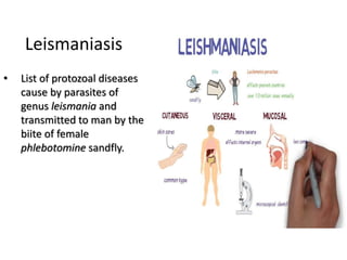
1. • List of protozoal diseases
cause by parasites of
genus leismania and
transmitted to man by the
biite of female
phlebotomine sandfly.
Leismaniasis
2. Various syndromes:
• Kala-azar or visceral leismaniasis.(VL)
• Cutaneous leismaniasis.(CL) or oriental sore.
• Muco-cutaneous leismaniasis.
• Anthroponotic cutaneous leismaniasis.
• Zoonotic cutaneous leismaniasis.
• Post kala-azar dermal leismaniasis.
Most of leismaniasis are zoonoses involving wild or domestic mammals
(rodents,canines). Some forms (indian kala-azar) are considered as non-
zoonotic infections.
*zoonoses=diseases which can be transmitted from animals(vertebrate) to
human.eg:Rabies,anthrax(splenic fever),cysticercosis etc
For more information:infonet-biovision.org/HumanHealth/Examples-
zoonoses.
3. Epidemiological Determinants
a)Agents
Leismania are intracellular parasites i.e. they infect and divide
within macrophages . Atleast 19 diff. leismania parasites have
been associated with human infections.
L. donovani ----- kala-azar(VL)
L. tropica ----- Cutaneous leismaniasis (oriental sore)
L.Brazilliensis --- Muco-cutaneous leismaniasis.
Life cycle of these agents are completed in two different hosts
.Vertebrates( amastigote form or leismania body)
.Insects ( promastogote forms, flagellated)
5. Host factors
a) Age
Kala-azar can occur in all age grops including infants below the age of one year. In
india (can be consider as a reference for Nepal as well) peak age is 5-9 years.
b) Sex
Male are more affected than female(2:1)
c)Population movement
Migrants,labours,tourists between endemic and non-endemic areas can results in the
apread of infections.
d) Socio-economic status
Poor are more affected.
e) Occupation
Farming parasites,forestry,mining,fishing,animal husbandary
f) Immunity
Recovery from kala-azar and oriental sore gives a lasting immunity.During active phase
of kala-azar,there is impairment of of cell mediated immunity,this is reflected in
negative skin rteaction to leismanin test.
6. Environmental factor
1. Altitude---- 2000feet(600m)
2. Season ---- after rain (november and march –april)
3. Vectors: P. argentipes ---kala-azar
P.sergenti and P.papatasi--- cutaneous
leismaniasis.
4. Developments projects: Forest clearing,cultivation
projects,large water resources schemes,colonization and
resettlement programmes.
7. MOT
• Female phlebotomine sandfly,P.argentipes
(anthrophilic).
• P.sergenti and P.papatasi
Extrinsic incubation period :6 to 9 days.(this is
the time required for the development of the
parasites in the insect vector)
Incubation period:1 to 4 months;range is 10
days to 2 years.
8. Clinical features
1.Kala-azar (VL,Black-sickness, dum-dum fever)
Fever,splenomegaly,hepatomegaly,accompanied by anaemia,weight loss.Darkenig of skin of face,
hands,feet and abdomen.
Lymphadenopathy may also occur.
If not treated, it has high mortality rate.
PKDL(Post-Kala-azar Dermal Leismaniasis)is caused by L. donovani , it appears one to several
years after apparent cure of Kala-azar. The lesions consists of multiple nodular infiltrations of the
skin,usually without ulceration.
9. 2. Cutaneous Leismaniasis
Several forms of leimaniasis:
Anthroponotic or urban Uutaneous Leismaniasis(ACL), Zoonotic or rural
Cutaneous Leismaniasis (ZCL),Diffuse Cutaneous Leismaniasis(DCL) etc.
The disease may be mistaken for leprosy.The agent is restricted to the skin.Its
is characterised by painful ulcers in the part of the exposed body part to the
sandfly.
11. Laboratory Diagnosis
1. Parasitological diagnosis
The demonstration of the parasite LD bodis in
the aspirates of the spleen, liver, bone marrow,
lymph nodes or in then skin ( in then case of CL)
is the only way to confirm VL or CL conclusively.
The parasites must be isolated in culture to
confirm the identity of the parasite.
12. 2.Aldehyde test
1 to 2 ml of sample of serum is isolated from
patients blood(venous) and a drop of formalin is
added.
If the sample converted into boiled egg like
structure within 2 to 20 mins than it is cosidered as
strong positive test if not then it is said to be
negative.
This is not good for diagnosis but good for
surveillance because test become positive 2 to 3
months after onset of disease, and reverts to
negative 6 months after cure.
13. Other tests
3.Serological tests (ELISA,DAT,IFAT)
4.Leismanin (Montenegro) test :base on skin
reaction
5.Haematological findings;ESR
increased,progressive
leucopenia,anamia,reversed albumin-globulin
ratio with greatly increased IgG.
WBC:RBC=1:1500(2000) (normal 1:750)
14. Control measure
1.Control of reservoir.
In Nepal man is the only reservoir of kala-azar. Awareness and treatment can
control the reservoir.
a)First line of drugs-short form
->SSG(sodium stibogluconate)
->Amphotericin B
b)First line drug-long term
->Miltefosine
->SSG IV or IM
c)Second line of drug
SSG failure ->Amphotericine B with increase dose
SSG and Miltefosine failure -> Liposomal Amphotericine B(repeated)
Infected Zoones control programmes.
16. facts
• Leishmaniasis is a blood borne parasite in all of it’s forms
• It is not known why some forms visceralize and some normally do not
• Leishmaniasis can remain dormant in a healthy person for up to twenty years
• Leishmaniasis can live in stored blood for up to thirty days
• Leishmaniasis is normally transmitted by the bite of an infected female sandfly
• Leishmaniasis is also transmittable sexually, congenitally, and by blood
transfusion or sharing needles.
• There is NO Sterile cure for leishmaniasis
• Leishmaniasis has been at epidemic levels in various parts of Afghanistan and
Iraq for the duration of the wars
• There is a one year ban on blood donations from persons having been to Iraq or
Afghanistan
• Leishmaniasis is a very variable bug and there is still much we do not know about
it
• Cutaneous species are showing signs of visceralizing