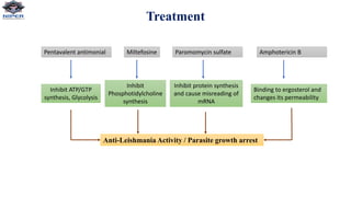
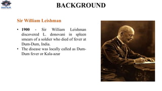
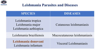
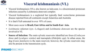
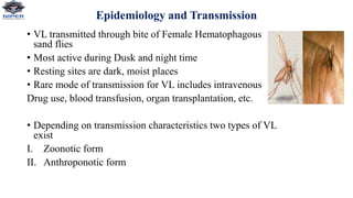
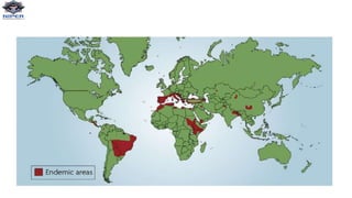
Shivam Kumar Pandey
Ph.D. Scholar
Department of Pharmacology & Toxicology
National Institute of Pharmaceutical Education and Research, Raebareli
New Transit Campus, Lucknow (U.P.)
Leishmania Donovani
BACKGROUND
Sir William Leishman
• 1900 - Sir William Leishman
discovered L. donovani in spleen
smears of a soldier who died of fever at
Dum-Dum, India.
• The disease was locally called as Dum-
Dum fever or Kala-azar
Leishmaniases : Neglected Tropical disease
• Disease mainly affects poor people in Africa, Asia and Latin America, and is
associated with malnutrition, population displacement, poor housing, weak
immune system and lack of resources.
• The leishmaniases are a group of diseases caused by the protozoa
parasite Leishmania.
• 20 Leishmania species known to be infective to humans are transmitted by the bite
of infected female phlebotomine sandflies.
• There are three main types of leishmaniasis:
i) Visceral, often known as kala-azar and the most serious form of the disease (VL);
ii) Cutaneous, the most common (CL);
iii) Mucocutaneous.
Leishmania Parasites and Diseases
SPECIES DISEASES
Leishmania tropica
Leishmania major
Leishmania aethiopica
Cutaneous leishmaniasis
Leishmania braziliensis Mucocutaneous leishmaniasis
Leishmania donovani
Leishmania infantum
Visceral Leishmaniasis
Visceral leishmaniasis (VL)
• Visceral leishmaniasis (VL), also known as kala-azar, is a disseminated protozoan
infection caused by Leishmania donovani complex
• Visceral leishmaniasis is a neglected but typically fatal vector-borne protozoan
disease reported from all continents except Antarctica and Australia
• It is fatal if left untreated in over 95% of cases.
• Most cases occur in Brazil, East Africa and in South-East Asia.
• Leishmania infantum (syn. L.chagasi) and Leishmania donovani are the species
involved in VL
• Source of Infection: The main sylvatic reservoirs identified are foxes (Cerdocyon
thous and Lycalopex vetulus) and marsupials (Didelphis spp). In urban areas, the
dog (Canis familiaris) is the main reservoir, however, the sylvatic reservoirs may
also be present in the transmission cycle.
Epidemiology and Transmission
• VL transmitted through bite of Female Hematophagous
sand flies
• Most active during Dusk and night time
• Resting sites are dark, moist places
• Rare mode of transmission for VL includes intravenous
Drug use, blood transfusion, organ transplantation, etc.
• Depending on transmission characteristics two types of VL
exist
I. Zoonotic form
II. Anthroponotic form
Pathogenesis
Life Cycle
Clinical Presentation
• Incubation period for VL ranges from 2-6 months but can vary from
weeks to several years
• Patients presents with insidious onset fever, weight loss and
organomegaly that persist of months.
• Splenomegaly is prominent
• Darkening of skin is mainly in south Asia, where it got name Kala-
azar (meaning black fever in Hindi)
• Anaemia, Thrombocytopenia, and Neutropenia usually seen,
reflecting bone marrow suppression
• Hepatic dysfunction, jaundice and ascites occur in advanced disease.
Post-Kala-azar Dermal Leishmaniasis(PKDL)
• Chronic skin rash that appears after effective treatment of VL
due to L. donovani
• PKDL very common (50%-60%) in Sudan occurring within
6 months of VL treatment while, less frequent (5%-10%) in
India and occur years after treatment
• Starts with erythematous macules and papules around the
perinasal areas and progress to plaques and nodules,
spreading to shoulder, trunk and extremities.
• Since parasite detected in skin lesions, such patents can
potentially have role in transmission of disease, acting as
reservoir
Diagnosis
1. Parasitology Diagnosis
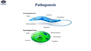
• Current gold standard for diagnosis relies on visualization of amastigote form of parasite within macrophages by
microscopic examination of tissue aspirates (spleen, Bone marrow or lymph nodes) after Giemsa staining
• In-vitro culture- require special media (Novy-MacNeal-Nicolle[NNN] media) and is not widely available in most endemic
regions
2. Serologic Diagnosis
• Direct agglutination test (sensitivity 95%, specificity 86%)
• rK39- based immunochromatographic strip test (sensitivity 94%, specificity 95%)
• IFA(Immunofluorescent assay), ELISA, Immunoblotting
3. Antigen Detection Test
• Kala-azar latex agglutination test (KAtex), detecting a heat-stable leishmania antigen in the urine of VL patient.
4. Molecular Diagnosis
• PCR-based assay to detect parasite DNA
• PCR on peripheral blood has been recommended as non-invasive first line screening test for both immunocompetent and
immunocompromised patients
NOTE: Diagnosis of PKDL relies on microscopic demonstration of parasites in skin specimens
Drug targets in Leishmania
Sterol biosynthetic pathway
Terbenafin
Zaragozic acids
Azasterols
Purine salvage pathway
Purine salvage pathway of Leishmania species. The enzymes involved in salvage of purines are (1) phosphoribosyltransferase (2) adenine
deaminase (3) guanine deaminase (4) adenosine deaminase (5) nucleoside kinase (6) nucleotidase (7) AMP deaminase (8) AMP kinase (9)
GMP kinase (10) IMP dehydrogenase (11) GMP synthetase (12) GMP reductase. AMP adenosine monophosphate; ADP adenosine
diphosphate; IMP inosine monophosphate; XMP xanthine monophosphate; GMP guanosine monophosphate; GDP guanosine diphosphate
present in both promastigote
and amastigotes
LdNT1
present in both promastigote
and amastigotes
LdNT2
present in amastigotes
Treatment
Pentavalent antimonial Miltefosine Paromomycin sulfate Amphotericin B
Inhibit ATP/GTP
synthesis, Glycolysis
Inhibit
Phosphotidylcholine
synthesis
Inhibit protein synthesis
and cause misreading of
mRNA
Binding to ergosterol and
changes its permeability
Anti-Leishmania Activity / Parasite growth arrest
Prevention and Control
• There is no vaccine to prevent infection.
• For the human population: Recommended measures for personal protection are aimed at
reducing contact with vectors, in particular: avoid outdoor activities from dusk to dawn, the use of
mosquito nets; use of protective clothing and insect repellents. People with clinical manifestations
of the disease should be treated as early as possible.
• For vector control: Preventive measures are directed towards integrated management actions in
environmental sanitation, same as those for cutaneous leishmaniasis.
• To control the urban reservoirs: Recommended preventive measures are the use of mosquito-
proof meshes in dog kennels to also keep out the sandflies and the use on dogs of personal
protective collars impregnated with insecticide.
NOTE: According to endemic transmission conducting serological surveys in dogs recommended
that if dog is positive for the parasite, humane euthanasia is indicated.
Leshmania.pptx

Leshmania.pptx

  • 1.
    Shivam Kumar Pandey Ph.D.Scholar Department of Pharmacology & Toxicology National Institute of Pharmaceutical Education and Research, Raebareli New Transit Campus, Lucknow (U.P.) Leishmania Donovani
  • 2.
    BACKGROUND Sir William Leishman •1900 - Sir William Leishman discovered L. donovani in spleen smears of a soldier who died of fever at Dum-Dum, India. • The disease was locally called as Dum- Dum fever or Kala-azar
  • 3.
    Leishmaniases : NeglectedTropical disease • Disease mainly affects poor people in Africa, Asia and Latin America, and is associated with malnutrition, population displacement, poor housing, weak immune system and lack of resources. • The leishmaniases are a group of diseases caused by the protozoa parasite Leishmania. • 20 Leishmania species known to be infective to humans are transmitted by the bite of infected female phlebotomine sandflies. • There are three main types of leishmaniasis: i) Visceral, often known as kala-azar and the most serious form of the disease (VL); ii) Cutaneous, the most common (CL); iii) Mucocutaneous.
  • 4.
    Leishmania Parasites andDiseases SPECIES DISEASES Leishmania tropica Leishmania major Leishmania aethiopica Cutaneous leishmaniasis Leishmania braziliensis Mucocutaneous leishmaniasis Leishmania donovani Leishmania infantum Visceral Leishmaniasis
  • 5.
    Visceral leishmaniasis (VL) •Visceral leishmaniasis (VL), also known as kala-azar, is a disseminated protozoan infection caused by Leishmania donovani complex • Visceral leishmaniasis is a neglected but typically fatal vector-borne protozoan disease reported from all continents except Antarctica and Australia • It is fatal if left untreated in over 95% of cases. • Most cases occur in Brazil, East Africa and in South-East Asia. • Leishmania infantum (syn. L.chagasi) and Leishmania donovani are the species involved in VL • Source of Infection: The main sylvatic reservoirs identified are foxes (Cerdocyon thous and Lycalopex vetulus) and marsupials (Didelphis spp). In urban areas, the dog (Canis familiaris) is the main reservoir, however, the sylvatic reservoirs may also be present in the transmission cycle.
  • 6.
    Epidemiology and Transmission •VL transmitted through bite of Female Hematophagous sand flies • Most active during Dusk and night time • Resting sites are dark, moist places • Rare mode of transmission for VL includes intravenous Drug use, blood transfusion, organ transplantation, etc. • Depending on transmission characteristics two types of VL exist I. Zoonotic form II. Anthroponotic form
  • 8.
  • 9.
  • 10.
    Clinical Presentation • Incubationperiod for VL ranges from 2-6 months but can vary from weeks to several years • Patients presents with insidious onset fever, weight loss and organomegaly that persist of months. • Splenomegaly is prominent • Darkening of skin is mainly in south Asia, where it got name Kala- azar (meaning black fever in Hindi) • Anaemia, Thrombocytopenia, and Neutropenia usually seen, reflecting bone marrow suppression • Hepatic dysfunction, jaundice and ascites occur in advanced disease.
  • 11.
    Post-Kala-azar Dermal Leishmaniasis(PKDL) •Chronic skin rash that appears after effective treatment of VL due to L. donovani • PKDL very common (50%-60%) in Sudan occurring within 6 months of VL treatment while, less frequent (5%-10%) in India and occur years after treatment • Starts with erythematous macules and papules around the perinasal areas and progress to plaques and nodules, spreading to shoulder, trunk and extremities. • Since parasite detected in skin lesions, such patents can potentially have role in transmission of disease, acting as reservoir
  • 12.
    Diagnosis 1. Parasitology Diagnosis •Current gold standard for diagnosis relies on visualization of amastigote form of parasite within macrophages by microscopic examination of tissue aspirates (spleen, Bone marrow or lymph nodes) after Giemsa staining • In-vitro culture- require special media (Novy-MacNeal-Nicolle[NNN] media) and is not widely available in most endemic regions 2. Serologic Diagnosis • Direct agglutination test (sensitivity 95%, specificity 86%) • rK39- based immunochromatographic strip test (sensitivity 94%, specificity 95%) • IFA(Immunofluorescent assay), ELISA, Immunoblotting 3. Antigen Detection Test • Kala-azar latex agglutination test (KAtex), detecting a heat-stable leishmania antigen in the urine of VL patient. 4. Molecular Diagnosis • PCR-based assay to detect parasite DNA • PCR on peripheral blood has been recommended as non-invasive first line screening test for both immunocompetent and immunocompromised patients NOTE: Diagnosis of PKDL relies on microscopic demonstration of parasites in skin specimens
  • 13.
    Drug targets inLeishmania Sterol biosynthetic pathway Terbenafin Zaragozic acids Azasterols
  • 14.
    Purine salvage pathway Purinesalvage pathway of Leishmania species. The enzymes involved in salvage of purines are (1) phosphoribosyltransferase (2) adenine deaminase (3) guanine deaminase (4) adenosine deaminase (5) nucleoside kinase (6) nucleotidase (7) AMP deaminase (8) AMP kinase (9) GMP kinase (10) IMP dehydrogenase (11) GMP synthetase (12) GMP reductase. AMP adenosine monophosphate; ADP adenosine diphosphate; IMP inosine monophosphate; XMP xanthine monophosphate; GMP guanosine monophosphate; GDP guanosine diphosphate present in both promastigote and amastigotes LdNT1 present in both promastigote and amastigotes LdNT2 present in amastigotes
  • 15.
    Treatment Pentavalent antimonial MiltefosineParomomycin sulfate Amphotericin B Inhibit ATP/GTP synthesis, Glycolysis Inhibit Phosphotidylcholine synthesis Inhibit protein synthesis and cause misreading of mRNA Binding to ergosterol and changes its permeability Anti-Leishmania Activity / Parasite growth arrest
  • 16.
    Prevention and Control •There is no vaccine to prevent infection. • For the human population: Recommended measures for personal protection are aimed at reducing contact with vectors, in particular: avoid outdoor activities from dusk to dawn, the use of mosquito nets; use of protective clothing and insect repellents. People with clinical manifestations of the disease should be treated as early as possible. • For vector control: Preventive measures are directed towards integrated management actions in environmental sanitation, same as those for cutaneous leishmaniasis. • To control the urban reservoirs: Recommended preventive measures are the use of mosquito- proof meshes in dog kennels to also keep out the sandflies and the use on dogs of personal protective collars impregnated with insecticide. NOTE: According to endemic transmission conducting serological surveys in dogs recommended that if dog is positive for the parasite, humane euthanasia is indicated.