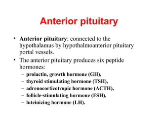
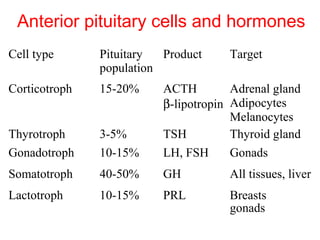
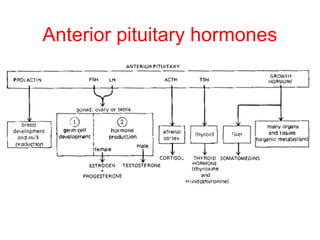
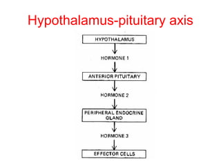
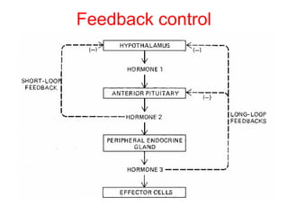
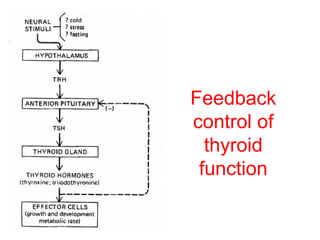
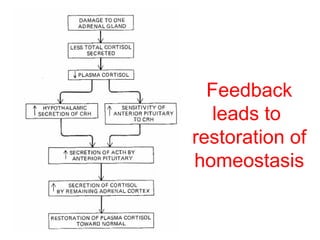
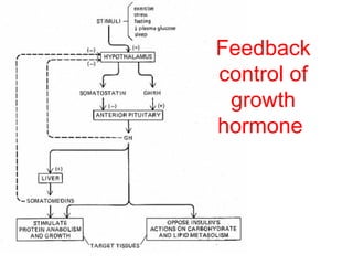
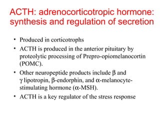
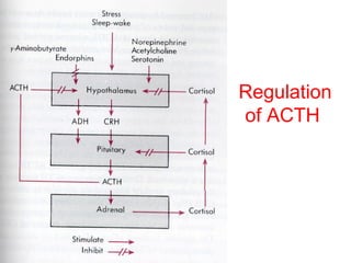
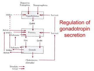
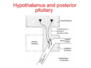
Endocrine Physiology
lecture 3
Dale Buchanan Hales, PhD
Department of Physiology & Biophysics
Anterior pituitary
• Anterior pituitary: connected to the
hypothalamus by hypothalmoanterior pituitary
portal vessels.
• The anterior pituitary produces six peptide
hormones:
–
–
–
–
–

prolactin, growth hormone (GH),
thyroid stimulating hormone (TSH),
adrenocorticotropic hormone (ACTH),
follicle-stimulating hormone (FSH),
luteinizing hormone (LH).
Anterior pituitary cells and hormones
Cell type

Pituitary
Product
population

Target

Corticotroph

15-20%

Thyrotroph
Gonadotroph

3-5%
10-15%

ACTH
Adrenal gland
β-lipotropin Adipocytes
Melanocytes
TSH
Thyroid gland
LH, FSH
Gonads

Somatotroph

40-50%

GH

All tissues, liver

Lactotroph

10-15%

PRL

Breasts
gonads
Anterior pituitary hormones
Feedback regulation of
hypothalmus/pituitary
A prominent feature of each of the hormonal
sequences initiated by the hypothalamic
releasing hormones is negative feedback
exerted upon the hypothalamic-pituitary
system by the hormones whose production
are stimulated in the sequence.
Hypothalamus-pituitary axis
Feedback control
Feedback
control of
thyroid
function
Feedback
leads to
restoration of
homeostasis
Feedback
control of
growth
hormone
Regulation of Growth
Hormone Secretion
• GH secretion controlled primarily by
hypothalamic GHRH stimulation and
somatostatin inhibition
• Neurotransmitters involved in control of
GH secretion– via regulation of GHRH and
somatostatin
Regulation of Growth
Hormone Secretion
• Neurotransmitter systems that stimulate
GHRH and/or inhibit somatostatin
– Catecholamines acting via α2-adrenergic
receptors
– Dopamine acting via D1 or D2 receptors
– Excitatory amino acids acting via both NMDA
and non-NMDA receptors
Regulation of Growth
Hormone Secretion
 β-adrenergic receptors stimulate
somatostatin release and inhibit GH
 β-adrenergic receptors inhibit hypothalamic
release of GHRH
Regulation of Growth
Hormone Secretion
• Additional central mechanisms that control
GH secretion include an ultra-short
feedback loop exerted by both somatostatin
and GHRH on their own secretion
Growth hormone vs.
metabolic state
• When protein and energy intake are adequate, it is
appropriate to convert amino acids to protein and stimulate
growth. hence GH and insulin promote anabolic reactions
during protein intake
• During carbohydrate intake, GH antagonizes insulin
effects-- blocks glucose uptake to prevent hypoglycemia.
(if there is too much insulin, all the glucose would be taken
up).
• When there is adequate glucose as during absorptive
phase, and glucose uptake is required, then GH secretion is
inhibited so it won't counter act insulin action.
Growth hormone vs.
metabolic state
• During fasting, GH antagonizes insulin action and helps
mediate glucose sparing, ie stimulates gluconeogenesis
• In general, during anabolic or absorptive phase, GH
facilitates insulin action, to promote growth.
• during fasting or post-absorptive phase, GH opposes
insulin action, to promote catabolism or glucose sparing
Growth
hormone
and
metabolic
state
Clinical assessment of GH
• Random serum samples not useful due to pulsatile
pattern of release
• Provocative tests necessary
– GH measurement after 90 min exercise
– GH measurement immediately after onset of sleep

• Definitive tests
– GH measurement after insulin-induced hypoglycemia
– Glucose suppresses GH levels 30-90 min after
administration– patients with GH excess do not suppress
– Measurement of IGF-1 to assess GH excess
Acromegaly and Gigantism
• Caused by eosinophilic adenomas of somatotrophs
• Excess GH leads to development of gigantism if
hypersecretion is present during early life– a rare
condition
– Symmetrical enlargement of body resulting in true
giant with overgrowth of long bones, connective tissue
and visceral organs.

• Excess GH leads to acromegaly if hypersecretion
occurs after body growth has stopped.
– Elongation of long bones not possible so there is over
growth of cancellous bones– protruding jaw, thickening
of phalanges, and over growth of visceral organs
Acromegaly
Acromegaly
A) before
presentation;
B) at admission
Harvey Cushing’s
first reported case
Gigantism

Identical twins, 22 years old, excess GH secretion
ACTH: adrenocorticotropic hormone:
synthesis and regulation of secretion
• Produced in corticotrophs
• ACTH is produced in the anterior pituitary by
proteolytic processing of Prepro-opiomelanocortin
(POMC).
• Other neuropeptide products include β and
γ lipotropin, β-endorphin, and α-melanocytestimulating hormone (α-MSH).
• ACTH is a key regulator of the stress response
ACTH synthesis

ACTH

Processing and cleavage of pro-opiomelanocortin (POMC)
ACTH
 ACTH is made up of 39 amino acids
Regulates adrenal cortex and synthesis of
adrenocorticosteroids
α-MSH resides in first 13 AA of ACTH
α-MSH stimulates melanocytes and can darken
skin
Overproduction of ACTH may accompany
increased pigmentation due to α-MSH.
Addison’s Disease
• Disease in which patients lack cortisol from
zona fasiculata, and thus lacks negative
feedback that suppresses ACTH production
• Result: overproduction of ACTH
• Skin color will darken
• JFK had Addison’s disease and was treated
with cortisol injections
β-endorphin
•
•
•
•

Produced as a result of ACTH synthesis
Binds to opiate receptors
Results in “runner’s high”
Role in anterior pituitary not completely
understood
• One of many endogenous opioids such as
enkephalins
Melanocyte-stimulating hormone
(MSH)
• MSH peptides derived by proteolytic cleavage of
POMC
∀ α-MSH has antipyretic and anti-inflammatory
effects
– Also inhibits CRH and LHRH secretion

•
•
•
•

Four MSH receptors identified
May inhibit feeding behavior
ACTH has MSH-like activity
However– MSH has NO ACTH like activity
Regulation
of ACTH
secretion
Regulation of ACTH secretion
• Stimulation of release
– CRH and ADH
– Stress
– Hypoglycemia

• CRH and ADH both synthesized in hypothalamus
– ADH (a.k.a. vasopressin) is released by posterior
pituitary and reaches anterior pituitary via inferior
hypophyseal artery.
Regulation of ACTH secretion
• Deficiency of vasopressin (ADH) in
hereditary diabetes insipidus is
accompanied by decreased ACTH release.
• Vasopressin potentiates CRH at both
hypothalamic and pituitary levels.
• Many vasopressinergic neurons also contain
CRH resulting in co-release of two peptides
into portal blood.
ACTH
• Circadian pattern of release
– Highest levels of cortisol are in early AM
following ACTH release
– Depends on sleep-wake cycle, jet-lag can result
in alteration of pattern

• Opposes the circadian pattern of growth
hormone secretion
Regulation
of ACTH
ACTH
• Acts on adrenal cortex
– stimulates growth of cortex (trophic action)
– Stimulates steroid hormone synthesis

• Lack of negative feedback from cortisol results in
aberrantly high ACTH, elevated levels of other
adrenal corticosteroids– adrenal androgens
• Adrenogenital syndrome: masculization of female
fetus
Glycoprotein hormones
 LH, FSH, TSH and hCG
α and β subunits
Each subunit encoded by different gene
 α subunit is identical for all hormones
 β subunit are unique and provide biological

specificity
Glycoprotein hormones
Glycoprotein hormones contain two subunits, a
common α subunit and a distinct β subunit:
TSH, LH, FSH and hCG.
Gonadotrophs
• Cells in anterior pituitary that produce LH and
FSH
• Synthesis and secretion stimulated by GnRH–
major effect on LH
• FSH secretion controlled by inhibin
• Pulsitile secretion of GnRH and inhibin cause
distinct patterns of LH and FSH secretion
LH/FSH
• Pulsatile pattern of secretion
– LH pulses are biphasic (every 1 minute, then large
pulse at 1 hour)
– FSH pulses are uniphasic

• Diurnal– LH/FSH more pronounced during
puberty
• Cyclic in females– ovarian cycle with LH surge at
time of ovulation
• Males are not cyclic, but constant pulses of LH
cause pulses of testosterone to be produced
Pulsitile secretion of GnRH and LH
Regulation of LH/FSH
• Negative feed-back
– Inhibin produced by testes and ovaries Decreases FSH
β-subunit expression
– Testosterone from Leydig cells– synthesis stimulated
by LH, feedsback to inhibit GnRH production from
hypothalamus and down-regulates GnRH receptors
– Progesterone– suppresses ovulation, basis for oral
contraceptives. Works at both the level of pituitary and
hypothalamus.
Regulation of LH/FSH
• Dopamine, endorphin, and prolactin inhibit GnRH
release.
– Prolactin inhibition affords post-partum contraceptive
effect

• Overproduction of prolactin via pituitary tumor
can cause amenorrhea– shuts off GnRH
– Treated with bromocryptine (dopamine agonist)
– Surgical removal of pituitary tumor
Regulation of LH/FSH
• Positive feedback
– Estradiol at high plasma concentrations in late
follicular phase of ovarian cycle stimulates
GnRH and LH surge– triggers ovulation
Regulation of
gonadotropin
secretion
Thyrotrophs
•
•
•
•

Site of TSH synthesis
Pattern of secretion is relatively steady
TSH secretion stimulated by TRH
Feedback control by T3 (thyroid hormone)
Feedback
control of
thyroid
function
Grave’s disease
• Hyperthyroidism caused by circulating
antibodies to the TSH receptor.
• Associated with diffuse goiter.
• Autoantibodies bind to TSH receptor and
mimic the action of TSH itself leads to
persistent stimulation of thyroid and
elevated levels of thyroid hormones.
Lacotrophs
• Site of production of prolactin
• Lactogenesis (milk synthesis) requires prolactin
• Tonically inhibited
– Of the anterior pituitary hormones, the only one
– Multifactoral control, balance favors inhibition

• Dopamine inhibits prolactin
• Prolactin releasing hormone is TRH
– Ocytocin also stimulates prolactin release
– Estradiol enhances prolactin synthesis
Prolactin
• Stimulates breast development and
lactogenesis
• May be involved in development of Leydig
cells in pre-pubertal males
• Immunomodulatory effects– stimulates T
cell functions
– Prolactin receptors in thymus
Clinical assessment of PRL
• Single basal serum PRL measurement sufficient to
determine excess
– PRL deficiency not a usual clinical concern

• PRL is only anterior pituitary with predominant
negative control by hypothalamus– often elevated
by lesions that interfere with portal blood flow.
• Elevated by primary PRL adenomas of pituitary
Posterior pituitary hormones:
ADH (AVP) and Oxytocin
(hypothalamic hormones)
 Both are synthesized in the cell bodies of
hypothalamic neurons
 ADH: supraoptic nucleus
 Oxytocin: paraventricular nucleus
 Both are synthesized as preprohormones and
processed into nonapeptides (nine amino acids).
 They are released from the termini in response to
an action potential which travels from the axon
body in the hypothalamus
Hypothalamus and posterior
pituitary
Structures of ADH and oxytocin
Oxytocin: stimulates
myoepithelial contractions
 In uterus during parturition
 In mammary gland during
lactation
Oxytocin: milk ejection from
lactating mammary gland
 suckling is major stimulus for release.
 sensory receptors in nipple connect with
nerve fibers to the spine, then impulses
are relayed through brain to PVN where
cholinergic synapses fire on oxytocin
neurons and stimulate release.
Oxytocin: uterine contractions
• Reflexes originating in the cervical, vaginal
and uterus stimulate oxytocin synthesis and
release via neural input to hypothalamus
• Increases in plasma at time of ovulation,
parturition, and coitus
• Estrogen increases synthesis and lowers
threshold for release
Oxytocin secretion is stimulated
by nursing

Lecture3

  • 1.
    Endocrine Physiology lecture 3 DaleBuchanan Hales, PhD Department of Physiology & Biophysics
  • 2.
    Anterior pituitary • Anteriorpituitary: connected to the hypothalamus by hypothalmoanterior pituitary portal vessels. • The anterior pituitary produces six peptide hormones: – – – – – prolactin, growth hormone (GH), thyroid stimulating hormone (TSH), adrenocorticotropic hormone (ACTH), follicle-stimulating hormone (FSH), luteinizing hormone (LH).
  • 3.
    Anterior pituitary cellsand hormones Cell type Pituitary Product population Target Corticotroph 15-20% Thyrotroph Gonadotroph 3-5% 10-15% ACTH Adrenal gland β-lipotropin Adipocytes Melanocytes TSH Thyroid gland LH, FSH Gonads Somatotroph 40-50% GH All tissues, liver Lactotroph 10-15% PRL Breasts gonads
  • 4.
  • 5.
    Feedback regulation of hypothalmus/pituitary Aprominent feature of each of the hormonal sequences initiated by the hypothalamic releasing hormones is negative feedback exerted upon the hypothalamic-pituitary system by the hormones whose production are stimulated in the sequence.
  • 6.
  • 7.
  • 8.
  • 9.
  • 10.
  • 11.
    Regulation of Growth HormoneSecretion • GH secretion controlled primarily by hypothalamic GHRH stimulation and somatostatin inhibition • Neurotransmitters involved in control of GH secretion– via regulation of GHRH and somatostatin
  • 12.
    Regulation of Growth HormoneSecretion • Neurotransmitter systems that stimulate GHRH and/or inhibit somatostatin – Catecholamines acting via α2-adrenergic receptors – Dopamine acting via D1 or D2 receptors – Excitatory amino acids acting via both NMDA and non-NMDA receptors
  • 13.
    Regulation of Growth HormoneSecretion  β-adrenergic receptors stimulate somatostatin release and inhibit GH  β-adrenergic receptors inhibit hypothalamic release of GHRH
  • 14.
    Regulation of Growth HormoneSecretion • Additional central mechanisms that control GH secretion include an ultra-short feedback loop exerted by both somatostatin and GHRH on their own secretion
  • 15.
    Growth hormone vs. metabolicstate • When protein and energy intake are adequate, it is appropriate to convert amino acids to protein and stimulate growth. hence GH and insulin promote anabolic reactions during protein intake • During carbohydrate intake, GH antagonizes insulin effects-- blocks glucose uptake to prevent hypoglycemia. (if there is too much insulin, all the glucose would be taken up). • When there is adequate glucose as during absorptive phase, and glucose uptake is required, then GH secretion is inhibited so it won't counter act insulin action.
  • 16.
    Growth hormone vs. metabolicstate • During fasting, GH antagonizes insulin action and helps mediate glucose sparing, ie stimulates gluconeogenesis • In general, during anabolic or absorptive phase, GH facilitates insulin action, to promote growth. • during fasting or post-absorptive phase, GH opposes insulin action, to promote catabolism or glucose sparing
  • 17.
  • 18.
    Clinical assessment ofGH • Random serum samples not useful due to pulsatile pattern of release • Provocative tests necessary – GH measurement after 90 min exercise – GH measurement immediately after onset of sleep • Definitive tests – GH measurement after insulin-induced hypoglycemia – Glucose suppresses GH levels 30-90 min after administration– patients with GH excess do not suppress – Measurement of IGF-1 to assess GH excess
  • 19.
    Acromegaly and Gigantism •Caused by eosinophilic adenomas of somatotrophs • Excess GH leads to development of gigantism if hypersecretion is present during early life– a rare condition – Symmetrical enlargement of body resulting in true giant with overgrowth of long bones, connective tissue and visceral organs. • Excess GH leads to acromegaly if hypersecretion occurs after body growth has stopped. – Elongation of long bones not possible so there is over growth of cancellous bones– protruding jaw, thickening of phalanges, and over growth of visceral organs
  • 20.
    Acromegaly Acromegaly A) before presentation; B) atadmission Harvey Cushing’s first reported case
  • 21.
    Gigantism Identical twins, 22years old, excess GH secretion
  • 22.
    ACTH: adrenocorticotropic hormone: synthesisand regulation of secretion • Produced in corticotrophs • ACTH is produced in the anterior pituitary by proteolytic processing of Prepro-opiomelanocortin (POMC). • Other neuropeptide products include β and γ lipotropin, β-endorphin, and α-melanocytestimulating hormone (α-MSH). • ACTH is a key regulator of the stress response
  • 23.
    ACTH synthesis ACTH Processing andcleavage of pro-opiomelanocortin (POMC)
  • 24.
    ACTH  ACTH ismade up of 39 amino acids Regulates adrenal cortex and synthesis of adrenocorticosteroids α-MSH resides in first 13 AA of ACTH α-MSH stimulates melanocytes and can darken skin Overproduction of ACTH may accompany increased pigmentation due to α-MSH.
  • 25.
    Addison’s Disease • Diseasein which patients lack cortisol from zona fasiculata, and thus lacks negative feedback that suppresses ACTH production • Result: overproduction of ACTH • Skin color will darken • JFK had Addison’s disease and was treated with cortisol injections
  • 26.
    β-endorphin • • • • Produced as aresult of ACTH synthesis Binds to opiate receptors Results in “runner’s high” Role in anterior pituitary not completely understood • One of many endogenous opioids such as enkephalins
  • 27.
    Melanocyte-stimulating hormone (MSH) • MSHpeptides derived by proteolytic cleavage of POMC ∀ α-MSH has antipyretic and anti-inflammatory effects – Also inhibits CRH and LHRH secretion • • • • Four MSH receptors identified May inhibit feeding behavior ACTH has MSH-like activity However– MSH has NO ACTH like activity
  • 28.
  • 29.
    Regulation of ACTHsecretion • Stimulation of release – CRH and ADH – Stress – Hypoglycemia • CRH and ADH both synthesized in hypothalamus – ADH (a.k.a. vasopressin) is released by posterior pituitary and reaches anterior pituitary via inferior hypophyseal artery.
  • 30.
    Regulation of ACTHsecretion • Deficiency of vasopressin (ADH) in hereditary diabetes insipidus is accompanied by decreased ACTH release. • Vasopressin potentiates CRH at both hypothalamic and pituitary levels. • Many vasopressinergic neurons also contain CRH resulting in co-release of two peptides into portal blood.
  • 31.
    ACTH • Circadian patternof release – Highest levels of cortisol are in early AM following ACTH release – Depends on sleep-wake cycle, jet-lag can result in alteration of pattern • Opposes the circadian pattern of growth hormone secretion
  • 32.
  • 33.
    ACTH • Acts onadrenal cortex – stimulates growth of cortex (trophic action) – Stimulates steroid hormone synthesis • Lack of negative feedback from cortisol results in aberrantly high ACTH, elevated levels of other adrenal corticosteroids– adrenal androgens • Adrenogenital syndrome: masculization of female fetus
  • 34.
    Glycoprotein hormones  LH,FSH, TSH and hCG α and β subunits Each subunit encoded by different gene  α subunit is identical for all hormones  β subunit are unique and provide biological specificity
  • 35.
    Glycoprotein hormones Glycoprotein hormonescontain two subunits, a common α subunit and a distinct β subunit: TSH, LH, FSH and hCG.
  • 36.
    Gonadotrophs • Cells inanterior pituitary that produce LH and FSH • Synthesis and secretion stimulated by GnRH– major effect on LH • FSH secretion controlled by inhibin • Pulsitile secretion of GnRH and inhibin cause distinct patterns of LH and FSH secretion
  • 37.
    LH/FSH • Pulsatile patternof secretion – LH pulses are biphasic (every 1 minute, then large pulse at 1 hour) – FSH pulses are uniphasic • Diurnal– LH/FSH more pronounced during puberty • Cyclic in females– ovarian cycle with LH surge at time of ovulation • Males are not cyclic, but constant pulses of LH cause pulses of testosterone to be produced
  • 38.
  • 39.
    Regulation of LH/FSH •Negative feed-back – Inhibin produced by testes and ovaries Decreases FSH β-subunit expression – Testosterone from Leydig cells– synthesis stimulated by LH, feedsback to inhibit GnRH production from hypothalamus and down-regulates GnRH receptors – Progesterone– suppresses ovulation, basis for oral contraceptives. Works at both the level of pituitary and hypothalamus.
  • 40.
    Regulation of LH/FSH •Dopamine, endorphin, and prolactin inhibit GnRH release. – Prolactin inhibition affords post-partum contraceptive effect • Overproduction of prolactin via pituitary tumor can cause amenorrhea– shuts off GnRH – Treated with bromocryptine (dopamine agonist) – Surgical removal of pituitary tumor
  • 41.
    Regulation of LH/FSH •Positive feedback – Estradiol at high plasma concentrations in late follicular phase of ovarian cycle stimulates GnRH and LH surge– triggers ovulation
  • 42.
  • 43.
    Thyrotrophs • • • • Site of TSHsynthesis Pattern of secretion is relatively steady TSH secretion stimulated by TRH Feedback control by T3 (thyroid hormone)
  • 44.
  • 45.
    Grave’s disease • Hyperthyroidismcaused by circulating antibodies to the TSH receptor. • Associated with diffuse goiter. • Autoantibodies bind to TSH receptor and mimic the action of TSH itself leads to persistent stimulation of thyroid and elevated levels of thyroid hormones.
  • 46.
    Lacotrophs • Site ofproduction of prolactin • Lactogenesis (milk synthesis) requires prolactin • Tonically inhibited – Of the anterior pituitary hormones, the only one – Multifactoral control, balance favors inhibition • Dopamine inhibits prolactin • Prolactin releasing hormone is TRH – Ocytocin also stimulates prolactin release – Estradiol enhances prolactin synthesis
  • 47.
    Prolactin • Stimulates breastdevelopment and lactogenesis • May be involved in development of Leydig cells in pre-pubertal males • Immunomodulatory effects– stimulates T cell functions – Prolactin receptors in thymus
  • 48.
    Clinical assessment ofPRL • Single basal serum PRL measurement sufficient to determine excess – PRL deficiency not a usual clinical concern • PRL is only anterior pituitary with predominant negative control by hypothalamus– often elevated by lesions that interfere with portal blood flow. • Elevated by primary PRL adenomas of pituitary
  • 49.
    Posterior pituitary hormones: ADH(AVP) and Oxytocin (hypothalamic hormones)  Both are synthesized in the cell bodies of hypothalamic neurons  ADH: supraoptic nucleus  Oxytocin: paraventricular nucleus  Both are synthesized as preprohormones and processed into nonapeptides (nine amino acids).  They are released from the termini in response to an action potential which travels from the axon body in the hypothalamus
  • 50.
  • 51.
    Structures of ADHand oxytocin
  • 52.
    Oxytocin: stimulates myoepithelial contractions In uterus during parturition  In mammary gland during lactation
  • 53.
    Oxytocin: milk ejectionfrom lactating mammary gland  suckling is major stimulus for release.  sensory receptors in nipple connect with nerve fibers to the spine, then impulses are relayed through brain to PVN where cholinergic synapses fire on oxytocin neurons and stimulate release.
  • 54.
    Oxytocin: uterine contractions •Reflexes originating in the cervical, vaginal and uterus stimulate oxytocin synthesis and release via neural input to hypothalamus • Increases in plasma at time of ovulation, parturition, and coitus • Estrogen increases synthesis and lowers threshold for release
  • 55.
    Oxytocin secretion isstimulated by nursing