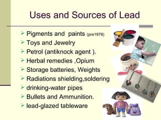
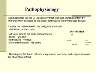
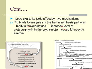
Lead Poisoning
Dr. Abdul Ghaffar Latiffi
Pediatric medicine 1st
Year Resident
FMIC
Ab.ghaffar.latiffi@gmail.com
Date:06.08.2013
“ Lead makes the mind give way.”
Greek Physician, 2000 B.C
o Heavy, soft, flexible, bluish, tasteless, odorless metal.
o wont break down over time, doesn't vaporize.
o Φ conduct electricity - resistant to corrosion.
o Abundant in the earth's crust (13g/ton or 0.0013% of the earth's crust)
o Throughout human history, over 350 million tons of lead
have been mined, used and ultimately discarded in the
environment
Introduction
Roman
metal
Lead Poisoning (Plumbism)
"lead poisoning contributed to
The decline of the Roman empire “
Jerome Nriagu,geochemist,1983
 Lead poisoning is a medical condition
caused by increased levels of lead in the
body.
APP currently defines lead poisoning as
a venous BLL ≥ 5μg/dL.
 first time lead poisoning in Childhood was
reported in Ceylon In 1967 .
 Lead paint prohibited in 1922.
 UNEP banned global use of leaded gasoline(1995)
 Still allowed in 6 nations. Afghanistan, Algeria,
Iraq, North Korea, Myanmar and Yemen
Epidemiology
Approx half million children in the USA have toxic BLL
A public health disaster and challenge
Children 6 yrs are more susceptible to leads toxic effects
 Increased prevalence in inner city children
 Children and pregnant women are at double jeopardy
due to increased absorption and heavy distribution.
 Nutritional deficiencies (Iron ,calcium, zinc, copper, and
protein ) result in greater lead absorption
August 14, 2007
Uses and Sources of Lead
 Pigments and paints (pre1978)
 Toys and Jewelry
 Petrol (antiknock agent ).
 Herbal remedies ,Opium
 Storage batteries, Weights
 Radiations shielding,soldering
 drinking-water pipes
 Bullets and Ammunition.
 lead-glazed tableware
Lead ores
Pathophysiology
Half life of lead in the body compartments
• Blood- 25 days
•Soft tissues - 40 days
•Mineralized tissues- >25 years
• Diets high in fat, low in calcium, magnesium, iron, zinc, and copper, increase
the absorption of lead.
Lead absorbed via the GI , respiratory tract ,skin and transplacentally by
the fetus then distribute to the blood, soft tissues and mineralized tissues
Lead is not metabolized in the body; it is absorbed,
distributed, and excreted
70%-80%
5-10%
1%
Distribution
Bone
Soft tissues
Blood
Cont….
► Lead exerts its toxic effect by two mechanisms
a) Pb binds to enzymes in the heme synthesis pathway
Inhibits ferrochelatase increase level of
protoporphyrin in the erythrocyte cause Microcytic
anemia
Date
Cont….
b)Lead as a high potency surrogate is disrupting calcium
homeostasis and in interfering with calcium-dependent
intracellular functions.
Lead interferes with enzymes that help in the
synthesis of vitamin D
Lead alters the permeability of blood vessels
and collagen synthesis
Lead exposure has also been associated with a
decrease in activity of immune cells such as PMN
leukocytes
Interferes with mitochondrial oxidative
phosphorylation, ATPases
enhances oxidation and cell apoptosis.
Clinical Presentation
 lead exposure often occurs with vague and non
specific symptoms, it frequently goes unrecognized
 Acute lead poisoning while less common, shows up
more quickly and can be fatal.
 chronic lead poisoning take time to develop,
however. Children can appear healthy despite having
high levels of lead in their blood.
 Impairment of IQ occur at even lower levels of
exposure (1 µg/dL.)
Clinical Presentation
1. loss of appetite
2. vomiting
3. weight loss
4. constipation
5. anemia
6. Irritability
7. learning disabilities
8. behavior problems.
1. Cranial nerve paralysis
2. Encephalopathy
3. seizures
4. coma
Blood Lead Effects Level
Date
Degree of Lead Poisoning
Diagnosis
A. Usually made via screening program
B. In suspected cases , must obtain a detailed history
• Onset of symptoms
• History of Pica
• Assessment of potential sources
• Family history of lead poisoning
C. PhysiCal examination
 Pallor and hyperactivity
Burton lines on the gums
Decreased stature
wrist drop, and cognitive dysfunction
•signs of elevated iCP
Impaired consciousness,Bradycardia
Hypertension ,Respiratory depression
Papilledema ,Coma
Diagnosis
Lab and radiographic findings:
1. Increased blood lead level
2. CBC
•Microcytic Anemia
•Leukocytosis
• Basophilic stippling on RBC ( aggregation of ribosomal RNA in the cytoplasm of RBC)
3. Increased FEP concentration
4.CSF changes
• high proteins, Lymphocytic pleocytosis
5.Radiodensity at the distal metaphyseal area of long bones
(Lead lines ) usually in chronic lead poisoning
Long bones radiographs
“Lead Lines” in
five year old
male with
radiological
growth
retardation and
blood lead level
of 37.7µg/dl
“Lead Lines”
in 3 yrs old
girl with BLL of
10.6 µg/dl
Differential Diagnoses
 Acute and Chronic Anemia (IDA)
 Constipation
 Failure to Thrive
 Abdominal pain (peptic ulcer ,appendicitis)
 Heavy Metal Toxicity
 Hydrocarbon Inhalation Injury
 Mental retardation
 Seizure disorders
 Subdural hematoma
 Hematologic and CNS neoplasm
 Behavior and emotional disorders
Treatment & Management
Treatment of lead toxicity involves
 Prevention of further lead exposure
 Decontamination
 Chelation
 Supportive therapy.
Prevention of further lead exposure
 Identification and correction of exposure sources is critical
 Frequently hands and pacifiers washing.
 Use only cold water from the tap ,Hot water may contain more
lead.
 Avoid using home remedies (such as azarcon, greta, pay-loo-
ah,zafron) and cosmetics (such as kohl)
 Shower and change clothes after working
if your work or hobbies involve working with
lead-based products.
 Correction of dietary def in iron, calcium,
magnesium, and zinc.
 Vitamin C is a weak but natural chelating agent
Decontamination
 May be performed in patients with acute lead
ingestion in whom lead paint chips are
identified on plain abdominal radiographs.
 Polyethylene glycol solution (GoLytely) can be
used for lead densities in the stomach and/or
small intestine.
 Gastric lavage may be performed.
 Secure the airway before the initiation of
gastric lavage in an obtunded child with acute
lead ingestion.
 Charcoal binds poorly to lead, and no evidence
Chelating Agents
Supportive management
 Anticonvulsants may be needed.
 Mannitol or corticosteroids and volume
restriction are indicated in patients with
encephalopathy.
 A high-calcium, high-phosphorus diet and
large doses of vitamin D may remove lead
from the blood by depositing it in the bones.
Prognosis
 The adverse effects of lead are not reversible.
 In acute lead poisoning ( seizures and coma),
there is a high risk Of permanent brain damage
and death.
 The long-term effects of lower levels of lead can
also be permanent and severe.
 Asymptomatic patients tend to have a better
prognosis, with good improvement in intellectual
functions following lowering of the BLL.
 Sequelae of encephalopathy includes seizure
disorders, impaired mentation, and rarely
REFERENCES
1.Nelson - Textbook Of Pediatrics - 19th Edition
2.Rudolph pediatric
4.http://www.lead.org.au
6. Park’s textbook of preventive and social medicine
THANK
YOU

Lead poisoning in Pediatrics

  • 1.
    Lead Poisoning Dr. AbdulGhaffar Latiffi Pediatric medicine 1st Year Resident FMIC Ab.ghaffar.latiffi@gmail.com Date:06.08.2013
  • 2.
    “ Lead makesthe mind give way.” Greek Physician, 2000 B.C o Heavy, soft, flexible, bluish, tasteless, odorless metal. o wont break down over time, doesn't vaporize. o Φ conduct electricity - resistant to corrosion. o Abundant in the earth's crust (13g/ton or 0.0013% of the earth's crust) o Throughout human history, over 350 million tons of lead have been mined, used and ultimately discarded in the environment Introduction Roman metal
  • 3.
    Lead Poisoning (Plumbism) "leadpoisoning contributed to The decline of the Roman empire “ Jerome Nriagu,geochemist,1983  Lead poisoning is a medical condition caused by increased levels of lead in the body. APP currently defines lead poisoning as a venous BLL ≥ 5μg/dL.  first time lead poisoning in Childhood was reported in Ceylon In 1967 .  Lead paint prohibited in 1922.  UNEP banned global use of leaded gasoline(1995)  Still allowed in 6 nations. Afghanistan, Algeria, Iraq, North Korea, Myanmar and Yemen
  • 4.
    Epidemiology Approx half millionchildren in the USA have toxic BLL A public health disaster and challenge Children 6 yrs are more susceptible to leads toxic effects  Increased prevalence in inner city children  Children and pregnant women are at double jeopardy due to increased absorption and heavy distribution.  Nutritional deficiencies (Iron ,calcium, zinc, copper, and protein ) result in greater lead absorption
  • 5.
  • 6.
    Uses and Sourcesof Lead  Pigments and paints (pre1978)  Toys and Jewelry  Petrol (antiknock agent ).  Herbal remedies ,Opium  Storage batteries, Weights  Radiations shielding,soldering  drinking-water pipes  Bullets and Ammunition.  lead-glazed tableware
  • 8.
  • 9.
    Pathophysiology Half life oflead in the body compartments • Blood- 25 days •Soft tissues - 40 days •Mineralized tissues- >25 years • Diets high in fat, low in calcium, magnesium, iron, zinc, and copper, increase the absorption of lead. Lead absorbed via the GI , respiratory tract ,skin and transplacentally by the fetus then distribute to the blood, soft tissues and mineralized tissues Lead is not metabolized in the body; it is absorbed, distributed, and excreted 70%-80% 5-10% 1% Distribution Bone Soft tissues Blood
  • 10.
    Cont…. ► Lead exertsits toxic effect by two mechanisms a) Pb binds to enzymes in the heme synthesis pathway Inhibits ferrochelatase increase level of protoporphyrin in the erythrocyte cause Microcytic anemia Date
  • 11.
    Cont…. b)Lead as ahigh potency surrogate is disrupting calcium homeostasis and in interfering with calcium-dependent intracellular functions. Lead interferes with enzymes that help in the synthesis of vitamin D Lead alters the permeability of blood vessels and collagen synthesis Lead exposure has also been associated with a decrease in activity of immune cells such as PMN leukocytes Interferes with mitochondrial oxidative phosphorylation, ATPases enhances oxidation and cell apoptosis.
  • 12.
    Clinical Presentation  leadexposure often occurs with vague and non specific symptoms, it frequently goes unrecognized  Acute lead poisoning while less common, shows up more quickly and can be fatal.  chronic lead poisoning take time to develop, however. Children can appear healthy despite having high levels of lead in their blood.  Impairment of IQ occur at even lower levels of exposure (1 µg/dL.)
  • 13.
    Clinical Presentation 1. lossof appetite 2. vomiting 3. weight loss 4. constipation 5. anemia 6. Irritability 7. learning disabilities 8. behavior problems. 1. Cranial nerve paralysis 2. Encephalopathy 3. seizures 4. coma
  • 14.
  • 15.
    Degree of LeadPoisoning
  • 16.
    Diagnosis A. Usually madevia screening program B. In suspected cases , must obtain a detailed history • Onset of symptoms • History of Pica • Assessment of potential sources • Family history of lead poisoning C. PhysiCal examination  Pallor and hyperactivity Burton lines on the gums Decreased stature wrist drop, and cognitive dysfunction •signs of elevated iCP Impaired consciousness,Bradycardia Hypertension ,Respiratory depression Papilledema ,Coma
  • 17.
    Diagnosis Lab and radiographicfindings: 1. Increased blood lead level 2. CBC •Microcytic Anemia •Leukocytosis • Basophilic stippling on RBC ( aggregation of ribosomal RNA in the cytoplasm of RBC) 3. Increased FEP concentration 4.CSF changes • high proteins, Lymphocytic pleocytosis 5.Radiodensity at the distal metaphyseal area of long bones (Lead lines ) usually in chronic lead poisoning
  • 18.
    Long bones radiographs “LeadLines” in five year old male with radiological growth retardation and blood lead level of 37.7µg/dl “Lead Lines” in 3 yrs old girl with BLL of 10.6 µg/dl
  • 19.
    Differential Diagnoses  Acuteand Chronic Anemia (IDA)  Constipation  Failure to Thrive  Abdominal pain (peptic ulcer ,appendicitis)  Heavy Metal Toxicity  Hydrocarbon Inhalation Injury  Mental retardation  Seizure disorders  Subdural hematoma  Hematologic and CNS neoplasm  Behavior and emotional disorders
  • 20.
    Treatment & Management Treatmentof lead toxicity involves  Prevention of further lead exposure  Decontamination  Chelation  Supportive therapy.
  • 21.
    Prevention of furtherlead exposure  Identification and correction of exposure sources is critical  Frequently hands and pacifiers washing.  Use only cold water from the tap ,Hot water may contain more lead.  Avoid using home remedies (such as azarcon, greta, pay-loo- ah,zafron) and cosmetics (such as kohl)  Shower and change clothes after working if your work or hobbies involve working with lead-based products.  Correction of dietary def in iron, calcium, magnesium, and zinc.  Vitamin C is a weak but natural chelating agent
  • 22.
    Decontamination  May beperformed in patients with acute lead ingestion in whom lead paint chips are identified on plain abdominal radiographs.  Polyethylene glycol solution (GoLytely) can be used for lead densities in the stomach and/or small intestine.  Gastric lavage may be performed.  Secure the airway before the initiation of gastric lavage in an obtunded child with acute lead ingestion.  Charcoal binds poorly to lead, and no evidence
  • 23.
  • 24.
    Supportive management  Anticonvulsantsmay be needed.  Mannitol or corticosteroids and volume restriction are indicated in patients with encephalopathy.  A high-calcium, high-phosphorus diet and large doses of vitamin D may remove lead from the blood by depositing it in the bones.
  • 25.
    Prognosis  The adverseeffects of lead are not reversible.  In acute lead poisoning ( seizures and coma), there is a high risk Of permanent brain damage and death.  The long-term effects of lower levels of lead can also be permanent and severe.  Asymptomatic patients tend to have a better prognosis, with good improvement in intellectual functions following lowering of the BLL.  Sequelae of encephalopathy includes seizure disorders, impaired mentation, and rarely
  • 26.
    REFERENCES 1.Nelson - TextbookOf Pediatrics - 19th Edition 2.Rudolph pediatric 4.http://www.lead.org.au 6. Park’s textbook of preventive and social medicine
  • 27.