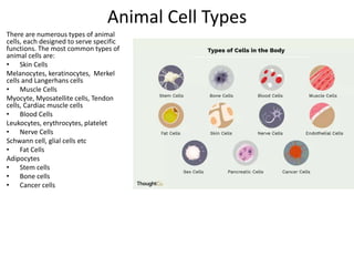
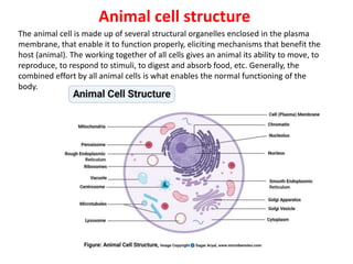
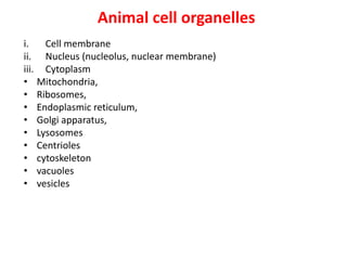
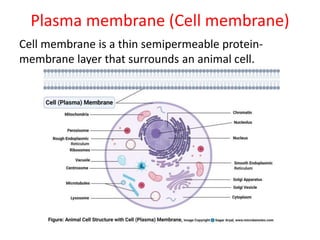
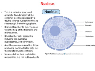
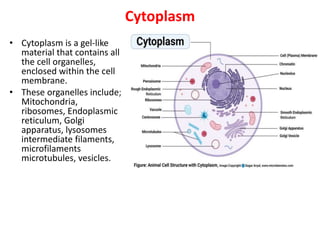
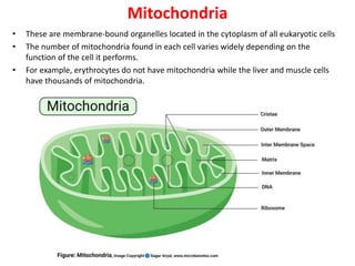
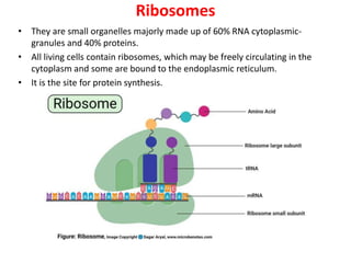
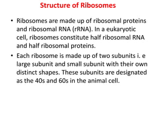
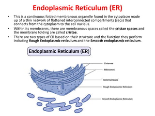
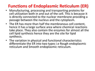
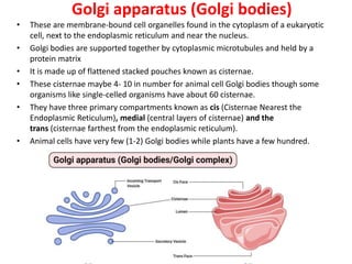
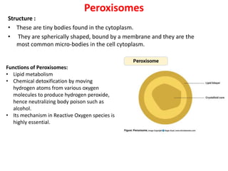
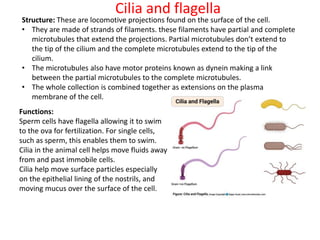
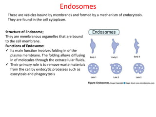
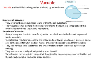
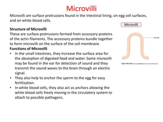
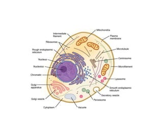
SlideShare a Scribd company logo
Introduction to animal cell
Unit-2
Cell component:
https://microbenotes.com/animal-cell-definition-structure-parts-
functions-and-diagram/#definition-of-animal-cell
Metabolism
https://www.britannica.com/science/metaboli
sm/Incomplete-oxidation
Biochemistry
https://wou.edu/chemistry/courses/online-chemistry-textbooks/ch450-and-
ch451-biochemistry-defining-life-at-the-molecular-level/chapter-1-the-
foundations-of-biochemistry/
Cell communication
https://rwu.pressbooks.pub/bio103/chapter/c
ell-communication/
Introduction
• Animals are a large group of diverse living organisms that make up to three-quarters of all
species on earth. With their ability to move, to respond to stimuli, respond to environmental
changes and adapt to different modes of feeding defense mechanisms and reproduction, all
these mechanisms are enhanced by their constituent elements in the body. However, animals
cannot manufacture their own food like plants and hence they depend on plants in one way or
another.
• All living things are made up of cells that make up their body structure. Some of these living
things are single-celled (unicellular) and other organisms are made up of more than one cell
(Multicellular).
• A cell is the smallest (microscopic) structural-functional unit of life of an organism. The cells
that constitute an animal are called animal cells and those that constitute plants are known as
plant cells.
• Most cells are covered by a protective membrane known as the cell wall which gives the cells
their shape and rigidity.
• An animal cell is a eukaryotic cell that lacks a cell wall, and it is enclosed by the plasma
membrane. The cell organelles are enclosed by the plasma membrane including the cell
nucleus. Unlike the animal cell lacking the cell wall, plant cells have a cell wall.
• Since animal cells lack a rigid cell wall it allows them to develop a great diversity of cell types,
tissues, and organs. The nerves and muscles are made up of specialized cells that plant cells
cannot evolve to form, hence giving these nerve and muscle cells have the ability to move.
Animal cell size and shape
• Animal cells come in all kinds of shapes and sizes, with their size ranging from a
few millimeters to micrometers. The largest animal cell is the ostrich egg which has
a 5-inch diameter, weighing about 1.2-1.4 kg and the smallest animal cells are the
neurons of about 100 microns in diameter.
• Animal cells are smaller than the plant cells and they are generally irregular in
shape taking various forms of shapes, due to lack of the cell wall. Some cells are
round, oval, flattened or rod-shaped, spherical, concave, rectangular. This is due to
the lack of a cell wall. Note: most of the cells are microscopic hence they can only
be seen under a microscope in order to study their anatomy.
• But animal cells share other cellular organelles with plant cells as both have
evolved from eukaryotic cells.
• As noted earlier, animal cells are eukaryotic cells with a membrane-bound nucleus.
therefore they have their genetic material in the form of DNA enclosed in the
nucleus. They also have several structural organelles within the plasma membrane
which perform various specific functions for proper cell function and generally to
maintain the body normal mechanisms.
Animal Cell Types
There are numerous types of animal
cells, each designed to serve specific
functions. The most common types of
animal cells are:
• Skin Cells
Melanocytes, keratinocytes, Merkel
cells and Langerhans cells
• Muscle Cells
Myocyte, Myosatellite cells, Tendon
cells, Cardiac muscle cells
• Blood Cells
Leukocytes, erythrocytes, platelet
• Nerve Cells
Schwann cell, glial cells etc
• Fat Cells
Adipocytes
• Stem cells
• Bone cells
• Cancer cells
Overview
• Humans are complex organisms made up of trillions of cells, each
with their own structure and function.
• Scientists have come a long way in estimating the number of cells in
the average human body. Most recent estimates put the number of
cells at around 30 trillion. Written out, that’s 30,000,000,000,000!
• These cells all work in harmony to carry out all the basic functions
necessary for humans to survive. But it’s not just human cells inside
your body. Scientists estimate that the number of bacterial cells in
the human body likely exceeds the number of human cells.
Cell types
• There are about 200 different types of cells in the body. Here are just a few examples:
• red blood cells (erythrocytes)
• skin cells
• neurons (nerve cells)
• fat cells
• Humans are multicellular, complex organisms. The cells inside our bodies are “specialized.” This
means that each type of cell performs a unique and special function. For this reason, each of the
200 different types of cells in the body has a different structure, size, shape, and function, and
contains different organelles.
For example:
• Cells in the brain may be longer in shape so they can transmit signals more efficiently.
• Cells of the heart have more mitochondria because they need a lot of energy.
• Cells in the respiratory system are responsible for taking up oxygen and releasing carbon dioxide.
• All the cells work together to keep the human body running efficiently.
Animal cell structure
The animal cell is made up of several structural organelles enclosed in the plasma
membrane, that enable it to function properly, eliciting mechanisms that benefit the
host (animal). The working together of all cells gives an animal its ability to move, to
reproduce, to respond to stimuli, to digest and absorb food, etc. Generally, the
combined effort by all animal cells is what enables the normal functioning of the
body.
Animal cell organelles
i. Cell membrane
ii. Nucleus (nucleolus, nuclear membrane)
iii. Cytoplasm
• Mitochondria,
• Ribosomes,
• Endoplasmic reticulum,
• Golgi apparatus,
• Lysosomes
• Centrioles
• cytoskeleton
• vacuoles
• vesicles
Plasma membrane (Cell membrane)
Cell membrane is a thin semipermeable protein-
membrane layer that surrounds an animal cell.
Structure of Plasma membrane (Cell
membrane)
• Thin semi-permeable membrane
• It contains a percentage of lipids making a
semi-permeable barrier between the cell and
its physical environment.
• It has some protein components
• It is very consistent around the cell
• All living cells have a plasma membrane.
Functions of Plasma membrane (Cell
membrane)
• Surrounds the cell
• Holds contents of cell inside (like skin)
• Keeps harmful substances out
• Controls what enters and leaves
• Water, oxygen, and nutrients are allowed to
enter
• Waste products are allowed to exit
Nucleus
• This is a spherical structured
organelle found majorly at the
center of a cell surrounded by a
double-layered nuclear membrane
separating it from the cytoplasm.
• It is held together to the cytoplasm
with the help of the filaments and
microtubules.
• It holds other cells organelles
including the nucleolus,
nucleosomes, and chromatins.
• A cell has one nucleus which divides
producing multinucleated cells e.g.
the skeletal muscle cell fibers.
• Some cells lose their nuclei after
maturations e.g. the red blood cells.
Structure of Nucleus
• The double-layered membrane is a continuous
channel of membranous from the endoplasmic
reticulum network.
• The membrane has pores which allow entry of large
molecule
• Nucleoli (Singular; nucleolus) are tiny/small bodies
found in the nucleus
• The nucleus and its component organelles are
suspended in the nucleoplasm (House of the
chromosomal DNA and genetic materials)
Functions of Nucleus
• The primary role of the nucleus is to control and
regulate cell activities of growth and maintain cell
metabolisms.
• It also carries the genes that have hereditary
information of the cell.
• The chromosomal DNA and genetic materials, which
are made up of genetic coded ultimately make up their
proteins’ amino acid sequences for use by the cell.
• Therefore, the nucleus is the information center.
• It is the site for Transcription (formation of mRNA from
DNA) and the mRNA is transported to the nuclear
envelope.
Cytoplasm
• Cytoplasm is a gel-like
material that contains all
the cell organelles,
enclosed within the cell
membrane.
• These organelles include;
Mitochondria,
ribosomes, Endoplasmic
reticulum, Golgi
apparatus, lysosomes
intermediate filaments,
microfilaments
microtubules, vesicles.
Mitochondria
• These are membrane-bound organelles located in the cytoplasm of all eukaryotic cells
• The number of mitochondria found in each cell varies widely depending on the
function of the cell it performs.
• For example, erythrocytes do not have mitochondria while the liver and muscle cells
have thousands of mitochondria.
Structure of Mitochondria
• They are rod-shaped or oval or spherically
shaped, with a size of 0.5 to 10 μm.
• Mitochondria have two special membranes –
outer and inner membrane.
• They have a mitochondrial gel-matric in the
central mass.
• The membranes bend into folds known
as cristae.
Functions of Mitochondria
• Primary function is to generate energy for the cell i.e they are the power generators,
producing energy in form of Adenosine Tri-phosphate (ATP), by converting nutrients and
oxygen into energy enabling the cell to perform its function and also release excess energy
from the cell.
• Mitochondria also store calcium which assists in cell signaling activity, generating cellular and
mechanical heat and mediating cellular growth and death.
• The outer membrane is permeable, allowing the transport of small molecules and a special
channel to transport large molecules.
• The inner mitochondrial membrane is less permeable thus allowing very small molecules into
the mitochondrial gel-matrix in the central mass. The gel matrix is composed of the
mitochondria DNA and enzymes for the Tricarboxylic Acid (TCA) cycle or the Kreb’s Cycle.
• The TCA cycle uses up the nutrients, converting them into by-products that the mitochondria
use for producing energy. These processes take place in the inner membrane because the
membrane bends into folds called the cristae, where the protein components used for the
main energy production system cells, known as the Electron Transport Chain (ETC). ETC is the
main source of ATP production in the body.
• The ETC involves several sequences of oxidation-reduction reactions to transport electrons
from one protein component to another, thus producing energy that is used for
phosphorylation of ADP (Adenosine diphosphate) to ATP. This process is called
the chemiosmotic coupling of oxidative phosphorylation. This mechanism gives energy to
most cellular activities including muscle movement and they power up the general brain
function.
• Some if not all proteins and molecules that make up the mitochondria come from the cell nucleus.
The mitochondrial nucleus genome has 37 genes of which 13 of these genes produce most of the
components of the ETC. However, the mitochondrial DNA is very vulnerable to mutations because
they don’t possess a large DNA repair mechanism, a common element found in other nuclear
DNAs.
• Moreover, Reactive Oxygen Species ((ROS)) also called free radicals are produced in the
mitochondrion, because of the preference for abnormal production of free electrons. These
electrons are neutralized by antioxidant proteins in the mitochondrion. However, some of the free
radicals can damage mitochondrial DNA (mtDNA).
• Equally, consumption of alcohol can cause damage to the mtDNA because excess ethanol in the
body causes saturation of the detoxifying enzymes leading to the production and leakage of highly
reactive electrons into the cytoplasmic membrane and into the mitochondrial matrix, combining
with other cellular molecules forming numerous radicals that significantly cause cell damage.
• Most organisms inherit the mtDNA from their mother. This is because the maternal egg donates
most of the cytoplasm to the embryo while the mitochondria inherited from the father’s sperm is
destroyed. This causes the origin of inherited and acquired mitochondrial diseases due to
mutations transmitted into the embryo from the maternal and paternal DNA or maternal mtDNA.
Such diseases include Alzheimer’s disease and Parkinson’s disease. When mutated mtDNA
accumulates over time has been linked to aging and the development of certain cancers and
diseases.
• Naturally, mitochondria play a major role in programmed cell death (apoptosis) and due to
mutations in the mtDNA can inhibit cell death-causing the development of cancer.
Functions of Mitochondria
Ribosomes
• They are small organelles majorly made up of 60% RNA cytoplasmic-
granules and 40% proteins.
• All living cells contain ribosomes, which may be freely circulating in the
cytoplasm and some are bound to the endoplasmic reticulum.
• It is the site for protein synthesis.
Structure of Ribosomes
• Ribosomes are made up of ribosomal proteins
and ribosomal RNA (rRNA). In a eukaryotic
cell, ribosomes constitute half ribosomal RNA
and half ribosomal proteins.
• Each ribosome is made up of two subunits i. e
large subunit and small subunit with their own
distinct shapes. These subunits are designated
as the 40s and 60s in the animal cell.
Functions of Ribosomes
• Ribosomes that occur as free particles are attached to the
endoplasmic reticulum membrane occurring in large numbers
accounting for about a quarter of the cell organelles. A single
replicated cell has about 10 million ribosomes.
• The ribosomal subunits are the site for genetic coding into proteins.
On the ribosomes, the mRNA helps determine the coding for
Transfer RNA (tRNA) which also determines the protein amino acid
sequences. This leads to the formation of the rRNA which are
involved in the catalyzation of peptidyl transferase creating the
peptide bond found between the amino acid sequences that
develop the proteins. The formed proteins then detach from the
ribosomes, migrating to other cell parts for utilization by the cell.
Endoplasmic Reticulum (ER)
• This is a continuous folded membranous organelle found in the cytoplasm made
up of a thin network of flattened interconnected compartments (sacs) that
connects from the cytoplasm to the cell nucleus.
• Within its membranes, there are membranous spaces called the cristae spaces and
the membrane folding are called cristae.
• There are two types of ER based on their structure and the function they perform
including Rough Endoplasmic reticulum and the Smooth endoplasmic reticulum.
Functions of Endoplasmic Reticulum (ER)
• Manufacturing, processing and transporting proteins for
cell utilization both in and out of the cell. This is because it
is directly connected to the nuclear membrane providing a
passage between the nucleus and the cytoplasm.
• The ER has more than half the membranous cell content,
hence it has a large surface area where chemical reactions
take place. They also contain the enzymes for almost all the
cell lipid synthesis hence they are the site for lipid
synthesis.
• The variation in physical and functional characteristics
differentiate the ER into two types i.e Rough endoplasmic
reticulum and Smooth endoplasmic reticulum.
Types of Endoplasmic Reticulum
• Rough Endoplasmic Reticulum (Rough ER) – Rough ER is called “rough”
because there surface is covered with ribosomes, giving it a rough
appearance. The function of the ribosomes on rough ER is to synthesis proteins
and they have a signaling sequence, directing them to the endoplasmic
reticulum for processing. Rough ER transports the proteins and lipids through
the cell into the cristae. They are then sent into the Golgi bodies or inserted
into the cell membrane.
• Smooth Endoplasmic Reticulum (Smooth ER) – Smooth ER is not associated
with ribosomes and their unction is different from that of the rough
endoplasmic reticulum, despite lying adjacent to the rough endoplasmic
reticulum. Its function is to synthesis lipids (cholesterol and phospholipids) that
are utilized for producing new cellular membranes. They are also involved in
the synthesis of steroid hormones from cholesterol for certain cell types. It also
contributes to the detoxification of the liver after the intake of drugs and toxic
chemicals.
• There is also a specialized type of smooth ER known as the sarcoplasmic
reticulum. Its function is to regulate the concentration of Calcium ions in the
muscle cell cytoplasm.
Golgi apparatus (Golgi bodies)
• These are membrane-bound cell organelles found in the cytoplasm of a eukaryotic
cell, next to the endoplasmic reticulum and near the nucleus.
• Golgi bodies are supported together by cytoplasmic microtubules and held by a
protein matrix
• It is made up of flattened stacked pouches known as cisternae.
• These cisternae maybe 4- 10 in number for animal cell Golgi bodies though some
organisms like single-celled organisms have about 60 cisternae.
• They have three primary compartments known as cis (Cisternae Nearest the
Endoplasmic Reticulum), medial (central layers of cisternae) and the
trans (cisternae farthest from the endoplasmic reticulum).
• Animal cells have very few (1-2) Golgi bodies while plants have a few hundred.
Functions of Golgi apparatus (Golgi bodies)
• Their primary function is to transport, modify and pack proteins and lipids into the Golgi
vesicles to deliver them to their target sites. Animal cells contain one or more Golgi bodies
while plants have a few hundred.
• Cis and trans Golgi network make up the outer layer of cisternae at the cis and trans face and
they are responsible for sorting proteins and lipids received at the cis face and released by
the trans face, by the Golgi bodies.
• The cis face collects the proteins and lipids, of fused vesicles in clusters. The fused vesicles
move along the microtubules through a specialized compartment known as the vesicular-
tubular cluster. This compartment is found between the endoplasmic reticulum and the
Golgi apparatus.
• The vesicle clusters fuse with the cis Golgi network, delivering the proteins and lipids into the
cis face cisternae and as they move from the cis face to the trans face, they get modified to
functional units. These functional units get delivered to intracellular and extracellular
components of the cell.
– Modification mechanisms include:
– Cleaving of oligosaccharides chains
– Attachment of sugar moieties of different side chains
• Adding fatty acids and/or phosphate groups by phosphorylation, and/or removing
monosaccharides e.g. the removal of the mannose moieties takes place in the cis and the
medial cisternae while adding of galactose takes place in the trans cisternae.
• Sorting of the modified proteins and lipids occurs in the trans-Golgi network and packed into
the trans vesicles, which then delivers them to the lysosomes or sometimes to the cell
membrane for exocytosis. Assisted by ligands bound to receptors triggering fusion and
Lysosomes
Lysosomes were discovered by Christian Rene de Duve, a Belgian cytologist in the
1950s. It is also known as cell vesicles.
Structure of Lysosomes
• They are round subcellular organelle found in almost all eukaryotic cells
• Lysosomes are very acidic organelles containing the digestive enzymes and
therefore each of the lysosomes is surrounded by a membrane to protect it from
the outer environment.
Functions of Lysosomes
• This is the site for digestion of cell nutrients, excretion, and cell renewal.
• Lysosomes break down macromolecules components from the outside of
the cell into simpler elements that are transported into the cytoplasm via
a proton pump to build new cell materials.
• These macromolecule components include old cells and parts, cell waste
products, microorganisms, and cell debris.
• The digestive enzymes found in the lysosomes are called hydrolytic
enzymes or acid hydrolases, breaking down large molecules into smaller
molecules that can be utilized by the cell.
• These enzymes also break down large molecules e. g proteins,
carbohydrates, lipids, into small molecules e.g. amino acids and simple
sugars, fatty acids, respectively.
• Note: The enzymes are active only on the inside of the acidic lysosome
and their acidity protects the cell from degrading itself when there is
lysosomal leakage because the cell pH is neutral to slightly alkaline.
Cytoskeleton
Structure :
• This is a fibrous network that’s formed from and by different proteins of long
chains of amino acids.
• These proteins are found in the cell cytoplasm of the eukaryotic cells.
• They are also made up of 3 types of tiny filaments: Actin filaments
(Microfilaments), Microtubules, Intermediate filaments.
Functions of Cytoskeleton
• The cytoskeleton functions to create a network organizing the cell components
and to also maintain the cell shape.
• It also provided a uniform movement of the cell and its organelles, by the filament
system network found in the cell’s cytoplasm.
• It also organizes some of the cell components maintaining the cell shape
• It plays a major role in the movement of the cell and some cell organelles in the
cytoplasm.
• The tiny filaments include:
– Actin filaments; also known as microfilaments; it’s a meshwork of fibers
running parallel to each other and they play a primary role in giving the cell its
shape; they change consistently, helping the cell to move and to also mediate
certain cell activities such as adherence ability to substrates and cleavage
mechanisms during mitotic cell division
– Microtubules- these are long filaments that assist in mitosis moving daughter
chromosomes to new forming daughter cells.
– Intermediate filaments– they are more stable filaments in comparison to the
actin and microtubules. They form the true skeleton of the cell, and the hold
the nucleus in its rightful position within the cell.
– It also allows the cell’s elasticity factor enabling it to endure physical tension.
• Other proteins that may be added as part of the cytoskeleton of the cell include
septin ((assembles the filaments) and spectrin (help maintain the structure of the
Microtubules
Structure:
• These are long, straight, hollow cylinders filaments that are constructed from 13-
15 sub-filaments (protofilament) strand of a special globular protein
called tubulin, found only in eukaryotic cells.
• They are found throughout the cytoplasm of the animal cell.
Functions of Microtubules
• Transportation of some organelles like the mitochondria and the vesicles
i.e. transporting vesicles from the neuron cell body to the axon tips, and
back to the cell body
• Structural support, they give characteristic support to the Golgi bodies,
holding them within the gel-matrix of the cytoplasm.
• They provide the rigid and organized component of the cytoskeleton of
the cell, enabling a cell to take up a particular shape.
• They are the main elements that make up the locomotive projections of a
cell (cilia and flagella)
• They also play a role in forming the spindle fibers of the chromosome of
the cell during mitotic cell division.
Centrioles
This is distinctly found in the animal cell, which has the ability to replicate or make copies by itself. It is
made up of 9 microtubule bundles and their primary function is to assist in organizing the cell division
process.
Structure of Centrioles
• It is a small structure that is made up of 9 sets of
microtubules, placed in groups of three hence they are
triplet microtubules.
• As triplets, they remain very strong together hence
they have been observed to be in structures like cilia
and flagella.
• The triplet microtubules are held together by proteins,
giving the centriole its shape.
• They are found in the centrosome, creating and
holding microtubules within the cell.
• The triplet microtubules are surrounded by a
pericentriolar matrix containing molecules that build
up the microtubules.
• Each microtubule within the triplet microtubule
complex is made up of tubulin subunits that join
together forming long hollow tubes that look like straw
(microtubules).
Functions of Centrioles
• The centriole microtubules allow the transportation of substances
that are linked together with a glycoprotein to any cell location. the
glycoprotein linkage acts as a signaling unit to move specific
proteins.
• The centrioles anchor the microtubules that extend from it and
contain the factors needed to create more tubules.
• Mitosis is achieved by replication of each centriole which makes
duplicates of each centriole (4 centrioles). The newly formed
centrioles divide into two centrosomes, each centriole at an angle
to the second centriole. The microtubules between the
centrosomes, push the pairs of centrioles apart, to the opposite
ends of the cell. When the centrioles are in place, the microtubules
extend to the cell cytoplasm, to seek for the chromosome. The
microtubules then bind to the chromosome at the centromere. The
microtubules are then unassembled fro the centriole moving the
chromosomes apart.
Peroxisomes
Structure :
• These are tiny bodies found in the cytoplasm.
• They are spherically shaped, bound by a membrane and they are the
most common micro-bodies in the cell cytoplasm.
Functions of Peroxisomes:
• Lipid metabolism
• Chemical detoxification by moving
hydrogen atoms from various oxygen
molecules to produce hydrogen peroxide,
hence neutralizing body poison such as
alcohol.
• Its mechanism in Reactive Oxygen species is
highly essential.
Cilia and flagella
Functions:
Sperm cells have flagella allowing it to swim
to the ova for fertilization. For single cells,
such as sperm, this enables them to swim.
Cilia in the animal cell helps move fluids away
from and past immobile cells.
Cilia help move surface particles especially
on the epithelial lining of the nostrils, and
moving mucus over the surface of the cell.
Structure: These are locomotive projections found on the surface of the cell.
• They are made of strands of filaments. these filaments have partial and complete
microtubules that extend the projections. Partial microtubules don’t extend to
the tip of the cilium and the complete microtubules extend to the tip of the
cilium.
• The microtubules also have motor proteins known as dynein making a link
between the partial microtubules to the complete microtubules.
• The whole collection is combined together as extensions on the plasma
membrane of the cell.
Endosomes
These are vesicles bound by membranes and formed by a mechanism of endocytosis.
They are found in the cell cytoplasm.
Structure of Endosome;
They are membranous organelles that are bound
to the cell membrane.
Functions of Endosome:
 Its main function involves folding in of the
plasma membrane. The folding allows diffusing
in of molecules through the extracellular fluids.
 Their primary role is to remove waste materials
from the cell by endocytic processes such as
exocytosis and phagocytosis
Vacuole
Vacuole are fluid-filled cell organelles enclosed by a membrane.
Structure of Vacuoles:
• They are membrane-bound sacs found within the cell cytoplasm.
• The vacuole sac has a single membrane surrounding it known as a tonoplast and this
membrane resembles the plasma membrane.
Functions of Vacuoles:
• their primary function is to store food, water, carbohydrates in the form of sugars and
waste materials.
• Tonoplast is a regulator controlling the inflow and outflow of small across a protein pump
• acts as the guard for what kinds of matter are allowed passage to and from vacuoles
• They also remove toxic substances and waste materials from the cell as a protection
strategy.
• They also remove poorly folded proteins from the cell.
• Vacuoles also can be able to change their functionality to provide necessary roles that suit
the cell, by being able to change shape and size.
Microvilli
Microvilli are surface protrusions found in the intestinal lining, on egg cell surfaces,
and on white blood cells.
Structure of Microvilli
These are surface protrusions formed from accessory proteins
of the actin filaments. The accessory proteins bundle together
to form microvilli on the surface of the cell membrane
Functions of Microvilli
• In the small intestines, they increase the surface area for
the absorption of digested food and water. Some microvilli
may be found in the ear for detection of sound and they
transmit the sound waves to the brain through an electric
signal.
• They also help to anchor the sperm to the egg for easy
fertilization.
• In white blood cells, they also act as anchors allowing the
white blood cells freely moving in the circulatory system to
attach to possible pathogens.
Composition of cell
Cell organic components
Metabolic pathways
There are two main reasons for studying a
metabolic pathway:
(1) to describe, in quantitative terms, the
chemical changes catalyzed by the
component enzymes of the route; and
(2) to describe the various intracellular controls
that govern the rate at which the pathway
functions.
biochemistry
• Cellular biochemistry is the study of all sorts of processes
that occur with in a biological cell and also interactions
between different cells. Studies include biomolecular
structures, biochemical mechanisms i.e., metabolic
pathways, their control, physiological importance and
clinical relevance.
• Biochemistry is both life science and a chemical science - it
explores the chemistry of living organisms and the
molecular basis for the changes occurring in living cells. It
uses the methods of chemistry, physics, molecular biology,
and immunology to study the structure and behaviour of
the complex molecules found in biological material and the
ways these molecules interact to form cells, tissues, and
whole organisms.
Cell communication
• Cell communication is the process by which a cell detects and responds to signals in its
environment. Most single-celled organisms can perceive changes in nutrient availability
and adapt their metabolism as needed.
• The study of cell communication focuses on how a cell gives and receives messages
with its environment and with itself. Indeed, cells do not live in isolation.
• Their survival depends on receiving and processing information from the outside
environment, whether that information pertains to the availability of nutrients, changes
in temperature, or variations in light levels.
• Cells can also communicate directly with one another — and change their own internal
workings in response — by way of a variety of chemical and mechanical signals.
• In multicellular organisms, cell signaling allows for specialization of groups of cells.
Multiple cell types can then join together to form tissues such as muscle, blood, and
brain tissue.
• In single-celled organisms, signaling allows populations of cells to coordinate with one
another and work like a team to accomplish tasks no single cell could carry out on its
own.
The study of cell signaling touches multiple biological disciplines, such as
developmental biology, neurobiology, and endocrinology.
Cont.
• Despite technical advances, global understanding of signal transduction, its internal
hierarchies, and its highly integrated and extremely dynamic nature remains largely
mysterious.
• A potential breakthrough in the field arose recently when scientists realized that there
are striking analogies between signaling networks in biological systems and electronic
circuits; both of them involve hierarchies, switches, modularity, redundancy, and the
existence of powerful feedback mechanisms. Such a realization gave impetus to the
field of computational biology as applied to cellular signaling.
• Today, the study of cell signaling is not restricted to biologists; with the contribution of
engineers and biophysicists, scientists can now create computational algorithms that
model the structure of a signaling network based on biological measurements, and
these models can be used to predict the outcome of otherwise physically impossible
experimental conditions.
• As it turns out, we are just beginning to appreciate that many of the designs and
strategies we have developed to manipulate information, particularly within the digital
world, are actually present in biological networks, having already been invented over
the course of a hundred million years of evolution.
Cellular Design and the Blueprint of Life
• The design for a cell mostly resides in the blueprint for the cell, the genetic
code, which is comprised of deoxyribonucleic acid (DNA) housed in the cell
nucleus and a small amount in the mitochondria.
• Of course, the DNA blueprint must be read out or transcribed into
ribonucleic acid (RNA) and then translated to proteins by ribozome
structures, which themselves were encoded by the DNA and contain a
combination of RNA and protein subunits.
• The genetic code has the master plan that determines the sequence of all
cellular proteins, which then perform almost all other activities in the cell,
including enzymatic functions, motility, architectural structure, transport,
etc.
• In contrast to DNA, RNA and protein polymers, the formation of the other
two major macromolecules (carbohydrates and lipids) are not driven by such
a template but rather by the enzymes that catalyzes the synthesis.
Cellular Import and Export of Molecules
• Many of the chemical constituents of the cell arise not from direct synthesis but
from import of both small and large molecules.
• The imported molecules must pass through the nonpolar lipid bilayer that
forms the cell membrane, and in some cases through additional membranes if
they need to reside inside membrane-bound organelles.
• Molecules can move into the cell by two major processes:
I. Diffusion The process of diffusion moves molecules down their concentration
gradient from an area of high concentration to an area of low concentration
and does not require an input of energy.
II. Active transport, on the other hand, requires energy to move molecules
against their concentration gradient from an area of low concentration to an
area of high concentration.
• Diffusion across the plasma membrane can either be passive or facilitated. In passive
diffusion, small, nonpolar molecules (such as CO2 and O2) move across the membrane
directly across the membrane. Larger and/or polar molecules move by facilitated
diffusion, which requires a channel or carrier protein

More Related Content

What's hot

Nucleus
NucleusNucleus
Nucleus
MAULIK PATEL
 
Different type of plant cell in tissue system of plant
Different type of plant cell in tissue system of plantDifferent type of plant cell in tissue system of plant
Different type of plant cell in tissue system of plant
Zuby Gohar Ansari
 
Plant tissue
Plant tissuePlant tissue
Plant tissue
stemegypt.edu.eg
 
Cell Membranes
Cell MembranesCell Membranes
Cell Membranes
shabeel pn
 
Cell membrane and cell wall
Cell membrane and cell wallCell membrane and cell wall
Cell membrane and cell wall
Jan Del Rosario
 
Cytosol (cytoplasm)
Cytosol (cytoplasm)Cytosol (cytoplasm)
Cytosol (cytoplasm)
hsalemme
 
Overview of Microscopic techniques
Overview of Microscopic techniques  Overview of Microscopic techniques
Overview of Microscopic techniques
Sir Parashurambhau College, Pune
 
Nuclear envelope
Nuclear envelope Nuclear envelope
Nuclear envelope
Nuria Cano
 
Nucleus Ppt
Nucleus PptNucleus Ppt
Nucleus Ppt
yshtiwa
 
Cell structure
Cell structureCell structure
Cell structure
Michele Nardella
 
Cell structure and its function
Cell structure and its functionCell structure and its function
Cell structure and its function
Shaista Jabeen
 
Cell Growth and Mitosis
Cell Growth and MitosisCell Growth and Mitosis
Cell Growth and Mitosis
jlehmkuhler
 
Cell structure and functions
Cell structure and functionsCell structure and functions
Cell structure and functions
Joy Batang Ü
 
Chapter four
Chapter fourChapter four
Chapter four
dquevedo1
 
Cell Biology Ppt || Centrioles and Centrosome
Cell Biology Ppt || Centrioles and CentrosomeCell Biology Ppt || Centrioles and Centrosome
Cell Biology Ppt || Centrioles and Centrosome
MOHAMMEDVALIKARIMWAL
 
Diversity of cell shape and size
Diversity of cell shape and sizeDiversity of cell shape and size
Diversity of cell shape and size
Akanksha Golchha
 
Cytohistological characters of meristematic tissue
Cytohistological characters of meristematic tissueCytohistological characters of meristematic tissue
Cytohistological characters of meristematic tissue
Rabbiasehar1
 
Extracellllular matrix
Extracellllular matrixExtracellllular matrix
Extracellllular matrix
DR MUKESH SAH
 
Cell Junctions
Cell JunctionsCell Junctions
Cell Junctions
Dr ABU SURAIH SAKHRI
 
Cell biology
Cell biologyCell biology

What's hot (20)

Nucleus
NucleusNucleus
Nucleus
 
Different type of plant cell in tissue system of plant
Different type of plant cell in tissue system of plantDifferent type of plant cell in tissue system of plant
Different type of plant cell in tissue system of plant
 
Plant tissue
Plant tissuePlant tissue
Plant tissue
 
Cell Membranes
Cell MembranesCell Membranes
Cell Membranes
 
Cell membrane and cell wall
Cell membrane and cell wallCell membrane and cell wall
Cell membrane and cell wall
 
Cytosol (cytoplasm)
Cytosol (cytoplasm)Cytosol (cytoplasm)
Cytosol (cytoplasm)
 
Overview of Microscopic techniques
Overview of Microscopic techniques  Overview of Microscopic techniques
Overview of Microscopic techniques
 
Nuclear envelope
Nuclear envelope Nuclear envelope
Nuclear envelope
 
Nucleus Ppt
Nucleus PptNucleus Ppt
Nucleus Ppt
 
Cell structure
Cell structureCell structure
Cell structure
 
Cell structure and its function
Cell structure and its functionCell structure and its function
Cell structure and its function
 
Cell Growth and Mitosis
Cell Growth and MitosisCell Growth and Mitosis
Cell Growth and Mitosis
 
Cell structure and functions
Cell structure and functionsCell structure and functions
Cell structure and functions
 
Chapter four
Chapter fourChapter four
Chapter four
 
Cell Biology Ppt || Centrioles and Centrosome
Cell Biology Ppt || Centrioles and CentrosomeCell Biology Ppt || Centrioles and Centrosome
Cell Biology Ppt || Centrioles and Centrosome
 
Diversity of cell shape and size
Diversity of cell shape and sizeDiversity of cell shape and size
Diversity of cell shape and size
 
Cytohistological characters of meristematic tissue
Cytohistological characters of meristematic tissueCytohistological characters of meristematic tissue
Cytohistological characters of meristematic tissue
 
Extracellllular matrix
Extracellllular matrixExtracellllular matrix
Extracellllular matrix
 
Cell Junctions
Cell JunctionsCell Junctions
Cell Junctions
 
Cell biology
Cell biologyCell biology
Cell biology
 

Similar to Introduction to animal cell.pptx

Cell organelles, from Dr. Himani Singh
Cell organelles, from Dr. Himani Singh Cell organelles, from Dr. Himani Singh
Cell organelles, from Dr. Himani Singh
DR. HIMANI SINGH
 
The cell: Animal cell, plant cell
The cell: Animal cell, plant cellThe cell: Animal cell, plant cell
The cell: Animal cell, plant cell
DR. HIMANI SINGH
 
All about cells !!!!!!!!!!!!!!!!!!!!!!!!!!!!!
All about cells !!!!!!!!!!!!!!!!!!!!!!!!!!!!!All about cells !!!!!!!!!!!!!!!!!!!!!!!!!!!!!
All about cells !!!!!!!!!!!!!!!!!!!!!!!!!!!!!
arivuselvi3
 
cells structure.pptx
cells structure.pptxcells structure.pptx
cells structure.pptx
SanamShaikh10
 
3. ultrastructure of prokaryotes
3. ultrastructure of prokaryotes3. ultrastructure of prokaryotes
3. ultrastructure of prokaryotes
MeenakshiKumari30
 
basics of anatomy and physiology-1.pptx
basics of anatomy and physiology-1.pptxbasics of anatomy and physiology-1.pptx
basics of anatomy and physiology-1.pptx
NavyaPS2
 
Biology chapter 2
Biology chapter 2Biology chapter 2
Biology chapter 2
Amna Khalid
 
Cell-biology-1.pptx (1).pdf
Cell-biology-1.pptx (1).pdfCell-biology-1.pptx (1).pdf
Cell-biology-1.pptx (1).pdf
SachinPokhrel2
 
D) introduction to cells and tissues
D) introduction to cells and tissues D) introduction to cells and tissues
D) introduction to cells and tissues
Sachin Parsekar
 
CELL STRUCTURE AND TRANSPORT.pptx
CELL STRUCTURE AND TRANSPORT.pptxCELL STRUCTURE AND TRANSPORT.pptx
CELL STRUCTURE AND TRANSPORT.pptx
KavithaAnandhan2
 
Cell organele AIMC
Cell organele  AIMCCell organele  AIMC
Cell organele AIMC
Shamim Akram
 
Cell biology
Cell biology Cell biology
Cell biology
MonishKumarRam
 
Lecture 3 Cell_structure_function.ppt
Lecture 3 Cell_structure_function.pptLecture 3 Cell_structure_function.ppt
Lecture 3 Cell_structure_function.ppt
MuhammadAbbasWali
 
1 GNM - Anatimy Unit - 2 - cell & tissue.pptx
1 GNM - Anatimy Unit - 2 - cell & tissue.pptx1 GNM - Anatimy Unit - 2 - cell & tissue.pptx
1 GNM - Anatimy Unit - 2 - cell & tissue.pptx
thiru murugan
 
Cellular Organizations | Class 8 | Science
Cellular Organizations | Class 8 | ScienceCellular Organizations | Class 8 | Science
Cellular Organizations | Class 8 | Science
Vijay Meena
 
Class8 Science Chapter8 Cell
Class8 Science Chapter8 Cell Class8 Science Chapter8 Cell
Class8 Science Chapter8 Cell
ChandraShekharMaurya5
 
The cell
The cellThe cell
The cell
SaneeshVm
 
Structure of cell
Structure of cellStructure of cell
Structure of cell
Praveen Garg
 
cellthefundamentalunitoflife-180820182355 (1)_1.pptx
cellthefundamentalunitoflife-180820182355 (1)_1.pptxcellthefundamentalunitoflife-180820182355 (1)_1.pptx
cellthefundamentalunitoflife-180820182355 (1)_1.pptx
EktaBhatt10
 
Cell for Yoga Net jrf by Dr Shivam Mishra.pptx
Cell for Yoga Net jrf by Dr Shivam Mishra.pptxCell for Yoga Net jrf by Dr Shivam Mishra.pptx
Cell for Yoga Net jrf by Dr Shivam Mishra.pptx
Dr Shivam Mishra
 

Similar to Introduction to animal cell.pptx (20)

Cell organelles, from Dr. Himani Singh
Cell organelles, from Dr. Himani Singh Cell organelles, from Dr. Himani Singh
Cell organelles, from Dr. Himani Singh
 
The cell: Animal cell, plant cell
The cell: Animal cell, plant cellThe cell: Animal cell, plant cell
The cell: Animal cell, plant cell
 
All about cells !!!!!!!!!!!!!!!!!!!!!!!!!!!!!
All about cells !!!!!!!!!!!!!!!!!!!!!!!!!!!!!All about cells !!!!!!!!!!!!!!!!!!!!!!!!!!!!!
All about cells !!!!!!!!!!!!!!!!!!!!!!!!!!!!!
 
cells structure.pptx
cells structure.pptxcells structure.pptx
cells structure.pptx
 
3. ultrastructure of prokaryotes
3. ultrastructure of prokaryotes3. ultrastructure of prokaryotes
3. ultrastructure of prokaryotes
 
basics of anatomy and physiology-1.pptx
basics of anatomy and physiology-1.pptxbasics of anatomy and physiology-1.pptx
basics of anatomy and physiology-1.pptx
 
Biology chapter 2
Biology chapter 2Biology chapter 2
Biology chapter 2
 
Cell-biology-1.pptx (1).pdf
Cell-biology-1.pptx (1).pdfCell-biology-1.pptx (1).pdf
Cell-biology-1.pptx (1).pdf
 
D) introduction to cells and tissues
D) introduction to cells and tissues D) introduction to cells and tissues
D) introduction to cells and tissues
 
CELL STRUCTURE AND TRANSPORT.pptx
CELL STRUCTURE AND TRANSPORT.pptxCELL STRUCTURE AND TRANSPORT.pptx
CELL STRUCTURE AND TRANSPORT.pptx
 
Cell organele AIMC
Cell organele  AIMCCell organele  AIMC
Cell organele AIMC
 
Cell biology
Cell biology Cell biology
Cell biology
 
Lecture 3 Cell_structure_function.ppt
Lecture 3 Cell_structure_function.pptLecture 3 Cell_structure_function.ppt
Lecture 3 Cell_structure_function.ppt
 
1 GNM - Anatimy Unit - 2 - cell & tissue.pptx
1 GNM - Anatimy Unit - 2 - cell & tissue.pptx1 GNM - Anatimy Unit - 2 - cell & tissue.pptx
1 GNM - Anatimy Unit - 2 - cell & tissue.pptx
 
Cellular Organizations | Class 8 | Science
Cellular Organizations | Class 8 | ScienceCellular Organizations | Class 8 | Science
Cellular Organizations | Class 8 | Science
 
Class8 Science Chapter8 Cell
Class8 Science Chapter8 Cell Class8 Science Chapter8 Cell
Class8 Science Chapter8 Cell
 
The cell
The cellThe cell
The cell
 
Structure of cell
Structure of cellStructure of cell
Structure of cell
 
cellthefundamentalunitoflife-180820182355 (1)_1.pptx
cellthefundamentalunitoflife-180820182355 (1)_1.pptxcellthefundamentalunitoflife-180820182355 (1)_1.pptx
cellthefundamentalunitoflife-180820182355 (1)_1.pptx
 
Cell for Yoga Net jrf by Dr Shivam Mishra.pptx
Cell for Yoga Net jrf by Dr Shivam Mishra.pptxCell for Yoga Net jrf by Dr Shivam Mishra.pptx
Cell for Yoga Net jrf by Dr Shivam Mishra.pptx
 

More from ssuserdeaeaf

defense mechanism.pdf
defense mechanism.pdfdefense mechanism.pdf
defense mechanism.pdf
ssuserdeaeaf
 
Case study on different types of animal intelligence.pdf
Case study on different types of animal intelligence.pdfCase study on different types of animal intelligence.pdf
Case study on different types of animal intelligence.pdf
ssuserdeaeaf
 
Different Types of Intelligent body systems.pdf
Different Types of Intelligent body systems.pdfDifferent Types of Intelligent body systems.pdf
Different Types of Intelligent body systems.pdf
ssuserdeaeaf
 
Chemical thermodynamics and electrochemistry.pdf
Chemical thermodynamics and electrochemistry.pdfChemical thermodynamics and electrochemistry.pdf
Chemical thermodynamics and electrochemistry.pdf
ssuserdeaeaf
 
SPECTROSCOPY.pdf
SPECTROSCOPY.pdfSPECTROSCOPY.pdf
SPECTROSCOPY.pdf
ssuserdeaeaf
 
Lubricants.pdf
Lubricants.pdfLubricants.pdf
Lubricants.pdf
ssuserdeaeaf
 
Introductions Polymers .pdf
Introductions Polymers .pdfIntroductions Polymers .pdf
Introductions Polymers .pdf
ssuserdeaeaf
 
WATER and ITS TREATMENT.pdf
WATER and ITS TREATMENT.pdfWATER and ITS TREATMENT.pdf
WATER and ITS TREATMENT.pdf
ssuserdeaeaf
 
Unit-1 Atomic Structures and theory-final.pdf
Unit-1 Atomic Structures and theory-final.pdfUnit-1 Atomic Structures and theory-final.pdf
Unit-1 Atomic Structures and theory-final.pdf
ssuserdeaeaf
 

More from ssuserdeaeaf (9)

defense mechanism.pdf
defense mechanism.pdfdefense mechanism.pdf
defense mechanism.pdf
 
Case study on different types of animal intelligence.pdf
Case study on different types of animal intelligence.pdfCase study on different types of animal intelligence.pdf
Case study on different types of animal intelligence.pdf
 
Different Types of Intelligent body systems.pdf
Different Types of Intelligent body systems.pdfDifferent Types of Intelligent body systems.pdf
Different Types of Intelligent body systems.pdf
 
Chemical thermodynamics and electrochemistry.pdf
Chemical thermodynamics and electrochemistry.pdfChemical thermodynamics and electrochemistry.pdf
Chemical thermodynamics and electrochemistry.pdf
 
SPECTROSCOPY.pdf
SPECTROSCOPY.pdfSPECTROSCOPY.pdf
SPECTROSCOPY.pdf
 
Lubricants.pdf
Lubricants.pdfLubricants.pdf
Lubricants.pdf
 
Introductions Polymers .pdf
Introductions Polymers .pdfIntroductions Polymers .pdf
Introductions Polymers .pdf
 
WATER and ITS TREATMENT.pdf
WATER and ITS TREATMENT.pdfWATER and ITS TREATMENT.pdf
WATER and ITS TREATMENT.pdf
 
Unit-1 Atomic Structures and theory-final.pdf
Unit-1 Atomic Structures and theory-final.pdfUnit-1 Atomic Structures and theory-final.pdf
Unit-1 Atomic Structures and theory-final.pdf
 

Recently uploaded

PCOS corelations and management through Ayurveda.
PCOS corelations and management through Ayurveda.PCOS corelations and management through Ayurveda.
PCOS corelations and management through Ayurveda.
Dr. Shivangi Singh Parihar
 
C1 Rubenstein AP HuG xxxxxxxxxxxxxx.pptx
C1 Rubenstein AP HuG xxxxxxxxxxxxxx.pptxC1 Rubenstein AP HuG xxxxxxxxxxxxxx.pptx
C1 Rubenstein AP HuG xxxxxxxxxxxxxx.pptx
mulvey2
 
Main Java[All of the Base Concepts}.docx
Main Java[All of the Base Concepts}.docxMain Java[All of the Base Concepts}.docx
Main Java[All of the Base Concepts}.docx
adhitya5119
 
RPMS TEMPLATE FOR SCHOOL YEAR 2023-2024 FOR TEACHER 1 TO TEACHER 3
RPMS TEMPLATE FOR SCHOOL YEAR 2023-2024 FOR TEACHER 1 TO TEACHER 3RPMS TEMPLATE FOR SCHOOL YEAR 2023-2024 FOR TEACHER 1 TO TEACHER 3
RPMS TEMPLATE FOR SCHOOL YEAR 2023-2024 FOR TEACHER 1 TO TEACHER 3
IreneSebastianRueco1
 
clinical examination of hip joint (1).pdf
clinical examination of hip joint (1).pdfclinical examination of hip joint (1).pdf
clinical examination of hip joint (1).pdf
Priyankaranawat4
 
Digital Artifact 1 - 10VCD Environments Unit
Digital Artifact 1 - 10VCD Environments UnitDigital Artifact 1 - 10VCD Environments Unit
Digital Artifact 1 - 10VCD Environments Unit
chanes7
 
The simplified electron and muon model, Oscillating Spacetime: The Foundation...
The simplified electron and muon model, Oscillating Spacetime: The Foundation...The simplified electron and muon model, Oscillating Spacetime: The Foundation...
The simplified electron and muon model, Oscillating Spacetime: The Foundation...
RitikBhardwaj56
 
World environment day ppt For 5 June 2024
World environment day ppt For 5 June 2024World environment day ppt For 5 June 2024
World environment day ppt For 5 June 2024
ak6969907
 
Chapter 4 - Islamic Financial Institutions in Malaysia.pptx
Chapter 4 - Islamic Financial Institutions in Malaysia.pptxChapter 4 - Islamic Financial Institutions in Malaysia.pptx
Chapter 4 - Islamic Financial Institutions in Malaysia.pptx
Mohd Adib Abd Muin, Senior Lecturer at Universiti Utara Malaysia
 
MARY JANE WILSON, A “BOA MÃE” .
MARY JANE WILSON, A “BOA MÃE”           .MARY JANE WILSON, A “BOA MÃE”           .
MARY JANE WILSON, A “BOA MÃE” .
Colégio Santa Teresinha
 
Digital Artefact 1 - Tiny Home Environmental Design
Digital Artefact 1 - Tiny Home Environmental DesignDigital Artefact 1 - Tiny Home Environmental Design
Digital Artefact 1 - Tiny Home Environmental Design
amberjdewit93
 
How to Fix the Import Error in the Odoo 17
How to Fix the Import Error in the Odoo 17How to Fix the Import Error in the Odoo 17
How to Fix the Import Error in the Odoo 17
Celine George
 
Lapbook sobre os Regimes Totalitários.pdf
Lapbook sobre os Regimes Totalitários.pdfLapbook sobre os Regimes Totalitários.pdf
Lapbook sobre os Regimes Totalitários.pdf
Jean Carlos Nunes Paixão
 
A Survey of Techniques for Maximizing LLM Performance.pptx
A Survey of Techniques for Maximizing LLM Performance.pptxA Survey of Techniques for Maximizing LLM Performance.pptx
A Survey of Techniques for Maximizing LLM Performance.pptx
thanhdowork
 
ANATOMY AND BIOMECHANICS OF HIP JOINT.pdf
ANATOMY AND BIOMECHANICS OF HIP JOINT.pdfANATOMY AND BIOMECHANICS OF HIP JOINT.pdf
ANATOMY AND BIOMECHANICS OF HIP JOINT.pdf
Priyankaranawat4
 
Pollock and Snow "DEIA in the Scholarly Landscape, Session One: Setting Expec...
Pollock and Snow "DEIA in the Scholarly Landscape, Session One: Setting Expec...Pollock and Snow "DEIA in the Scholarly Landscape, Session One: Setting Expec...
Pollock and Snow "DEIA in the Scholarly Landscape, Session One: Setting Expec...
National Information Standards Organization (NISO)
 
Hindi varnamala | hindi alphabet PPT.pdf
Hindi varnamala | hindi alphabet PPT.pdfHindi varnamala | hindi alphabet PPT.pdf
Hindi varnamala | hindi alphabet PPT.pdf
Dr. Mulla Adam Ali
 
Introduction to AI for Nonprofits with Tapp Network
Introduction to AI for Nonprofits with Tapp NetworkIntroduction to AI for Nonprofits with Tapp Network
Introduction to AI for Nonprofits with Tapp Network
TechSoup
 
Film vocab for eal 3 students: Australia the movie
Film vocab for eal 3 students: Australia the movieFilm vocab for eal 3 students: Australia the movie
Film vocab for eal 3 students: Australia the movie
Nicholas Montgomery
 
How to Add Chatter in the odoo 17 ERP Module
How to Add Chatter in the odoo 17 ERP ModuleHow to Add Chatter in the odoo 17 ERP Module
How to Add Chatter in the odoo 17 ERP Module
Celine George
 

Recently uploaded (20)

PCOS corelations and management through Ayurveda.
PCOS corelations and management through Ayurveda.PCOS corelations and management through Ayurveda.
PCOS corelations and management through Ayurveda.
 
C1 Rubenstein AP HuG xxxxxxxxxxxxxx.pptx
C1 Rubenstein AP HuG xxxxxxxxxxxxxx.pptxC1 Rubenstein AP HuG xxxxxxxxxxxxxx.pptx
C1 Rubenstein AP HuG xxxxxxxxxxxxxx.pptx
 
Main Java[All of the Base Concepts}.docx
Main Java[All of the Base Concepts}.docxMain Java[All of the Base Concepts}.docx
Main Java[All of the Base Concepts}.docx
 
RPMS TEMPLATE FOR SCHOOL YEAR 2023-2024 FOR TEACHER 1 TO TEACHER 3
RPMS TEMPLATE FOR SCHOOL YEAR 2023-2024 FOR TEACHER 1 TO TEACHER 3RPMS TEMPLATE FOR SCHOOL YEAR 2023-2024 FOR TEACHER 1 TO TEACHER 3
RPMS TEMPLATE FOR SCHOOL YEAR 2023-2024 FOR TEACHER 1 TO TEACHER 3
 
clinical examination of hip joint (1).pdf
clinical examination of hip joint (1).pdfclinical examination of hip joint (1).pdf
clinical examination of hip joint (1).pdf
 
Digital Artifact 1 - 10VCD Environments Unit
Digital Artifact 1 - 10VCD Environments UnitDigital Artifact 1 - 10VCD Environments Unit
Digital Artifact 1 - 10VCD Environments Unit
 
The simplified electron and muon model, Oscillating Spacetime: The Foundation...
The simplified electron and muon model, Oscillating Spacetime: The Foundation...The simplified electron and muon model, Oscillating Spacetime: The Foundation...
The simplified electron and muon model, Oscillating Spacetime: The Foundation...
 
World environment day ppt For 5 June 2024
World environment day ppt For 5 June 2024World environment day ppt For 5 June 2024
World environment day ppt For 5 June 2024
 
Chapter 4 - Islamic Financial Institutions in Malaysia.pptx
Chapter 4 - Islamic Financial Institutions in Malaysia.pptxChapter 4 - Islamic Financial Institutions in Malaysia.pptx
Chapter 4 - Islamic Financial Institutions in Malaysia.pptx
 
MARY JANE WILSON, A “BOA MÃE” .
MARY JANE WILSON, A “BOA MÃE”           .MARY JANE WILSON, A “BOA MÃE”           .
MARY JANE WILSON, A “BOA MÃE” .
 
Digital Artefact 1 - Tiny Home Environmental Design
Digital Artefact 1 - Tiny Home Environmental DesignDigital Artefact 1 - Tiny Home Environmental Design
Digital Artefact 1 - Tiny Home Environmental Design
 
How to Fix the Import Error in the Odoo 17
How to Fix the Import Error in the Odoo 17How to Fix the Import Error in the Odoo 17
How to Fix the Import Error in the Odoo 17
 
Lapbook sobre os Regimes Totalitários.pdf
Lapbook sobre os Regimes Totalitários.pdfLapbook sobre os Regimes Totalitários.pdf
Lapbook sobre os Regimes Totalitários.pdf
 
A Survey of Techniques for Maximizing LLM Performance.pptx
A Survey of Techniques for Maximizing LLM Performance.pptxA Survey of Techniques for Maximizing LLM Performance.pptx
A Survey of Techniques for Maximizing LLM Performance.pptx
 
ANATOMY AND BIOMECHANICS OF HIP JOINT.pdf
ANATOMY AND BIOMECHANICS OF HIP JOINT.pdfANATOMY AND BIOMECHANICS OF HIP JOINT.pdf
ANATOMY AND BIOMECHANICS OF HIP JOINT.pdf
 
Pollock and Snow "DEIA in the Scholarly Landscape, Session One: Setting Expec...
Pollock and Snow "DEIA in the Scholarly Landscape, Session One: Setting Expec...Pollock and Snow "DEIA in the Scholarly Landscape, Session One: Setting Expec...
Pollock and Snow "DEIA in the Scholarly Landscape, Session One: Setting Expec...
 
Hindi varnamala | hindi alphabet PPT.pdf
Hindi varnamala | hindi alphabet PPT.pdfHindi varnamala | hindi alphabet PPT.pdf
Hindi varnamala | hindi alphabet PPT.pdf
 
Introduction to AI for Nonprofits with Tapp Network
Introduction to AI for Nonprofits with Tapp NetworkIntroduction to AI for Nonprofits with Tapp Network
Introduction to AI for Nonprofits with Tapp Network
 
Film vocab for eal 3 students: Australia the movie
Film vocab for eal 3 students: Australia the movieFilm vocab for eal 3 students: Australia the movie
Film vocab for eal 3 students: Australia the movie
 
How to Add Chatter in the odoo 17 ERP Module
How to Add Chatter in the odoo 17 ERP ModuleHow to Add Chatter in the odoo 17 ERP Module
How to Add Chatter in the odoo 17 ERP Module
 

Introduction to animal cell.pptx

  • 1. Introduction to animal cell Unit-2 Cell component: https://microbenotes.com/animal-cell-definition-structure-parts- functions-and-diagram/#definition-of-animal-cell Metabolism https://www.britannica.com/science/metaboli sm/Incomplete-oxidation Biochemistry https://wou.edu/chemistry/courses/online-chemistry-textbooks/ch450-and- ch451-biochemistry-defining-life-at-the-molecular-level/chapter-1-the- foundations-of-biochemistry/ Cell communication https://rwu.pressbooks.pub/bio103/chapter/c ell-communication/
  • 2.
  • 3. Introduction • Animals are a large group of diverse living organisms that make up to three-quarters of all species on earth. With their ability to move, to respond to stimuli, respond to environmental changes and adapt to different modes of feeding defense mechanisms and reproduction, all these mechanisms are enhanced by their constituent elements in the body. However, animals cannot manufacture their own food like plants and hence they depend on plants in one way or another. • All living things are made up of cells that make up their body structure. Some of these living things are single-celled (unicellular) and other organisms are made up of more than one cell (Multicellular). • A cell is the smallest (microscopic) structural-functional unit of life of an organism. The cells that constitute an animal are called animal cells and those that constitute plants are known as plant cells. • Most cells are covered by a protective membrane known as the cell wall which gives the cells their shape and rigidity. • An animal cell is a eukaryotic cell that lacks a cell wall, and it is enclosed by the plasma membrane. The cell organelles are enclosed by the plasma membrane including the cell nucleus. Unlike the animal cell lacking the cell wall, plant cells have a cell wall. • Since animal cells lack a rigid cell wall it allows them to develop a great diversity of cell types, tissues, and organs. The nerves and muscles are made up of specialized cells that plant cells cannot evolve to form, hence giving these nerve and muscle cells have the ability to move.
  • 4. Animal cell size and shape • Animal cells come in all kinds of shapes and sizes, with their size ranging from a few millimeters to micrometers. The largest animal cell is the ostrich egg which has a 5-inch diameter, weighing about 1.2-1.4 kg and the smallest animal cells are the neurons of about 100 microns in diameter. • Animal cells are smaller than the plant cells and they are generally irregular in shape taking various forms of shapes, due to lack of the cell wall. Some cells are round, oval, flattened or rod-shaped, spherical, concave, rectangular. This is due to the lack of a cell wall. Note: most of the cells are microscopic hence they can only be seen under a microscope in order to study their anatomy. • But animal cells share other cellular organelles with plant cells as both have evolved from eukaryotic cells. • As noted earlier, animal cells are eukaryotic cells with a membrane-bound nucleus. therefore they have their genetic material in the form of DNA enclosed in the nucleus. They also have several structural organelles within the plasma membrane which perform various specific functions for proper cell function and generally to maintain the body normal mechanisms.
  • 5. Animal Cell Types There are numerous types of animal cells, each designed to serve specific functions. The most common types of animal cells are: • Skin Cells Melanocytes, keratinocytes, Merkel cells and Langerhans cells • Muscle Cells Myocyte, Myosatellite cells, Tendon cells, Cardiac muscle cells • Blood Cells Leukocytes, erythrocytes, platelet • Nerve Cells Schwann cell, glial cells etc • Fat Cells Adipocytes • Stem cells • Bone cells • Cancer cells
  • 6. Overview • Humans are complex organisms made up of trillions of cells, each with their own structure and function. • Scientists have come a long way in estimating the number of cells in the average human body. Most recent estimates put the number of cells at around 30 trillion. Written out, that’s 30,000,000,000,000! • These cells all work in harmony to carry out all the basic functions necessary for humans to survive. But it’s not just human cells inside your body. Scientists estimate that the number of bacterial cells in the human body likely exceeds the number of human cells.
  • 7. Cell types • There are about 200 different types of cells in the body. Here are just a few examples: • red blood cells (erythrocytes) • skin cells • neurons (nerve cells) • fat cells • Humans are multicellular, complex organisms. The cells inside our bodies are “specialized.” This means that each type of cell performs a unique and special function. For this reason, each of the 200 different types of cells in the body has a different structure, size, shape, and function, and contains different organelles. For example: • Cells in the brain may be longer in shape so they can transmit signals more efficiently. • Cells of the heart have more mitochondria because they need a lot of energy. • Cells in the respiratory system are responsible for taking up oxygen and releasing carbon dioxide. • All the cells work together to keep the human body running efficiently.
  • 8. Animal cell structure The animal cell is made up of several structural organelles enclosed in the plasma membrane, that enable it to function properly, eliciting mechanisms that benefit the host (animal). The working together of all cells gives an animal its ability to move, to reproduce, to respond to stimuli, to digest and absorb food, etc. Generally, the combined effort by all animal cells is what enables the normal functioning of the body.
  • 9. Animal cell organelles i. Cell membrane ii. Nucleus (nucleolus, nuclear membrane) iii. Cytoplasm • Mitochondria, • Ribosomes, • Endoplasmic reticulum, • Golgi apparatus, • Lysosomes • Centrioles • cytoskeleton • vacuoles • vesicles
  • 10. Plasma membrane (Cell membrane) Cell membrane is a thin semipermeable protein- membrane layer that surrounds an animal cell.
  • 11. Structure of Plasma membrane (Cell membrane) • Thin semi-permeable membrane • It contains a percentage of lipids making a semi-permeable barrier between the cell and its physical environment. • It has some protein components • It is very consistent around the cell • All living cells have a plasma membrane.
  • 12. Functions of Plasma membrane (Cell membrane) • Surrounds the cell • Holds contents of cell inside (like skin) • Keeps harmful substances out • Controls what enters and leaves • Water, oxygen, and nutrients are allowed to enter • Waste products are allowed to exit
  • 13. Nucleus • This is a spherical structured organelle found majorly at the center of a cell surrounded by a double-layered nuclear membrane separating it from the cytoplasm. • It is held together to the cytoplasm with the help of the filaments and microtubules. • It holds other cells organelles including the nucleolus, nucleosomes, and chromatins. • A cell has one nucleus which divides producing multinucleated cells e.g. the skeletal muscle cell fibers. • Some cells lose their nuclei after maturations e.g. the red blood cells.
  • 14. Structure of Nucleus • The double-layered membrane is a continuous channel of membranous from the endoplasmic reticulum network. • The membrane has pores which allow entry of large molecule • Nucleoli (Singular; nucleolus) are tiny/small bodies found in the nucleus • The nucleus and its component organelles are suspended in the nucleoplasm (House of the chromosomal DNA and genetic materials)
  • 15. Functions of Nucleus • The primary role of the nucleus is to control and regulate cell activities of growth and maintain cell metabolisms. • It also carries the genes that have hereditary information of the cell. • The chromosomal DNA and genetic materials, which are made up of genetic coded ultimately make up their proteins’ amino acid sequences for use by the cell. • Therefore, the nucleus is the information center. • It is the site for Transcription (formation of mRNA from DNA) and the mRNA is transported to the nuclear envelope.
  • 16. Cytoplasm • Cytoplasm is a gel-like material that contains all the cell organelles, enclosed within the cell membrane. • These organelles include; Mitochondria, ribosomes, Endoplasmic reticulum, Golgi apparatus, lysosomes intermediate filaments, microfilaments microtubules, vesicles.
  • 17. Mitochondria • These are membrane-bound organelles located in the cytoplasm of all eukaryotic cells • The number of mitochondria found in each cell varies widely depending on the function of the cell it performs. • For example, erythrocytes do not have mitochondria while the liver and muscle cells have thousands of mitochondria.
  • 18. Structure of Mitochondria • They are rod-shaped or oval or spherically shaped, with a size of 0.5 to 10 μm. • Mitochondria have two special membranes – outer and inner membrane. • They have a mitochondrial gel-matric in the central mass. • The membranes bend into folds known as cristae.
  • 19. Functions of Mitochondria • Primary function is to generate energy for the cell i.e they are the power generators, producing energy in form of Adenosine Tri-phosphate (ATP), by converting nutrients and oxygen into energy enabling the cell to perform its function and also release excess energy from the cell. • Mitochondria also store calcium which assists in cell signaling activity, generating cellular and mechanical heat and mediating cellular growth and death. • The outer membrane is permeable, allowing the transport of small molecules and a special channel to transport large molecules. • The inner mitochondrial membrane is less permeable thus allowing very small molecules into the mitochondrial gel-matrix in the central mass. The gel matrix is composed of the mitochondria DNA and enzymes for the Tricarboxylic Acid (TCA) cycle or the Kreb’s Cycle. • The TCA cycle uses up the nutrients, converting them into by-products that the mitochondria use for producing energy. These processes take place in the inner membrane because the membrane bends into folds called the cristae, where the protein components used for the main energy production system cells, known as the Electron Transport Chain (ETC). ETC is the main source of ATP production in the body. • The ETC involves several sequences of oxidation-reduction reactions to transport electrons from one protein component to another, thus producing energy that is used for phosphorylation of ADP (Adenosine diphosphate) to ATP. This process is called the chemiosmotic coupling of oxidative phosphorylation. This mechanism gives energy to most cellular activities including muscle movement and they power up the general brain function.
  • 20. • Some if not all proteins and molecules that make up the mitochondria come from the cell nucleus. The mitochondrial nucleus genome has 37 genes of which 13 of these genes produce most of the components of the ETC. However, the mitochondrial DNA is very vulnerable to mutations because they don’t possess a large DNA repair mechanism, a common element found in other nuclear DNAs. • Moreover, Reactive Oxygen Species ((ROS)) also called free radicals are produced in the mitochondrion, because of the preference for abnormal production of free electrons. These electrons are neutralized by antioxidant proteins in the mitochondrion. However, some of the free radicals can damage mitochondrial DNA (mtDNA). • Equally, consumption of alcohol can cause damage to the mtDNA because excess ethanol in the body causes saturation of the detoxifying enzymes leading to the production and leakage of highly reactive electrons into the cytoplasmic membrane and into the mitochondrial matrix, combining with other cellular molecules forming numerous radicals that significantly cause cell damage. • Most organisms inherit the mtDNA from their mother. This is because the maternal egg donates most of the cytoplasm to the embryo while the mitochondria inherited from the father’s sperm is destroyed. This causes the origin of inherited and acquired mitochondrial diseases due to mutations transmitted into the embryo from the maternal and paternal DNA or maternal mtDNA. Such diseases include Alzheimer’s disease and Parkinson’s disease. When mutated mtDNA accumulates over time has been linked to aging and the development of certain cancers and diseases. • Naturally, mitochondria play a major role in programmed cell death (apoptosis) and due to mutations in the mtDNA can inhibit cell death-causing the development of cancer. Functions of Mitochondria
  • 21. Ribosomes • They are small organelles majorly made up of 60% RNA cytoplasmic- granules and 40% proteins. • All living cells contain ribosomes, which may be freely circulating in the cytoplasm and some are bound to the endoplasmic reticulum. • It is the site for protein synthesis.
  • 22. Structure of Ribosomes • Ribosomes are made up of ribosomal proteins and ribosomal RNA (rRNA). In a eukaryotic cell, ribosomes constitute half ribosomal RNA and half ribosomal proteins. • Each ribosome is made up of two subunits i. e large subunit and small subunit with their own distinct shapes. These subunits are designated as the 40s and 60s in the animal cell.
  • 23. Functions of Ribosomes • Ribosomes that occur as free particles are attached to the endoplasmic reticulum membrane occurring in large numbers accounting for about a quarter of the cell organelles. A single replicated cell has about 10 million ribosomes. • The ribosomal subunits are the site for genetic coding into proteins. On the ribosomes, the mRNA helps determine the coding for Transfer RNA (tRNA) which also determines the protein amino acid sequences. This leads to the formation of the rRNA which are involved in the catalyzation of peptidyl transferase creating the peptide bond found between the amino acid sequences that develop the proteins. The formed proteins then detach from the ribosomes, migrating to other cell parts for utilization by the cell.
  • 24. Endoplasmic Reticulum (ER) • This is a continuous folded membranous organelle found in the cytoplasm made up of a thin network of flattened interconnected compartments (sacs) that connects from the cytoplasm to the cell nucleus. • Within its membranes, there are membranous spaces called the cristae spaces and the membrane folding are called cristae. • There are two types of ER based on their structure and the function they perform including Rough Endoplasmic reticulum and the Smooth endoplasmic reticulum.
  • 25. Functions of Endoplasmic Reticulum (ER) • Manufacturing, processing and transporting proteins for cell utilization both in and out of the cell. This is because it is directly connected to the nuclear membrane providing a passage between the nucleus and the cytoplasm. • The ER has more than half the membranous cell content, hence it has a large surface area where chemical reactions take place. They also contain the enzymes for almost all the cell lipid synthesis hence they are the site for lipid synthesis. • The variation in physical and functional characteristics differentiate the ER into two types i.e Rough endoplasmic reticulum and Smooth endoplasmic reticulum.
  • 26. Types of Endoplasmic Reticulum • Rough Endoplasmic Reticulum (Rough ER) – Rough ER is called “rough” because there surface is covered with ribosomes, giving it a rough appearance. The function of the ribosomes on rough ER is to synthesis proteins and they have a signaling sequence, directing them to the endoplasmic reticulum for processing. Rough ER transports the proteins and lipids through the cell into the cristae. They are then sent into the Golgi bodies or inserted into the cell membrane. • Smooth Endoplasmic Reticulum (Smooth ER) – Smooth ER is not associated with ribosomes and their unction is different from that of the rough endoplasmic reticulum, despite lying adjacent to the rough endoplasmic reticulum. Its function is to synthesis lipids (cholesterol and phospholipids) that are utilized for producing new cellular membranes. They are also involved in the synthesis of steroid hormones from cholesterol for certain cell types. It also contributes to the detoxification of the liver after the intake of drugs and toxic chemicals. • There is also a specialized type of smooth ER known as the sarcoplasmic reticulum. Its function is to regulate the concentration of Calcium ions in the muscle cell cytoplasm.
  • 27. Golgi apparatus (Golgi bodies) • These are membrane-bound cell organelles found in the cytoplasm of a eukaryotic cell, next to the endoplasmic reticulum and near the nucleus. • Golgi bodies are supported together by cytoplasmic microtubules and held by a protein matrix • It is made up of flattened stacked pouches known as cisternae. • These cisternae maybe 4- 10 in number for animal cell Golgi bodies though some organisms like single-celled organisms have about 60 cisternae. • They have three primary compartments known as cis (Cisternae Nearest the Endoplasmic Reticulum), medial (central layers of cisternae) and the trans (cisternae farthest from the endoplasmic reticulum). • Animal cells have very few (1-2) Golgi bodies while plants have a few hundred.
  • 28. Functions of Golgi apparatus (Golgi bodies) • Their primary function is to transport, modify and pack proteins and lipids into the Golgi vesicles to deliver them to their target sites. Animal cells contain one or more Golgi bodies while plants have a few hundred. • Cis and trans Golgi network make up the outer layer of cisternae at the cis and trans face and they are responsible for sorting proteins and lipids received at the cis face and released by the trans face, by the Golgi bodies. • The cis face collects the proteins and lipids, of fused vesicles in clusters. The fused vesicles move along the microtubules through a specialized compartment known as the vesicular- tubular cluster. This compartment is found between the endoplasmic reticulum and the Golgi apparatus. • The vesicle clusters fuse with the cis Golgi network, delivering the proteins and lipids into the cis face cisternae and as they move from the cis face to the trans face, they get modified to functional units. These functional units get delivered to intracellular and extracellular components of the cell. – Modification mechanisms include: – Cleaving of oligosaccharides chains – Attachment of sugar moieties of different side chains • Adding fatty acids and/or phosphate groups by phosphorylation, and/or removing monosaccharides e.g. the removal of the mannose moieties takes place in the cis and the medial cisternae while adding of galactose takes place in the trans cisternae. • Sorting of the modified proteins and lipids occurs in the trans-Golgi network and packed into the trans vesicles, which then delivers them to the lysosomes or sometimes to the cell membrane for exocytosis. Assisted by ligands bound to receptors triggering fusion and
  • 29. Lysosomes Lysosomes were discovered by Christian Rene de Duve, a Belgian cytologist in the 1950s. It is also known as cell vesicles. Structure of Lysosomes • They are round subcellular organelle found in almost all eukaryotic cells • Lysosomes are very acidic organelles containing the digestive enzymes and therefore each of the lysosomes is surrounded by a membrane to protect it from the outer environment.
  • 30. Functions of Lysosomes • This is the site for digestion of cell nutrients, excretion, and cell renewal. • Lysosomes break down macromolecules components from the outside of the cell into simpler elements that are transported into the cytoplasm via a proton pump to build new cell materials. • These macromolecule components include old cells and parts, cell waste products, microorganisms, and cell debris. • The digestive enzymes found in the lysosomes are called hydrolytic enzymes or acid hydrolases, breaking down large molecules into smaller molecules that can be utilized by the cell. • These enzymes also break down large molecules e. g proteins, carbohydrates, lipids, into small molecules e.g. amino acids and simple sugars, fatty acids, respectively. • Note: The enzymes are active only on the inside of the acidic lysosome and their acidity protects the cell from degrading itself when there is lysosomal leakage because the cell pH is neutral to slightly alkaline.
  • 31. Cytoskeleton Structure : • This is a fibrous network that’s formed from and by different proteins of long chains of amino acids. • These proteins are found in the cell cytoplasm of the eukaryotic cells. • They are also made up of 3 types of tiny filaments: Actin filaments (Microfilaments), Microtubules, Intermediate filaments.
  • 32. Functions of Cytoskeleton • The cytoskeleton functions to create a network organizing the cell components and to also maintain the cell shape. • It also provided a uniform movement of the cell and its organelles, by the filament system network found in the cell’s cytoplasm. • It also organizes some of the cell components maintaining the cell shape • It plays a major role in the movement of the cell and some cell organelles in the cytoplasm. • The tiny filaments include: – Actin filaments; also known as microfilaments; it’s a meshwork of fibers running parallel to each other and they play a primary role in giving the cell its shape; they change consistently, helping the cell to move and to also mediate certain cell activities such as adherence ability to substrates and cleavage mechanisms during mitotic cell division – Microtubules- these are long filaments that assist in mitosis moving daughter chromosomes to new forming daughter cells. – Intermediate filaments– they are more stable filaments in comparison to the actin and microtubules. They form the true skeleton of the cell, and the hold the nucleus in its rightful position within the cell. – It also allows the cell’s elasticity factor enabling it to endure physical tension. • Other proteins that may be added as part of the cytoskeleton of the cell include septin ((assembles the filaments) and spectrin (help maintain the structure of the
  • 33. Microtubules Structure: • These are long, straight, hollow cylinders filaments that are constructed from 13- 15 sub-filaments (protofilament) strand of a special globular protein called tubulin, found only in eukaryotic cells. • They are found throughout the cytoplasm of the animal cell.
  • 34. Functions of Microtubules • Transportation of some organelles like the mitochondria and the vesicles i.e. transporting vesicles from the neuron cell body to the axon tips, and back to the cell body • Structural support, they give characteristic support to the Golgi bodies, holding them within the gel-matrix of the cytoplasm. • They provide the rigid and organized component of the cytoskeleton of the cell, enabling a cell to take up a particular shape. • They are the main elements that make up the locomotive projections of a cell (cilia and flagella) • They also play a role in forming the spindle fibers of the chromosome of the cell during mitotic cell division.
  • 35. Centrioles This is distinctly found in the animal cell, which has the ability to replicate or make copies by itself. It is made up of 9 microtubule bundles and their primary function is to assist in organizing the cell division process. Structure of Centrioles • It is a small structure that is made up of 9 sets of microtubules, placed in groups of three hence they are triplet microtubules. • As triplets, they remain very strong together hence they have been observed to be in structures like cilia and flagella. • The triplet microtubules are held together by proteins, giving the centriole its shape. • They are found in the centrosome, creating and holding microtubules within the cell. • The triplet microtubules are surrounded by a pericentriolar matrix containing molecules that build up the microtubules. • Each microtubule within the triplet microtubule complex is made up of tubulin subunits that join together forming long hollow tubes that look like straw (microtubules).
  • 36. Functions of Centrioles • The centriole microtubules allow the transportation of substances that are linked together with a glycoprotein to any cell location. the glycoprotein linkage acts as a signaling unit to move specific proteins. • The centrioles anchor the microtubules that extend from it and contain the factors needed to create more tubules. • Mitosis is achieved by replication of each centriole which makes duplicates of each centriole (4 centrioles). The newly formed centrioles divide into two centrosomes, each centriole at an angle to the second centriole. The microtubules between the centrosomes, push the pairs of centrioles apart, to the opposite ends of the cell. When the centrioles are in place, the microtubules extend to the cell cytoplasm, to seek for the chromosome. The microtubules then bind to the chromosome at the centromere. The microtubules are then unassembled fro the centriole moving the chromosomes apart.
  • 37. Peroxisomes Structure : • These are tiny bodies found in the cytoplasm. • They are spherically shaped, bound by a membrane and they are the most common micro-bodies in the cell cytoplasm. Functions of Peroxisomes: • Lipid metabolism • Chemical detoxification by moving hydrogen atoms from various oxygen molecules to produce hydrogen peroxide, hence neutralizing body poison such as alcohol. • Its mechanism in Reactive Oxygen species is highly essential.
  • 38. Cilia and flagella Functions: Sperm cells have flagella allowing it to swim to the ova for fertilization. For single cells, such as sperm, this enables them to swim. Cilia in the animal cell helps move fluids away from and past immobile cells. Cilia help move surface particles especially on the epithelial lining of the nostrils, and moving mucus over the surface of the cell. Structure: These are locomotive projections found on the surface of the cell. • They are made of strands of filaments. these filaments have partial and complete microtubules that extend the projections. Partial microtubules don’t extend to the tip of the cilium and the complete microtubules extend to the tip of the cilium. • The microtubules also have motor proteins known as dynein making a link between the partial microtubules to the complete microtubules. • The whole collection is combined together as extensions on the plasma membrane of the cell.
  • 39. Endosomes These are vesicles bound by membranes and formed by a mechanism of endocytosis. They are found in the cell cytoplasm. Structure of Endosome; They are membranous organelles that are bound to the cell membrane. Functions of Endosome:  Its main function involves folding in of the plasma membrane. The folding allows diffusing in of molecules through the extracellular fluids.  Their primary role is to remove waste materials from the cell by endocytic processes such as exocytosis and phagocytosis
  • 40. Vacuole Vacuole are fluid-filled cell organelles enclosed by a membrane. Structure of Vacuoles: • They are membrane-bound sacs found within the cell cytoplasm. • The vacuole sac has a single membrane surrounding it known as a tonoplast and this membrane resembles the plasma membrane. Functions of Vacuoles: • their primary function is to store food, water, carbohydrates in the form of sugars and waste materials. • Tonoplast is a regulator controlling the inflow and outflow of small across a protein pump • acts as the guard for what kinds of matter are allowed passage to and from vacuoles • They also remove toxic substances and waste materials from the cell as a protection strategy. • They also remove poorly folded proteins from the cell. • Vacuoles also can be able to change their functionality to provide necessary roles that suit the cell, by being able to change shape and size.
  • 41. Microvilli Microvilli are surface protrusions found in the intestinal lining, on egg cell surfaces, and on white blood cells. Structure of Microvilli These are surface protrusions formed from accessory proteins of the actin filaments. The accessory proteins bundle together to form microvilli on the surface of the cell membrane Functions of Microvilli • In the small intestines, they increase the surface area for the absorption of digested food and water. Some microvilli may be found in the ear for detection of sound and they transmit the sound waves to the brain through an electric signal. • They also help to anchor the sperm to the egg for easy fertilization. • In white blood cells, they also act as anchors allowing the white blood cells freely moving in the circulatory system to attach to possible pathogens.
  • 44.
  • 45. Metabolic pathways There are two main reasons for studying a metabolic pathway: (1) to describe, in quantitative terms, the chemical changes catalyzed by the component enzymes of the route; and (2) to describe the various intracellular controls that govern the rate at which the pathway functions.
  • 46. biochemistry • Cellular biochemistry is the study of all sorts of processes that occur with in a biological cell and also interactions between different cells. Studies include biomolecular structures, biochemical mechanisms i.e., metabolic pathways, their control, physiological importance and clinical relevance. • Biochemistry is both life science and a chemical science - it explores the chemistry of living organisms and the molecular basis for the changes occurring in living cells. It uses the methods of chemistry, physics, molecular biology, and immunology to study the structure and behaviour of the complex molecules found in biological material and the ways these molecules interact to form cells, tissues, and whole organisms.
  • 47. Cell communication • Cell communication is the process by which a cell detects and responds to signals in its environment. Most single-celled organisms can perceive changes in nutrient availability and adapt their metabolism as needed. • The study of cell communication focuses on how a cell gives and receives messages with its environment and with itself. Indeed, cells do not live in isolation. • Their survival depends on receiving and processing information from the outside environment, whether that information pertains to the availability of nutrients, changes in temperature, or variations in light levels. • Cells can also communicate directly with one another — and change their own internal workings in response — by way of a variety of chemical and mechanical signals. • In multicellular organisms, cell signaling allows for specialization of groups of cells. Multiple cell types can then join together to form tissues such as muscle, blood, and brain tissue. • In single-celled organisms, signaling allows populations of cells to coordinate with one another and work like a team to accomplish tasks no single cell could carry out on its own. The study of cell signaling touches multiple biological disciplines, such as developmental biology, neurobiology, and endocrinology.
  • 48. Cont. • Despite technical advances, global understanding of signal transduction, its internal hierarchies, and its highly integrated and extremely dynamic nature remains largely mysterious. • A potential breakthrough in the field arose recently when scientists realized that there are striking analogies between signaling networks in biological systems and electronic circuits; both of them involve hierarchies, switches, modularity, redundancy, and the existence of powerful feedback mechanisms. Such a realization gave impetus to the field of computational biology as applied to cellular signaling. • Today, the study of cell signaling is not restricted to biologists; with the contribution of engineers and biophysicists, scientists can now create computational algorithms that model the structure of a signaling network based on biological measurements, and these models can be used to predict the outcome of otherwise physically impossible experimental conditions. • As it turns out, we are just beginning to appreciate that many of the designs and strategies we have developed to manipulate information, particularly within the digital world, are actually present in biological networks, having already been invented over the course of a hundred million years of evolution.
  • 49.
  • 50. Cellular Design and the Blueprint of Life • The design for a cell mostly resides in the blueprint for the cell, the genetic code, which is comprised of deoxyribonucleic acid (DNA) housed in the cell nucleus and a small amount in the mitochondria. • Of course, the DNA blueprint must be read out or transcribed into ribonucleic acid (RNA) and then translated to proteins by ribozome structures, which themselves were encoded by the DNA and contain a combination of RNA and protein subunits. • The genetic code has the master plan that determines the sequence of all cellular proteins, which then perform almost all other activities in the cell, including enzymatic functions, motility, architectural structure, transport, etc. • In contrast to DNA, RNA and protein polymers, the formation of the other two major macromolecules (carbohydrates and lipids) are not driven by such a template but rather by the enzymes that catalyzes the synthesis.
  • 51. Cellular Import and Export of Molecules • Many of the chemical constituents of the cell arise not from direct synthesis but from import of both small and large molecules. • The imported molecules must pass through the nonpolar lipid bilayer that forms the cell membrane, and in some cases through additional membranes if they need to reside inside membrane-bound organelles. • Molecules can move into the cell by two major processes: I. Diffusion The process of diffusion moves molecules down their concentration gradient from an area of high concentration to an area of low concentration and does not require an input of energy. II. Active transport, on the other hand, requires energy to move molecules against their concentration gradient from an area of low concentration to an area of high concentration. • Diffusion across the plasma membrane can either be passive or facilitated. In passive diffusion, small, nonpolar molecules (such as CO2 and O2) move across the membrane directly across the membrane. Larger and/or polar molecules move by facilitated diffusion, which requires a channel or carrier protein