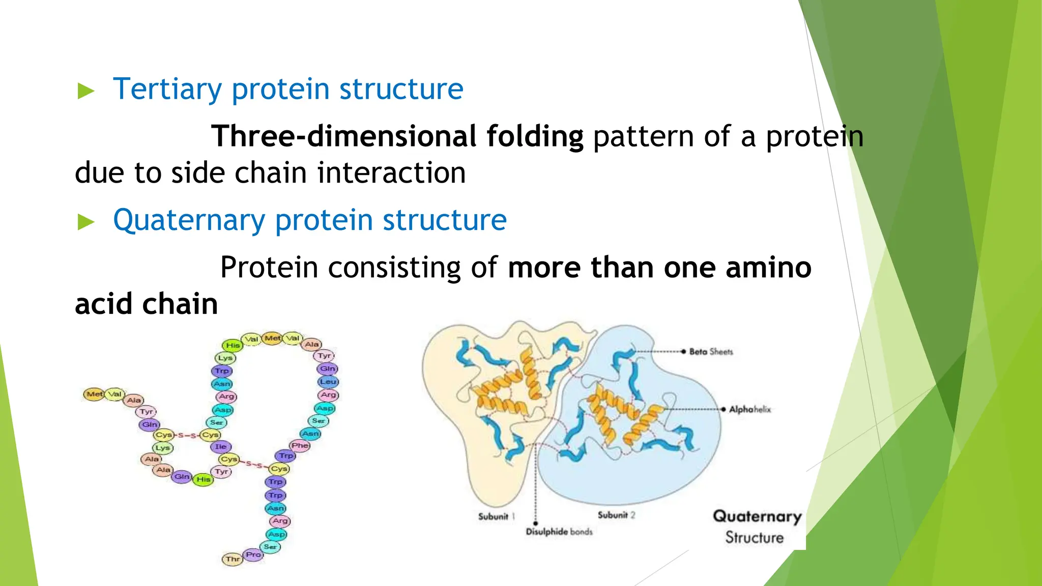
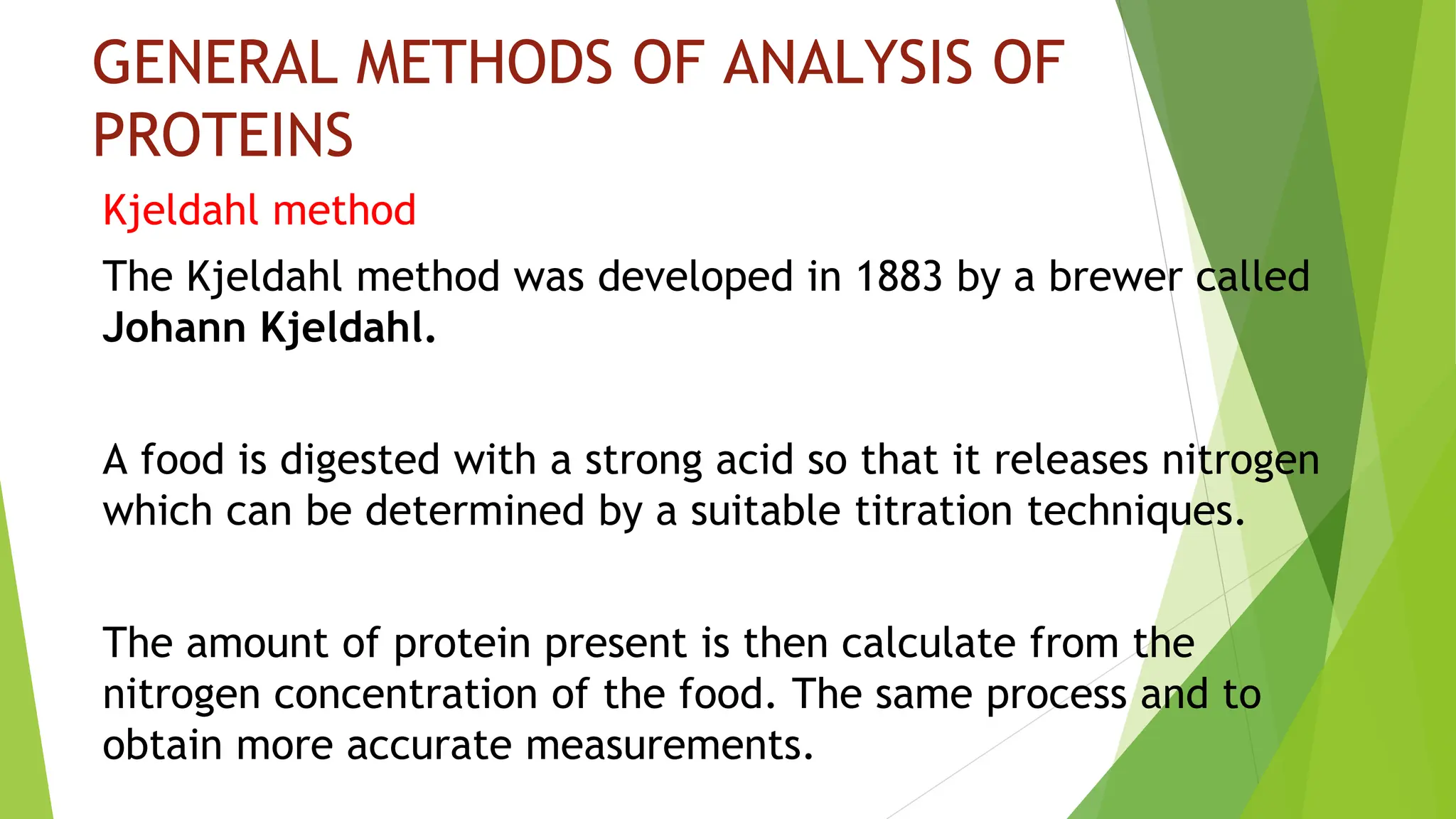
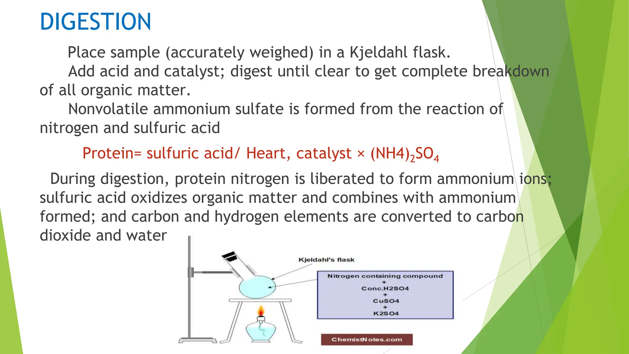
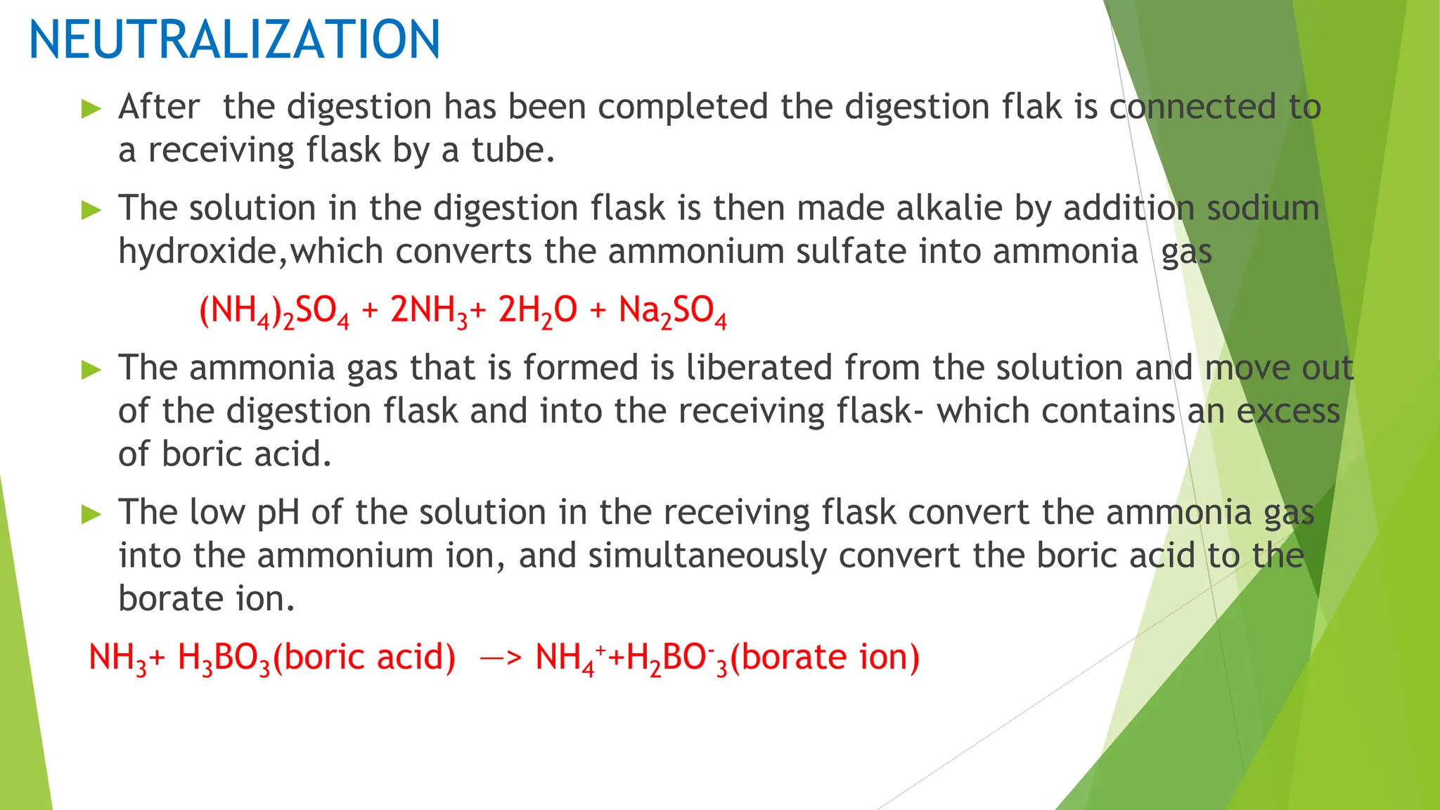
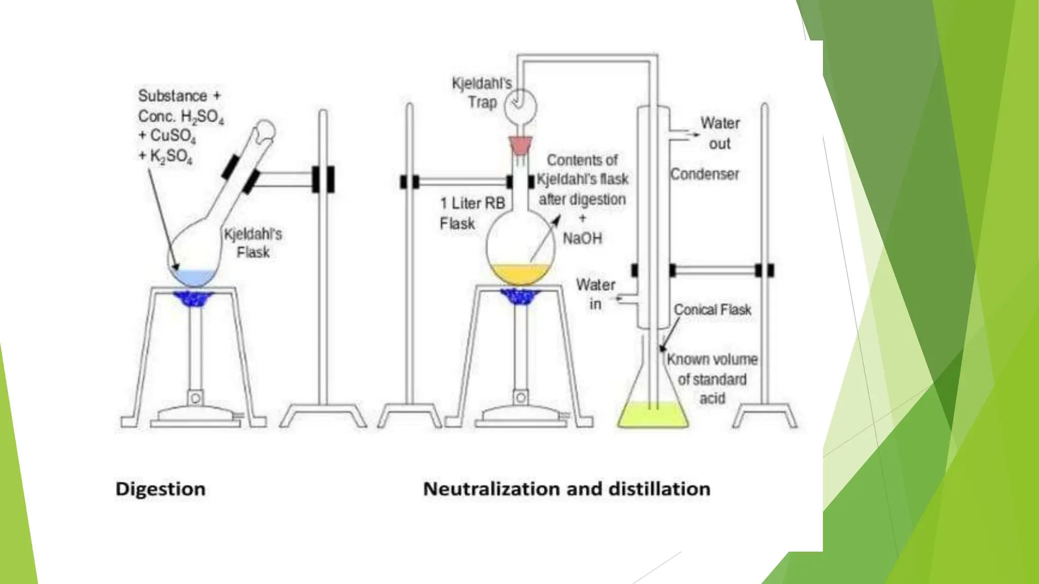
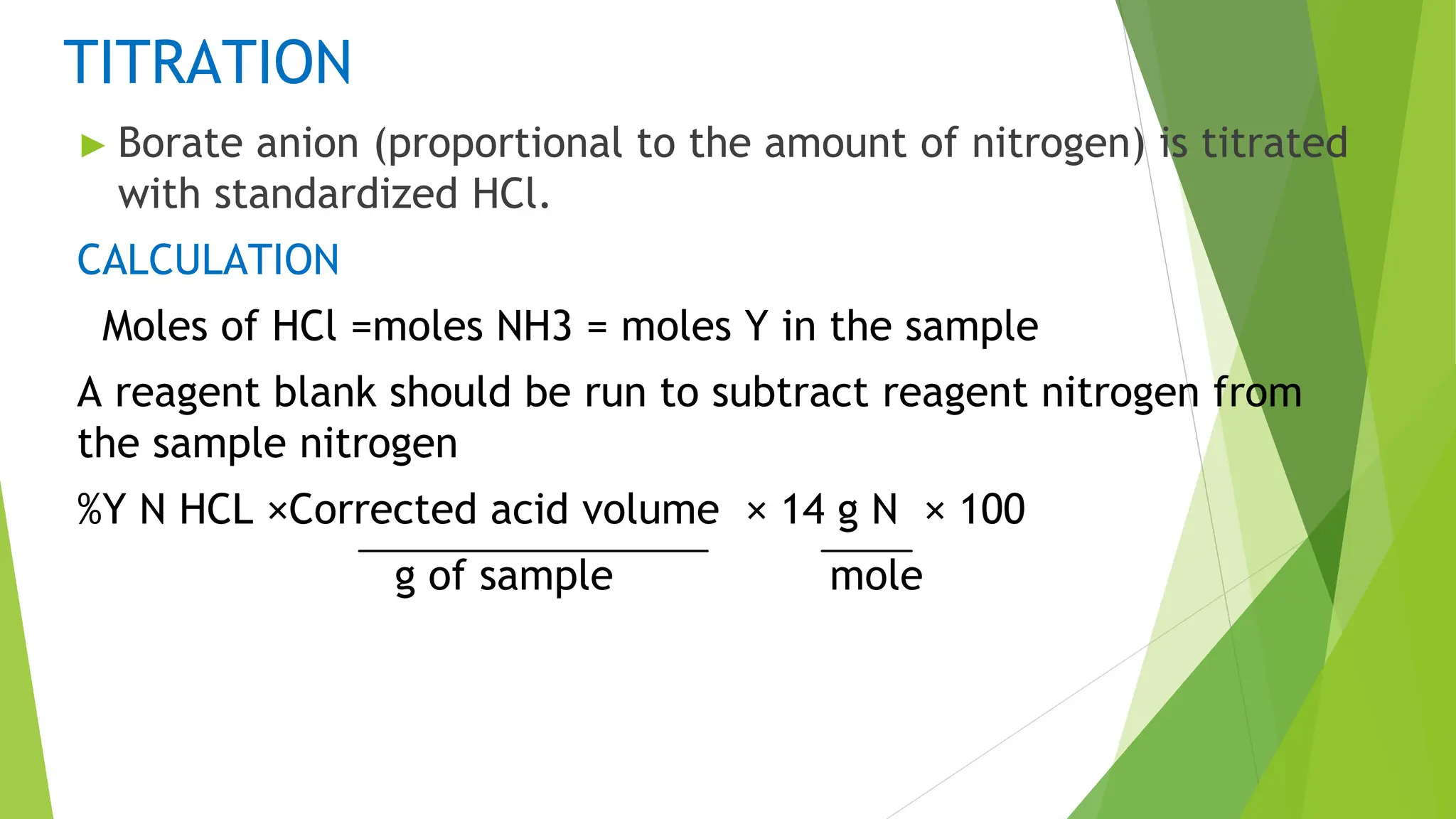
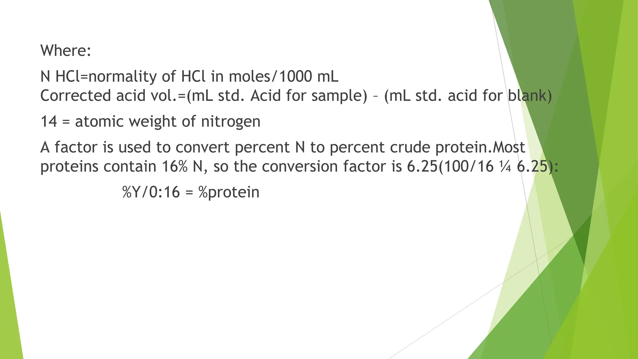
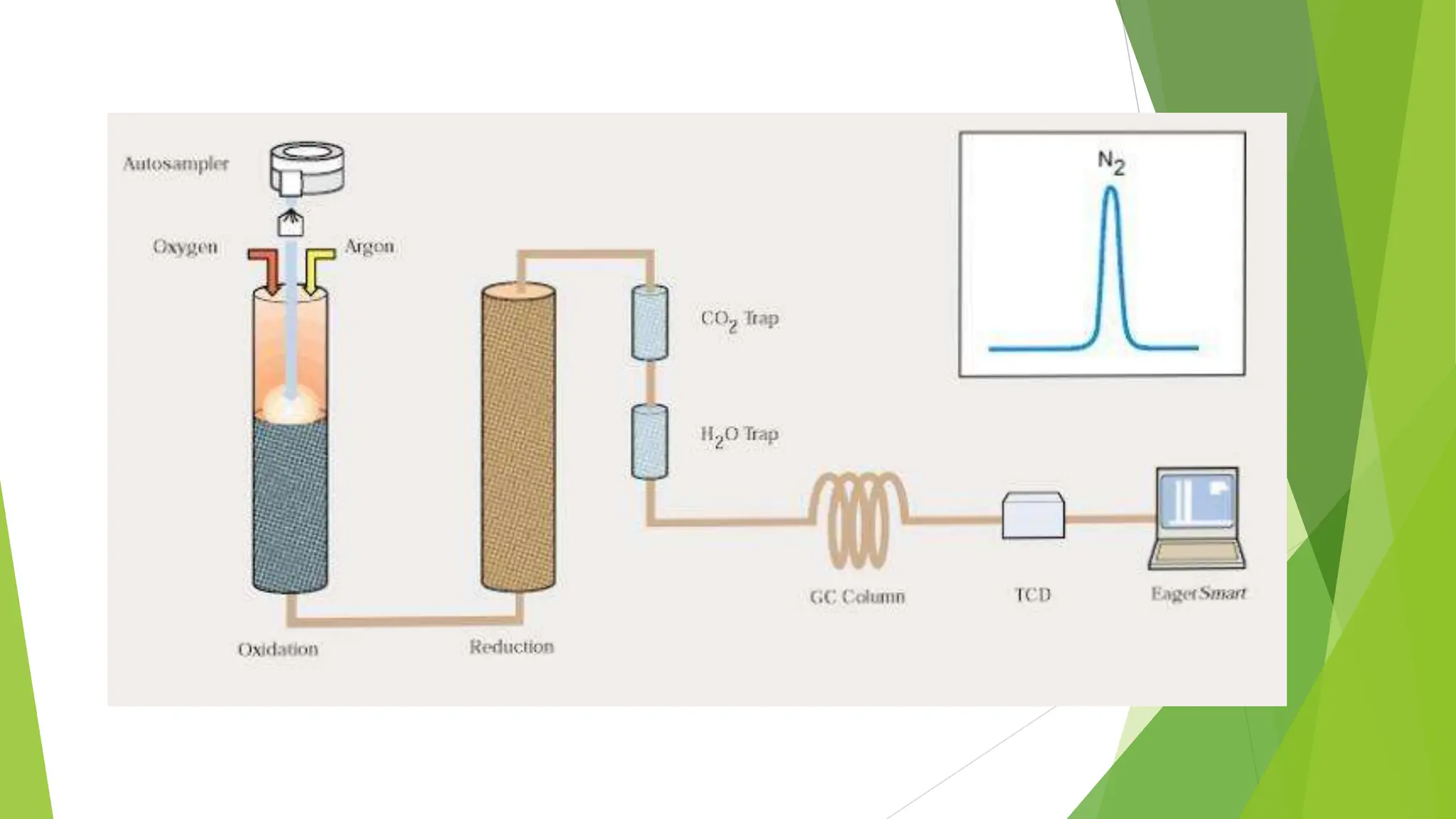
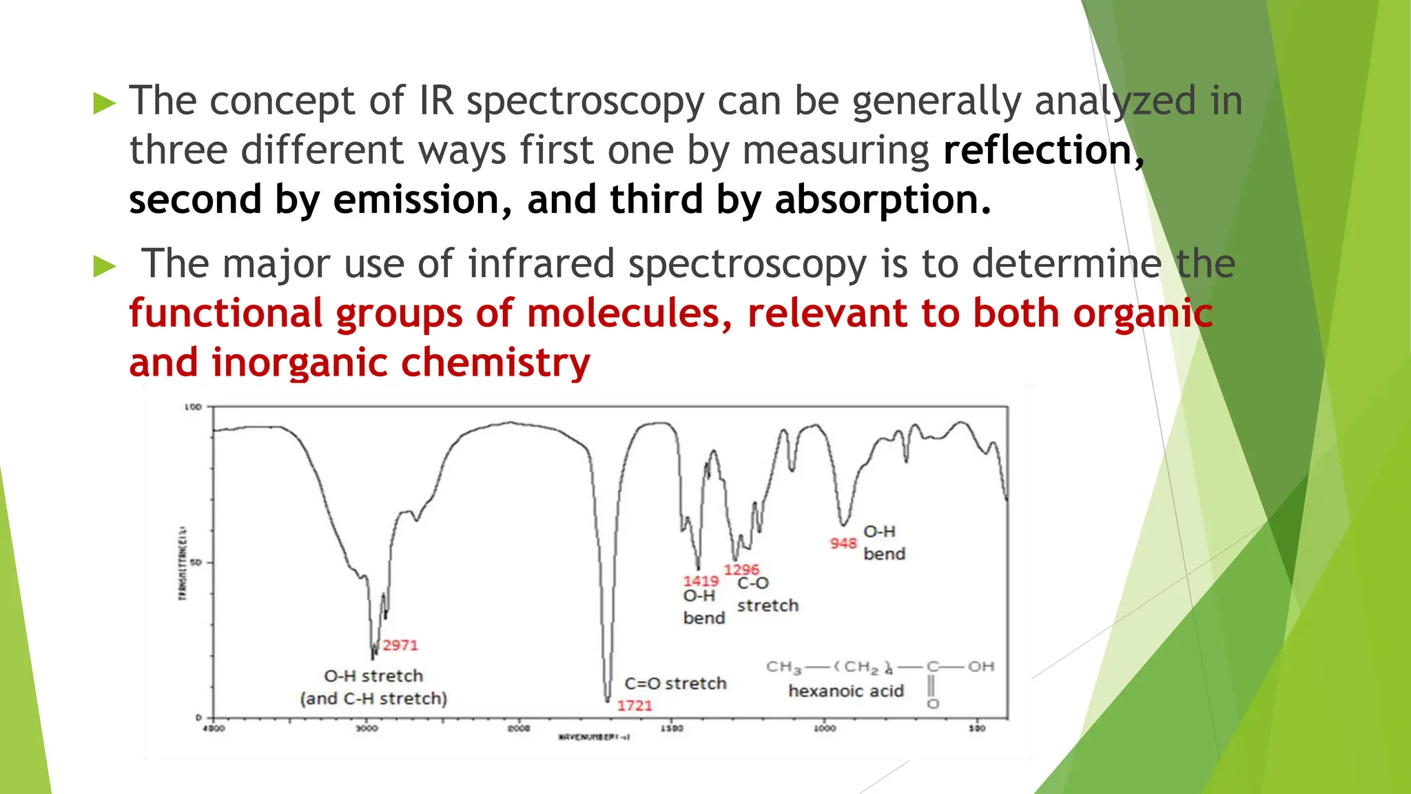
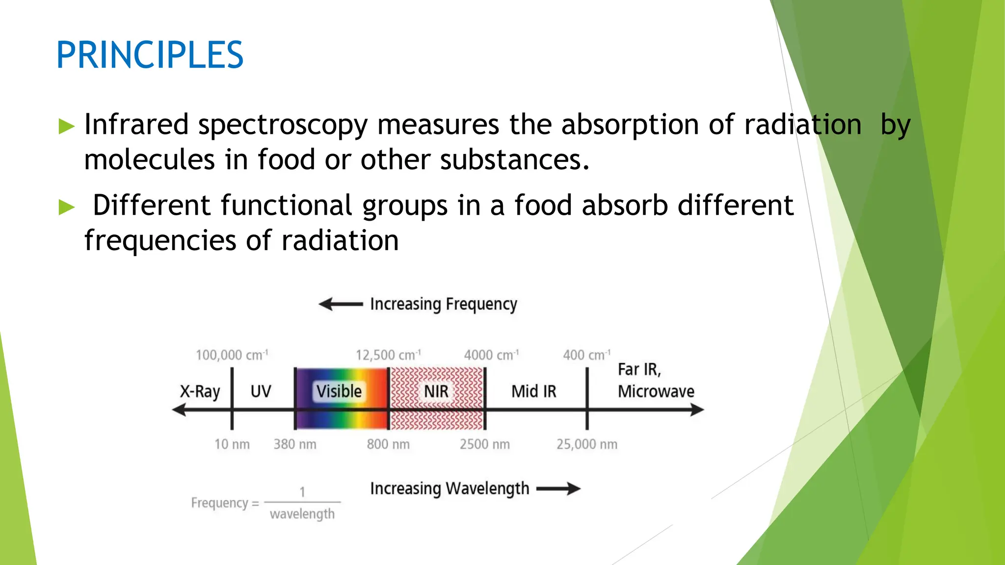
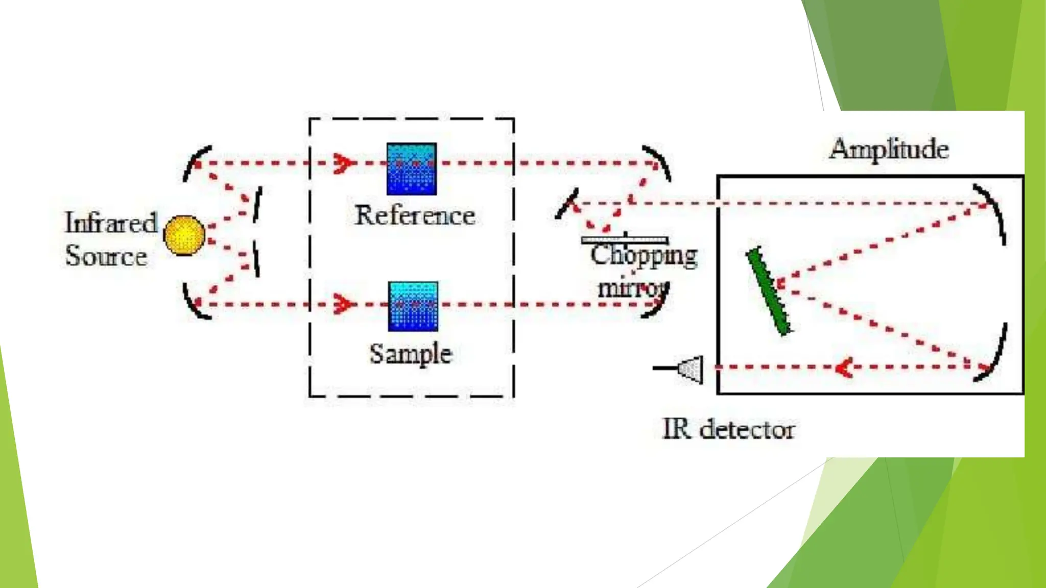
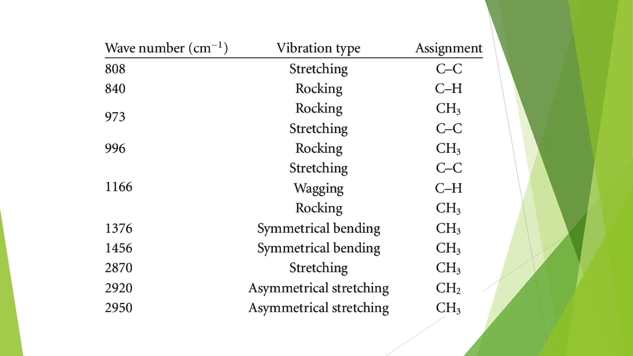
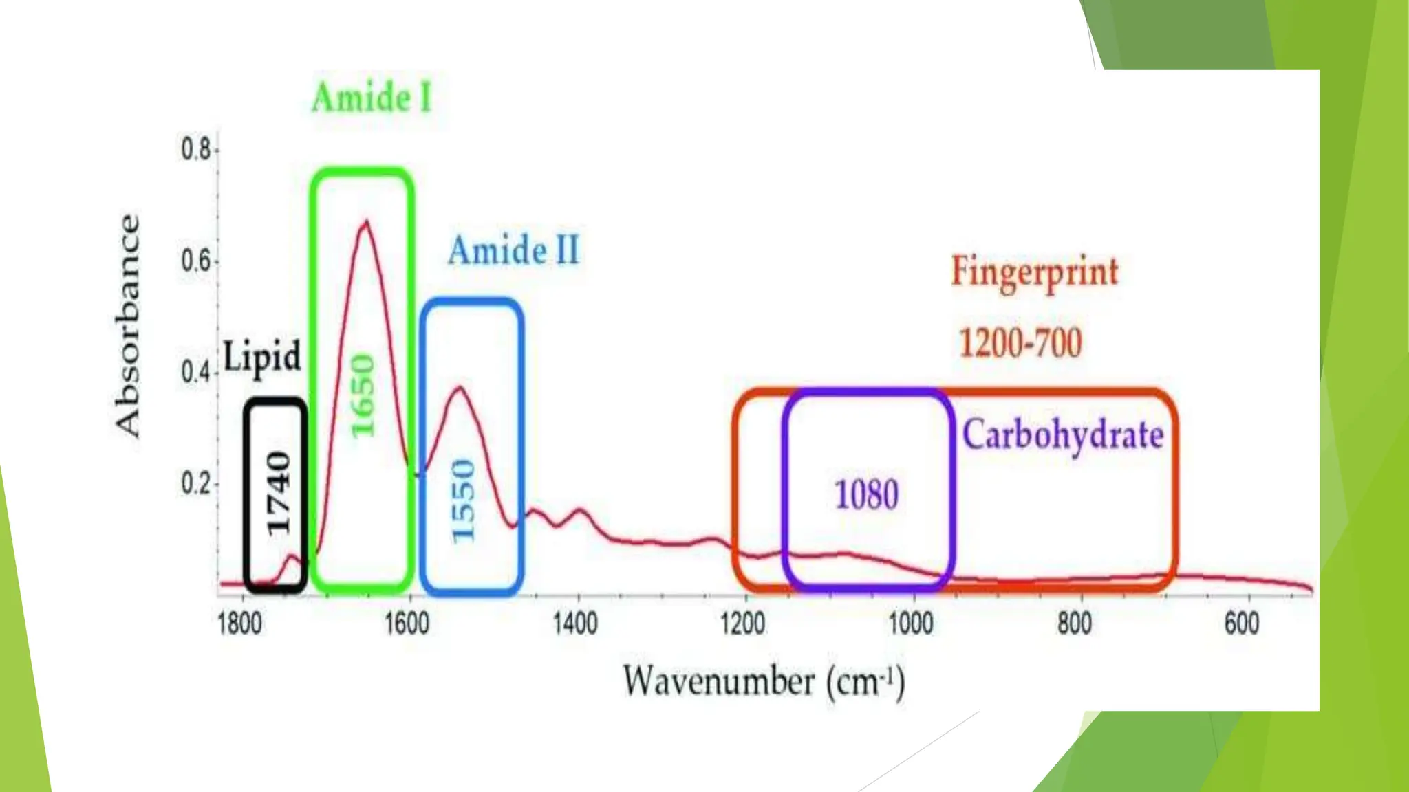
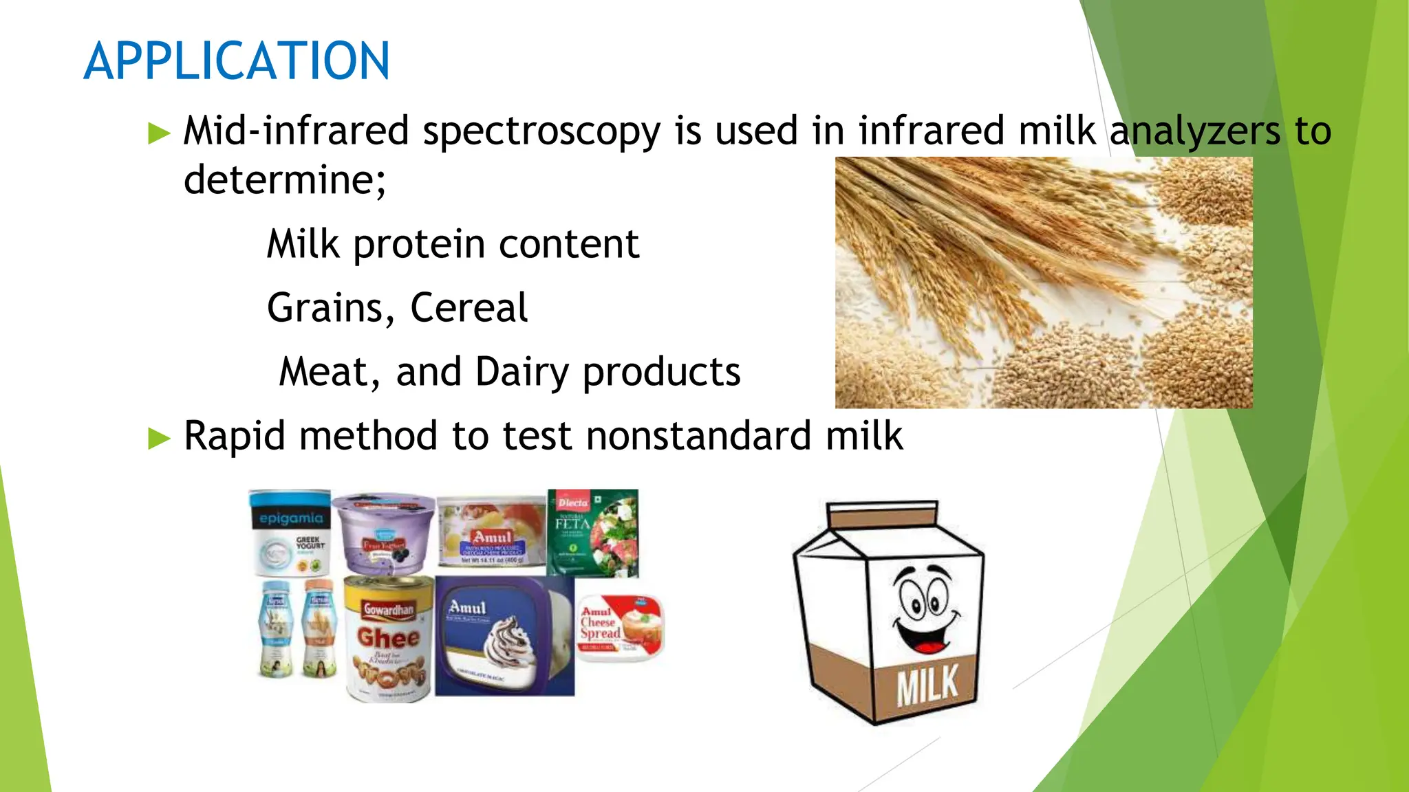
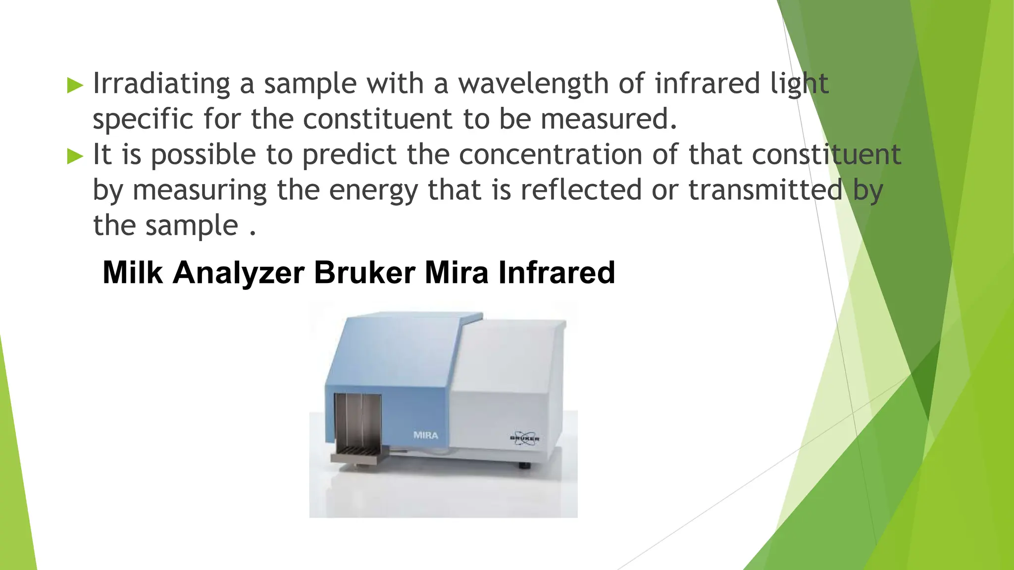
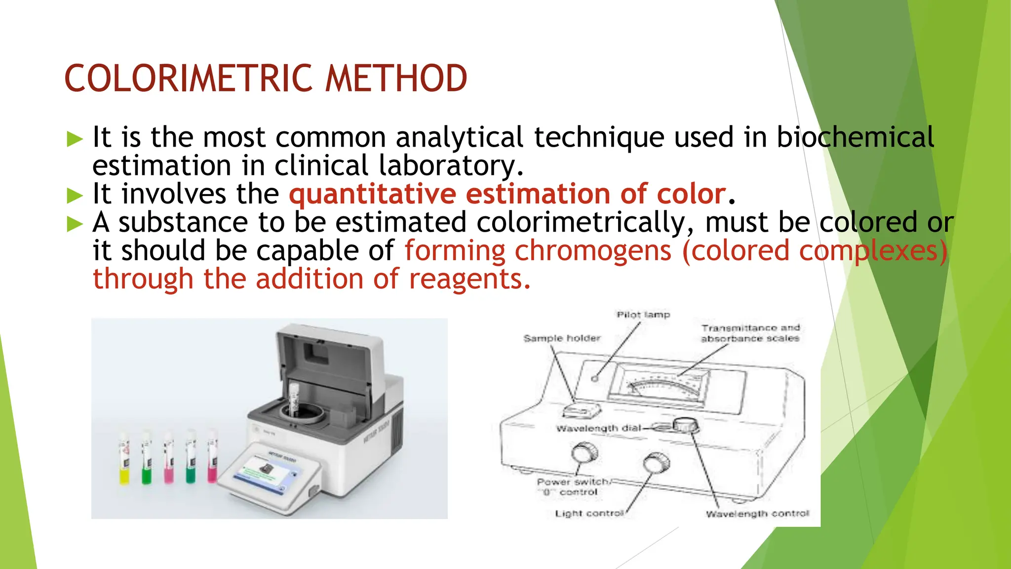
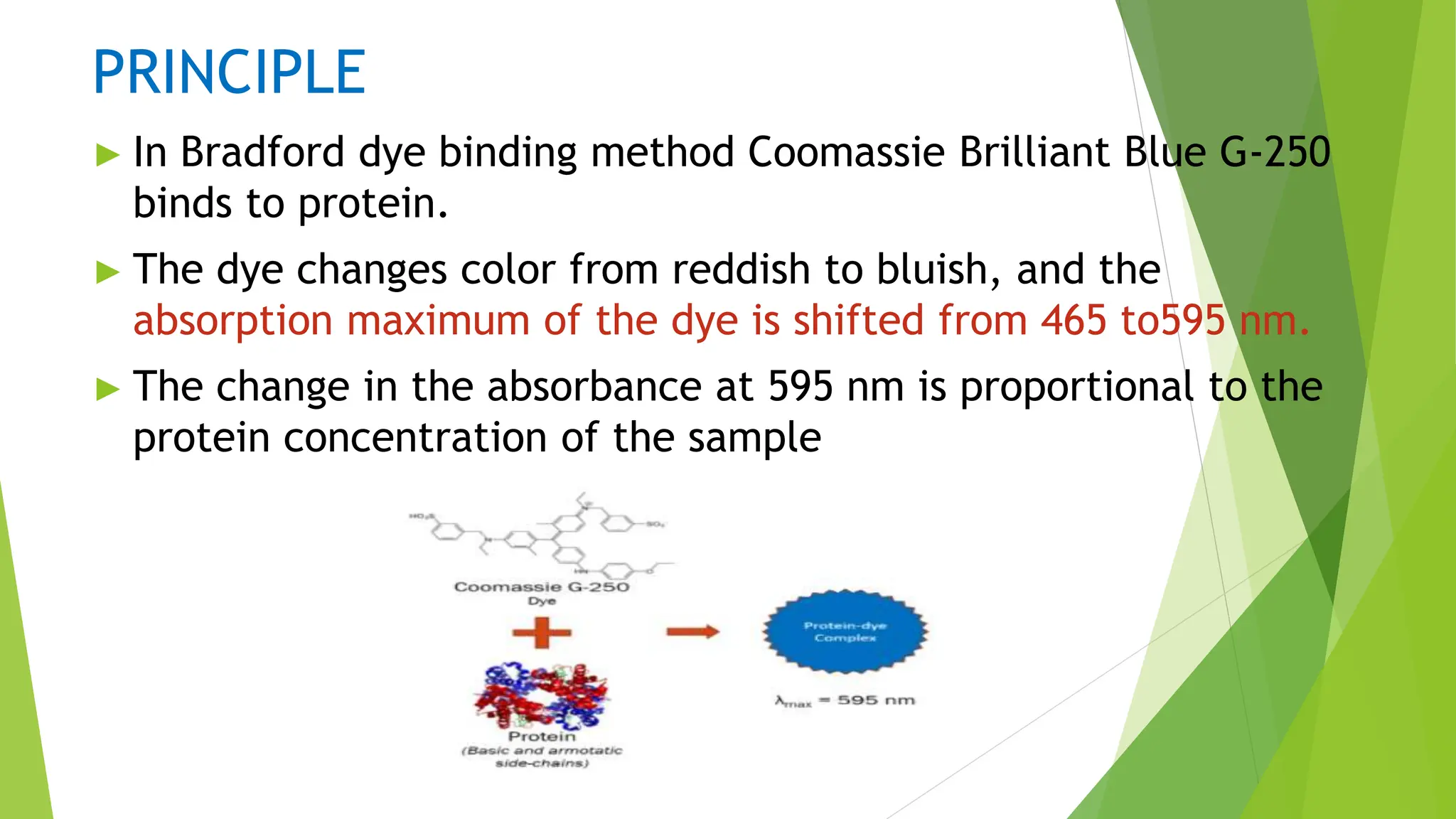
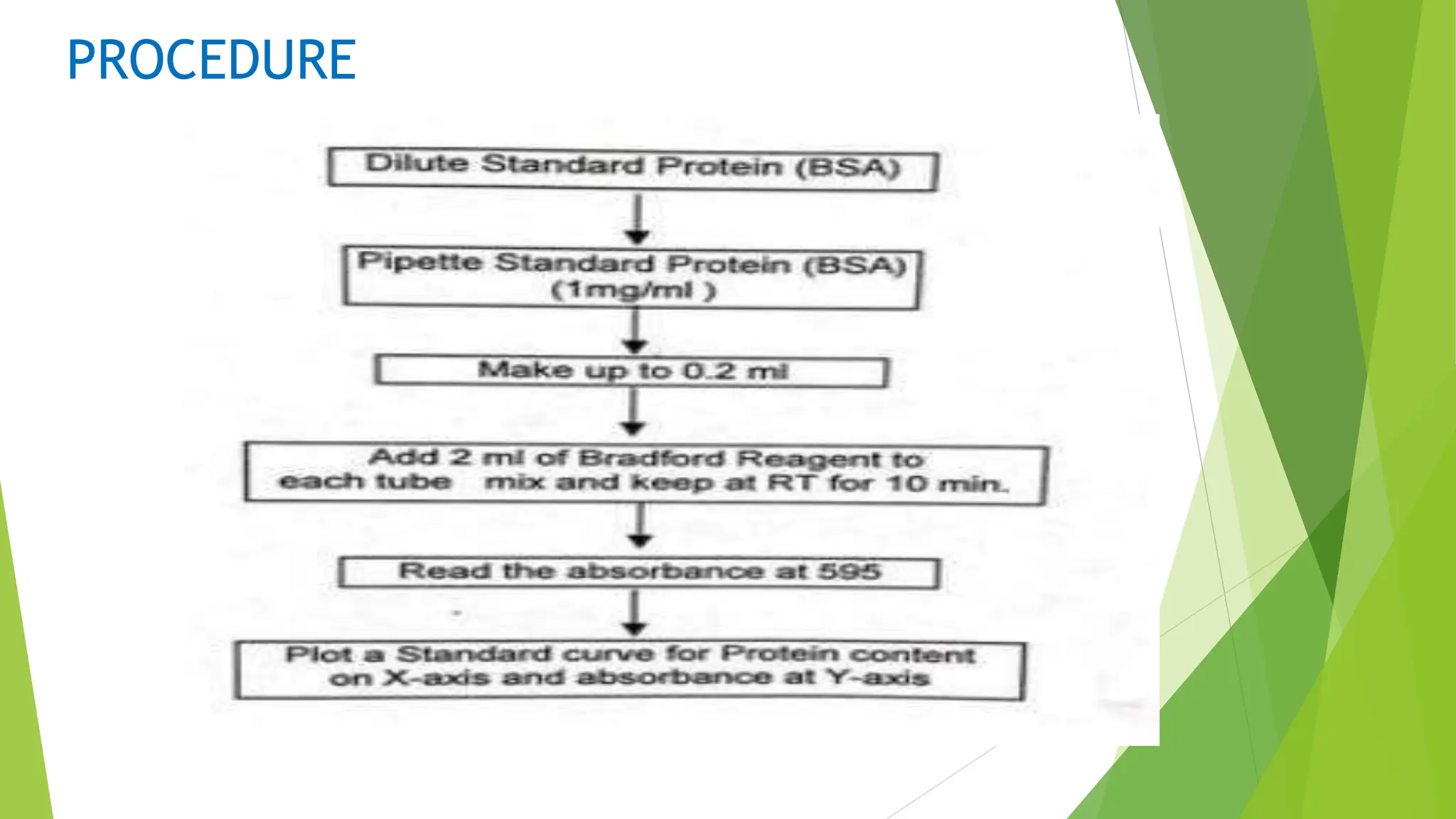
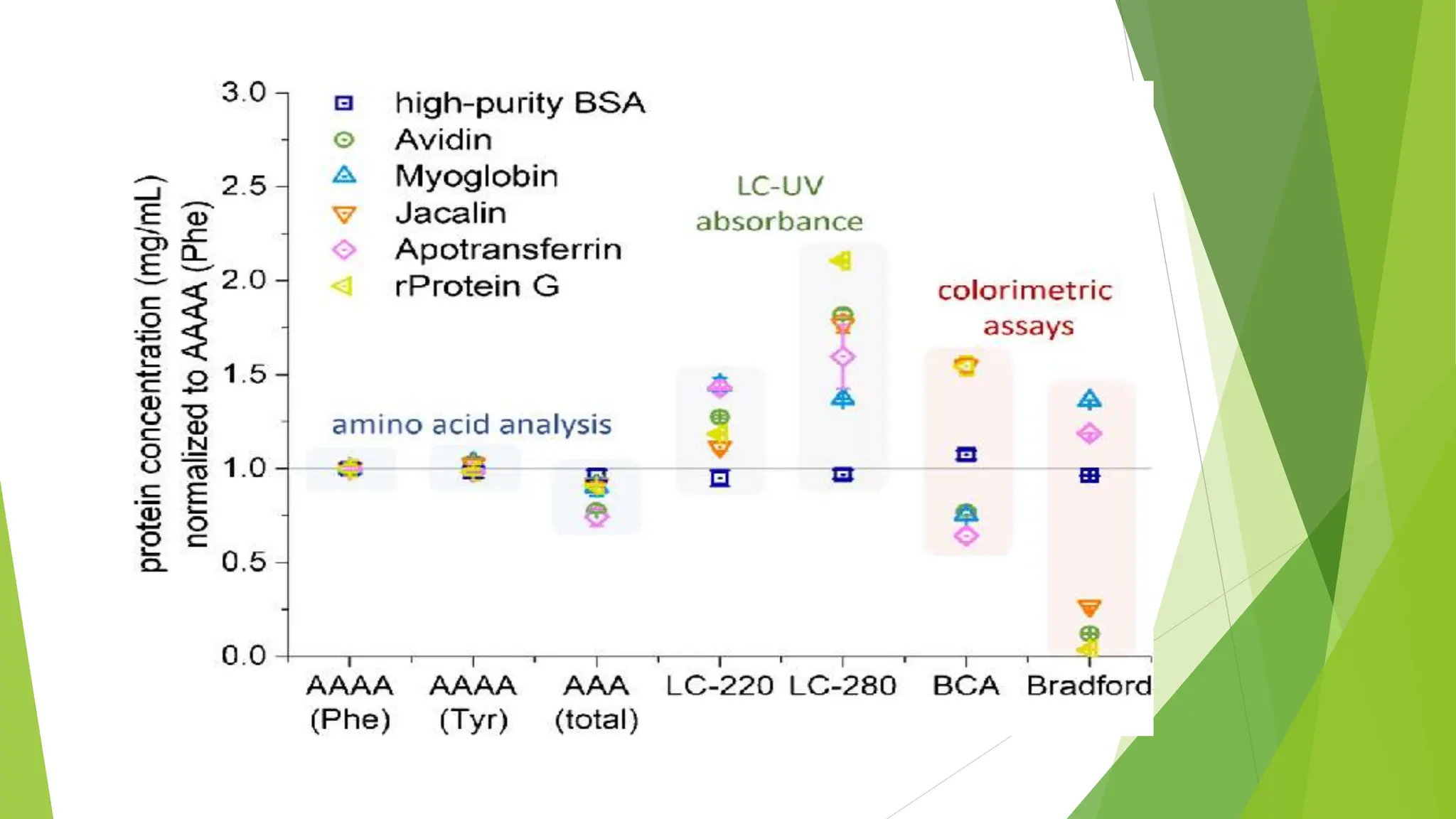
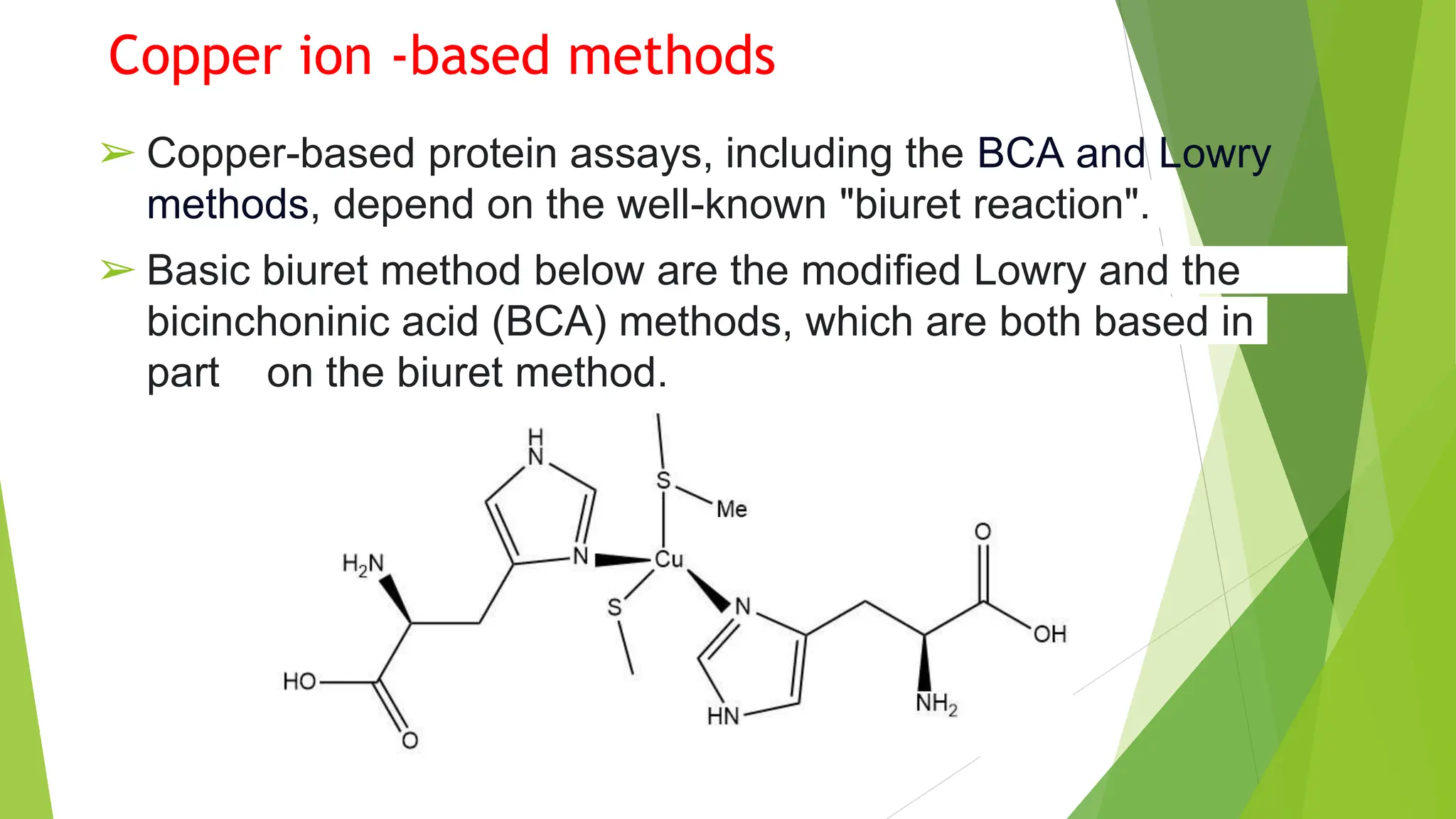
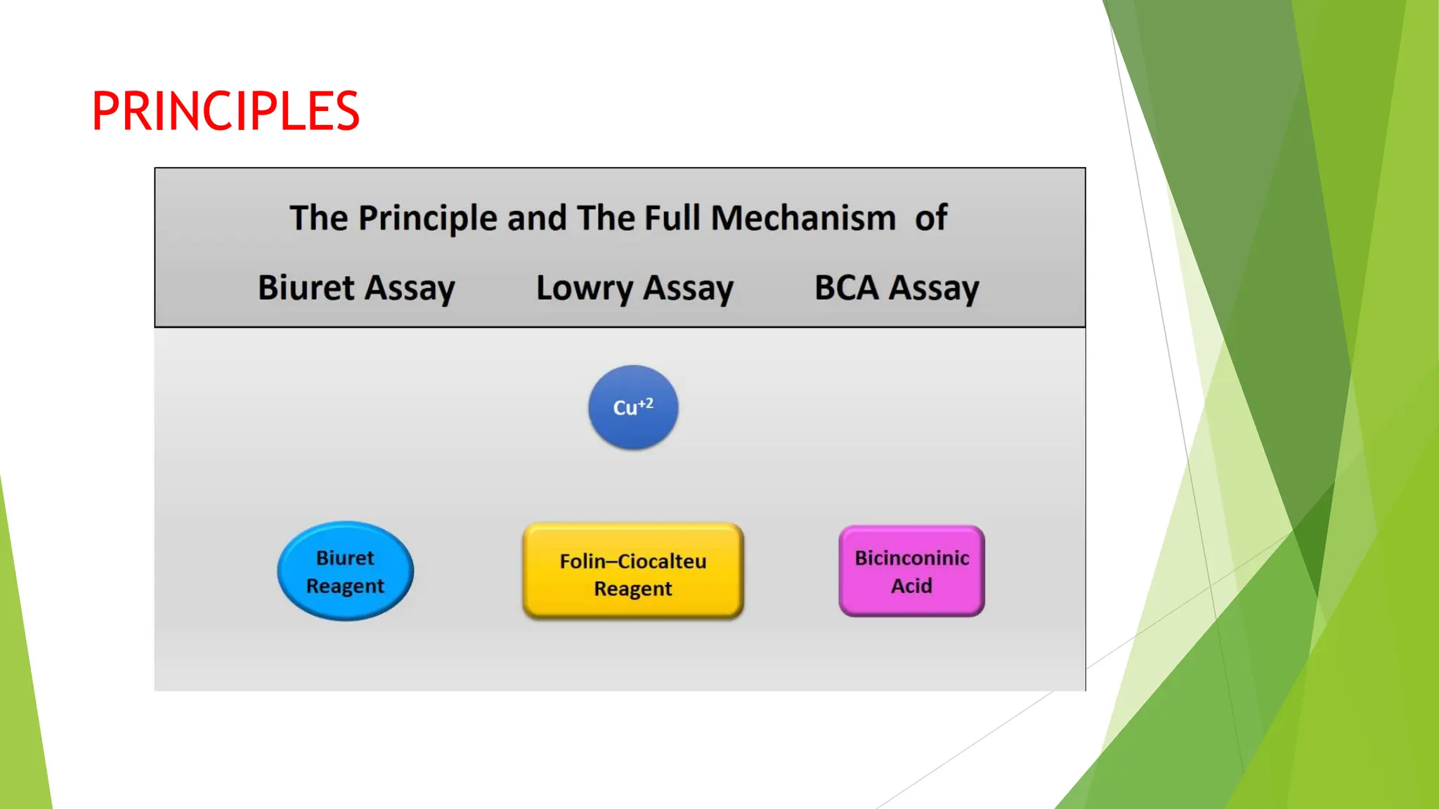
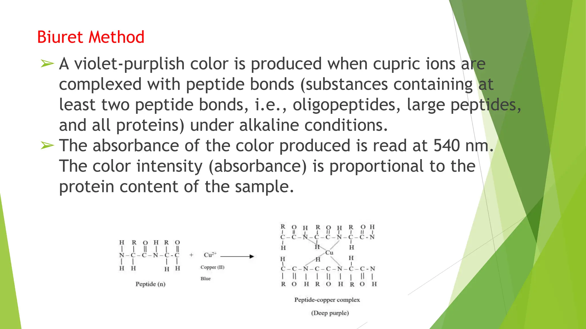
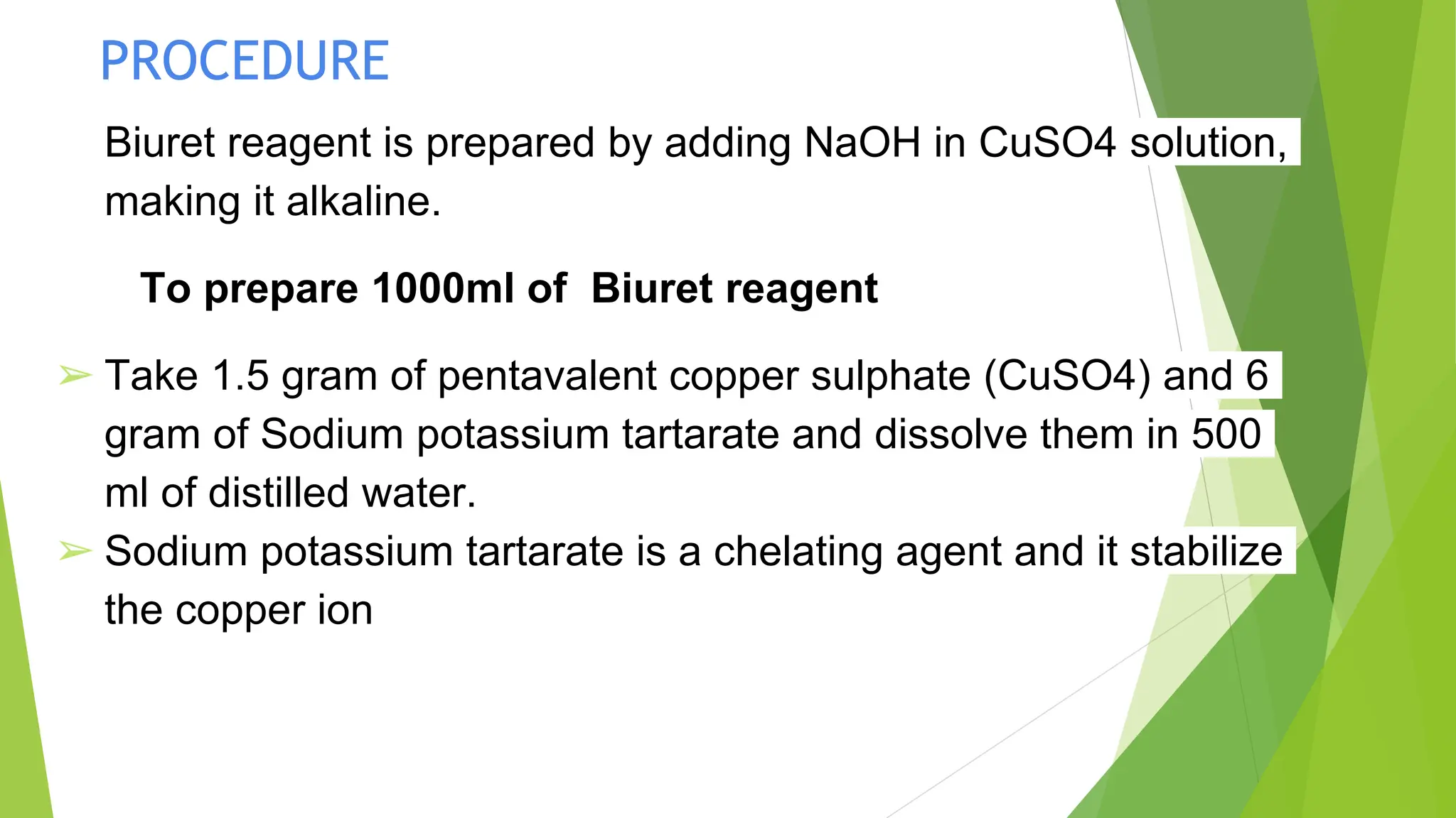
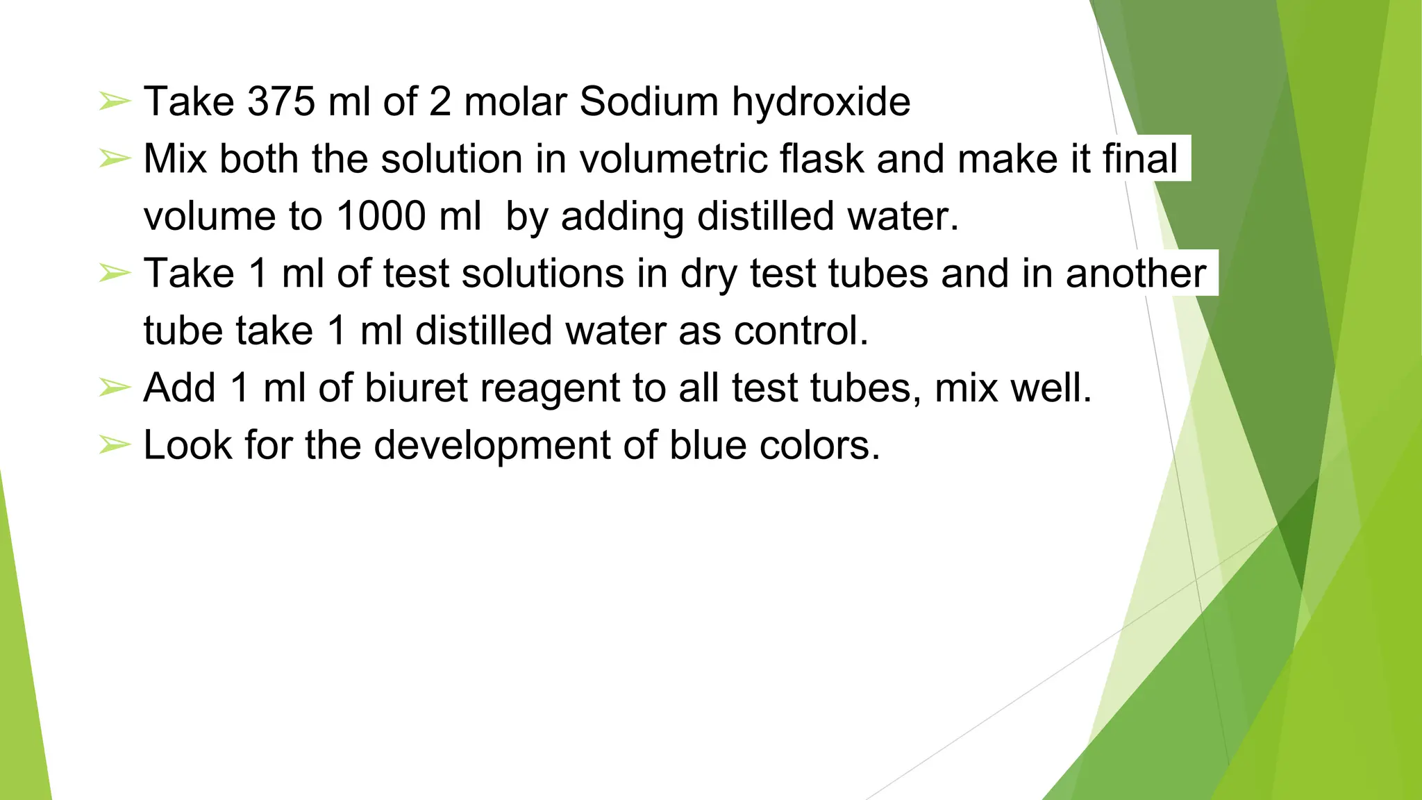
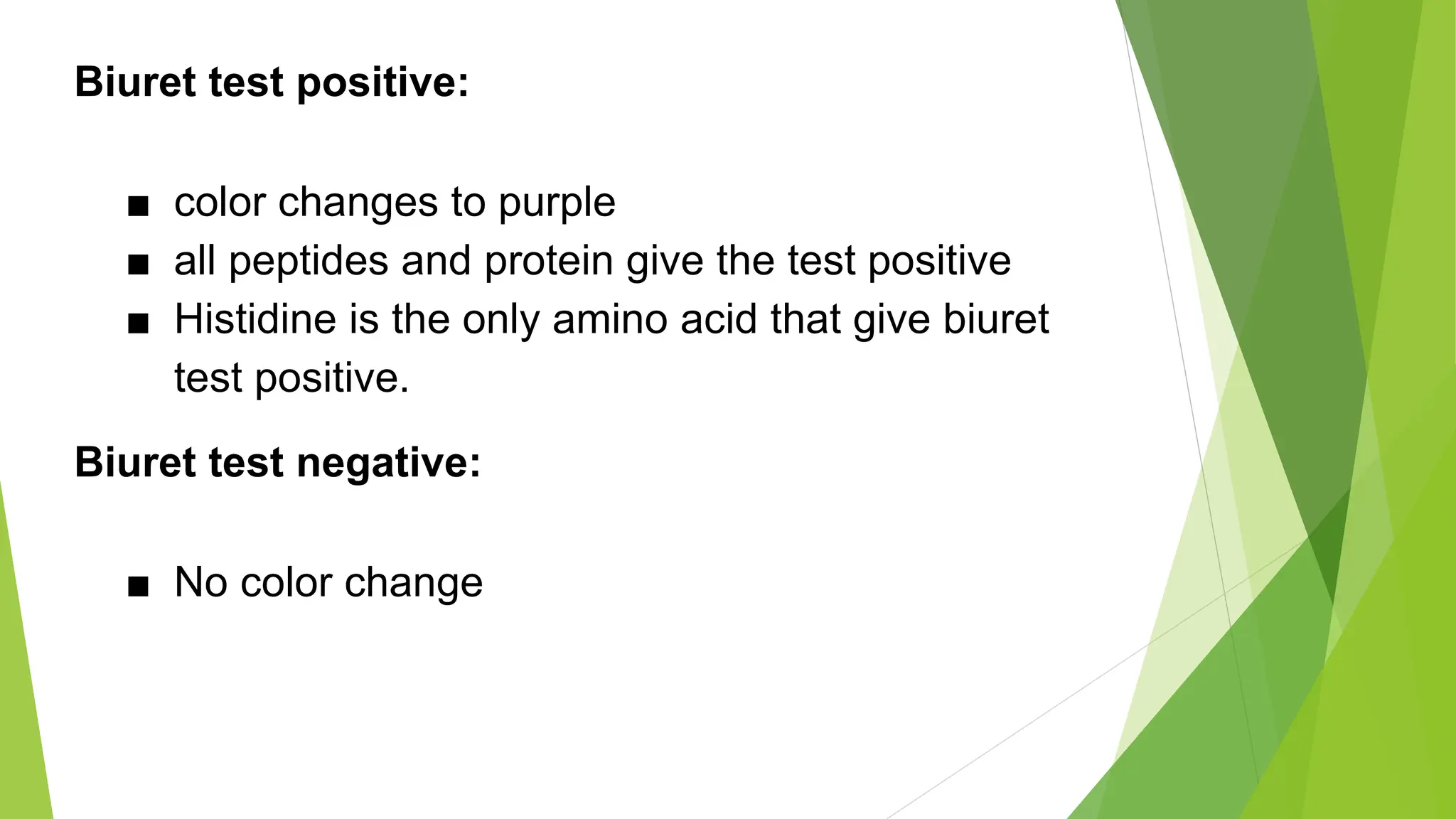
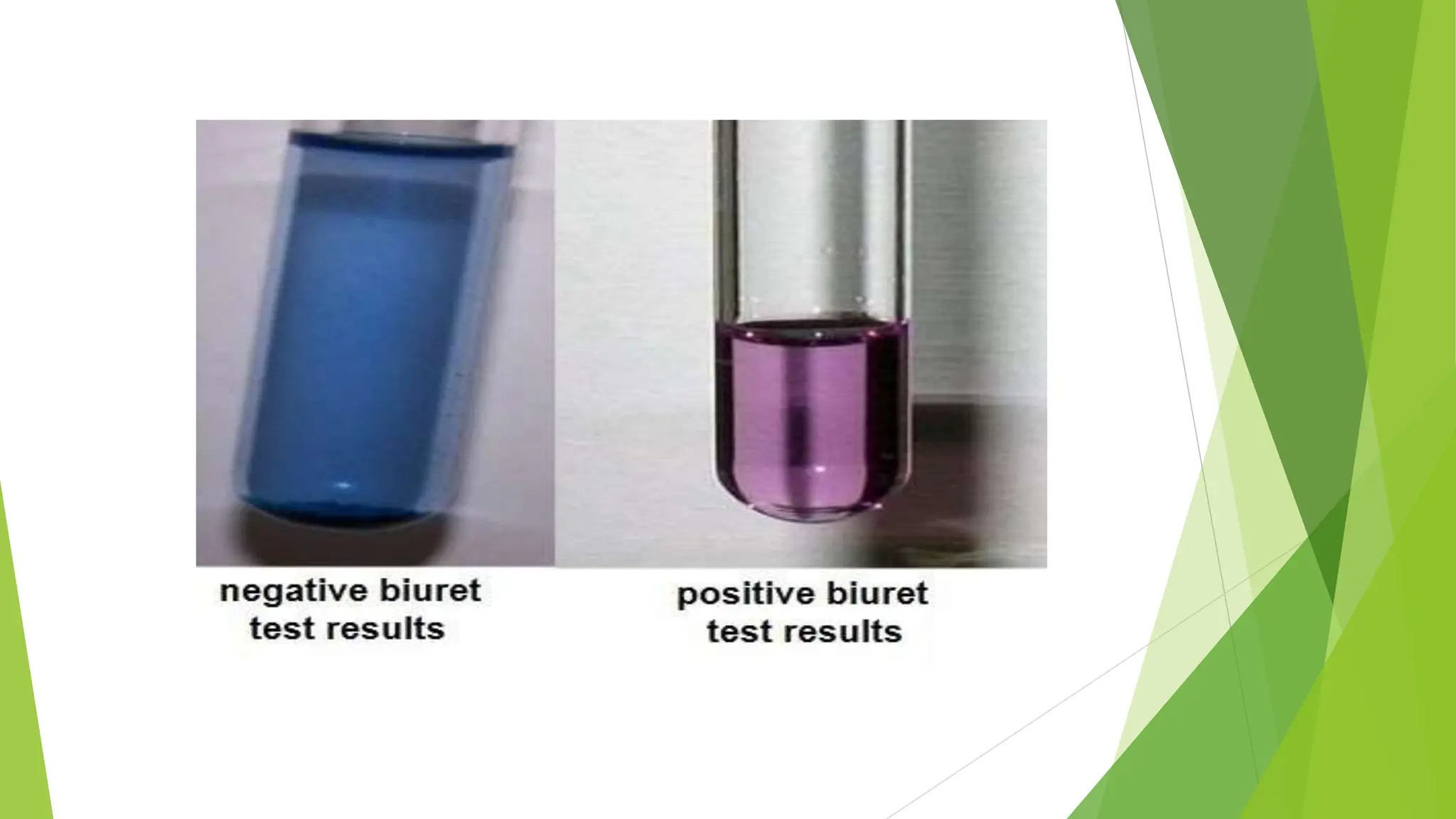
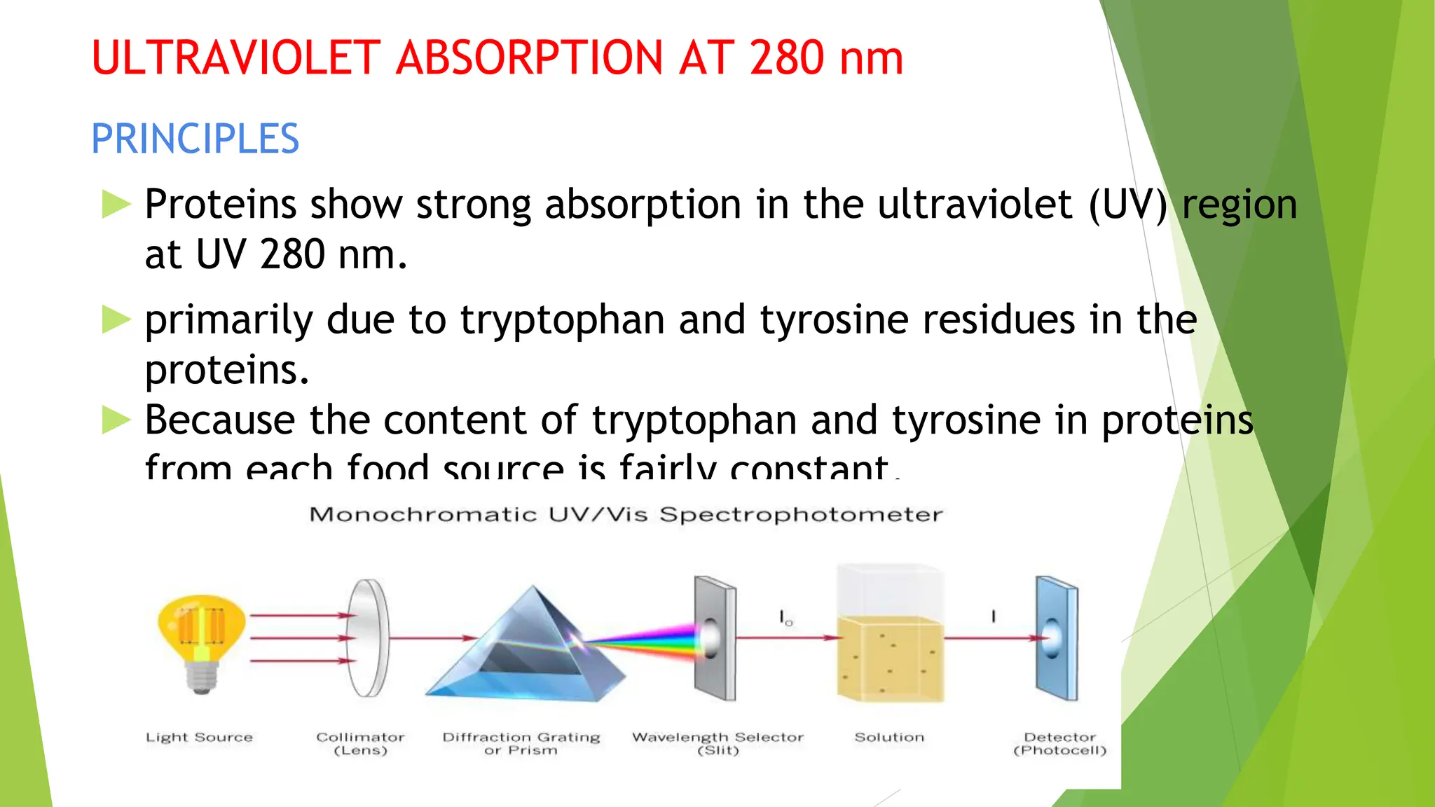
General methods of analysis
of Proteins
Department of Presented by:
Pharmaceutical N.ABINAYA
Analysis K. SUPRIYA
HOD: M.SOWMIYA
Dr.S.S.RAJENDRAN
INTRODUCTION
►Proteins are known as building blocks of life.
►Proteins are the most abundant intracellular macro-
molecules. They provide structure, protection to the
body of multicellular organisms in the form of skin,
hair, callus, cartilage, ligaments, muscles,
tendons.
STRUCTURE
► Primary protein structure
Sequence of a chain of amino acids
► Secondary protein structure
Hydrogen bonding of the peptide backbone cause the
amino acid to fold into a repeating pattern.
► Tertiary protein structure
Three-dimensional folding pattern of a protein
due to side chain interaction
► Quaternary protein structure
Protein consisting of more than one amino
acid chain
GENERAL METHODS OF ANALYSIS OF
PROTEINS
Kjeldahl method
The Kjeldahl method was developed in 1883 by a brewer called
Johann Kjeldahl.
A food is digested with a strong acid so that it releases nitrogen
which can be determined by a suitable titration techniques.
The amount of protein present is then calculate from the
nitrogen concentration of the food. The same process and to
obtain more accurate measurements.
PRINCIPLES
1. DIGESTION
2. NEUTRALIZATION
3. TITRATION
DIGESTION
Place sample (accurately weighed) in a Kjeldahl flask.
Add acid and catalyst; digest until clear to get complete breakdown
of all organic matter.
Nonvolatile ammonium sulfate is formed from the reaction of
nitrogen and sulfuric acid
Protein= sulfuric acid/ Heart, catalyst × (NH4)2SO4
During digestion, protein nitrogen is liberated to form ammonium ions;
sulfuric acid oxidizes organic matter and combines with ammonium
formed; and carbon and hydrogen elements are converted to carbon
dioxide and water.
NEUTRALIZATION
► After the digestion has been completed the digestion flak is connected to
a receiving flask by a tube.
► The solution in the digestion flask is then made alkalie by addition sodium
hydroxide,which converts the ammonium sulfate into ammonia gas
(NH4)2SO4 + 2NH3+ 2H2O + Na2SO4
► The ammonia gas that is formed is liberated from the solution and move out
of the digestion flask and into the receiving flask- which contains an excess
of boric acid.
► The low pH of the solution in the receiving flask convert the ammonia gas
into the ammonium ion, and simultaneously convert the boric acid to the
borate ion.
NH3+ H3BO3(boric acid) —> NH4
++H2BO-
3(borate ion)
TITRATION
► Borate anion (proportional to the amount of nitrogen) is titrated
with standardized HCl.
CALCULATION
Moles of HCl =moles NH3 = moles Y in the sample
A reagent blank should be run to subtract reagent nitrogen from
the sample nitrogen
%Y N HCL ×Corrected acid volume × 14 g N × 100
g of sample mole
Where:
N HCl=normality of HCl in moles/1000 mL
Corrected acid vol.=(mL std. Acid for sample) – (mL std. acid for blank)
14 = atomic weight of nitrogen
A factor is used to convert percent N to percent crude protein.Most
proteins contain 16% N, so the conversion factor is 6.25(100/16 ¼ 6.25):
%Y/0:16 = %protein
APPLICATION
The Kjeldahl method is an AOAC official method for crude
protein content and has been the basis for evaluation of many
other protein methods. The Kjeldahl method is still used for
some applications, but nowadays its use is limited in many
countries due to the availability and advantages of automated
nitrogen combustion (Dumas) systems.
DUMAS (NITROGEN COMBUSTION) METHOD
PRINCIPLES
► The combustion method was introduced in 1831 by Jean-
Baptiste Dumas.
► It has been modified and automated to improve accuracy since
that time.
► Samples are combusted at high temperatures (700–1000 C) with
a flow of pure oxygen.
► All carbon in the sample is converted to carbon dioxide during
the flash combustion.
► Nitrogen-containing components produced include N2 and
nitrogen oxides.
► Nitrogen oxides are reduced to nitrogen in a copper reduction
column at a high temperature (600 C).
► The total nitrogen (including inorganic fraction, i.e., nitrate and
nitrite) released is carried by pure helium and quantitated by gas
chromatography using a thermal conductivity detector (TCD).
► Ultrahigh purity acetanilide and EDTA
(ethylenediaminetetraacetate) may be used as the standards for
the calibration of the nitrogen analyzer.
► The nitrogen determined is converted to protein content in the
sample using a protein conversion factor.
PROCEDURE
► Samples (approximately 100–500 mg) are weighed into a tin
capsule and introduced to a combustion reactor in
automated equipment.
► The nitrogen released is measured by a built-in gas
chromatograph.
APPLICATION
► The combustion method is a faster and safer alternative to the
Kjeldahl method and is suitable for all types of foods.As an
AOAC method, the Dumas method is widely used for official
purposes, but its speed also allows for quality control
applications. The industry uses different units/systems,
depending on sample size and protein content. Freeze-drying
can be used to concentrate diluted liquid samples, e.g., waste
steam samples
INFRARED SPECTROSCOPY
INTRODUCTION
►Infrared spectroscopy is a technique for analysing the
interaction of molecules with infrared light.
►The range of infrared region can be divided into
Near infrared -0.8µ - 2.5µ
Mid infrared -25µ - 2.5µ
Far infrared -25µ- 1000µ
► The concept of IR spectroscopy can be generally analyzed in
three different ways first one by measuring reflection,
second by emission, and third by absorption.
► The major use of infrared spectroscopy is to determine the
functional groups of molecules, relevant to both organic
and inorganic chemistry
PRINCIPLES
► Infrared spectroscopy measures the absorption of radiation by
molecules in food or other substances.
► Different functional groups in a food absorb different
frequencies of radiation
APPLICATION
► Mid-infrared spectroscopy is used in infrared milk analyzers to
determine;
Milk protein content
Grains, Cereal
Meat, and Dairy products
► Rapid method to test nonstandard milk
► Irradiating a sample with a wavelength of infrared light
specific for the constituent to be measured.
► It is possible to predict the concentration of that constituent
by measuring the energy that is reflected or transmitted by
the sample .
Milk Analyzer Bruker Mira Infrared
COLORIMETRIC METHOD
► It is the most common analytical technique used in biochemical
estimation in clinical laboratory.
► It involves the quantitative estimation of color.
► A substance to be estimated colorimetrically, must be colored or
it should be capable of forming chromogens (colored complexes)
through the addition of reagents.
DYE- BINDING METHOD
ANIONIC DYE- BINDING METHOD
► The protein content sample mixed with anionic dye in buffer
solution
► It forms a insoluble complex and it’s removed by centrifugation,
excess insoluble complex separate by using anionic sulfonic acid
dye
► The amount of unbound dye is inversely related to the protein
content of the sample.
PRINCIPLES
► In this method protein-containing sample is mixed with a known
excess amount of anionic dye in a buffered solution.
► Proteins bind the dye to form an insoluble complex.
► Excess insoluble complex dye is separated using anionic sulfonic
acid dye, including acid orange 12, orange G, and Amido Black
10B
► Binds cationic groups of the basic amino acid residues,and the free
amino-terminal group of the protein
PROCEDURE
The sample is finely ground and added to an excess dye solution
with known concentration
Then it is vigorously Shaken and centrifuged to remove insoluble
substance
Absorbance of the unbound dye solution in the filtrate or
supernatant is measured and dye concentration estimated
from a dye standard curve
A straight calibration curve can be obtained by plotting the unbound
dye concentration vs total nitrogen(as determined by Kjeldahl
method) of a given food covering a wide range of protein content
Protein content of the unknown sample of the same food type can be
estimated from the calibration curve
APPLICATION
► The anionic dye-binding method may be used to estimate the
changes in available lysine content of cereal products during
processing
►Used to estimate protein contentmilk,wheatflour,soyproduct,and
meats.
BRADFORD’S DYE- BINDING METHOD
► It is one of the method to determine the protein concentration
in food.
► Bradford method relies on the amphoteric nature of proteins
► In the case of the Bradford method, the dye bound to protein
has a change in absorbance spectrum relative to the unbound
dye
PRINCIPLE
► In Bradford dye binding method Coomassie Brilliant Blue G-250
binds to protein.
► The dye changes color from reddish to bluish, and the
absorption maximum of the dye is shifted from 465 to595 nm.
► The change in the absorbance at 595 nm is proportional to the
protein concentration of the sample
PROCEDURE
APPLICATION
► This procedure has been used to measure microgram quantities of
protein.
► The Bradford method has been used successfully to determine
protein content in worts and beer products and in potato tubers.
Copper ion -based methods
➢ Copper-based protein assays, including the BCA and Lowry
methods, depend on the well-known "biuret reaction".
➢ Basic biuret method below are the modified Lowry and the
bicinchoninic acid (BCA) methods, which are both based in
part on the biuret method.
PRINCIPLES
Biuret Method
➢ A violet-purplish color is produced when cupric ions are
complexed with peptide bonds (substances containing at
least two peptide bonds, i.e., oligopeptides, large peptides,
and all proteins) under alkaline conditions.
➢ The absorbance of the color produced is read at 540 nm.
The color intensity (absorbance) is proportional to the
protein content of the sample.
PROCEDURE
Biuret reagent is prepared by adding NaOH in CuSO4 solution,
making it alkaline.
To prepare 1000ml of Biuret reagent
➢ Take 1.5 gram of pentavalent copper sulphate (CuSO4) and 6
gram of Sodium potassium tartarate and dissolve them in 500
ml of distilled water.
➢ Sodium potassium tartarate is a chelating agent and it stabilize
the copper ion
➢ Take 375 ml of 2 molar Sodium hydroxide
➢ Mix both the solution in volumetric flask and make it final
volume to 1000 ml by adding distilled water.
➢ Take 1 ml of test solutions in dry test tubes and in another
tube take 1 ml distilled water as control.
➢ Add 1 ml of biuret reagent to all test tubes, mix well.
➢ Look for the development of blue colors.
Biuret test positive:
■ color changes to purple
■ all peptides and protein give the test positive
■ Histidine is the only amino acid that give biuret
test positive.
Biuret test negative:
■ No color change
APPLICATION
► The biuret method has been used to determine proteins
in cereal, meat, and soybean proteins and as a
qualitative test for animal feed.
► The biuret method also can be used to measure the
protein content of isolated proteins.
LOWRY METHOD
► The Lowry method combines the biuret reaction with
the reduction of the Folin-Ciocalteu phenol reagent
(phosphomolybdic-phosphotungstic acid) by tyrosine
and tryptophan residues in the proteins.
► The bluish color developed is read at 750 nm(high
sensitivity for low protein concentration) or 500 nm
(low sensitivity for high protein concentration).
PROCEDURE
► Proteins to be analyzed are diluted to an appropriate range
(20–100 μg).
► K Na tartrate-Na2CO3 solution is added after cooling and then
incubated at room temperature for 10 min.
► CuSO4-K Na tartrate-NaOH solution is added after cooling and
then incubated at room temperature for 10 min.
► Freshly prepared Folin’s reagent is added, and then the
reaction mixture is mixed and incubated at 50 C for 10 min.
► Absorbance is read at 650 nm.
► A standard curve of BSA is carefully constructed for estimating
protein concentration of the unknown
APPLICATION
► Determination of microgram amounts of protein in
influenza vaccine.
► Because of its simplicity and sensitivity.
► Most widely used to determine proteins in food systems
without first extracting the proteins from the food mixture.
BICINCHONINIC ACID METHOD
► Proteins and peptides reduce cupric ions to cuprous ions
under alkaline conditions.
► The cuprous ion then reacts with the apple-greenish
bicinchoninic acid reagent (BCA) to form a purplish complex
► The color measured at 562 nm .
► Peptide bonds and four amino acids (cysteine, cystine,
tryptophan, and tyrosine) contribute to the color formation
with BCA
PROCEDURE
► Mix (one step) the protein solution with the BCA
reagent, BCA sodium salt,sodium carbonate, NaOH,
and copper sulfate.
► Incubate at 37 C for 30 min or room temperature for
2 h or 60 C for 30 min. A higher temperature gives a
greater color response.
► Read the solution at 562 nm .
APPLICATION
► The BCA method is widely used in protein isolation and
purification due to its advantage of being compatible
with samples containing up to 5% detergents.
► While most dye-binding methods are faster, the BCA
method is less affected by protein compositional
differences
► so there is better protein-to protein uniformity
ULTRAVIOLET ABSORPTION AT 280 nm
PRINCIPLES
► Proteins show strong absorption in the ultraviolet (UV) region
at UV 280 nm.
► primarily due to tryptophan and tyrosine residues in the
proteins.
► Because the content of tryptophan and tyrosine in proteins
from each food source is fairly constant.
PROCEDURE
➢ Proteins are solubilized in buffer or alkali.
➢ Protein concentration is calculated according to
the following equation the absorbance at 280 nm
could be used to estimate the concentration of
proteins, using Beer’s law.
A = abc
where A = absorbance;
a =absorptivity;
b = cell or cuvette path length;
c =concentration.
APPLICATION
➢ The UV 280 nm method has been used to
determine the protein contents of milk and meat
products.
➢ It has not been used widely in food systems.
➢ Although peptide bonds in proteins absorb more
strongly at 190–220 nm than at 280 nm, the low
UV region is more difficult to measure.
PEPTIDE MEASUREMENT AT 190-220 nm
➢ Peptides with or without low level of tyrosine or
tryptophan residues can be quantified at 190–220
nm at which peptide bonds have maximum
absorption.
➢ Protein can also be measured in this UV range.
REFERENCE:
Aakanchha Jain et al. Basic Techniques in Biochemistry, Microbiology and
Molecular Biology: Principles and Techniques, Springer Protocols Handbooks,
https://doi.org/10.1007/978-1-4939-9861-6, © Springer Science+Business Media,
LLC, part of Springer Nature 2020
Rutherfurd SM accurate determination of amino acid content of selected
feedstuff .int.J.foodsci.Nutr.2009,60;53
Introduction about protein and General method of analysis of protein

Introduction about protein and General method of analysis of protein

  • 1.
    General methods ofanalysis of Proteins Department of Presented by: Pharmaceutical N.ABINAYA Analysis K. SUPRIYA HOD: M.SOWMIYA Dr.S.S.RAJENDRAN
  • 2.
    INTRODUCTION ►Proteins are knownas building blocks of life. ►Proteins are the most abundant intracellular macro- molecules. They provide structure, protection to the body of multicellular organisms in the form of skin, hair, callus, cartilage, ligaments, muscles, tendons.
  • 3.
  • 4.
    ► Primary proteinstructure Sequence of a chain of amino acids ► Secondary protein structure Hydrogen bonding of the peptide backbone cause the amino acid to fold into a repeating pattern.
  • 5.
    ► Tertiary proteinstructure Three-dimensional folding pattern of a protein due to side chain interaction ► Quaternary protein structure Protein consisting of more than one amino acid chain
  • 6.
    GENERAL METHODS OFANALYSIS OF PROTEINS Kjeldahl method The Kjeldahl method was developed in 1883 by a brewer called Johann Kjeldahl. A food is digested with a strong acid so that it releases nitrogen which can be determined by a suitable titration techniques. The amount of protein present is then calculate from the nitrogen concentration of the food. The same process and to obtain more accurate measurements.
  • 7.
  • 8.
    DIGESTION Place sample (accuratelyweighed) in a Kjeldahl flask. Add acid and catalyst; digest until clear to get complete breakdown of all organic matter. Nonvolatile ammonium sulfate is formed from the reaction of nitrogen and sulfuric acid Protein= sulfuric acid/ Heart, catalyst × (NH4)2SO4 During digestion, protein nitrogen is liberated to form ammonium ions; sulfuric acid oxidizes organic matter and combines with ammonium formed; and carbon and hydrogen elements are converted to carbon dioxide and water.
  • 9.
    NEUTRALIZATION ► After thedigestion has been completed the digestion flak is connected to a receiving flask by a tube. ► The solution in the digestion flask is then made alkalie by addition sodium hydroxide,which converts the ammonium sulfate into ammonia gas (NH4)2SO4 + 2NH3+ 2H2O + Na2SO4 ► The ammonia gas that is formed is liberated from the solution and move out of the digestion flask and into the receiving flask- which contains an excess of boric acid. ► The low pH of the solution in the receiving flask convert the ammonia gas into the ammonium ion, and simultaneously convert the boric acid to the borate ion. NH3+ H3BO3(boric acid) —> NH4 ++H2BO- 3(borate ion)
  • 11.
    TITRATION ► Borate anion(proportional to the amount of nitrogen) is titrated with standardized HCl. CALCULATION Moles of HCl =moles NH3 = moles Y in the sample A reagent blank should be run to subtract reagent nitrogen from the sample nitrogen %Y N HCL ×Corrected acid volume × 14 g N × 100 g of sample mole
  • 12.
    Where: N HCl=normality ofHCl in moles/1000 mL Corrected acid vol.=(mL std. Acid for sample) – (mL std. acid for blank) 14 = atomic weight of nitrogen A factor is used to convert percent N to percent crude protein.Most proteins contain 16% N, so the conversion factor is 6.25(100/16 ¼ 6.25): %Y/0:16 = %protein
  • 13.
    APPLICATION The Kjeldahl methodis an AOAC official method for crude protein content and has been the basis for evaluation of many other protein methods. The Kjeldahl method is still used for some applications, but nowadays its use is limited in many countries due to the availability and advantages of automated nitrogen combustion (Dumas) systems.
  • 14.
    DUMAS (NITROGEN COMBUSTION)METHOD PRINCIPLES ► The combustion method was introduced in 1831 by Jean- Baptiste Dumas. ► It has been modified and automated to improve accuracy since that time. ► Samples are combusted at high temperatures (700–1000 C) with a flow of pure oxygen. ► All carbon in the sample is converted to carbon dioxide during the flash combustion. ► Nitrogen-containing components produced include N2 and nitrogen oxides. ► Nitrogen oxides are reduced to nitrogen in a copper reduction column at a high temperature (600 C).
  • 15.
    ► The totalnitrogen (including inorganic fraction, i.e., nitrate and nitrite) released is carried by pure helium and quantitated by gas chromatography using a thermal conductivity detector (TCD). ► Ultrahigh purity acetanilide and EDTA (ethylenediaminetetraacetate) may be used as the standards for the calibration of the nitrogen analyzer. ► The nitrogen determined is converted to protein content in the sample using a protein conversion factor.
  • 16.
    PROCEDURE ► Samples (approximately100–500 mg) are weighed into a tin capsule and introduced to a combustion reactor in automated equipment. ► The nitrogen released is measured by a built-in gas chromatograph.
  • 18.
    APPLICATION ► The combustionmethod is a faster and safer alternative to the Kjeldahl method and is suitable for all types of foods.As an AOAC method, the Dumas method is widely used for official purposes, but its speed also allows for quality control applications. The industry uses different units/systems, depending on sample size and protein content. Freeze-drying can be used to concentrate diluted liquid samples, e.g., waste steam samples
  • 19.
    INFRARED SPECTROSCOPY INTRODUCTION ►Infrared spectroscopyis a technique for analysing the interaction of molecules with infrared light. ►The range of infrared region can be divided into Near infrared -0.8µ - 2.5µ Mid infrared -25µ - 2.5µ Far infrared -25µ- 1000µ
  • 20.
    ► The conceptof IR spectroscopy can be generally analyzed in three different ways first one by measuring reflection, second by emission, and third by absorption. ► The major use of infrared spectroscopy is to determine the functional groups of molecules, relevant to both organic and inorganic chemistry
  • 22.
    PRINCIPLES ► Infrared spectroscopymeasures the absorption of radiation by molecules in food or other substances. ► Different functional groups in a food absorb different frequencies of radiation
  • 26.
    APPLICATION ► Mid-infrared spectroscopyis used in infrared milk analyzers to determine; Milk protein content Grains, Cereal Meat, and Dairy products ► Rapid method to test nonstandard milk
  • 27.
    ► Irradiating asample with a wavelength of infrared light specific for the constituent to be measured. ► It is possible to predict the concentration of that constituent by measuring the energy that is reflected or transmitted by the sample . Milk Analyzer Bruker Mira Infrared
  • 28.
    COLORIMETRIC METHOD ► Itis the most common analytical technique used in biochemical estimation in clinical laboratory. ► It involves the quantitative estimation of color. ► A substance to be estimated colorimetrically, must be colored or it should be capable of forming chromogens (colored complexes) through the addition of reagents.
  • 29.
    DYE- BINDING METHOD ANIONICDYE- BINDING METHOD ► The protein content sample mixed with anionic dye in buffer solution ► It forms a insoluble complex and it’s removed by centrifugation, excess insoluble complex separate by using anionic sulfonic acid dye ► The amount of unbound dye is inversely related to the protein content of the sample.
  • 30.
    PRINCIPLES ► In thismethod protein-containing sample is mixed with a known excess amount of anionic dye in a buffered solution. ► Proteins bind the dye to form an insoluble complex. ► Excess insoluble complex dye is separated using anionic sulfonic acid dye, including acid orange 12, orange G, and Amido Black 10B ► Binds cationic groups of the basic amino acid residues,and the free amino-terminal group of the protein
  • 31.
    PROCEDURE The sample isfinely ground and added to an excess dye solution with known concentration Then it is vigorously Shaken and centrifuged to remove insoluble substance Absorbance of the unbound dye solution in the filtrate or supernatant is measured and dye concentration estimated from a dye standard curve
  • 32.
    A straight calibrationcurve can be obtained by plotting the unbound dye concentration vs total nitrogen(as determined by Kjeldahl method) of a given food covering a wide range of protein content Protein content of the unknown sample of the same food type can be estimated from the calibration curve
  • 33.
    APPLICATION ► The anionicdye-binding method may be used to estimate the changes in available lysine content of cereal products during processing ►Used to estimate protein contentmilk,wheatflour,soyproduct,and meats.
  • 34.
    BRADFORD’S DYE- BINDINGMETHOD ► It is one of the method to determine the protein concentration in food. ► Bradford method relies on the amphoteric nature of proteins ► In the case of the Bradford method, the dye bound to protein has a change in absorbance spectrum relative to the unbound dye
  • 35.
    PRINCIPLE ► In Bradforddye binding method Coomassie Brilliant Blue G-250 binds to protein. ► The dye changes color from reddish to bluish, and the absorption maximum of the dye is shifted from 465 to595 nm. ► The change in the absorbance at 595 nm is proportional to the protein concentration of the sample
  • 36.
  • 38.
    APPLICATION ► This procedurehas been used to measure microgram quantities of protein. ► The Bradford method has been used successfully to determine protein content in worts and beer products and in potato tubers.
  • 39.
    Copper ion -basedmethods ➢ Copper-based protein assays, including the BCA and Lowry methods, depend on the well-known "biuret reaction". ➢ Basic biuret method below are the modified Lowry and the bicinchoninic acid (BCA) methods, which are both based in part on the biuret method.
  • 40.
  • 41.
    Biuret Method ➢ Aviolet-purplish color is produced when cupric ions are complexed with peptide bonds (substances containing at least two peptide bonds, i.e., oligopeptides, large peptides, and all proteins) under alkaline conditions. ➢ The absorbance of the color produced is read at 540 nm. The color intensity (absorbance) is proportional to the protein content of the sample.
  • 42.
    PROCEDURE Biuret reagent isprepared by adding NaOH in CuSO4 solution, making it alkaline. To prepare 1000ml of Biuret reagent ➢ Take 1.5 gram of pentavalent copper sulphate (CuSO4) and 6 gram of Sodium potassium tartarate and dissolve them in 500 ml of distilled water. ➢ Sodium potassium tartarate is a chelating agent and it stabilize the copper ion
  • 43.
    ➢ Take 375ml of 2 molar Sodium hydroxide ➢ Mix both the solution in volumetric flask and make it final volume to 1000 ml by adding distilled water. ➢ Take 1 ml of test solutions in dry test tubes and in another tube take 1 ml distilled water as control. ➢ Add 1 ml of biuret reagent to all test tubes, mix well. ➢ Look for the development of blue colors.
  • 44.
    Biuret test positive: ■color changes to purple ■ all peptides and protein give the test positive ■ Histidine is the only amino acid that give biuret test positive. Biuret test negative: ■ No color change
  • 46.
    APPLICATION ► The biuretmethod has been used to determine proteins in cereal, meat, and soybean proteins and as a qualitative test for animal feed. ► The biuret method also can be used to measure the protein content of isolated proteins.
  • 47.
    LOWRY METHOD ► TheLowry method combines the biuret reaction with the reduction of the Folin-Ciocalteu phenol reagent (phosphomolybdic-phosphotungstic acid) by tyrosine and tryptophan residues in the proteins. ► The bluish color developed is read at 750 nm(high sensitivity for low protein concentration) or 500 nm (low sensitivity for high protein concentration).
  • 48.
    PROCEDURE ► Proteins tobe analyzed are diluted to an appropriate range (20–100 μg). ► K Na tartrate-Na2CO3 solution is added after cooling and then incubated at room temperature for 10 min. ► CuSO4-K Na tartrate-NaOH solution is added after cooling and then incubated at room temperature for 10 min. ► Freshly prepared Folin’s reagent is added, and then the reaction mixture is mixed and incubated at 50 C for 10 min. ► Absorbance is read at 650 nm. ► A standard curve of BSA is carefully constructed for estimating protein concentration of the unknown
  • 49.
    APPLICATION ► Determination ofmicrogram amounts of protein in influenza vaccine. ► Because of its simplicity and sensitivity. ► Most widely used to determine proteins in food systems without first extracting the proteins from the food mixture.
  • 50.
    BICINCHONINIC ACID METHOD ►Proteins and peptides reduce cupric ions to cuprous ions under alkaline conditions. ► The cuprous ion then reacts with the apple-greenish bicinchoninic acid reagent (BCA) to form a purplish complex ► The color measured at 562 nm . ► Peptide bonds and four amino acids (cysteine, cystine, tryptophan, and tyrosine) contribute to the color formation with BCA
  • 51.
    PROCEDURE ► Mix (onestep) the protein solution with the BCA reagent, BCA sodium salt,sodium carbonate, NaOH, and copper sulfate. ► Incubate at 37 C for 30 min or room temperature for 2 h or 60 C for 30 min. A higher temperature gives a greater color response. ► Read the solution at 562 nm .
  • 53.
    APPLICATION ► The BCAmethod is widely used in protein isolation and purification due to its advantage of being compatible with samples containing up to 5% detergents. ► While most dye-binding methods are faster, the BCA method is less affected by protein compositional differences ► so there is better protein-to protein uniformity
  • 54.
    ULTRAVIOLET ABSORPTION AT280 nm PRINCIPLES ► Proteins show strong absorption in the ultraviolet (UV) region at UV 280 nm. ► primarily due to tryptophan and tyrosine residues in the proteins. ► Because the content of tryptophan and tyrosine in proteins from each food source is fairly constant.
  • 55.
    PROCEDURE ➢ Proteins aresolubilized in buffer or alkali. ➢ Protein concentration is calculated according to the following equation the absorbance at 280 nm could be used to estimate the concentration of proteins, using Beer’s law. A = abc where A = absorbance; a =absorptivity; b = cell or cuvette path length; c =concentration.
  • 56.
    APPLICATION ➢ The UV280 nm method has been used to determine the protein contents of milk and meat products. ➢ It has not been used widely in food systems. ➢ Although peptide bonds in proteins absorb more strongly at 190–220 nm than at 280 nm, the low UV region is more difficult to measure.
  • 57.
    PEPTIDE MEASUREMENT AT190-220 nm ➢ Peptides with or without low level of tyrosine or tryptophan residues can be quantified at 190–220 nm at which peptide bonds have maximum absorption. ➢ Protein can also be measured in this UV range.
  • 58.
    REFERENCE: Aakanchha Jain etal. Basic Techniques in Biochemistry, Microbiology and Molecular Biology: Principles and Techniques, Springer Protocols Handbooks, https://doi.org/10.1007/978-1-4939-9861-6, © Springer Science+Business Media, LLC, part of Springer Nature 2020 Rutherfurd SM accurate determination of amino acid content of selected feedstuff .int.J.foodsci.Nutr.2009,60;53