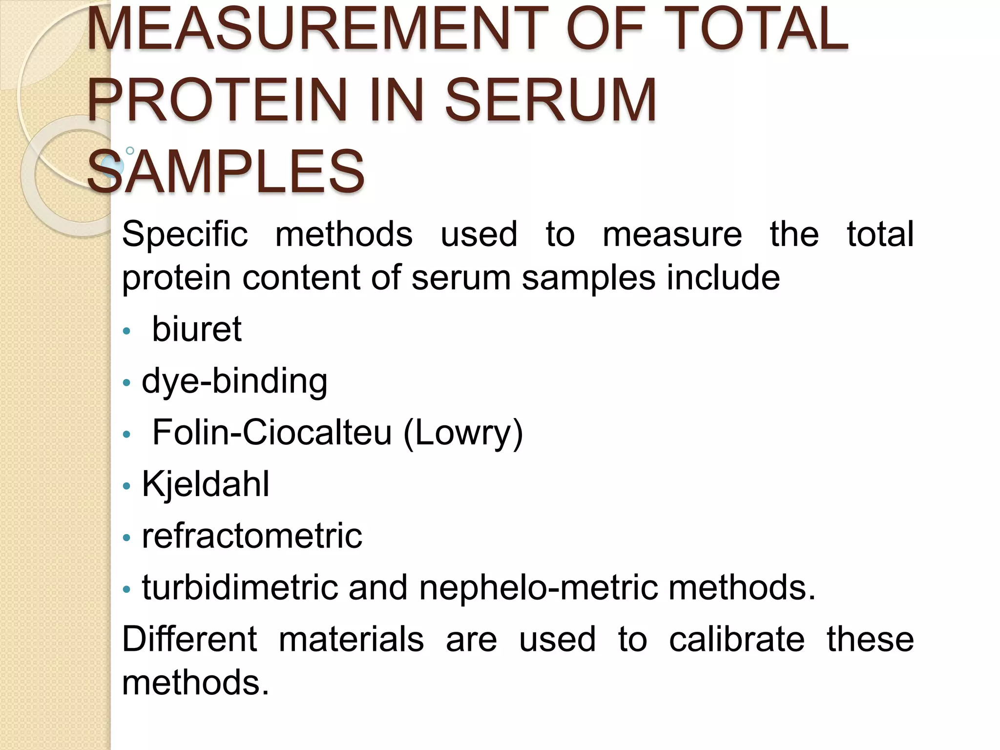
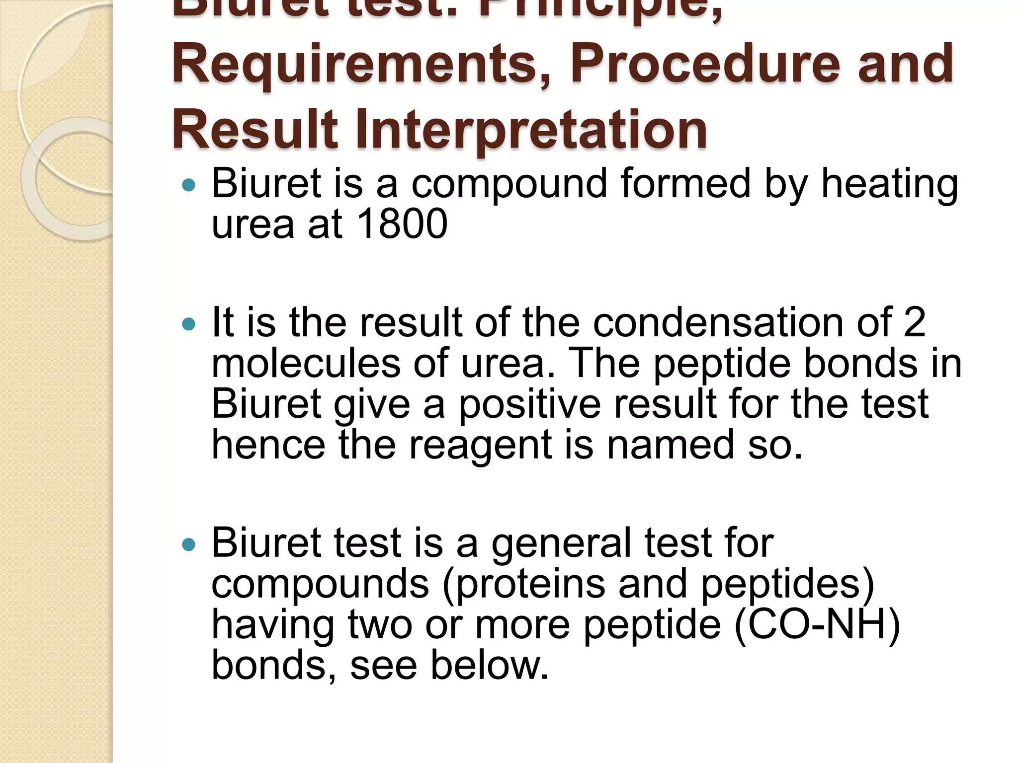
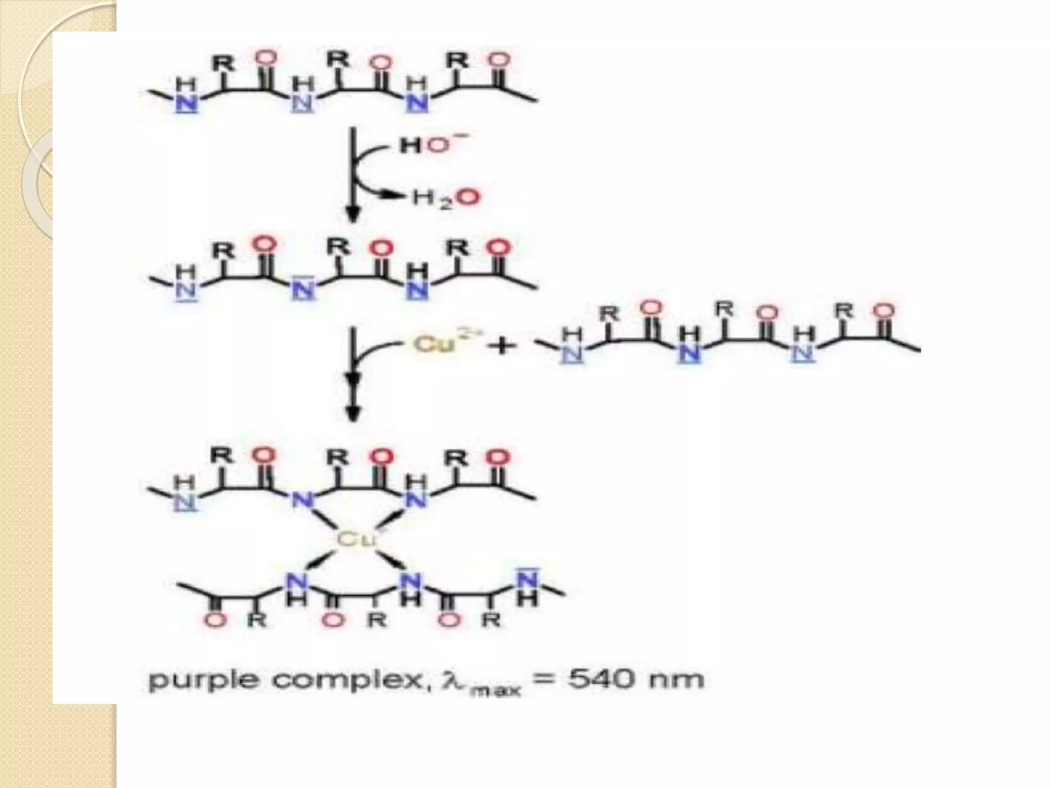
ESTIMATION OF PROTEIN
 Plasma normally contains about 6.5 to
8.5 g/dL protein, and serum about 4%
less
 The determination of amount of protein
present in a solution is a widely used
procedure in biochemistry.
 Important to know the amount of
protein in different stages of purification
to calculate the specific activity of the
protein.
MEASUREMENT OF TOTAL
PROTEIN IN SERUM
SAMPLES
Specific methods used to measure the total
protein content of serum samples include
• biuret
• dye-binding
• Folin-Ciocalteu (Lowry)
• Kjeldahl
• refractometric
• turbidimetric and nephelo-metric methods.
Different materials are used to calibrate these
methods.
Biuret test: Principle,
Requirements, Procedure and
Result Interpretation
 Biuret is a compound formed by heating
urea at 1800
 It is the result of the condensation of 2
molecules of urea. The peptide bonds in
Biuret give a positive result for the test
hence the reagent is named so.
 Biuret test is a general test for
compounds (proteins and peptides)
having two or more peptide (CO-NH)
bonds, see below.
Principle:
 When biuret is treated with dilute copper sulfate in alkaline
medium, a purple colored compound is formed. It is believed
that the color is due to the formation of a copper co- ordinated
complex or chelate complex.
 The chelate complex absorbs light at 540 nm and hence
appears violet. The color change from blue to violet indicates
the presence of proteins.
 The greater the number of peptide bonds in a protein, the
greater the color intensity. Hence, the color change is from
blue to pink if the concentration of peptide bonds is low like in
short-chain peptides.
 The principle of biuret test is conveniently used to detect the
presence of proteins in biological uids.
Cont,,
Requirements
 Biuret reagent i.e. mixture of Hydrated Copper sulphate, Potassium
hydroxide solution and Potassium sodium tartrate ,Test tubes (clean
and dry), De-ionized water (negative control), Albumin or egg white
(positive control)
Procedure
 Take 3 clean and dry test tubes.
 Add 1-2 ml of the test solution, egg albumin and deionized water in the
respective test tubes.
 Add 1-2 ml of Biuret reagent to all the test tubes.
Shake well and allow the mixtures to stand for 5 minutes. Observe for
any color change.
Interpretation
 No color change (solution remains blue ) means Proteins are not
present
 The solution turns from blue to violet (deep purple) means Proteins are
present
 The solution turns from blue to pink means Peptides are present
(Peptides or peptones are short chains of amino acid residues which
Interestingly,
DYE BINDING METHOD
 Dye-binding methods are based o n the ability of
proteins to bind dyes such as Amido black 10B
and CBB
 The dye-binding method of greatest con-
temporary interest, particularly for assay of total
protein in CSF and urine, uses CBB G-250
 CBB binds to protonated amine groups of amino
acid residues in the polypeptide chain, and the
absorbance maximum for the bound species of
the dye decreases at 465 nm and increases at
595 nm
 The method is simple, fast, and linear up to 150
mg/dL.
FOLIN-CIOCALTEU (LOWRY)
Principle
The principle of this method is based on two reactions leading to colour
complex formation
Firstly, the Biuret reaction in which Cu2+ of the reaction mixture reacts
with the peptide bond of proteins under alkaline conditions resulting in
their reduction to cuprous ions (Cu+), and Lowry’s reaction in which the
Folin Ciocaltaeu reagent, which contains phosphomolybdic complex
which is a mixture of sodium tungstate, sodium molybdate and
phosphate, along with copper sulphate solution and the protein forms a
blue purple colour which can be assessed by measuring the
absorbance at 650-700nm
The phenolic group of the aminoacid (tyrosine and tryptophan) residues
will produce a blue purple colour due to the reduction of
phosphomolybdotungstate to hetero-polymolybdenum blue by the
copper catalysed oxidation of the amino acids and its intensity
depends on the amount of these aromatic amino acids present.
The blue purple colour formed thus differ from protein to protein.The blue
purple color is formed of aromatic amino acids tryptophan and tyrosine.
The reaction is pH dependent and works best in alkaline conditions with
pH between 9 and 10.5
KJELDAHL METHOD
OBJECTIVE
“ QUALITATIVE DETERMINATION OF ORGANIC AS WELL AS INORGANIC NITROGEN
(N2) IN THE SUBSTANCES .”
THE PROCEDURE INVOLVES THREE MAJOR STEPS
 DIGESTION: For decomposition of nitrogen sample utilizing a concentrated acid
solution(H2SO4) a catalyst(CuSO4) and K2SO4 for boiling point increasing agent. As a
result Ammonium Sulphate produce.
ie..Nitrogenous Compound + H2SO4 +CuSO4 + K2SO4 = (NH4)2SO4 + H2O + CO2 +
Other by-products
 DISTILLATION: Adding excess base to the acid digestion mixture to convert NH4+ to
NH3, followed by boiling and condensation of the NH3 gas in a receiving solution.
(NH4)2SO4 + 2NaOH = 2NH3 ↑ + Na2SO4 + 2H2O TITRATION: To quantify the amount
of ammonia in the receiving solution.
 TITRATION
principle
 the sample is first acid digested to convert
nitrogen in the protein to ammonium ion
 The con- centration of ammonia nitrogen
then is evaluated by titration or
nesslerization, a correction is made for
nitrogen contributed by nonprotein
compounds also present in serum, and the
ammonia nitrogen value is multiplied by a
factor of 6.25 (100%/16%) to express
protein nitrogen as total protein
TITRATION
Reaction with Hydrochloric Acid (HCl): [Back
titration]
 The ammonia is captured by a, carefully measured
excess of a standardized acid solution in the receiving
flask. The excess of acid in the receiving solution keeps
the pH low, and the indicator does not change color.
 NH3 + HCl (in excess) = NH4+Cl- + HCl (left back)
 The excess acid solution is exactly neutralized by a
carefully
measured standardized alkaline base solution such as
sodium hydroxide. A color change is produced at the
end point of the titration.
 HCl + NaOH = NaCl + H2O
 pg 7
 V ml of X (N) NaOH = V ml of X (N) HCl = V ml of X (N)
NH3
REFRACTOMETRY METHOD
 Refractometry is a quick alternative to chemical analysis
for serum total protein when a rapid estimate is required
 At orotein concentrations less than 3.5 g/dL,
refractometric results are likely to be inac- curate.
 A t a concentration greater than 11.0 g/dL, a valid result
is obtained by dilution of the serum with equal parts of
water, followed by a reading of the diluted sample.
 A day-to-day coefficicnt of variation (CV) of less than
2.0% is acceptable precision.
TURBIDIMETRIC AND
NEPHELO-METRIC METHODS
 Precipitation of protein for turbidimetric or
nephelomenic assays is achieved with,
 sulfosalicylic acid alone, with sulfosali- cylic acid
in combination with sodium sulfate or
trichloroace- tic acid (TCA),or with TCA alone
 Precipitation methods for total protein assay
depend on formation of a fine precipitate of
uniform, insoluble protein particles, which scatter
incident light in suspension.
 Albumin and globulins often give different
reactivities in precipitation methods
THANK YOU

serum protein estimation

  • 1.
    ESTIMATION OF PROTEIN Plasma normally contains about 6.5 to 8.5 g/dL protein, and serum about 4% less  The determination of amount of protein present in a solution is a widely used procedure in biochemistry.  Important to know the amount of protein in different stages of purification to calculate the specific activity of the protein.
  • 2.
    MEASUREMENT OF TOTAL PROTEININ SERUM SAMPLES Specific methods used to measure the total protein content of serum samples include • biuret • dye-binding • Folin-Ciocalteu (Lowry) • Kjeldahl • refractometric • turbidimetric and nephelo-metric methods. Different materials are used to calibrate these methods.
  • 3.
    Biuret test: Principle, Requirements,Procedure and Result Interpretation  Biuret is a compound formed by heating urea at 1800  It is the result of the condensation of 2 molecules of urea. The peptide bonds in Biuret give a positive result for the test hence the reagent is named so.  Biuret test is a general test for compounds (proteins and peptides) having two or more peptide (CO-NH) bonds, see below.
  • 5.
    Principle:  When biuretis treated with dilute copper sulfate in alkaline medium, a purple colored compound is formed. It is believed that the color is due to the formation of a copper co- ordinated complex or chelate complex.  The chelate complex absorbs light at 540 nm and hence appears violet. The color change from blue to violet indicates the presence of proteins.  The greater the number of peptide bonds in a protein, the greater the color intensity. Hence, the color change is from blue to pink if the concentration of peptide bonds is low like in short-chain peptides.  The principle of biuret test is conveniently used to detect the presence of proteins in biological uids.
  • 6.
    Cont,, Requirements  Biuret reagenti.e. mixture of Hydrated Copper sulphate, Potassium hydroxide solution and Potassium sodium tartrate ,Test tubes (clean and dry), De-ionized water (negative control), Albumin or egg white (positive control) Procedure  Take 3 clean and dry test tubes.  Add 1-2 ml of the test solution, egg albumin and deionized water in the respective test tubes.  Add 1-2 ml of Biuret reagent to all the test tubes. Shake well and allow the mixtures to stand for 5 minutes. Observe for any color change. Interpretation  No color change (solution remains blue ) means Proteins are not present  The solution turns from blue to violet (deep purple) means Proteins are present  The solution turns from blue to pink means Peptides are present (Peptides or peptones are short chains of amino acid residues which
  • 7.
  • 8.
    DYE BINDING METHOD Dye-binding methods are based o n the ability of proteins to bind dyes such as Amido black 10B and CBB  The dye-binding method of greatest con- temporary interest, particularly for assay of total protein in CSF and urine, uses CBB G-250  CBB binds to protonated amine groups of amino acid residues in the polypeptide chain, and the absorbance maximum for the bound species of the dye decreases at 465 nm and increases at 595 nm  The method is simple, fast, and linear up to 150 mg/dL.
  • 9.
    FOLIN-CIOCALTEU (LOWRY) Principle The principleof this method is based on two reactions leading to colour complex formation Firstly, the Biuret reaction in which Cu2+ of the reaction mixture reacts with the peptide bond of proteins under alkaline conditions resulting in their reduction to cuprous ions (Cu+), and Lowry’s reaction in which the Folin Ciocaltaeu reagent, which contains phosphomolybdic complex which is a mixture of sodium tungstate, sodium molybdate and phosphate, along with copper sulphate solution and the protein forms a blue purple colour which can be assessed by measuring the absorbance at 650-700nm The phenolic group of the aminoacid (tyrosine and tryptophan) residues will produce a blue purple colour due to the reduction of phosphomolybdotungstate to hetero-polymolybdenum blue by the copper catalysed oxidation of the amino acids and its intensity depends on the amount of these aromatic amino acids present. The blue purple colour formed thus differ from protein to protein.The blue purple color is formed of aromatic amino acids tryptophan and tyrosine. The reaction is pH dependent and works best in alkaline conditions with pH between 9 and 10.5
  • 10.
    KJELDAHL METHOD OBJECTIVE “ QUALITATIVEDETERMINATION OF ORGANIC AS WELL AS INORGANIC NITROGEN (N2) IN THE SUBSTANCES .” THE PROCEDURE INVOLVES THREE MAJOR STEPS  DIGESTION: For decomposition of nitrogen sample utilizing a concentrated acid solution(H2SO4) a catalyst(CuSO4) and K2SO4 for boiling point increasing agent. As a result Ammonium Sulphate produce. ie..Nitrogenous Compound + H2SO4 +CuSO4 + K2SO4 = (NH4)2SO4 + H2O + CO2 + Other by-products  DISTILLATION: Adding excess base to the acid digestion mixture to convert NH4+ to NH3, followed by boiling and condensation of the NH3 gas in a receiving solution. (NH4)2SO4 + 2NaOH = 2NH3 ↑ + Na2SO4 + 2H2O TITRATION: To quantify the amount of ammonia in the receiving solution.  TITRATION
  • 11.
    principle  the sampleis first acid digested to convert nitrogen in the protein to ammonium ion  The con- centration of ammonia nitrogen then is evaluated by titration or nesslerization, a correction is made for nitrogen contributed by nonprotein compounds also present in serum, and the ammonia nitrogen value is multiplied by a factor of 6.25 (100%/16%) to express protein nitrogen as total protein
  • 12.
    TITRATION Reaction with HydrochloricAcid (HCl): [Back titration]  The ammonia is captured by a, carefully measured excess of a standardized acid solution in the receiving flask. The excess of acid in the receiving solution keeps the pH low, and the indicator does not change color.  NH3 + HCl (in excess) = NH4+Cl- + HCl (left back)  The excess acid solution is exactly neutralized by a carefully measured standardized alkaline base solution such as sodium hydroxide. A color change is produced at the end point of the titration.  HCl + NaOH = NaCl + H2O  pg 7  V ml of X (N) NaOH = V ml of X (N) HCl = V ml of X (N) NH3
  • 14.
    REFRACTOMETRY METHOD  Refractometryis a quick alternative to chemical analysis for serum total protein when a rapid estimate is required  At orotein concentrations less than 3.5 g/dL, refractometric results are likely to be inac- curate.  A t a concentration greater than 11.0 g/dL, a valid result is obtained by dilution of the serum with equal parts of water, followed by a reading of the diluted sample.  A day-to-day coefficicnt of variation (CV) of less than 2.0% is acceptable precision.
  • 15.
    TURBIDIMETRIC AND NEPHELO-METRIC METHODS Precipitation of protein for turbidimetric or nephelomenic assays is achieved with,  sulfosalicylic acid alone, with sulfosali- cylic acid in combination with sodium sulfate or trichloroace- tic acid (TCA),or with TCA alone  Precipitation methods for total protein assay depend on formation of a fine precipitate of uniform, insoluble protein particles, which scatter incident light in suspension.  Albumin and globulins often give different reactivities in precipitation methods
  • 16.