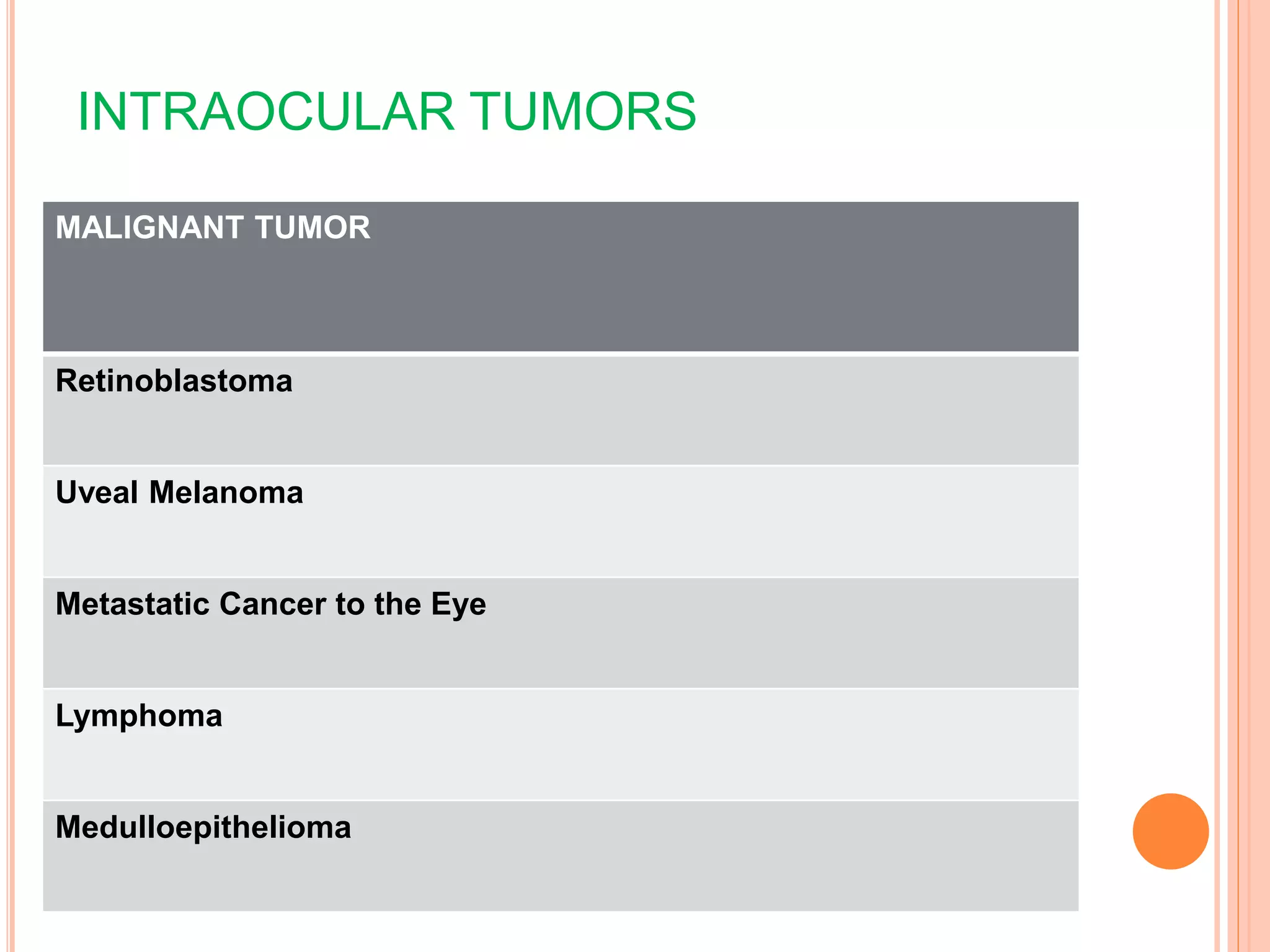
SEMINAR
MALIGNANT INTRAOCULAR
TUMORS
Presenter-Dr. Kriti Chandra
INTRAOCULAR TUMORS
MALIGNANT TUMOR
Retinoblastoma
Uveal Melanoma
Metastatic Cancer to the Eye
Lymphoma
Medulloepithelioma
INTRODUCTION
 Most common intraocular malignancy in children ( 1
in 15,000 to 1 in 18,000 live births).
 No racial or gender predisposition.
 Bilateral in 25 to 35% cases.
 Unilateral cases around 24 months & bilateral before
12 months.
RETINOBLASTOMA
GENETICS
 Deletion of long arm of chromosome 13, 13q14, which is a tumor
suppressor gene termed as RB gene.
 The deletion of 13q chromosome may be associated with other dysmorphic features
such as microcephaly, broad prominent nasal bridge, hypertelorism,
microphthalmos, epicanthus, toe abnormalities, and psychomotor and mental
retardation.
 This gene is the first gene in its class of human cancer ‘suppressor’ gene, which
actually prevents the abnormal uncontrolled proliferation of immature retinal
cells called the retinoblasts.
SPORADIC FAMILIAL
Unilateral,unifocal 85% bilateral,multifocal
60% of all cases 40% of all cases
Present later Present earlier
Children of affected are normal Children of the affected have 45%
chances of inheritance
Chromosomal anomaly is a
somatic mutation
Chromosomal anomaly is a germline
mutation
Relatives have a low risk of RB
development
Relatives have a high risk of RB
development
Increased risk for second malignancies
AD with high penetrance
ALFRED KNUDSON’S TWO HIT
HYPOTHESIS (1971)
 Retinoblastoma appears to result from loss or inactivation of both
normal alleles of the retinoblastoma gene
 The timing of the loss or inactivation of the two normal alleles
determines whether the disease is germinal or somatic .
 In germinal retinoblastoma, at least one normal allele must be lost or
inactivated prior to the first mitotic division of embryogenesis.
 In somatic retinoblastoma, both alleles are present and active beyond
the stage of the fertilized egg, but subsequent mutations occur to delete
or inactivate both alleles in at least one immature retinal cell
(retinoblast).
CLINICAL FEATURES
 Leukocoria (white pupillary reflex): 56% ,
commonest
 Strabismus : 20%
 Red painful eye (secondary glaucoma)
 Loss of vision
 Proptosis: extra ocular invasion
 (less common are
buphthalmos,discoloration of iris
hyphema,sterile orbital cellulitis)
 Trilateral retinoblastoma; bil RB+
pineoblastoma
 Distant metastases
CLINICAL SIGNS OF RETINOBLASTOMA DEPENDS ON THE STAGE OF THE
DISEASE
 Early intraretinal tumor-flat lesion which appear as a tranalucent or white
fluffy retinal mass.
 Child may present with strabismus if the tumor involves macula or with reduced
visual acuity.
 Moderately advance lesions
As the tumor grows further,three patterns usually seen. Endophytic-
White to cream-colored mass breaks through internal limiting
membrane
No surface vessels or small, irregular tumor vessels
Associated with vitreous seeding
 Exophytic-Yellow-white lesion in subretinal space
Overlying retinal vessels increased in caliber and tortuosity
Associated subretinal fluid and RD that can obscure the tumour
 Diffuse infiltrating tumor-tumor diffusely involves the retina causing just a placoid
thickness of the retina and not a mass,associated with vitreous seeding and
extension of tumor cells into the anterior chamber aqueous.No intratumoral
calification.
 Advanced tumors
manifest with proptosis secondary to optic nerve extension or orbital extension and
systemic metastasis.
Systemic metastasis occurs to the brain,skull,distant bones and lymph nodes.
RETINOMA
gray-white color,
clumped calcification
Limited vascularity
surrounding chorioretinal
atrophy with retinal pigment
epithelial hyperplasia
DIAGNOSTIC WORKUP
 Diagnosis of retinoblastoma is made without pathologic confirmation and is
based on a clinical examination.
 slit lamp examination- to assess the anterior chamber and iris for evidence of
tumor cells and iris neovascularization and to evaluate the clarity of the lens and
retrolental vitreous, may also disclose finely dispersed cells or tumor cell clumps
(seeds) in the vitreous.
 Direct ophthalmoscopy: white reflex
 Indirect ophthalmoscopy:
 RB seen as projecting into vitreous, creamy white in color,
neovascularisation seen on surface, calcification gives
cottage cheese appearance (glistening white).
(A) A fundus photograph of an eye with
retinoblastoma
(B) The corresponding sketch of the disease in
the eye diagram.
Ophthalmic USG: Non-invasive, safe, repeatable, and immediately interpretable.
 B-scan USG reveals a 2D cross-sectional view, confirms presence and the relationship, the
size and shape of the tumors.
Orbital involvement, optic nerve invasion can be seen, extrascleral extension, and calcification.
 The presence of high amplitude spikes which persist on low gain confirm the presence
of calcium within the tumor. USG is also used to assess the response to systemic
chemotherapy and focal treatment.
.
CT scan, dense heterogenous lesion with hyper dense foci corresponding to
calcification.
 for assessing extraocular extension and invasion of the optic nerve.
A computed tomography scan of a
large calcified retinoblastoma in
the right eye.
 MRI is not as specific for retinoblastoma as CT because of its lack of
sensitivity in detecting calcification.
MRI
MRI scan of the brain and orbits (with gadolinium enhancement ) done for two
reasons.
First, to evaluate whether there is extraocular extension and optic nerve
invasion by the tumor ideally before the staging examination under anesthesia,
and
second, to eliminate the possibility of the “trilateral” retinoblastoma syndrome.
OTHER TESTS
 Assays of aqueous humor enzyme levels could offer useful information to
patients with suspected retinoblastoma.
 Lactate dehydrogenase (LDH) is a glycolytic enzyme that uses glucose as
an energy source. It is present in high concentrations within metabolically
active cells.
 Normally, its concentration in serum and aqueous humor is low and the ratio
of aqueous humor to serum LDH is less than 1.0 in patients with ocular
disease other than retinoblastoma. However, aqueous humor for eyes with
retinoblastoma exhibits increased LDH activity expressed as an aqueous
humor/LDH ratio of greater than 1.0.
 CSF cytology
 Bone marrow biopsy &aspiration
 Bone scan
 Lab tests: Hemogram, KFT, LFT.
HISTOPATHOLOGY
 Poorly differentiated tumors consist of small to medium sized round cells
with large hyperchromatic nuclei and scanty cytoplasm with mitotic figures.
 Well differentiated tumors show the presence of rosettes and
fleurettes.These can be of various types.
 1) Flexner-Wintersteiner rosettes-highly specific for retinoblastoma,consist of
columnar cells arranged around a central Lumen.
 2) Homer Wright rosettes- consist of cells arranged around a central
neuromuscular tangle.This is also found in neuroblastoma medulloblastoma
and medulloepitheliomas.
 3) Fleurettes-eosinophilic structures composed of tumor cells with pear
shaped eosinophilic processes projecting through a fenestrated membrane.
STAGING
REESE-ELLSWORTH CLASSIFICATION
 International Classification System for Intraocular Retinoblastoma
 Group A: Very Low Risk Eyes with small intra-retinal tumors away from critical structures
 1. Tumors are 3 mm or smaller in greatest dimension
 2. Confi ned to the retina
 3. Located further than 3mm from the foveola and 1.5 mm from the optic disc
 4. No seeding Group
 Group B: Low Risk Eyes with no seeding. Tumor(s) are discrete but any size or location
 1 All other tumours confined to the retina not in Group A.
 2. Tumour-associated subretinal fluid less than 3 mm from the tumour with no subretinal seeding
 Group C: Moderate Risk Discrete local disease with minimal subretinal or vitreous seeding
 1. Local fine vitreous seeding close to discrete tumor or
 2. Local sub-retinal seeding < 3mm from the tumor
 3. Or both
 Group D: High Risk Eyes with diffuse vitreous or subretinal seeding
 1. More extensive seeding than Group C
 2. Diffuse vitreous seeding or subretinal seeding (>3mm from tumor margin) or both
 Group E: Very High Risk Presence of any one or more of the following poor prognostic features:
 1. Irreversible neo-vascular glaucoma
 2. Opaque media due to hemorrhage
 3. Aseptic orbital cellulitis
 4. Tumor anterior to anterior vitreous face
 5. Tumor touching the lens
 6. Diffuse infi ltrating retinoblastoma
 7. Phthisis bulbi
International Retinoblastoma Staging System
 Stage 0 Patients treated conservatively
 Stage I Eye enucleated, tumor completely resected on histopathology examination
 Stage II Eye enucleated, microscopic residual tumor present in the form of
1. Tumor invasion into extrascleral tissue
2. Tumor invasion into cut end of optic nerve
 Stage III Regional extension
a. Overt orbital disease
b. Preauricular or cervical lymph node extension
 Stage IV Metastatic
a. Hematogenous metastasis (without CNS extension) 1) single lesion 2) multiple
lesion
b. Central nervous system extension (with or without any other site of regional or metastatic
disease)
1. Prechiasmatic lesion 2. Central nervous system mass 3. leptomeningeal and csf
disease
TREATMENT OF RETINOBLASTOMA
 Primary goal
 to ensure the survival of children.
 retention of eyes and vision.
 Avoidance of side effects- second malignancies, facial bony
deformities, or other physical changes that can affect functional
well-being.
 Treatment approaches are guided by the presence of
intraocular or extraocular disease.
TREATMENT MODALITIES
Surgery
• enucleation,
exenteration
Local therapy
• cryotherapy, photocoagulation,
laser hyperthermia
Radiotherapy
• EBRT, radioactive plaque
Chemotherapy
• Systemic Chemotherapy,
intra-arterial
CURRENT SUGGESTED PROTOCOL FOR TREATMENT
 A. Intraocular tumor, International Classification Group A to C,
Unilateral or Bilateral
 1. Focal therapy (cryotherapy or transpupillary thermotherapy) alone for smaller
tumors (< 3mm in diameter and height) located in visually noncrucial areas
 2. Standard 6 cycle chemoreduction and sequential aggressive focal therapy for
larger tumors and those located in visually crucial areas
 3. Transpupillary thermotherapy or plaque brachytherapy for residual tumor in
the macular and juxtapapillary areas >6 cycles.
 4. Focal therapy for small residual tumor, and plaque brachytherapy/ external
beam radiotherapy (>12 months age) for large residual tumor if bilateral, and
enucleation if unilateral.
 B.Intraocular tumor, International Classification Group D, Unilateral or
Bilateral
 1. High dose chemotherapy and sequential aggressive focal therapy
 2. Periocular carboplatin for vitreous seeds
 3. Consider primary enucleation if unilateral, specially in eyes with no visual prognosis
 C. Intraocular tumor, International Classification Group E, Unilateral or
Bilateral
 1. Primary enucleation
 2. Evaluate histopathology for high risk factors
 D. High risk factors on histopathology, International Staging, Stage 2
 1. Baseline systemic evaluation for metastasis
 2. Standard 6 cycle adjuvant chemotherapy
 3. High dose adjuvant chemotherapy and orbital external beam radiotherapy in patients
with scleral infiltration, extraocular extension, and optic nerve extension to transection.
 E. Extraocular tumor, International Staging, Stage 3A
 1. Baseline systemic evaluation for metastasis
 2. High dose chemotherapy for 3-6 cycles, followed by enucleation or
extended enucleation, external beam radiotherapy, and continued high
dose chemotherapy for 12 cycles
 F. Regional Lymph Node Metastasis, International Staging, Stage 3B
 1. Baseline evaluation for systemic metastasis
 2. Neck dissection, high dose chemotherapy for 6 cycles, followed by
external beam radiotherapy, and continued high dose chemotherapy for
12 cycles
 G. Hematogenous or Central Nervous System Metastasis, International
Staging, Stage 4
 1. Palliative therapy in discussion with the family
 2. High dose chemotherapy with bone marrow rescue for hematogenous
metastasis
 3. High dose chemotherapy with intrathecal chemotherapy for central
nervous system metastasis
FOLLOW UP
 1st 6 months: 4-6 week intervals
 Upto 3 years: at 4-6 month intervals
 Later, yearly
 Family history positive: all family members [other children at
birth] should be examined yearly
UVEAL MELANOMA
 Uveal melanoma is a malignant neoplasm that arises from neuroectodermal
melanocytes within the choroid, ciliary body, or iris.
 Uveal melanoma has a cumulative lifetime incidence of approximately 1 in
2000–2500 individuals.
 It can arise from any portion of the uveal tract, but choroidal involvement is by
far the most common
 The average age at detection of melanomas of the choroid or ciliary body is
about 55–60years.
 Choroidal melanomas are the most common primary
intraocular malignancies in adults.
 It is the second most common type of primary malignant
melanoma in the body.
 No strong familial inheritance pattern exists for this neoplasm.
 Several somatic chromosomal abnormalities have been
encountered frequently in uveal melanoma cells, including
A) monosomy 3
B) partial duplications of the long (q) arm of chromosome 8,
C) complementary gains of material on chromosome 6p and loss
of material from chromosome 6q, and
these cytogenetic abnormalities appear to be associated with
an increased risk of metastasis and melanoma-related death
HISTOLOGICAL FINDINGS
 3 Cell Types:
 Spindle A cells
 Spindle B cells
 Epitheloid cells
Spindle-A cells
•Slender nuclei
•Delicate chromatin
•Ill-defined or absent
nucleoli
•No mitotic activity
•Resemble naevus
Spindle-B cells
•Plump nuclei
•Small but
prominent nucleoli
•Coarse chromatin
•Mitotic figures
common
Epitheloid cells
•Eosinophilic cytoplasms
•Cell sizes variable:
larger and pleomorphic
•May be multinucleated
•Chromatin coarse
clumping
•Mitotic figures abundant
•No cohesiveness.
CALLENDER CLASSIFICATION
 Modified Callender’s Classification:
 Spindle cell nevi
 Spindle cell melanoma
 Epitheloid cell melanoma
 Mixed cell melanoma
 Necrotic melanoma
ETIOLOGY
 Risk factors are people with light-colored iris, whites,
median age-55 yrs, men
 Sunlight exposure is a contributory factor.
 Predisposing diseases
1)family history of uveal melanoma
2) uveal nevus
3)congenital ocular melanocytosis
4)dysplastic nevus syndrome
5) xeroderma pigmentosum.
CLINICAL FEATURES
1. Quiescent stage :
 Symptoms depend upon the location and size of tumour.
 Small tumour
 located in the periphery may not produce any symptoms
 tumours arising from the post. pole present with early visual loss.
 Sign: appearances of orange patches in the pigment epithelium.
 large tumour
 penetrate through Bruch’s membrane grows in subretinal space
characterised by exudative retinal detachment
 produces marked loss of vision and gradually tumour fills the whole eye.
 Other associated features : sub/intraretinal hemorrhage, choroidal folds, &
vitrous hemorrhage.
2. Glaucomatous stage :
 Glaucoma may develop due to obstruction of venous outflow by
pressure on vortex veins, blockage of the angle of ant. chamber
 Sx : severe pain, redness, watering
 Signs : chemosed & congested conjunctiva, oedematous cornea,
shallow anterior chamber, fixed & dilated pupil,lens opaque,
increase IOP (stony hard eye), features of iridocyclitis.
3. Stage of extraocular lesion :
 the tumor may burst through sclera usually at the limbus.
 Rapid fungation and involvement of extraocular tissues resulting in
marked proptosis.
4. Stage of distant metastasis :
 Hematologic spread commonly occurs in liver & commonest cause
of death.
Highly
pimented
melanoma
Orange
pigment on
surface
Diffuse melanoma
Amelanotic
melanoma
Collar stud
With exudative
RD
Clinical features
DIAGNOSIS
 Clinical appearance
 FFA
 Ultrasound
 Radiography
 Laboratory tests
 Invasive technique
 Histopathology
 Immunohistochemistry
 New diagnostic tests
FUNDUS FLUORESCEIN ANGIOGRAPHY
Fluorescein angiography and are not diagnostic. They heip to differentiate
between choroidal melanoma and simulating lesions such as choroidal
hemangioma and haemorrhagic lesions.
 Small choroidal melanomas with intact RPE shows no changes.
 Larger melanomas with disrupted RPE may show
Arterial phase-mottled hyperfluorescence
Venous phase-pinpoint hyperfluorescence
Late venous phase- late staining due to SRF
A SCAN
 A-scan ultrasonography is useful for tumors thicker than 2-3 mm.
 Choroidal melanoma shows an initial prominent spike, followed by low-to-
medium internal reflectivity with diminishing amplitude and a significant echo.
 Performing sequential A-scans, with accurate dimension measurements, in
cases of diagnostic uncertainty is important.
B-SCAN:
 B-scan is a routine test used in the evaluation of any posterior segment
mass..
 B-scan helps in
-establishing the diagnosis,
-to evaluate possible extraocular extension
-to estimate tumor size for periodic observation
-to plan therapeutic intervention.
Intraocular melanomas have several distinctive features on B scan:
 Low-to-medium reflectivity
 Excavation of underlying uveal tissue
 Shadowing of subjacent soft tissues
 Internal vascularity
 An acoustic quiet zone at the base of the tumor called acoustic hollowing
 Indocyanin green angiography-
 Indocyanine green angiography of choroidal melanomas shows most tumors
to be quite hypofluorescent throughout the study.
Ultrasound biomicroscopy (UBM)
 It can differentiate very anterior choroidal melanomas from those of
ciliary body origin.
 It is also helpful in assessing angle-closure glaucoma.
Computed Tomography
 CT scan is not as sensitive as ultrasonography.
 It is useful for visualizing extraocular extension and may help
differentiate between choroidal or retinal detachment and a solid
tumor.
Magnetic Resonance Imaging
 less sensitive .
 Use of surface coil imaging and gadolinium as a contrast material greatly
improves its resolution.
 Pigmented melanomas are seen as a high-density image in T1 and as a
low-density image in T2 .
 MRI also can be used to determine extrascleral extension and distinguish
surrounding fluid from the tumor.
 Invasive technique
Fine-needle biopsy and incisional biopsy are not usually required but may be
helpful in case where diagnosis is not established.
 particularly for distinguishing amelanotic melanomas from metastatic tumors.
 Fine-needle biopsy is increasingly being performed for prognostic purposes
 In opaque medias, ultrasound guided approach is essential.
IMMUNOHISTOCHEMICAL FEATURES
 The most important immunohistochemical markers expressed by the tumor
cells are HMB45, S100, Melan A, MITF, and tyrosinase. HMB 45 and
Melan A are melanocytic markers.
 While Melan A stains melanocytes in general, HMB45 is predominantly
expressed in “activated” melanocytes and is therefore more suggestive of
malignant melanocytic lesions.
 S100 is expressed in different types of cells including melanocytes and very
often used in combination with HMB45 as a marker for uveal melanoma.
 Microphthalmia transcriptioni s essential for the development and survival of
melanocytes and therefore expressed in various melanocytic lesions including
uveal melanoma.
 Tyrosinase is an enzyme that is involved in the metabolism of melanocytes
and was recently introduced as a melanoma marker.
 At least two markers (e.g., HMB45 and S100) should be employed for the
diagnosis of uveal melanoma if the diagnosis cannot be made on histologic
features alone.
COLLABORATIVE OCULAR MELANOMA STUDY
(COMS)
 MEDIUM SIZE MELANOMA –
 No statistically significant difference in 5 year survival
rate following brachytherapy or enucleation
 LARGE SIZE MELANOMA –
 Pre - enucleation radiotherapy did not improve 5 year
survival rate over enucleation alone
CLASSIFICATION OF MELANOMA ACCORDING TO SIZE
(COMS)
Size Apical Height Longest basal diameter
Small
1.5-2.4 mm 5-16 mm
Medium 2.5-10 mm
5-16 mm
Large >10 mm >16 mm
TREATMENT DECISION OF MELANOMA
DEPENDS ON
 Size, shape and location of tumor
 Vision in affected and fellow eye
 General systemic condition of patient
MANAGEMENT OPTION BEFORE
METASTASIS DIVIDED AS
Eye conserving Eye destructive
 Observation
 MicrosurgicalResection
 Transpupillary
Thermotherapy
 Laser photocogulation
 Brachytherapy
 External Beam Irradiation
 Enucleation
 Exentration
OBSERVATION
 In whom the differentiation between nevus and melanoma cannot be
made with reasonable certainty
 Advisable for those who have coexistent life-threatening medical
conditions that preclude surgical intervention.
Microsurgical resection
Most frequently employed trans-scleral resection technique- a partial-
thickness scleral flap is made directly over the tumor; the tumor is cut out of the
eye, along with some of the adjacent normal uveal tissue, using microscissors;
and then scleral opening is closed with multiple interrupted sutures.
Trans-scleral resection appears to be most applicable for patients who are
relatively young and systemically healthy and who have thick malignant
melanomas of the ciliary body or peripheral choroid that are no more than 12–
13mm in basal dimension.
Endoresection method- a complete pars plana vitrectomy, followed by internal
tumor resection using vitreoretinal instruments.
small choroidal melanomas adjacent to or partly overlying the optic disc
Transpupillary thermotherapy
 Transpupillary thermotherapy uses an infrared laser beam to
induce tumour cell death by hyperthermia but not
coagulation.
 Indications :1)Small, pigmented choroidal tumour(<3mm),
away from macula.
 Upto 3.5 mm in thickness and basal diameter< 10 mm
 choroidal melanoma when radiotherapy is inappropriate because
of poor general health or reduced life expectancy.
Laser photocoagulation
It is used to treat selected small choroidal melanomas.
when the lesions are located away from the fovea and are less than or
equal to 2mm,<10mm in diameter.
 with limited or no associated retinal detachment that are located
posterior to the ocular equator in eyes with clear optical media
Brachytherapy
 Brachytherapy (episcleral plaque radiotherapy) with ruthenium-106 or
an iodine-125 applicator is usually the treatment of first choice
 Indications
-Tumours less than 20 mm in basal diameter in which there is a
reasonable chance of salvaging vision.
-5 mm thick with a ruthenium plaque
-10 mm thick with an iodine plaque.
2)Technique
a.The tumour is localized by transillumination or binocular
indirect ophthalmoscopy.
b. A template consisting of a transparent plastic dummy or
metal ring with eyelets is sutured to the sclera
c.The sutures are loosened and used to secure the radioactive
plaque.
d. The plaque is removed once the appropriate dose has been
delivered, usually within 3–7 days. At least 80 Gy should be
delivered to the tumour apex.
3)Tumour response is usually gradual
Tumour regression starts about 1–2 months after treatment and continues for
several years, leaving a flat or dome-shaped pigmented scar.
4)Complications
Excessive irradiation causes cataract, papillopathy and maculopathy. The
irradiated tumour can cause macular edema , retinal hard exudates, serous
retinal detachment, rubeosis and neovascular glaucoma (‘toxic tumour
syndrome’).
External beam radiotherapy
 Irradiation with charged particles such as protons achieves a high dose in the
tumour with a relatively small dose in the superficial tissues, it provide a
collimated beam that would limit the radiotherapy to the precise area of the
tumor.
Indications: tumours unsuitable for brachytherapy either because of large size or
posterior location making positioning of a plaque unreliable.
Survival results: similar to brachytherapy or enucleation
Enucleation
Indications:
-large tumour size,( greater than 15 mm in diameter and greater than 10 mm in thickness)
-optic disc invasion,
-extensive involvement of the ciliary body or angle,
-irreversible loss of vision
Modified Technique:
 many juxtapapillary melanomas, that have not invaded the nerve, can often be managed by custom
designed notched radioactive plaques.The so-called “no touch enucleation” was introduced to
minimize the amount of surgical trauma and to lessen the chance of tumor dissemination at the time of
surgery.
 An essential aspect of this technique was to freeze the venous drainage from the tumor prior to cutting
the optic nerve.
 The “no touch” technique has recently fallen into disuse at most centers because it is cumbersome and
its benefits are only theoretical.
Orbital exenteration
Reserved for cases with widespread orbital extension.
Patients with such advanced melanomas are likely to have
extensive distant metastases and poor prognoses.
The procedure should be considered only in rare cases where
marked discomfort is associated with massive orbital spread of
the melanoma.
METASTATIC CANCER TO THE EYES
 Primary malignant extra-ophthalmic neoplasm that spreads
hematogenously to the eye.
 Most intraocular matastasic tumors involve the choroid,but similar lesions also
affect the iris,ciliary body,optic nerve, and retina in some patients.
 The incidence of clinically detected metastatic intraocular tumors is
approximately 0.1% (1 in 1000) to 0.25% (1 in 400).
 In women, the most common malignancy that gives rise to metastatic
carcinoma to the eye is breast cancer. In men, the most common primary
cancer type appears to be lung cancer
Ocular manifestation
The principal symptom caused by metastatic
carcinoma to the eye is blurred or distorted vision.
Pain is usually not a symptom of metastatic cancer to
the eye, except in patients who have extensive
intraocular tumor and marked visual impairment,
frequently in association with bullous retinal
detachment and secondary glaucoma.
The typical metastatic carcinoma to the choroid from
breast, lung, or gastrointestinal tract appears as a
golden yellow to yellowish white round to oval
lesion .
Metastatic carcinoma to the optic disc can appear either as a swollen disc
without a distinct mass or as a discohesive cellular infiltration of the
superficial aspects of the optic disc .
A metastatic carcinoma to the optic disc is often associated with
profound visual loss, which may or may not be reversible.
 Metastatic carcinoma to the iris typically appears as a solid,
amelanotic iris mass .
 Some metastases to the iris are quite discohesive and shed cells
that form a pseudohypopyon in the anterior chamber. The shed
tumor cells can clog the trabecular meshwork and cause a
secondary open-angle glaucoma. Metastatic iris tumors can also
cause spontaneous hyphema.
 Ciliary body metastatic carcinoma occasionally presents as
diffuse and sometimes multinodular masses, often associated
with extensive retinal detachment and occasionally severe ocular
pain.
DIAGNOSIS
 Fluorescein angiography of metastatic carcinoma to the choroid-
relative hypofluorescence of the lesion in the early frames, and
diffuse hyperfluorescence of the lesion in the late frames.
 Indocyanine green angiography of metastatic carcinoma to
the choroid frequently reveals subtle metastatic lesions that are
not evident on fluorescein angiography. Most metastatic tumors
appear hypofluorescent throughout the study.
 B-scan ultrasonography of metastatic carcinoma to the choroid
shows the typical tumor to have a relatively flat cross-sectional shape
compared with its basal diameters .
 In contrast to choroidal melanomas, metastatic choroidal tumors usually
appear relatively sonoreflective (bright) rather than sonolucent internally
 Fine-needle aspiration biopsy appears to be a reliable and safe technique
for establishing the diagnosis in selected cases.
 The most appropriate indication for such a procedure is suspicion of
metastatic carcinoma in a patient with no history of prior nonocular
cancer or no concurrent extraophthalmic foci of possible primary or
metastatic malignancy that would be more amenable to biopsy than the
ocular lesion.
 For iris tumors, a limbal puncture usually is employed. For choroidal
metastatic tumors, either a direct trans-scleral puncture or an indirect
transvitreal puncture in the pars plana region is employed
 TREATMENT
 The principal management options for patients who have
metastatic carcinoma to the eye include radiotherapy,
chemotherapy, and hormonal therapy .
 Radiotherapy for intraocular metastatic carcinoma is
generally given as an outpatient course of linear
accelerator beam treatment.
 This form of treatment can be used to deliver a relatively
uniform dose of radiation (usually in the range of 35–50Gy)
to all affected portions of the eye while giving a limited
radiation dose to other tissues.
 To maximize treatment effectiveness and minimize
complications, such treatment is usually given in multiple
fractions over 3–5weeks.
 External beam radiation therapy is particularly applicable to
patients who have large tumors that involve the optic nerve or
macula and cause substantial visual disturbance or affect multiple
areas in both eyes.
 In contrast, individuals who have single small to medium-sized tumors can
occasionally be treated effectively by radioactive plaque therapy.
 This treatment consists of suturing a radioactive device (plaque) to the
sclera directly overlying the intraocular tumor. The plaque is left in place
for several days, generally until a radiation dose of 40–50Gy has been
delivered to the apex of the tumor, and then removed.
 Most metastatic carcinomas are responsive to the doses of
radiation given by external beam and plaque methods.
 Metastatic intraocular tumors generally show rapid regression
(shrinkage) after radiation therapy , and vision in the eye is
frequently stabilized if not improved (especially if the cause of
visual disturbance was subretinal fluid that involved the macula).
 If vision is not severely affected, and particularly if other sites are
found to have concurrent metastatic tumors, chemotherapy or
hormonal therapy may be recommended as a first-line treatment.
 Many patients who develop metastatic carcinoma to the
eye are managed by both radiation therapy and
chemotherapy.
 The particular treatment regimen advocated for an individual depends
on: the type of tumor; the size, extent, and location of the intraocular
tumor; the number of tumors; the laterality of involvement; the effects
on associated intraocular tissues; the visual status of the affected eye or
eyes; the visual status of the fellow eye in unilateral cases; the extent of
extraocular disease; and the age and general health of the patient
PRIMARY INTRAOCULAR LYMPHOMA
 Primary intraocular lymphoma is lymphocytic neoplasia that arises
diffusely or multicentrically within the retina or uvea of one or both eyes.
 Two principal subtypes of primary intraocular lymphoma are currently
recognized.
 A) Primary vitreoretinal lymphoma is characterized by vitreous cells and
geographical subretinal pigment epithelial infiltrative masses. This subtype is
regularly associated with independent nonmetastatic foci of primary
central nervous system (CNS) lymphoma.
 B) Primary uveal lymphoma is characterized by diffuse or multifocal
creamy yellow choroidal infiltrates in one or both eyes associated with
systemic NHL and also with involvement of orbital structures. It is
typically small B-cell proliferation (low-grade lymphoma) and usually
occurs with advanced systemic disease.
 DIAGNOSIS
 Fluorescein angiography of a typical subretinal pigment epithelial lesion in
primary vitreoretinal lymphoma characteristically shows the mass to be
hypofluorescent early in the study and hyperfluorescent late.
 Indocyanine green angiography of such lesions has not yet been reported.
 Fluorescein angiography of primary uveal lymphoma typically shows an
irregular pattern of fluorescence blockage by the orange pigment clumps or
disrupted retinal pigment epithelium.
 Ultrasonography can show diffuse uveal thickening, prominent infiltrative
subretinal retinal masses, and intravitreal cells in primary intraocular
lymphoma, but these findings are not diagnostic.
 Because several non-neoplastic conditions can simulate primary
intraocular lymphoma quite closely, biopsy of an appropriate tissue is
usually performed to confirm the diagnosis before treatment.
 If a cellular infiltrate in the vitreous is a prominent aspect of the
disorder, the diagnosis can be confirmed by cytological examination
of vitreous fluid removed by pars plana vitrectomy.
 An alternative method that may be useful for confirming primary
intraocular lymphoma in uncertain cases is determination of the
relative concentrations of interleukin-10 and interleukin-6 in the
fluid specimen.
 An interleukin-10-to-interleukin-6 ratio substantially greater than
1.0 is strongly suggestive of lymphoma, while a ratio
substantially less than 1.0 is more suggestive of intraocular
inflammation
 TREATMENT
 Treatment of the intraocular lesions of primary intraocular lymphoma generally
consists of whole eye irradiation by fractionated external beam radiation therapy .
 The typical target dose of radiation is 30–45Gy. The fundus lesions usually respond
promptly to radiotherapy.
 If concurrent CNS lymphoma exists, aggressive intravenous and intrathecal
chemotherapy and occasionally whole brain irradiation are usually
recommended.
 The role of chemotherapy in primary intraocular lymphoma without evidence of
CNS lymphoma or visceral lymphoma is controversial.
 Intravitreal chemotherapy has also been used to treat a limited number of patients
with primary or recurrent vitreoretinal lymphoma. The benefits and risks of this
form of therapy relative to those associated with systemic chemotherapy and ocular
irradiation have not yet been determined
MEDULLOEPITHELIOMA
 Primary intraocular neoplasm derived from immature medullary
epithelial cells of embryonic optic cup.
 Male and female equally affected.
 It is usually a congenital or infantile tumor
 Average age of the affected individual is about 5 years
 Not genetically transmitted
 No known risk factors exist for the development of this neoplasm
 The usual presenting symptoms of medulloepithelioma are a red
eye, change in color of the iris, visible mass in the iris, and (in adults
and some older children) visual impairment.
 Medulloepithelioma of the ciliary body typically appears as a tan to
white lesion of the extreme peripheral fundus.
Tumor involving iris appear as a tan to pink tumor that replaces the
peripheral iris and fills the angle
Diagnosis Pathology Treatment
B SCAN-Large tumor
Unable to demostrate
small ciliary tumor
B Scan or ultrasound
biomicroscopy
Intraocular
medulloepitheliomas are
characterized by cords of
primitive neuroepithelial
cells that resemble the
embryonic retina or neural
tube, surrounded by a
loose mesenchymal
tissue rich in hyaluronic
acid
Microsurgical
excision(iridocyclectomy
or cyclectomy) for
small,circumscribed
tumors
Enucleation for large
lesions,recurrent lesions
after excision,blind eyes
REFERENCES
 Yanoff & Duker: Ophthalmology, 5th ed. London: Elsevier
Saunders;2013.p.157-160
 Ryan stephenes J . 5th ed los angeles. Elseiver Saunders; 2013.
 bowling j. clinical ophthalmology. 7th ed. Uk: Elsevier Saunders; 2011.
 Chintagumpala M,Chevez –Barrios P,Paysse E,Hurwitz
R.Retinoblastoma:Review of Current Management.The
Oncologist.2007;12(10):1237-1246.
 Fundamentals and principles of ophthalmology.San
Francisco,CA:American Academy of ophthalomology;2009
Intraocular tumors

Intraocular tumors

  • 1.
  • 2.
    INTRAOCULAR TUMORS MALIGNANT TUMOR Retinoblastoma UvealMelanoma Metastatic Cancer to the Eye Lymphoma Medulloepithelioma
  • 3.
    INTRODUCTION  Most commonintraocular malignancy in children ( 1 in 15,000 to 1 in 18,000 live births).  No racial or gender predisposition.  Bilateral in 25 to 35% cases.  Unilateral cases around 24 months & bilateral before 12 months. RETINOBLASTOMA
  • 4.
    GENETICS  Deletion oflong arm of chromosome 13, 13q14, which is a tumor suppressor gene termed as RB gene.  The deletion of 13q chromosome may be associated with other dysmorphic features such as microcephaly, broad prominent nasal bridge, hypertelorism, microphthalmos, epicanthus, toe abnormalities, and psychomotor and mental retardation.  This gene is the first gene in its class of human cancer ‘suppressor’ gene, which actually prevents the abnormal uncontrolled proliferation of immature retinal cells called the retinoblasts.
  • 5.
    SPORADIC FAMILIAL Unilateral,unifocal 85%bilateral,multifocal 60% of all cases 40% of all cases Present later Present earlier Children of affected are normal Children of the affected have 45% chances of inheritance Chromosomal anomaly is a somatic mutation Chromosomal anomaly is a germline mutation Relatives have a low risk of RB development Relatives have a high risk of RB development Increased risk for second malignancies AD with high penetrance
  • 6.
    ALFRED KNUDSON’S TWOHIT HYPOTHESIS (1971)  Retinoblastoma appears to result from loss or inactivation of both normal alleles of the retinoblastoma gene  The timing of the loss or inactivation of the two normal alleles determines whether the disease is germinal or somatic .  In germinal retinoblastoma, at least one normal allele must be lost or inactivated prior to the first mitotic division of embryogenesis.  In somatic retinoblastoma, both alleles are present and active beyond the stage of the fertilized egg, but subsequent mutations occur to delete or inactivate both alleles in at least one immature retinal cell (retinoblast).
  • 7.
    CLINICAL FEATURES  Leukocoria(white pupillary reflex): 56% , commonest  Strabismus : 20%  Red painful eye (secondary glaucoma)  Loss of vision  Proptosis: extra ocular invasion  (less common are buphthalmos,discoloration of iris hyphema,sterile orbital cellulitis)  Trilateral retinoblastoma; bil RB+ pineoblastoma  Distant metastases
  • 8.
    CLINICAL SIGNS OFRETINOBLASTOMA DEPENDS ON THE STAGE OF THE DISEASE  Early intraretinal tumor-flat lesion which appear as a tranalucent or white fluffy retinal mass.  Child may present with strabismus if the tumor involves macula or with reduced visual acuity.  Moderately advance lesions As the tumor grows further,three patterns usually seen. Endophytic- White to cream-colored mass breaks through internal limiting membrane No surface vessels or small, irregular tumor vessels Associated with vitreous seeding
  • 9.
     Exophytic-Yellow-white lesionin subretinal space Overlying retinal vessels increased in caliber and tortuosity Associated subretinal fluid and RD that can obscure the tumour  Diffuse infiltrating tumor-tumor diffusely involves the retina causing just a placoid thickness of the retina and not a mass,associated with vitreous seeding and extension of tumor cells into the anterior chamber aqueous.No intratumoral calification.  Advanced tumors manifest with proptosis secondary to optic nerve extension or orbital extension and systemic metastasis. Systemic metastasis occurs to the brain,skull,distant bones and lymph nodes.
  • 10.
    RETINOMA gray-white color, clumped calcification Limitedvascularity surrounding chorioretinal atrophy with retinal pigment epithelial hyperplasia
  • 11.
    DIAGNOSTIC WORKUP  Diagnosisof retinoblastoma is made without pathologic confirmation and is based on a clinical examination.  slit lamp examination- to assess the anterior chamber and iris for evidence of tumor cells and iris neovascularization and to evaluate the clarity of the lens and retrolental vitreous, may also disclose finely dispersed cells or tumor cell clumps (seeds) in the vitreous.  Direct ophthalmoscopy: white reflex  Indirect ophthalmoscopy:  RB seen as projecting into vitreous, creamy white in color, neovascularisation seen on surface, calcification gives cottage cheese appearance (glistening white). (A) A fundus photograph of an eye with retinoblastoma (B) The corresponding sketch of the disease in the eye diagram.
  • 12.
    Ophthalmic USG: Non-invasive,safe, repeatable, and immediately interpretable.  B-scan USG reveals a 2D cross-sectional view, confirms presence and the relationship, the size and shape of the tumors. Orbital involvement, optic nerve invasion can be seen, extrascleral extension, and calcification.  The presence of high amplitude spikes which persist on low gain confirm the presence of calcium within the tumor. USG is also used to assess the response to systemic chemotherapy and focal treatment. .
  • 13.
    CT scan, denseheterogenous lesion with hyper dense foci corresponding to calcification.  for assessing extraocular extension and invasion of the optic nerve. A computed tomography scan of a large calcified retinoblastoma in the right eye.
  • 14.
     MRI isnot as specific for retinoblastoma as CT because of its lack of sensitivity in detecting calcification. MRI MRI scan of the brain and orbits (with gadolinium enhancement ) done for two reasons. First, to evaluate whether there is extraocular extension and optic nerve invasion by the tumor ideally before the staging examination under anesthesia, and second, to eliminate the possibility of the “trilateral” retinoblastoma syndrome.
  • 15.
    OTHER TESTS  Assaysof aqueous humor enzyme levels could offer useful information to patients with suspected retinoblastoma.  Lactate dehydrogenase (LDH) is a glycolytic enzyme that uses glucose as an energy source. It is present in high concentrations within metabolically active cells.  Normally, its concentration in serum and aqueous humor is low and the ratio of aqueous humor to serum LDH is less than 1.0 in patients with ocular disease other than retinoblastoma. However, aqueous humor for eyes with retinoblastoma exhibits increased LDH activity expressed as an aqueous humor/LDH ratio of greater than 1.0.  CSF cytology  Bone marrow biopsy &aspiration  Bone scan  Lab tests: Hemogram, KFT, LFT.
  • 16.
    HISTOPATHOLOGY  Poorly differentiatedtumors consist of small to medium sized round cells with large hyperchromatic nuclei and scanty cytoplasm with mitotic figures.  Well differentiated tumors show the presence of rosettes and fleurettes.These can be of various types.  1) Flexner-Wintersteiner rosettes-highly specific for retinoblastoma,consist of columnar cells arranged around a central Lumen.  2) Homer Wright rosettes- consist of cells arranged around a central neuromuscular tangle.This is also found in neuroblastoma medulloblastoma and medulloepitheliomas.  3) Fleurettes-eosinophilic structures composed of tumor cells with pear shaped eosinophilic processes projecting through a fenestrated membrane.
  • 17.
  • 18.
  • 19.
     International ClassificationSystem for Intraocular Retinoblastoma  Group A: Very Low Risk Eyes with small intra-retinal tumors away from critical structures  1. Tumors are 3 mm or smaller in greatest dimension  2. Confi ned to the retina  3. Located further than 3mm from the foveola and 1.5 mm from the optic disc  4. No seeding Group  Group B: Low Risk Eyes with no seeding. Tumor(s) are discrete but any size or location  1 All other tumours confined to the retina not in Group A.  2. Tumour-associated subretinal fluid less than 3 mm from the tumour with no subretinal seeding  Group C: Moderate Risk Discrete local disease with minimal subretinal or vitreous seeding  1. Local fine vitreous seeding close to discrete tumor or  2. Local sub-retinal seeding < 3mm from the tumor  3. Or both  Group D: High Risk Eyes with diffuse vitreous or subretinal seeding  1. More extensive seeding than Group C  2. Diffuse vitreous seeding or subretinal seeding (>3mm from tumor margin) or both  Group E: Very High Risk Presence of any one or more of the following poor prognostic features:  1. Irreversible neo-vascular glaucoma  2. Opaque media due to hemorrhage  3. Aseptic orbital cellulitis  4. Tumor anterior to anterior vitreous face  5. Tumor touching the lens  6. Diffuse infi ltrating retinoblastoma  7. Phthisis bulbi
  • 20.
    International Retinoblastoma StagingSystem  Stage 0 Patients treated conservatively  Stage I Eye enucleated, tumor completely resected on histopathology examination  Stage II Eye enucleated, microscopic residual tumor present in the form of 1. Tumor invasion into extrascleral tissue 2. Tumor invasion into cut end of optic nerve  Stage III Regional extension a. Overt orbital disease b. Preauricular or cervical lymph node extension  Stage IV Metastatic a. Hematogenous metastasis (without CNS extension) 1) single lesion 2) multiple lesion b. Central nervous system extension (with or without any other site of regional or metastatic disease) 1. Prechiasmatic lesion 2. Central nervous system mass 3. leptomeningeal and csf disease
  • 21.
    TREATMENT OF RETINOBLASTOMA Primary goal  to ensure the survival of children.  retention of eyes and vision.  Avoidance of side effects- second malignancies, facial bony deformities, or other physical changes that can affect functional well-being.  Treatment approaches are guided by the presence of intraocular or extraocular disease.
  • 22.
    TREATMENT MODALITIES Surgery • enucleation, exenteration Localtherapy • cryotherapy, photocoagulation, laser hyperthermia Radiotherapy • EBRT, radioactive plaque Chemotherapy • Systemic Chemotherapy, intra-arterial
  • 23.
    CURRENT SUGGESTED PROTOCOLFOR TREATMENT  A. Intraocular tumor, International Classification Group A to C, Unilateral or Bilateral  1. Focal therapy (cryotherapy or transpupillary thermotherapy) alone for smaller tumors (< 3mm in diameter and height) located in visually noncrucial areas  2. Standard 6 cycle chemoreduction and sequential aggressive focal therapy for larger tumors and those located in visually crucial areas  3. Transpupillary thermotherapy or plaque brachytherapy for residual tumor in the macular and juxtapapillary areas >6 cycles.  4. Focal therapy for small residual tumor, and plaque brachytherapy/ external beam radiotherapy (>12 months age) for large residual tumor if bilateral, and enucleation if unilateral.
  • 24.
     B.Intraocular tumor,International Classification Group D, Unilateral or Bilateral  1. High dose chemotherapy and sequential aggressive focal therapy  2. Periocular carboplatin for vitreous seeds  3. Consider primary enucleation if unilateral, specially in eyes with no visual prognosis  C. Intraocular tumor, International Classification Group E, Unilateral or Bilateral  1. Primary enucleation  2. Evaluate histopathology for high risk factors  D. High risk factors on histopathology, International Staging, Stage 2  1. Baseline systemic evaluation for metastasis  2. Standard 6 cycle adjuvant chemotherapy  3. High dose adjuvant chemotherapy and orbital external beam radiotherapy in patients with scleral infiltration, extraocular extension, and optic nerve extension to transection.
  • 25.
     E. Extraoculartumor, International Staging, Stage 3A  1. Baseline systemic evaluation for metastasis  2. High dose chemotherapy for 3-6 cycles, followed by enucleation or extended enucleation, external beam radiotherapy, and continued high dose chemotherapy for 12 cycles  F. Regional Lymph Node Metastasis, International Staging, Stage 3B  1. Baseline evaluation for systemic metastasis  2. Neck dissection, high dose chemotherapy for 6 cycles, followed by external beam radiotherapy, and continued high dose chemotherapy for 12 cycles  G. Hematogenous or Central Nervous System Metastasis, International Staging, Stage 4  1. Palliative therapy in discussion with the family  2. High dose chemotherapy with bone marrow rescue for hematogenous metastasis  3. High dose chemotherapy with intrathecal chemotherapy for central nervous system metastasis
  • 26.
    FOLLOW UP  1st6 months: 4-6 week intervals  Upto 3 years: at 4-6 month intervals  Later, yearly  Family history positive: all family members [other children at birth] should be examined yearly
  • 27.
    UVEAL MELANOMA  Uvealmelanoma is a malignant neoplasm that arises from neuroectodermal melanocytes within the choroid, ciliary body, or iris.  Uveal melanoma has a cumulative lifetime incidence of approximately 1 in 2000–2500 individuals.  It can arise from any portion of the uveal tract, but choroidal involvement is by far the most common  The average age at detection of melanomas of the choroid or ciliary body is about 55–60years.  Choroidal melanomas are the most common primary intraocular malignancies in adults.  It is the second most common type of primary malignant melanoma in the body.
  • 28.
     No strongfamilial inheritance pattern exists for this neoplasm.  Several somatic chromosomal abnormalities have been encountered frequently in uveal melanoma cells, including A) monosomy 3 B) partial duplications of the long (q) arm of chromosome 8, C) complementary gains of material on chromosome 6p and loss of material from chromosome 6q, and these cytogenetic abnormalities appear to be associated with an increased risk of metastasis and melanoma-related death
  • 29.
    HISTOLOGICAL FINDINGS  3Cell Types:  Spindle A cells  Spindle B cells  Epitheloid cells Spindle-A cells •Slender nuclei •Delicate chromatin •Ill-defined or absent nucleoli •No mitotic activity •Resemble naevus Spindle-B cells •Plump nuclei •Small but prominent nucleoli •Coarse chromatin •Mitotic figures common Epitheloid cells •Eosinophilic cytoplasms •Cell sizes variable: larger and pleomorphic •May be multinucleated •Chromatin coarse clumping •Mitotic figures abundant •No cohesiveness.
  • 30.
    CALLENDER CLASSIFICATION  ModifiedCallender’s Classification:  Spindle cell nevi  Spindle cell melanoma  Epitheloid cell melanoma  Mixed cell melanoma  Necrotic melanoma
  • 31.
    ETIOLOGY  Risk factorsare people with light-colored iris, whites, median age-55 yrs, men  Sunlight exposure is a contributory factor.  Predisposing diseases 1)family history of uveal melanoma 2) uveal nevus 3)congenital ocular melanocytosis 4)dysplastic nevus syndrome 5) xeroderma pigmentosum.
  • 32.
    CLINICAL FEATURES 1. Quiescentstage :  Symptoms depend upon the location and size of tumour.  Small tumour  located in the periphery may not produce any symptoms  tumours arising from the post. pole present with early visual loss.  Sign: appearances of orange patches in the pigment epithelium.  large tumour  penetrate through Bruch’s membrane grows in subretinal space characterised by exudative retinal detachment  produces marked loss of vision and gradually tumour fills the whole eye.  Other associated features : sub/intraretinal hemorrhage, choroidal folds, & vitrous hemorrhage.
  • 33.
    2. Glaucomatous stage:  Glaucoma may develop due to obstruction of venous outflow by pressure on vortex veins, blockage of the angle of ant. chamber  Sx : severe pain, redness, watering  Signs : chemosed & congested conjunctiva, oedematous cornea, shallow anterior chamber, fixed & dilated pupil,lens opaque, increase IOP (stony hard eye), features of iridocyclitis. 3. Stage of extraocular lesion :  the tumor may burst through sclera usually at the limbus.  Rapid fungation and involvement of extraocular tissues resulting in marked proptosis. 4. Stage of distant metastasis :  Hematologic spread commonly occurs in liver & commonest cause of death.
  • 34.
  • 35.
    DIAGNOSIS  Clinical appearance FFA  Ultrasound  Radiography  Laboratory tests  Invasive technique  Histopathology  Immunohistochemistry  New diagnostic tests
  • 36.
    FUNDUS FLUORESCEIN ANGIOGRAPHY Fluoresceinangiography and are not diagnostic. They heip to differentiate between choroidal melanoma and simulating lesions such as choroidal hemangioma and haemorrhagic lesions.  Small choroidal melanomas with intact RPE shows no changes.  Larger melanomas with disrupted RPE may show Arterial phase-mottled hyperfluorescence Venous phase-pinpoint hyperfluorescence Late venous phase- late staining due to SRF
  • 37.
    A SCAN  A-scanultrasonography is useful for tumors thicker than 2-3 mm.  Choroidal melanoma shows an initial prominent spike, followed by low-to- medium internal reflectivity with diminishing amplitude and a significant echo.  Performing sequential A-scans, with accurate dimension measurements, in cases of diagnostic uncertainty is important.
  • 38.
    B-SCAN:  B-scan isa routine test used in the evaluation of any posterior segment mass..  B-scan helps in -establishing the diagnosis, -to evaluate possible extraocular extension -to estimate tumor size for periodic observation -to plan therapeutic intervention. Intraocular melanomas have several distinctive features on B scan:  Low-to-medium reflectivity  Excavation of underlying uveal tissue  Shadowing of subjacent soft tissues  Internal vascularity  An acoustic quiet zone at the base of the tumor called acoustic hollowing
  • 39.
     Indocyanin greenangiography-  Indocyanine green angiography of choroidal melanomas shows most tumors to be quite hypofluorescent throughout the study. Ultrasound biomicroscopy (UBM)  It can differentiate very anterior choroidal melanomas from those of ciliary body origin.  It is also helpful in assessing angle-closure glaucoma. Computed Tomography  CT scan is not as sensitive as ultrasonography.  It is useful for visualizing extraocular extension and may help differentiate between choroidal or retinal detachment and a solid tumor.
  • 40.
    Magnetic Resonance Imaging less sensitive .  Use of surface coil imaging and gadolinium as a contrast material greatly improves its resolution.  Pigmented melanomas are seen as a high-density image in T1 and as a low-density image in T2 .  MRI also can be used to determine extrascleral extension and distinguish surrounding fluid from the tumor.  Invasive technique Fine-needle biopsy and incisional biopsy are not usually required but may be helpful in case where diagnosis is not established.  particularly for distinguishing amelanotic melanomas from metastatic tumors.  Fine-needle biopsy is increasingly being performed for prognostic purposes  In opaque medias, ultrasound guided approach is essential.
  • 41.
    IMMUNOHISTOCHEMICAL FEATURES  Themost important immunohistochemical markers expressed by the tumor cells are HMB45, S100, Melan A, MITF, and tyrosinase. HMB 45 and Melan A are melanocytic markers.  While Melan A stains melanocytes in general, HMB45 is predominantly expressed in “activated” melanocytes and is therefore more suggestive of malignant melanocytic lesions.  S100 is expressed in different types of cells including melanocytes and very often used in combination with HMB45 as a marker for uveal melanoma.  Microphthalmia transcriptioni s essential for the development and survival of melanocytes and therefore expressed in various melanocytic lesions including uveal melanoma.  Tyrosinase is an enzyme that is involved in the metabolism of melanocytes and was recently introduced as a melanoma marker.  At least two markers (e.g., HMB45 and S100) should be employed for the diagnosis of uveal melanoma if the diagnosis cannot be made on histologic features alone.
  • 42.
    COLLABORATIVE OCULAR MELANOMASTUDY (COMS)  MEDIUM SIZE MELANOMA –  No statistically significant difference in 5 year survival rate following brachytherapy or enucleation  LARGE SIZE MELANOMA –  Pre - enucleation radiotherapy did not improve 5 year survival rate over enucleation alone
  • 43.
    CLASSIFICATION OF MELANOMAACCORDING TO SIZE (COMS) Size Apical Height Longest basal diameter Small 1.5-2.4 mm 5-16 mm Medium 2.5-10 mm 5-16 mm Large >10 mm >16 mm
  • 44.
    TREATMENT DECISION OFMELANOMA DEPENDS ON  Size, shape and location of tumor  Vision in affected and fellow eye  General systemic condition of patient
  • 45.
    MANAGEMENT OPTION BEFORE METASTASISDIVIDED AS Eye conserving Eye destructive  Observation  MicrosurgicalResection  Transpupillary Thermotherapy  Laser photocogulation  Brachytherapy  External Beam Irradiation  Enucleation  Exentration
  • 46.
    OBSERVATION  In whomthe differentiation between nevus and melanoma cannot be made with reasonable certainty  Advisable for those who have coexistent life-threatening medical conditions that preclude surgical intervention. Microsurgical resection Most frequently employed trans-scleral resection technique- a partial- thickness scleral flap is made directly over the tumor; the tumor is cut out of the eye, along with some of the adjacent normal uveal tissue, using microscissors; and then scleral opening is closed with multiple interrupted sutures. Trans-scleral resection appears to be most applicable for patients who are relatively young and systemically healthy and who have thick malignant melanomas of the ciliary body or peripheral choroid that are no more than 12– 13mm in basal dimension. Endoresection method- a complete pars plana vitrectomy, followed by internal tumor resection using vitreoretinal instruments. small choroidal melanomas adjacent to or partly overlying the optic disc
  • 47.
    Transpupillary thermotherapy  Transpupillarythermotherapy uses an infrared laser beam to induce tumour cell death by hyperthermia but not coagulation.  Indications :1)Small, pigmented choroidal tumour(<3mm), away from macula.  Upto 3.5 mm in thickness and basal diameter< 10 mm  choroidal melanoma when radiotherapy is inappropriate because of poor general health or reduced life expectancy.
  • 48.
    Laser photocoagulation It isused to treat selected small choroidal melanomas. when the lesions are located away from the fovea and are less than or equal to 2mm,<10mm in diameter.  with limited or no associated retinal detachment that are located posterior to the ocular equator in eyes with clear optical media
  • 49.
    Brachytherapy  Brachytherapy (episcleralplaque radiotherapy) with ruthenium-106 or an iodine-125 applicator is usually the treatment of first choice  Indications -Tumours less than 20 mm in basal diameter in which there is a reasonable chance of salvaging vision. -5 mm thick with a ruthenium plaque -10 mm thick with an iodine plaque.
  • 50.
    2)Technique a.The tumour islocalized by transillumination or binocular indirect ophthalmoscopy. b. A template consisting of a transparent plastic dummy or metal ring with eyelets is sutured to the sclera c.The sutures are loosened and used to secure the radioactive plaque. d. The plaque is removed once the appropriate dose has been delivered, usually within 3–7 days. At least 80 Gy should be delivered to the tumour apex.
  • 51.
    3)Tumour response isusually gradual Tumour regression starts about 1–2 months after treatment and continues for several years, leaving a flat or dome-shaped pigmented scar. 4)Complications Excessive irradiation causes cataract, papillopathy and maculopathy. The irradiated tumour can cause macular edema , retinal hard exudates, serous retinal detachment, rubeosis and neovascular glaucoma (‘toxic tumour syndrome’).
  • 52.
    External beam radiotherapy Irradiation with charged particles such as protons achieves a high dose in the tumour with a relatively small dose in the superficial tissues, it provide a collimated beam that would limit the radiotherapy to the precise area of the tumor. Indications: tumours unsuitable for brachytherapy either because of large size or posterior location making positioning of a plaque unreliable. Survival results: similar to brachytherapy or enucleation
  • 53.
    Enucleation Indications: -large tumour size,(greater than 15 mm in diameter and greater than 10 mm in thickness) -optic disc invasion, -extensive involvement of the ciliary body or angle, -irreversible loss of vision Modified Technique:  many juxtapapillary melanomas, that have not invaded the nerve, can often be managed by custom designed notched radioactive plaques.The so-called “no touch enucleation” was introduced to minimize the amount of surgical trauma and to lessen the chance of tumor dissemination at the time of surgery.  An essential aspect of this technique was to freeze the venous drainage from the tumor prior to cutting the optic nerve.  The “no touch” technique has recently fallen into disuse at most centers because it is cumbersome and its benefits are only theoretical.
  • 54.
    Orbital exenteration Reserved forcases with widespread orbital extension. Patients with such advanced melanomas are likely to have extensive distant metastases and poor prognoses. The procedure should be considered only in rare cases where marked discomfort is associated with massive orbital spread of the melanoma.
  • 55.
    METASTATIC CANCER TOTHE EYES  Primary malignant extra-ophthalmic neoplasm that spreads hematogenously to the eye.  Most intraocular matastasic tumors involve the choroid,but similar lesions also affect the iris,ciliary body,optic nerve, and retina in some patients.  The incidence of clinically detected metastatic intraocular tumors is approximately 0.1% (1 in 1000) to 0.25% (1 in 400).  In women, the most common malignancy that gives rise to metastatic carcinoma to the eye is breast cancer. In men, the most common primary cancer type appears to be lung cancer
  • 56.
    Ocular manifestation The principalsymptom caused by metastatic carcinoma to the eye is blurred or distorted vision. Pain is usually not a symptom of metastatic cancer to the eye, except in patients who have extensive intraocular tumor and marked visual impairment, frequently in association with bullous retinal detachment and secondary glaucoma. The typical metastatic carcinoma to the choroid from breast, lung, or gastrointestinal tract appears as a golden yellow to yellowish white round to oval lesion . Metastatic carcinoma to the optic disc can appear either as a swollen disc without a distinct mass or as a discohesive cellular infiltration of the superficial aspects of the optic disc . A metastatic carcinoma to the optic disc is often associated with profound visual loss, which may or may not be reversible.
  • 57.
     Metastatic carcinomato the iris typically appears as a solid, amelanotic iris mass .  Some metastases to the iris are quite discohesive and shed cells that form a pseudohypopyon in the anterior chamber. The shed tumor cells can clog the trabecular meshwork and cause a secondary open-angle glaucoma. Metastatic iris tumors can also cause spontaneous hyphema.  Ciliary body metastatic carcinoma occasionally presents as diffuse and sometimes multinodular masses, often associated with extensive retinal detachment and occasionally severe ocular pain.
  • 58.
    DIAGNOSIS  Fluorescein angiographyof metastatic carcinoma to the choroid- relative hypofluorescence of the lesion in the early frames, and diffuse hyperfluorescence of the lesion in the late frames.  Indocyanine green angiography of metastatic carcinoma to the choroid frequently reveals subtle metastatic lesions that are not evident on fluorescein angiography. Most metastatic tumors appear hypofluorescent throughout the study.  B-scan ultrasonography of metastatic carcinoma to the choroid shows the typical tumor to have a relatively flat cross-sectional shape compared with its basal diameters .  In contrast to choroidal melanomas, metastatic choroidal tumors usually appear relatively sonoreflective (bright) rather than sonolucent internally
  • 59.
     Fine-needle aspirationbiopsy appears to be a reliable and safe technique for establishing the diagnosis in selected cases.  The most appropriate indication for such a procedure is suspicion of metastatic carcinoma in a patient with no history of prior nonocular cancer or no concurrent extraophthalmic foci of possible primary or metastatic malignancy that would be more amenable to biopsy than the ocular lesion.  For iris tumors, a limbal puncture usually is employed. For choroidal metastatic tumors, either a direct trans-scleral puncture or an indirect transvitreal puncture in the pars plana region is employed
  • 60.
     TREATMENT  Theprincipal management options for patients who have metastatic carcinoma to the eye include radiotherapy, chemotherapy, and hormonal therapy .  Radiotherapy for intraocular metastatic carcinoma is generally given as an outpatient course of linear accelerator beam treatment.  This form of treatment can be used to deliver a relatively uniform dose of radiation (usually in the range of 35–50Gy) to all affected portions of the eye while giving a limited radiation dose to other tissues.  To maximize treatment effectiveness and minimize complications, such treatment is usually given in multiple fractions over 3–5weeks.  External beam radiation therapy is particularly applicable to patients who have large tumors that involve the optic nerve or macula and cause substantial visual disturbance or affect multiple areas in both eyes.
  • 61.
     In contrast,individuals who have single small to medium-sized tumors can occasionally be treated effectively by radioactive plaque therapy.  This treatment consists of suturing a radioactive device (plaque) to the sclera directly overlying the intraocular tumor. The plaque is left in place for several days, generally until a radiation dose of 40–50Gy has been delivered to the apex of the tumor, and then removed.  Most metastatic carcinomas are responsive to the doses of radiation given by external beam and plaque methods.  Metastatic intraocular tumors generally show rapid regression (shrinkage) after radiation therapy , and vision in the eye is frequently stabilized if not improved (especially if the cause of visual disturbance was subretinal fluid that involved the macula).
  • 62.
     If visionis not severely affected, and particularly if other sites are found to have concurrent metastatic tumors, chemotherapy or hormonal therapy may be recommended as a first-line treatment.  Many patients who develop metastatic carcinoma to the eye are managed by both radiation therapy and chemotherapy.  The particular treatment regimen advocated for an individual depends on: the type of tumor; the size, extent, and location of the intraocular tumor; the number of tumors; the laterality of involvement; the effects on associated intraocular tissues; the visual status of the affected eye or eyes; the visual status of the fellow eye in unilateral cases; the extent of extraocular disease; and the age and general health of the patient
  • 63.
    PRIMARY INTRAOCULAR LYMPHOMA Primary intraocular lymphoma is lymphocytic neoplasia that arises diffusely or multicentrically within the retina or uvea of one or both eyes.  Two principal subtypes of primary intraocular lymphoma are currently recognized.  A) Primary vitreoretinal lymphoma is characterized by vitreous cells and geographical subretinal pigment epithelial infiltrative masses. This subtype is regularly associated with independent nonmetastatic foci of primary central nervous system (CNS) lymphoma.  B) Primary uveal lymphoma is characterized by diffuse or multifocal creamy yellow choroidal infiltrates in one or both eyes associated with systemic NHL and also with involvement of orbital structures. It is typically small B-cell proliferation (low-grade lymphoma) and usually occurs with advanced systemic disease.
  • 64.
     DIAGNOSIS  Fluoresceinangiography of a typical subretinal pigment epithelial lesion in primary vitreoretinal lymphoma characteristically shows the mass to be hypofluorescent early in the study and hyperfluorescent late.  Indocyanine green angiography of such lesions has not yet been reported.  Fluorescein angiography of primary uveal lymphoma typically shows an irregular pattern of fluorescence blockage by the orange pigment clumps or disrupted retinal pigment epithelium.  Ultrasonography can show diffuse uveal thickening, prominent infiltrative subretinal retinal masses, and intravitreal cells in primary intraocular lymphoma, but these findings are not diagnostic.
  • 65.
     Because severalnon-neoplastic conditions can simulate primary intraocular lymphoma quite closely, biopsy of an appropriate tissue is usually performed to confirm the diagnosis before treatment.  If a cellular infiltrate in the vitreous is a prominent aspect of the disorder, the diagnosis can be confirmed by cytological examination of vitreous fluid removed by pars plana vitrectomy.  An alternative method that may be useful for confirming primary intraocular lymphoma in uncertain cases is determination of the relative concentrations of interleukin-10 and interleukin-6 in the fluid specimen.  An interleukin-10-to-interleukin-6 ratio substantially greater than 1.0 is strongly suggestive of lymphoma, while a ratio substantially less than 1.0 is more suggestive of intraocular inflammation
  • 66.
     TREATMENT  Treatmentof the intraocular lesions of primary intraocular lymphoma generally consists of whole eye irradiation by fractionated external beam radiation therapy .  The typical target dose of radiation is 30–45Gy. The fundus lesions usually respond promptly to radiotherapy.  If concurrent CNS lymphoma exists, aggressive intravenous and intrathecal chemotherapy and occasionally whole brain irradiation are usually recommended.  The role of chemotherapy in primary intraocular lymphoma without evidence of CNS lymphoma or visceral lymphoma is controversial.  Intravitreal chemotherapy has also been used to treat a limited number of patients with primary or recurrent vitreoretinal lymphoma. The benefits and risks of this form of therapy relative to those associated with systemic chemotherapy and ocular irradiation have not yet been determined
  • 67.
    MEDULLOEPITHELIOMA  Primary intraocularneoplasm derived from immature medullary epithelial cells of embryonic optic cup.  Male and female equally affected.  It is usually a congenital or infantile tumor  Average age of the affected individual is about 5 years  Not genetically transmitted  No known risk factors exist for the development of this neoplasm  The usual presenting symptoms of medulloepithelioma are a red eye, change in color of the iris, visible mass in the iris, and (in adults and some older children) visual impairment.  Medulloepithelioma of the ciliary body typically appears as a tan to white lesion of the extreme peripheral fundus. Tumor involving iris appear as a tan to pink tumor that replaces the peripheral iris and fills the angle
  • 68.
    Diagnosis Pathology Treatment BSCAN-Large tumor Unable to demostrate small ciliary tumor B Scan or ultrasound biomicroscopy Intraocular medulloepitheliomas are characterized by cords of primitive neuroepithelial cells that resemble the embryonic retina or neural tube, surrounded by a loose mesenchymal tissue rich in hyaluronic acid Microsurgical excision(iridocyclectomy or cyclectomy) for small,circumscribed tumors Enucleation for large lesions,recurrent lesions after excision,blind eyes
  • 69.
    REFERENCES  Yanoff &Duker: Ophthalmology, 5th ed. London: Elsevier Saunders;2013.p.157-160  Ryan stephenes J . 5th ed los angeles. Elseiver Saunders; 2013.  bowling j. clinical ophthalmology. 7th ed. Uk: Elsevier Saunders; 2011.  Chintagumpala M,Chevez –Barrios P,Paysse E,Hurwitz R.Retinoblastoma:Review of Current Management.The Oncologist.2007;12(10):1237-1246.  Fundamentals and principles of ophthalmology.San Francisco,CA:American Academy of ophthalomology;2009

Editor's Notes

  • #19 The Reese- Ellsworth classification, still the most widely used retinoblastoma organization system, is based on intraocular tumor staging and globe salvage prediction after EBRT; survival is not taken into account in this categorization.
  • #37 Venous phase frame showing mild hypofluorescence of most of tumor but with ill-defined hyperfluorescent focus near inferior margin. C. Later venous phase frame showing persistent generalized hypofluorescence of mass, increased smudgy and punctate hyperfluorescence near the inferior and temporal margins of lesion, and choroidal fluorescence blockage by lipofuscin pigment overlying mass. D. Late-phase frame showing diffuse hyperfluorescence of subretinal fluid and outer retina overlying mass, persistent hypofluorescence corresponding to the margin of the lesion, persistent choroidal fluorescence blockage corresponding to the lipofuscin pigment clumps, and several pinpoint dots of intense hyperfluorescence overlying the mass.