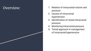
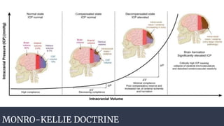
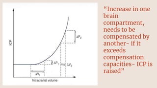
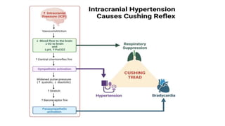
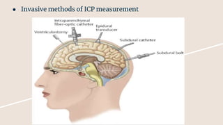
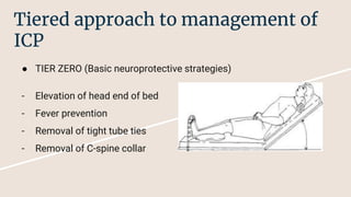
INTRACRANIAL PRESSURE
DR. DIKSHITA BHATTARAI
MEDICAL OFFICER
1. Relation of intracranial volume and
pressure
2. Causes of intracranial
hypertension
3. Identification of raised intracranial
pressure
4. Monitoring Intracranial pressure
5. Tiered approach in management
of intracranial hypertension
Overview:
Components of intracranial cavity
MONRO-KELLIE DOCTRINE
“Increase in one
brain
compartment,
needs to be
compensated by
another- if it
exceeds
compensation
capacities- ICP is
raised”
Causes of raised ICP
● Brain
- Tumors
- Abscesses
- Hematoma
- Cerebral edema
- Encephalitis
● CSF
- Hydrocephalus
- Choroid Plexus Tumors
- Obstruction
● Blood
- Vascular Malformations
- CVST
- Hypoxemia
- Hyperpyrexia
- Elevated CVP (i.e. Heart Failure)
Features of Raised ICP
Herniation Syndromes
CT findings of Raised ICP
ICP Monitoring
● Non-invasive-Ocular sonography:
1. Increased nerve sheath diameter (>5.0mm)
2. Discrete, anechoic fluid collection within optic nerve sheath
3. Elevation of optic disc (>6.0mm)
4. Visualization of ipsilateral MCA
● Invasive methods of ICP measurement
Tiered approach to management of
ICP
● TIER ZERO (Basic neuroprotective strategies)
- Elevation of head end of bed
- Fever prevention
- Removal of tight tube ties
- Removal of C-spine collar
● TIER ONE
- Increase Analgesia
- Increase Sedation
- Normo-Hypercarbic state (PCO2 35-38mmHg)
- Mannitol or hypertonic saline bolus (upto sodium of 155mmol/L)
- CSF drainage by EVD
● TIER TWO
- Hyperventilate upto 32-35mmHg
- Use neuromuscular paralysis
- Perform MAP challenge - if autoregulation is intact, use
fluids/vasopressors/inotropes to increase CPP to 60-70
● TIER THREE
- Thiopentone coma
- Decompressive Craniectomy
- Therapeutic hypothermia (35C - 36C)
Summary
● Increase in one cranial compartment needs to be compensated by another-the
failure of which causes raise in intracranial pressure
● ICP increases as a result in decompensation after increase in any one of the brain,
CSF of Blood compartments in the cranial vault
● Raised ICP manifests as headache, visual disturbances, nausea/vomiting,
convulsions, altered sensorium
● Non-invasive methods of ICP monitoring include Optic nerve sheath measurement
while invasive methods include Ventriculostomy, Epidural transducers, Subdural
bolts and catheters
● Tiered approach to management of Intracranial Hypertension:
1. Tier zero: Basic neuroprotective strategies
2. Tier 1: Osmotic agents, Hypercarbia, Analgesia and Sedation, EVD
3. Tier 2: Hyperventilation, Paralysing agents, MAP challenge
4. Tier 3: Thiopentone coma, Decompressive craniectomy
References
https://www.youtube.com/watch?v=K4QexcCqcCU&t=2535s- Principles of
Neurophysiology (Dr. Rebel), University of Kentucky Department of Anesthesiology
Pinto VL, Tadi P, Adeyinka A. Increased intracranial pressure. Nih.gov. Published July
31, 2023. https://www.ncbi.nlm.nih.gov/books/NBK482119/

INTRACRANIAL PRESSURE- pathogenesis and management

  • 1.
    INTRACRANIAL PRESSURE DR. DIKSHITABHATTARAI MEDICAL OFFICER
  • 2.
    1. Relation ofintracranial volume and pressure 2. Causes of intracranial hypertension 3. Identification of raised intracranial pressure 4. Monitoring Intracranial pressure 5. Tiered approach in management of intracranial hypertension Overview:
  • 3.
  • 4.
  • 5.
    “Increase in one brain compartment, needsto be compensated by another- if it exceeds compensation capacities- ICP is raised”
  • 6.
    Causes of raisedICP ● Brain - Tumors - Abscesses - Hematoma - Cerebral edema - Encephalitis
  • 7.
    ● CSF - Hydrocephalus -Choroid Plexus Tumors - Obstruction
  • 8.
    ● Blood - VascularMalformations - CVST - Hypoxemia - Hyperpyrexia - Elevated CVP (i.e. Heart Failure)
  • 9.
  • 11.
  • 12.
    CT findings ofRaised ICP
  • 13.
    ICP Monitoring ● Non-invasive-Ocularsonography: 1. Increased nerve sheath diameter (>5.0mm) 2. Discrete, anechoic fluid collection within optic nerve sheath 3. Elevation of optic disc (>6.0mm) 4. Visualization of ipsilateral MCA
  • 14.
    ● Invasive methodsof ICP measurement
  • 15.
    Tiered approach tomanagement of ICP ● TIER ZERO (Basic neuroprotective strategies) - Elevation of head end of bed - Fever prevention - Removal of tight tube ties - Removal of C-spine collar
  • 16.
    ● TIER ONE -Increase Analgesia - Increase Sedation - Normo-Hypercarbic state (PCO2 35-38mmHg) - Mannitol or hypertonic saline bolus (upto sodium of 155mmol/L) - CSF drainage by EVD
  • 17.
    ● TIER TWO -Hyperventilate upto 32-35mmHg - Use neuromuscular paralysis - Perform MAP challenge - if autoregulation is intact, use fluids/vasopressors/inotropes to increase CPP to 60-70
  • 18.
    ● TIER THREE -Thiopentone coma - Decompressive Craniectomy - Therapeutic hypothermia (35C - 36C)
  • 19.
    Summary ● Increase inone cranial compartment needs to be compensated by another-the failure of which causes raise in intracranial pressure ● ICP increases as a result in decompensation after increase in any one of the brain, CSF of Blood compartments in the cranial vault ● Raised ICP manifests as headache, visual disturbances, nausea/vomiting, convulsions, altered sensorium
  • 20.
    ● Non-invasive methodsof ICP monitoring include Optic nerve sheath measurement while invasive methods include Ventriculostomy, Epidural transducers, Subdural bolts and catheters ● Tiered approach to management of Intracranial Hypertension: 1. Tier zero: Basic neuroprotective strategies 2. Tier 1: Osmotic agents, Hypercarbia, Analgesia and Sedation, EVD 3. Tier 2: Hyperventilation, Paralysing agents, MAP challenge 4. Tier 3: Thiopentone coma, Decompressive craniectomy
  • 21.
    References https://www.youtube.com/watch?v=K4QexcCqcCU&t=2535s- Principles of Neurophysiology(Dr. Rebel), University of Kentucky Department of Anesthesiology Pinto VL, Tadi P, Adeyinka A. Increased intracranial pressure. Nih.gov. Published July 31, 2023. https://www.ncbi.nlm.nih.gov/books/NBK482119/

Editor's Notes

  • #4 The three constituents of the cranial vault exist in a dynamic equilibrium, maintaining a consistent intracranial pressure of approx 5-15mmHg. Any increase in volume by one constituent will be compensated with decrease in another constituent
  • #5 Pressure-volume curve for ICP. The pressure-volume curve has four 'zones': (1) baseline intracranial volume with good compensatory reserve and high compliance (blue); (2) gradual depletion of compensatory reserve as intracranial volume increases (yellow); (3) poor compensatory reserve and increased risk of cerebral ischemia and herniation (red); and (4) critically high ICP causing collapse of cerebral microvasculature and disturbed cerebrovascular reactivity (grey).
  • #13 Effacement of ventricles, basal cisterns and other CSF spaces Loss of grey-white differentiation Brain herniation
  • #14 Opening pressure during lumbar puncture and EVD placement