SlideShare a Scribd company logo
Calum C. Bain
Allan McI. Mowat
Macrophages in intestinal
homeostasis and inflammation
Authors’ address
Calum C. Bain1
, Allan McI. Mowat1
1
Centre for Immunobiology, Institute of Infection,
Immunity and Inflammation, University of Glasgow,
Glasgow, UK.
Correspondence to:
Allan Mowat
Institute for Infection, Immunity and Inflammation
University of Glasgow
Sir Graeme Davies Building, 120 University Place
Glasgow G12 8TA, UK
Tel.: +44 141 330 8414
Fax: +44 141 330 4297
e-mail: allan.mowat@glasgow.ac.uk
Acknowledgements
The work of the authors is supported by the Wellcome
Trust UK and Tenovus Scotland. The authors have no
Conflicts of Interest.
This is an open access article under the terms of the
Creative Commons Attribution License, which permits use,
distribution and reproduction in any medium, provided
the original work is properly cited.
This article is part of a series of reviews
covering Mucosal Immunity appearing in
Volume 260 of Immunological Reviews.
Summary: The intestine contains the largest pool of macrophages in
the body which are essential for maintaining mucosal homeostasis in
the face of the microbiota and the constant need for epithelial renewal
but are also important components of protective immunity and are
involved in the pathology of inflammatory bowel disease (IBD). How-
ever, defining the biological roles of intestinal macrophages has been
impeded by problems in defining the phenotype and origins of differ-
ent populations of myeloid cells in the mucosa. Here, we discuss how
multiple parameters can be used in combination to discriminate
between functionally distinct myeloid cells and discuss the roles of
macrophages during homeostasis and how these may change when
inflammation ensues. We also discuss the evidence that intestinal mac-
rophages do not fit the current paradigm that tissue-resident macro-
phages are derived from embryonic precursors that self-renew in situ,
but require constant replenishment by blood monocytes. We describe
our recent work demonstrating that classical monocytes constantly
enter the intestinal mucosa and how the environment dictates their
subsequent fate. We believe that understanding the factors that drive
intestinal macrophage development in the steady state and how these
may change in response to pathogens or inflammation could provide
important insights into the treatment of IBD.
Keywords: intestine, macrophages, monocytes, homeostasis, inflammation
Introduction
As discussed elsewhere in this volume, the intestine is the
largest mucosal surface of the body and is the single biggest
compartment of the immune system. It is exposed con-
stantly to an array of foreign antigens and must discriminate
between harmful and harmless antigens to ensure that the
appropriate response is mounted to each. Robust protective
immune responses must be generated in response to patho-
genic insult, but similar responses mounted against innocu-
ous dietary proteins or commensal bacteria can lead to the
development of chronic inflammatory disorders such as
celiac disease and Crohn’s disease, respectively.
Mononuclear phagocytes (MPs) comprising both macro-
phages (m/) and dendritic cells (DCs) are central to this
discrimination process and therefore hold promise as targets
for potential novel therapies to treat IBD. In this article, we
Immunological Reviews 2014
Vol. 260: 102–117
Printed in Singapore. All rights reserved
© 2014 The Authors Immunological Reviews Published by John Wiley
& Sons Ltd.
Immunological Reviews
0105-2896
© 2014 The Authors Immunological Reviews Published by John Wiley & Sons Ltd.
102 Immunological Reviews 260/2014
discuss the role of m/ in gut homeostasis and describe how
they change when inflammation or infection is present. Fur-
thermore, we discuss the ontogeny of intestinal m/ and
highlight that unlike many other tissue m/, those resident
in the gut wall must be replenished continually by blood
monocytes, whose fate is ultimately dictated by the environ-
ment into which they arrive. These unusual m/ are ideally
adapted to their surroundings and raise many questions
about current views of the MP system and its functions.
The challenges of identifying intestinal macrophages
MPs are one of the most abundant populations of leukocytes
in the healthy intestinal mucosa, and this is the largest pop-
ulation of m/ in the body (1, 2). However, our under-
standing of the roles that individual subsets of DCs and
macrophages play in maintaining homeostasis, and how
they change when protective immune responses or inflam-
mation occur has been stifled by the inability to identify
these cells accurately. In large part, this is due to the sub-
stantial overlap in surface phenotype of intestinal DCs, m/,
and other myeloid cells in the mucosa. For instance,
although DCs in mice are traditionally identified by their
co-expression of CD11c and major histocompatibility com-
plex class II (MHCII), it is clear that m/ of the gut wall
express both these markers at high levels (3). Furthermore,
CD11c has been shown to be expressed by plasma cells (4)
and intestinal eosinophils in mice (5, 6), with the latter also
expressing the pan- m/ marker F4/80 (7, 8). Expression of
the fractalkine receptor CX3CR1 has also been used exten-
sively to distinguish murine DCs from m/, with the pro-
posal that these cells exhibited mutually exclusive expression
of CD103, the aE integrin, and CX3CR1, respectively (9–
11). However, it is now clear that both m/ and some DCs
can express CX3CR1, albeit at different levels, while not all
bona fide mucosal DCs are CD103+
(12–14). Thus, many of
the surface markers regarded as ‘lineage-specific’ are now
known to be expressed by multiple cell types, which in the
past has led to the mischaracterization of mucosal MP. Some
studies still use CD11c and MHCII co-expression alone to
identify DC in non-lymphoid tissues, which makes the
interpretation of these studies complex (15). Therefore, it is
clear that unambiguous identification of intestinal m/ and
DC requires thorough multi-parameter analysis, with several
surface markers needing to be examined in combination.
A useful approach to this comes from recent work that
has demonstrated that expression of CD64 (the high affinity
FcRc1), F4/80 and MerTK (12, 16, 17), can be used in
combination with CD11c and MHCII to define functionally
distinct MP populations in mice. In the intestine, we have
shown that CD11c+
MHCII+
MPs expressing CD64 and/or
F4/80 are highly phagocytic cells with abundant foamy
cytoplasm (12). These cells do not migrate to the mesen-
teric lymph nodes (MLNs) (13, 14), cannot prime naive T
cells (14), and their development in vivo requires the CSF-1R
(18, 19), but not the DC-specific growth factor flt3 ligand
(flt3L) (12, 13, 16). Together, these features confirm the
classification of CD64+
MPs as m/.
When identified appropriately, murine intestinal m/ can
be shown to express a number of characteristic surface mol-
ecules in addition to F4/80 and CD64, including the hemo-
globin scavenger receptor CD163 and the mannose receptor
(CD206) (12). Mature intestinal m/ also express high levels
of CX3CR1, although as discussed below, this appears to be
acquired during local differentiation in situ and some cells of
the m/ lineage express lower levels of CX3CR1 (12). It is
important to note that CX3CR1 expression is not a charac-
teristic shared by all tissue m/. For instance, lung alveolar
m/, splenic red pulp m/, liver Kupffer cells, epidermal
Langerhans cells, and m/ of the peritoneal cavity all lack
CX3CR1 expression (20), although fate mapping studies
suggest they derive from a CX3CR1-expressing progenitor
(20). Retention and/or upregulation of CX3CR1 expression
only occurs in some m/. As well as those in the intestine,
this includes microglia of the CNS and m/ resident in the
kidney (20). It seems likely that this is driven by signals
from the environment (see below).
In contrast, CD64À
F4/80À
CD11c+
MHCII+
MPs display
poor phagocytic activity, expand markedly in response to
exogenous flt3L administration, and express the DC-specific
transcription factor Zbtb46 (12, 13, 21). As these
CD64À
F4/80À
MPs also migrate constitutively to the MLNs
in a CCR7-dependent manner and excel at priming naive T
cells (13, 14), they appear to represent classical migratory
DCs. As discussed in more detail by Agace and colleagues
elsewhere in this volume, it is important to mention that
the intestinal DC compartment is itself heterogeneous, with
discrete subsets identifiable on the basis of CD103 and
CD11b expression (13, 21, 22). Although it was once
thought that all mucosal DC expressed CD103, it is now
clear that bona fide CD103À
DCs exist (13, 21) and that
CD103 cannot be used as a de facto marker of mucosal DCs.
Importantly, CD103À
CD11b+
DCs also express intermediate
levels of CX3CR1, making them almost identical to m/ in
terms of their surface phenotype. This emphasizes the need
to use multiple markers to distinguish intestinal DCs from
m/, using markers such as CD64, as well as CD26 and
© 2014 The Authors Immunological Reviews Published by John Wiley & Sons Ltd.
Immunological Reviews 260/2014 103
Bain & Mowat Á Roles of intestinal macrophages
CD272, recently identified by the Immunological Genome
Project as DC-specific markers (23).
Characterization of m/ in the human gut can also prove
difficult, with investigators using different panels of pan-
myeloid cell markers, including CD33, CD14, and CD13
(24–26). Interestingly, more powerful multi-parameter
analysis has shown recently that there is conservation of sur-
face marker expression between mouse and human which
may prove useful. For instance, expression of CD64 also
appears to allow accurate identification of m/ in human gut
and, like those found in the mouse gut, they express high
levels of MHCII, CD163, and CD68 (12, 26, 27). Unlike
murine intestinal m/, those in the human mucosa essen-
tially lack CD11c expression, except for a small population
that also expresses high levels of the LPS co-receptor CD14,
normally found at low levels on mature macrophages (12).
As discussed below, these CD11c+
CD14hi
cells may repre-
sent recently arrived monocytes from blood. Importantly,
CX3CR1 has also been shown to be expressed by intestinal
macrophages in humans (28), although it is unclear
whether levels of CX3CR1 expression identify different sub-
sets of MP in man.
It has become commonplace to classify m/ into one of
two major subtypes based on their surface receptor expres-
sion and which mediators they produce – M1 and M2 (29).
However, gut-resident m/ do not fit readily into this ‘M1-
M2 paradigm’, having some of hallmarks of both M1 and
M2 m/. For instance, they express high levels of MHCII
and produce TNFa constitutively (12, 30), features normally
associated with M1 or ‘classically activated’ m/ (31). How-
ever as discussed above, they also express CD206, CD163,
and produce interleukin-10 (IL-10), features associated with
M2 or M2-like m/ (32). However, they fail to express argi-
nase which is a cardinal feature of M2 m/ (12). Thus, like
most tissue m/ in vivo, those resident in the gut wall adapt
to their local environment in complex and specific ways that
may not be reflected by the rigid classification of the M1-
M2 paradigm.
Anatomical distribution of intestinal macrophages
M/ are found in the mucosa throughout the entire GI tract
and are located mostly in the lamina propria (LP) in close
proximity to the epithelial monolayer (2). A discrete popu-
lation is also present in the smooth muscle layers of the gut
wall, playing important roles in regulating intestinal motility
(33, 34). The number of m/ in the LP varies between the
different parts of the GI tract, with more being found in the
colon than the small intestine of humans and rodents (35,
36, authors’ unpublished observations). Although it has
been reported that there is a continuous gradient in m/
numbers from proximal to distal ends of the entire mouse
intestine (36), this may not be the case in humans, where
the colon appears to have similar numbers of macrophages
throughout its length (35).
Functions of macrophages in the steady state mucosa
Like most tissue m/, those resident in the normal gut wall
play essential housekeeping functions, such as the clearance
of apoptotic or senescent cells, and tissue remodeling (35,
37, 38) (Fig. 1). This would be consistent with their expres-
sion of scavenger receptors such as CD36 that can bind and
engulf apoptotic cells (39). Furthermore, intestinal m/ are
known to produce a variety of cytokines and other soluble
factors that help maintain tissue homeostasis. One such fac-
tor is PGE2 (40), which allows local m/ to stimulate the
proliferation of epithelial progenitors in intestinal crypts
(41), thereby regulating the integrity of the epithelial bar-
rier. Reflecting the importance of these homeostatic func-
tions, selective depletion of m/ by targeting the CSF1R
leads to enhanced susceptibility to dextran sodium sulfate
(DSS)-induced colitis (42) and epithelial repair in this
model requires MyD88 signaling in myeloid cells (43).
However, it is in their role as innate immune effector
cells that m/ are best known. Resident intestinal m/ are
highly phagocytic and are actively bactericidal (44), which
together with their positioning immediately under the epi-
thelial monolayer, means they are ideally located to capture
and destroy any material breaching the epithelial barrier
(Fig. 1). They also express high levels of the TREM2 phago-
cytic receptor (30), which enhances their ability to engulf
bacteria (45). However, one of the most characteristic fea-
tures that distinguishes intestinal m/ from their counter-
parts elsewhere in the body is that exposure to bacteria or
their products does not trigger pro-inflammatory responses
by mucosal m/ (12, 44). Similarly, ingestion of bacteria
does not stimulate respiratory burst activity (46) or the gen-
eration of nitric oxide (47) by intestinal m/. There is also
no enhanced production of TNFa, IL-1, IL-6, or other
inflammatory cytokines and chemokines in response to liga-
tion of TLR or intracellular NOD receptors (44, 48). How-
ever, recent studies in mice indicate that intestinal m/ are
not completely anergic in terms of cytokine production, as
they show constitutive production of substantial amounts of
IL-10, as well as low levels of TNFa (12). Although it may
© 2014 The Authors Immunological Reviews Published by John Wiley & Sons Ltd.
104 Immunological Reviews 260/2014
Bain & Mowat Á Roles of intestinal macrophages
seem surprising that m/ constitutively produce this proto-
typical pro-inflammatory cytokine, TNFa may have much
wider effects. For instance, TNFa can regulate enterocyte
growth and alter the permeability of the epithelial barrier
(49). Furthermore, TNFa stimulates production of matrix
metalloproteinases and other tissue remodeling enzymes in
the intestinal mesenchymal cells that are central to regulat-
ing epithelial cell function (50).
Given their high expression of MHCII and their ability to
take up orally administered protein antigens (51–53) and
bacteria (37), intestinal m/ may also serve as
antigen-presenting cells, interacting with and influencing
the differentiation of CD4+
T cells (Fig. 1). It has also been
suggested that m/ may play a selective role in initiating
the differentiation of FoxP3+
regulatory T cells in the intes-
tine (54). However, as m/ do not migrate to the MLN
(14) and the intestinal mucosa is essentially devoid of naive
CD4+
T cells (55), they are unlikely to be involved in the
initial priming of naive CD4+
T cells. Furthermore, LP m/
are extremely poor at activating naive CD4+
T cells in vitro
(14). Thus if mucosal m/ do present antigens to CD4+
T
cells, it seems more likely that this involves lymphocytes
Commensal
bacteria
Ly6Chi blood monocyte
(Ly6ChiCX3CR1int
CD115+CCR2+)
IL10
Elimination of
invading bacteria
MyD88/CX3CR1-
dependent sampling of
luminal contents
IL10-dependent secondary
expansion of Treg
CCR2-dependent
replenishment by Ly6Chi
blood monocytes
Resident M
CX3CR1hi
F4/80hi CD11b+
MHCIIhi CD64+
Mono/M
Intermediate
CX3CR1int
Ly6C– MHCII+
CD64+
Maintenance
of epithelial
barrier integrity
Regulatory
T cells
Engulfment of
epithelial cell/
apoptotic material
Newly extravasated
monocyte
CX3CR1int Ly6C+
MHCII– CD64lo
Maturing monocyte
CX3CR1int Ly6C+
MHCII+CD64+
PGE2
IL10
Epithelial barrier
damage
Pathogenic
Bacteria
Aberrant responses
against commensal
bacteria
PGE2
Regulation of TNF & ROS
production by neutrophils
Neutrophil
TNF , IL12,
IL23, IL6, IL1
Maintenance of
effector T cells
Th1/Th17 T cell
TGF
CCL2
CCL7
?
Effector monocytes
CX3CR1int
Ly6C+ MHCII+ CD64+
CCL2, CCL3,
CCL11
Recruitment of innate
immune effector cells
Pro-inflammatory
M
CX3CR1int
Ly6C– MHCII+
CD64+
IL10
Retention of anti-
inflammatory signature
by CX3CRhiresident M
Eosinophils
TNF
Homeostasis Inflammation
CSF1
Fig. 1. Under steady state conditions, Ly6Chi
monocytes constitutively enter the intestinal mucosa and differentiate into mature CX3CR1hi
F4/80+
macrophages (m/) through a series of short-lived CX3CR1int
intermediaries. These CX3CR1hi
m/ are positioned immediately
beneath the epithelial barrier and contribute to its integrity by secreting PGE2 which stimulates proliferation of epithelial progenitors. This
positioning coupled with their high phagocytic capacity means they are poised to capture and destroy any invading commensals or pathogens, as
well as clearing apoptotic or senescent cells. They may also be able to directly sample the luminal contents by the extending processes between
the cells of the intestinal epithelial barrier. CX3CR1hi
m/ also produce interleukin-10 (IL-10) constitutively, which facilitates secondary expansion
of regulatory T cells in the mucosa and may also condition newly arrived monocytes. Regulatory T cells may also contribute to the conditioning
of newly extravasated monocytes through their production of TGFb. When homeostasis is perturbed by inflammation or infection, Ly6Chi
monocytes and their CX3CR1int
derivatives accumulate in large numbers and display enhanced pro-inflammatory characteristics. They produce
pro-inflammatory cytokines which may support the maintenance of other effector cells such as IFNc producing and IFNc+
IL-17 double
producing T cells. They also orchestrate the recruitment of other innate effector cells such as eosinophils through secretion of inflammatory
chemokines. Importantly, during inflammation, CX3CR1hi
m/ retain their anti-inflammatory signature, e.g. IL-10 production. Elicited classical
monocytes may also play a regulatory role by controlling the production of TNFa and ROS by neutrophils.
© 2014 The Authors Immunological Reviews Published by John Wiley & Sons Ltd.
Immunological Reviews 260/2014 105
Bain & Mowat Á Roles of intestinal macrophages
that have returned to the mucosa after being first primed in
the MLN. Consistent with this idea, Hadis et al. (56) have
recently shown that m/ facilitate the secondary expansion
and maintenance of antigen-specific FoxP3+
Treg in the
intestinal mucosa through their production of IL-10
(Fig. 1). The importance of this local fine tuning of T-cell
responses by m/ is highlighted by the loss of FoxP3
expression and suppressive capacity that occurs when T cells
are rendered unresponsive to IL-10 through deletion of the
IL-10R (57). Further evidence that intestinal m/ may con-
tribute to the generation of homeostatic Treg comes from
work showing that mice lacking TRAF6 expression in
CD11c+
MP develop a spontaneous Th2 cell-mediated enter-
itis. This is associated with a defective ability of mucosal
MP to induce FoxP3+
T cells in vitro and reduced numbers
of Treg in the LP (58). Although interpreted as reflecting a
role for ‘DCs’, the relevant myeloid cells were defined only
as CD11c+
and as we have noted, most of these cells in the
normal LP are in fact m/. Further work is needed to define
the precise cellular basis of TRAF6-mediated generation of
Treg in the LP.
Intestinal m/ may also contribute to the maintenance of
other T cells in the mucosa. For instance, the production of
IL1b in response to TLR stimulation may be relatively pre-
served in intestinal m/ and this has been reported to help
support the development of Th17 cells in the steady state
intestine (59). It remains to be determined whether these
effects of intestinal m/ on T cells require cognate interac-
tions via peptide-MHC complexes and the TCR, or whether
it involves non-specific factors such as polarizing cytokines
alone, as has been suggested for the generation of Th17
responses by mucosal CD103+
CD11b+
DCs (60).
Recent studies have proposed a further way in which
mucosal m/ may contribute to indirectly to T-cell priming,
through Connexin 43-mediated transfer of soluble antigen
obtained from the lumen to neighboring CD103+
DCs (52).
This cooperative process has been suggested to be important
for the development of oral tolerance and if confirmed,
these findings could indicate an important new role for m/
in the steady state intestine.
Exactly how m/ in the LP acquire luminal antigen
remains to be established with certainty. One possibility is
that they may extend transepithelial dendrites (TED) across
the epithelium to capture soluble antigen in the lumen
(Fig. 1). Originally thought to be derived from DCs on the
basis of the expression of MHCII and CD11c (61, 62), it
now seems clear that these TED originate from CX3CR1+
m/. Their formation requires CX3CR1 (61, 63) and indeed,
the induction of oral tolerance to protein antigens is
impaired in CX3CR1-deficient mice (52, 56), consistent
with a requirement for CX3CR1-mediated uptake of luminal
antigen in this process. However, the physiological signifi-
cance of such TED remains unclear. Not only did the origi-
nal reports disagree on which part of the small intestine
they were present in and if they were dependent on TLR
signaling (61, 62), but more recent studies have failed to
find TED anywhere in the steady state intestine (64). Fur-
thermore, earlier studies on the uptake of soluble protein
from the intestinal lumen reported that this process was not
dependent on the CX3CL1–CX3CR1 axis (14). Finally, as we
have noted, other work has also proposed that the defect in
oral tolerance in CX3CR1-deficient mice reflects an absence
of IL-10 production by intestinal m/ (56). If and how LP
m/ contribute to the uptake of soluble antigen and then
help shape subsequent immune responses clearly warrants
further investigation.
Despite their phagocytic activity and morphological
appearance, there is similar uncertainty about whether LP
m/ play a role in the capture of and induction of immunity
or tolerance to local bacteria. Although it is well established
that CD103+
DCs can present bacterial antigen to naive T
cells in the MLN (53), initial reports indicated that the TED
formed by MP in the mucosa could capture Salmonella organ-
isms in the lumen (61, 62). Furthermore, a very recent
study showing that CX3CR1+
cells may transport Salmonella in
lymph to the MLN was interpreted as showing a role for
migratory m/ in this process (65). However, this phenom-
enon was only revealed after depletion of the normal micro-
biota with antibiotics and the study did not exclude the
possibility that the antigen loaded CX3CR1+
MP belonged to
the population of bona fide CX3CR1 expressing DC that we
and others have observed in intestinal lymph (13, 14).
Again, these findings reveal how much is still to be under-
stood about how m/ contribute to the induction and shap-
ing of mucosal immune responses.
Ontogeny of intestinal macrophages in the steady
state
The mononuclear phagocyte system
Tissue m/ are traditionally viewed as part of the mononu-
clear phagocyte system (MPS). First described in 1972
(66), the concept of the MPS proposes that all tissue m/
are the terminally differentiated progeny of blood mono-
cytes, which constitutively enter tissues under steady state
conditions, and are themselves replaced by rapidly dividing
© 2014 The Authors Immunological Reviews Published by John Wiley & Sons Ltd.
106 Immunological Reviews 260/2014
Bain & Mowat Á Roles of intestinal macrophages
precursors in the bone marrow (BM). Originating from
the common myeloid progenitor, which also generates DCs
and granulocytes, a common macrophage and dendritic cell
progenitor gives rise to both committed DC precursors
(pre-DCs) and to monocytes (67, 68), with the latter pass-
ing through a recently identified common monocyte pro-
genitor (69). The development of monocytes via these
processes is dependent on the transcription factor PU.1 and
the growth factors CSF1 (M-CSF), CSF2 (GM-CSF), and
IL-3 (69).
It is now known that the monocyte pool is heterogeneous
and, in mice, CSF1R+
(CD115) monocyte subsets can be
distinguished by their expression of Ly6C/Gr1 and the
chemokine receptors CCR2 and CX3CR1 (70). The majority
of blood monocytes expresses high levels of Ly6C/Gr-1,
CCR2 and CD62L, but low levels of CX3CR1 (so-called
Ly6Chi
monocytes). The entry of Ly6Chi
monocytes into the
circulation from the BM is dependent on the CCL2-CCR2
chemokine axis and as a result, mice with genetic deletion
of either of these molecules have markedly fewer circulating
monocytes (71). Although initially referred to as ‘inflamma-
tory’ monocytes because of an apparent preference to
migrate to inflamed tissues, these Ly6Chi
monocytes are
now classified as ‘classical’ monocytes, as they fulfill all the
roles previously attributed to generic monocytes in the MPS.
In human, an analogous population of classical monocytes is
identified as CD14hi
CD16À
and these also express CCR2
(72).
A smaller population of murine monocytes expressing
lower levels of Ly6C and CCR2, but higher amounts of
CX3CR1 (Ly6Clo
monocytes) were initially proposed to be
the precursors of steady state tissue m/ (70). However, lit-
tle direct evidence exists to support this idea, and more
recently it has become clear that the primary function of
Ly6Clo
monocytes is to act as phagocytes in the blood-
stream, patrolling and maintaining the vasculature (73, 74).
As such, they are now commonly referred to as ‘patrolling’
or non-classical monocytes (75), and there is accumulating
evidence that there is a developmental relationship between
classical and non-classical blood monocytes, with the latter
being the CSF1R-dependent progeny of the former (20,
76). The equivalents of these non-classical Ly6Clo
monocytes
in human are identified as CD14lo
CD16+
, and these express
relatively high levels of MHCII (70). Whether they exert
similar patrolling functions to their murine counterparts is
not known, although gene expression analysis has revealed
many parallels between the monocyte subsets in mouse and
human (72).
Macrophage development independent of the MPS
The validity of the MPS has been questioned recently, with
multiple reports from independent groups proposing that
the majority of tissue m/ exists independently of blood
monocytes (77–79). Using a variety of techniques including
fate mapping, parabiosis, and analysis of proliferative capac-
ity, it has been shown that epidermal Langerhans cells (79,
80), lung alveolar m/ (20, 81), liver Kupffer cells (20,
77), microglia of the CNS (20, 77, 82, 83), and m/ of the
peritoneal cavity (78) derive from embryonic precursors
that seed tissues early in development and maintain them-
selves through in situ proliferation. Although there is debate
about whether these precursors originate in the mesen-
chyme of the yolk sac, or from the fetal liver, an assumption
has come about that all tissue m/ populations may be
derived from these early progenitors (84).
Our recent work shows that the intestine is an exception
to this paradigm, as m/ in the adult steady state mucosa
require continuous replenishment by Ly6Chi
blood mono-
cytes (12). Although earlier studies had shown that Ly6Chi
monocytes could enter the colonic mucosa following intense
depletion of resident MP (9), it was unclear whether such a
mechanism operated in the unmanipulated mucosa and if
the progeny represented m/ or DCs (10, 85). Using an
adoptive transfer approach, we could demonstrate that
Ly6Chi
monocytes constantly enter the normal colonic
mucosa and mature into CD64+
F4/80hi
CX3CR1hi
MHCII+
cells (12, 16), indistinguishable from endogenous resident
m/ (Fig. 1). The development of mature m/ involves a
local differentiation process, in which a series of intermedi-
aries lose Ly6C and upregulate F4/80, CX3CR1, CD163, and
CD206, as well as acquiring CD11c and MHCII expression
(12), features that account for their previous misclassifica-
tion as DCs (9, 10). Full maturation appears to take around
5 days and as well as adopting the phenotypic signature of
mature colonic m/, the differentiating monocytes begin to
acquire the characteristic functional signature of intestinal
m/. Thus they become increasingly phagocytic and begin
to produce IL-10, as well as developing resistance to TLR
stimulation (12).
There are multiple lines of evidence to support the idea
that monocyte recruitment is the principal mechanism of
replenishment of intestinal m/. Firstly, m/ in the adult
mucosa have very poor proliferative capacity (12, 86), sug-
gesting in situ self-renewal plays little or no role under nor-
mal physiological conditions. Secondly, mice in which the
CCL2–CCR2 axis has been disrupted by genetic deletion of
© 2014 The Authors Immunological Reviews Published by John Wiley & Sons Ltd.
Immunological Reviews 260/2014 107
Bain & Mowat Á Roles of intestinal macrophages
either molecule have significantly fewer colonic m/ (12,
87). Next, the intestinal m/ pool derives almost exclusively
from WT BM in WT:CCR2À/À
mixed BM chimeric mice
(16). Finally, cells matching the phenotype of intestinal m/
are completely eliminated by DT-mediated depletion in
CCR2-DTR mice (88). This requirement for the CCL2-CCR2
axis does not simply reflect its role in BM egress of mono-
cytes, as CCR2-sufficient Ly6Chi
monocytes fail to enter the
CCL2-deficient gut mucosa in WT-CCL2À/À
parabiotic mice
(87), demonstrating that the CCL2-CCR2 axis is needed for
entry of monocytes into the intestinal LP. Although the
source of CCL2 in the intestine is unclear, CX3CR1hi
resi-
dent m/ in the normal mucosa express high levels of the
CCR2 ligands CCL2 and CCL7 (87, 89) and therefore they
may control their own replenishment. This mechanism has
been shown to occur in the mouse uterus, where CSF1R-
signaling results in production of CCR2 ligands by uterine
m/ (90). Intestinal m/ are all labeled in MacGreen mice in
which GFP is expressed under control of the CSF1R pro-
moter (91, 92) and thus it is intriguing to speculate that
rather than being required for their local proliferation/sur-
vival, CSF1R-signalling in gut m/ may be required to elicit
monocyte recruitment to the mucosa, as has been suggested
in other tissues (90, 93).
As CCR2- and CCL2-deficient mice are not completely
devoid of intestinal m/ (12, 87), there may be other
mechanisms of replenishment in addition to the CCL2-
CCR2 axis. This is unlikely to involve non-classical Ly6Clo
monocytes, as these fail to migrate into non-lymphoid tis-
sues when adoptively transferred (9, 12), and as discussed
above, they seem to have a distinct effector role patrolling
the vasculature (73). In addition, although CX3CR1-defi-
cient mice have markedly fewer circulating Ly6Clo
mono-
cytes due to impaired survival (63, 94), the intestinal m/
compartment of these mice remains unaffected in size (56).
In vitro studies have also implicated TGFb and IL-8 in the
recruitment of monocytes to the intestinal mucosa (27,
86), but the role of these and other mediators has not been
addressed in vivo.
Why the gut mucosa should need continuous recruitment
and differentiation of Ly6Chi
monocytes is not completely
understood. An obvious possibility is that this is required to
monitor the constant exposure to the commensal microbiota
and other environmental agents found at barrier surfaces.
Although the abundance of differentiating monocytes is
reduced in the dermis of germ free (GF) mice (16), differ-
ent studies mice have reached conflicting conclusions on the
size of colonic m/ compartment in GF mice (11, 95, 96).
Furthermore, whether GF conditions or antibiotic treatment
alter monocyte recruitment to the gut mucosa and/or their
subsequent differentiation has never been formally exam-
ined.
An analogous monocyte differentiation process seems to
exist in human intestine. We recently showed that CD14hi
monocytes appear to differentiate into mature m/ through
a series of intermediaries which involves downregulation of
CD14 and CD11c, and upregulation of MHCII, CD209 and
CD163 (12). Together these findings indicate that classical
monocytes are the principal source of the resident intestinal
m/ pool in mouse and human.
Monocyte education in the mucosa
The environmental cues that shape monocyte differentiation
in the intestinal mucosa remain to be identified with cer-
tainty, and it is likely that multiple factors are responsible
for the imprinting of the phenotypic and functional charac-
teristics adopted by maturing monocytes (Fig. 1). These fac-
tors are likely to be specific to the intestinal mucosa, as
monocytes do not acquire the same characteristics after
entering other mucosal sites such as the lung (97). Multiple
growth factors are responsible for the maturation of cells of
the MPS, including flt3L, CSF1, and CSF2. As noted above,
intestinal m/ do not expand in response to exogenous flt3L
and remain equally abundant in flt3L-deficient mice (16,
authors’ unpublished data). Similarly, although some m/
such as lung alveolar m/ have been shown to depend on
the CSF2R for their development (78, 98), it is unlikely that
this applies to intestinal m/, as cells matching their pheno-
type are derived equally from CSF2R-deficient BM in WT:
CSF2R-deficient BM chimeras (99). The development of
intestinal m/ requires signaling through the CSF1R and as a
result, they are derived almost exclusively from WT BM in
WT:CSF1RÀ/À
mixed BM chimeric mice (19). There are
currently two identified ligands of the CSF1R: CSF1 and
IL-34. To date, only microglia and Langerhans cells of the
epidermis have been reported to depend on IL-34 and in
these cases, this appears to be the driver of in situ self-
renewal (100, 101), making it seem unlikely that IL-34 will
play a role in intestinal m/ homeostasis. However this has
never been examined directly. It is likely that CSF1 is the
predominant ligand required in the gut as evidenced by a
marked reduction in intestinal m/ in osteopetrotic Csf1op/op
mice, which have a mutation in the gene encoding CSF1
(102). It also remains to be determined at which stage of
intestinal m/ development CSF1R signalling is involved and
specifically whether it is important simply for the generation
© 2014 The Authors Immunological Reviews Published by John Wiley & Sons Ltd.
108 Immunological Reviews 260/2014
Bain & Mowat Á Roles of intestinal macrophages
of progenitors, or also for the differentiation of monocytes
that enter the mucosa. The loss of Ly6C and upregulation of
F4/80 that occurs during the transition of Ly6Chi
to Ly6Clo
monocytes in blood is dependent on CSF1-CSF1R interac-
tions (18, 20), as is the development of monocyte-derived
m/ in other tissues (18, 19). However, it seems unlikely
that CSF1R signalling alone can account for all aspects of
intestinal m/ development given the fact that adoptively
transferred monocytes generate entirely distinctive m/ in
this tissue. Thus, the local environment of the intestinal
mucosa must play the definitive role in local m/ develop-
ment.
A distinctive feature of intestinal m/ is their expression of
MHCII at levels even greater than those seen on mucosal DCs.
Indeed, acquisition of MHCII is one of the first phenotypic
changes to occur during monocyte maturation in the intes-
tine, although its functional significance remains unclear
(12). This does not appear to be a default property of mono-
cyte maturation, as it is not a universal feature of other mono-
cyte-derived tissue m/ (90, 103) or of the blood Ly6Clo
monocytes, which mature from Ly6Chi
monocytes (97).
IFNcR signaling is known to induce MHCII on m/ (104), but
MHCII expression by intestinal m/ is normal in IFNcR-defi-
cient mice (our unpublished observations). It also appears to
be independent of CSF2, IL-10, and lymphocytes or their
products (our unpublished observations). Recently, the Ran-
dolph laboratory has suggested that signals from vascular
endothelial cells may instruct upregulation of MHCII by
extravasating monocytes (97). However, whether this occurs
as monocytes enter the gut mucosa remains unclear.
As noted above, acquisition of the CX3CR1hi
phenotype
appears to be associated with the development of an anti-
inflammatory signature in fully differentiated intestinal m/.
Given that the majority of m/ in other tissues lack CX3CR1, it
is clear that the local environment of the gut must instruct
developing monocytes to upregulate this molecule. The factor
responsible has not been identified, although TGFb is a clear
candidate, as it is known to induce CX3CR1 expression by mi-
croglia in the brain (105). There is accumulating evidence
that CX3CR1 itself may play a role in controlling the differen-
tiation and function of intestinal m/. Firstly, as discussed
above, the CX3CL1–CX3CR1 axis has been shown to control
the ability of CX3CR1+
MPs to extend cellular protrusions into
the intestinal lumen (61, 63, 106). Although this remains an
area of contention, it is conceivable that the expression of
CX3CL1 by enterocytes (63) could influence the anatomical
positioning of m/ under the epithelial layer. However to
date, it has not been reported that CX3CR1-deficient m/
inhabit different anatomical locales than their WT counter-
parts. Next, the CX3CL1–CX3CR1 axis has been shown to be
required for optimal production of IL-10 by intestinal m/
(56) and finally, CX3CR1-deficiency has been reported to lead
to decreased numbers of intestinal m/ (107, 108) as well as
altering susceptibility to chemically induced colitis. However,
given that CX3CR1-deficient mice have been shown both to
be protected (11, 63) and more susceptible to experimental
colitis (107), further investigation of the CX3CR1–CX3CL1
axis is required to elucidate its involvement in the control of
m/ function in the intestine.
The IL-10–IL-10R axis plays a crucial role in conditioning
the behavior of m/ in the mucosa. One such effect is the
upregulation of scavenger receptors such as CD206 and
CD163, which is reduced in IL-10-deficient mice (96, 109).
Even more marked is the requirement for IL-10 in the hyp-
oresponsiveness of intestinal m/ to pro-inflammatory stim-
uli (44) (see below). The cellular source of the IL-10
remains unclear. Although m/ themselves produce IL-10
constitutively and this could act in an autocrine manner,
selective knockout of IL-10 in LysM+
cells appears not to
lead to spontaneous IBD in mice (110), suggesting other
cells are more important. This is likely to be IL-10-produc-
ing FoxP3+
CD4+
T cells, as these are abundant in the nor-
mal intestine and IBD occurs after specific deletion of IL-10
in these cells (111, 112). The induction of IL-10 in matur-
ing m/ may be a response to colonization of the intestinal
tract by the commensal microbiota, as colonic m/ from
mice reared in GF conditions produce less IL-10 (95, 96).
However given that this failure to upregulate IL-10 produc-
tion cannot be recapitulated in MyD88À/À
mice (95), the
mechanisms underlying this phenomenon require further
investigation. Interestingly, the production of protective
IL-10 by CD4+
T cells is also dependent on the microbiota
(113). While it has been reported that IL-10 may be impor-
tant for more general aspects of maturation of newly
recruited monocytes in the peritoneum, including upregula-
tion of MHCII (114), our work on inflamed intestine sug-
gests that the lack of MHCII may reflect the failure of
monocytes to differentiate fully due to the enhanced inflam-
mation found in the absence of IL-10, rather than a direct
effect of IL-10 (see below).
Mechanisms of inflammatory anergy in intestinal
macrophages
Hyporesponsiveness to activation via TLRs and other stim-
uli is a cardinal feature of resident intestinal m/. Early
work suggested that this was the result of a failure to
© 2014 The Authors Immunological Reviews Published by John Wiley & Sons Ltd.
Immunological Reviews 260/2014 109
Bain & Mowat Á Roles of intestinal macrophages
express pattern recognition receptors. However, it is now
clear that gut-resident m/ in mice and human express a
full range of TLR (12, 27), and it is now believed that
regulation of adapter molecules downstream of the TLR
may instead be responsible for the hyporesponsiveness. For
instance, signaling molecules such as CD14, MyD88,
TRAF-6, MD2, TRIF, and IRAK1 appear to be
downregulated in mature intestinal m/ (27, 44, 89). In
parallel, mechanisms that inhibit TLR signaling and/or NF-
jB activation (e.g. IRAK-M and IkBNS) appear to overex-
pressed in gut-resident m/ (27, 89, 109), suggesting that
molecules that propagate TLR signaling are targeted rather
than the TLR expression itself.
At least some of these inhibitory processes may be driven
by IL-10 (109). In mice, the hyporesponsiveness develops
progressively as monocytes mature in the mucosa and it cor-
relates with increasing IL-10 production (12). As a result,
colonic m/ from IL-10À/À
mice or mice with LysM-medi-
ated deletion of the IL-10R signaling molecule STAT3 dis-
play exaggerated pro-inflammatory responses to bacterial
products (95, 96, 109, 115), and the animals develop spon-
taneous colitis (116, 117). Importantly, IL-10R deficiency is
also associated with an early onset form of severe IBD in
human (118, 119). Thus, it is clear that IL-10 is one of the
most important factors ensuring m/ quiescence in the face
of environmental stimuli. However, other mechanisms are
likely to be involved. For instance, TGFb has been shown to
render blood monocytes hyporesponsive to a variety TLR
ligands (27) and active TGFb is abundant in the steady state
LP (120). The spontaneous, microbiota-dependent enteritis
that occurs in mice lacking expression of TRAF6 in CD11c+
MP could also reflect defective TGFbR-dependent signaling
in m/, as TRAF6 and its downstream partner TAK1 are crit-
ical components in this pathway (58).
The control of m/ activity may also involve dedicated
inhibitory receptors including SIRPa and CD200R1. Indeed,
the CD200–CD200R1 axis appears to play an important role
in regulating alveolar m/ activity (121). However, our
recent work suggests that the same regulatory loop is dis-
pensable in the intestinal mucosa, with CD200R1-deficient
colonic m/ showing no signs of dysregulation (8). Further-
more, deletion of CD200R1 or its only ligand, CD200 does
not render mice more susceptible to DSS-induced colitis, as
would be expected if m/ activity was not controlled appro-
priately (8). Although resident m/ express high levels of
SIRPa, whether this plays any role in controlling intestinal
m/ activity through interaction with its ligand CD47 has
never been formally tested.
The development of the hyporesponsive state appears to
involve recognition of microbial signals, as colonic MP from
GF mice respond robustly to stimulation with the TLR4 ago-
nist LPS (96). However, it must be noted that in this study
m/ were identified only as CD11b+
CD11cÀ
cells, which
will include many cell types and exclude the majority of res-
ident m/ which express CD11c. Therefore, the full impact
of the microbiota on the behavior of resident colonic m/
remains to be elucidated.
Together, these findings demonstrate that fully responsive
Ly6Chi
monocytes continuously enter the steady state intesti-
nal mucosa as a surveillance measure. In the absence of any
threat, the monocytes adopt an anti-inflammatory phenotype
which is imprinted by local factors in the mucosa. The nat-
ure and mechanisms of action of these mediators remain to
be defined fully.
Monocytes and macrophages in intestinal inflammation
It is well known that the composition of the human intesti-
nal m/ pool changes considerably when there is perturba-
tion of homeostasis. For instance, in the mucosa of Crohn’s
disease and ulcerative colitis patients, there is an accumula-
tion of pro-inflammatory macrophages. These can be identi-
fied by their high expression of CD14, in contrast to the
CD14lo/À
population of resident m/ (12, 24, 122, 123).
The ability of these cells to produce large amounts of medi-
ators such as IL1, IL6, TNFa, reactive oxygen intermediaries
and nitric oxide makes them quite distinct from the m/
found in healthy intestine and has led to interest in the idea
of targeting the monocyte-macrophage lineage for therapeu-
tic purposes. For these reasons, it has become important to
establish whether the inflammatory cells represent newly
arrived monocyte-derived cells, or are resident cells that
have altered their behavior in the presence of inflammation.
Mouse models of intestinal inflammation
There is similar infiltration of pro-inflammatory monocytes
and m/ in animal models of intestinal inflammation such as
colitis induced by administration of DSS (12, 89), by the
adoptive transfer of naive CD4+
T cells into lymphopenic
hosts (16, 30), or after infection with Helicobacter hepaticus
coupled with neutralization of IL-10 (our unpublished
observations). In all these cases, the infiltrate is characterized
by a reversal in the ratio of CX3CR1hi
and CX3CR1int
cells,
caused by the accumulation of Ly6Chi
monocytes and their
CX3CR1int
progeny (Fig. 1). The infiltrating CX3CR1int
cells
display typical pro-inflammatory characteristics, including
© 2014 The Authors Immunological Reviews Published by John Wiley & Sons Ltd.
110 Immunological Reviews 260/2014
Bain & Mowat Á Roles of intestinal macrophages
the production of TNFa, IL-6, IL-1b, IL-12, IL-23, and
expression of iNOS. In contrast, the remaining CX3CR1hi
m/ retain their anti-inflammatory signature, continuing to
produce high levels of IL-10 and being TLR hyporesponsive,
suggesting that resident m/ do not turn rogue during
inflammation (12, 16, 30, 89). Not unexpectedly, CCR2 is
required for the entry of pro-inflammatory cells into the
inflamed colonic mucosa, with considerably fewer CCR2À/À
cells entering the mucosa when co-transferred with their
WT counterparts (89, 124). A pathological role for these
elicited Ly6Chi
monocytes is evidenced by amelioration of
colitis in monocytopenic CCR2-deficient mice (124) and in
mice depleted of CCR2-expressing cells (89). Furthermore,
TNFa production by these cells appears to be central to dis-
ease progression, as the severity of DSS-induced colitis is
reduced in mice in which Ly6Chi
monocytes are deficient in
TNFa production (9). On the basis of our studies of DSS
colitis, we have proposed that the normal monocyte differ-
entiation process is arrested during inflammation, resulting
in the retention of cells that remain responsive to TLR stim-
ulation and produce copious amounts of pro-inflammatory
cytokines and chemokines (12, 30, 89). Why these pro-
cesses are disrupted in inflammation remains to be deter-
mined, but presumably reflects alterations in the local
microenvironment, leading to defects in the conditioning
mechanisms that normally specify maturation of monocytes
into anti-inflammatory m/. Clearly however, the resident
m/ themselves cannot be influenced by these changes, indi-
cating these are likely to be terminally differentiated cells
and raising the possibility that homeostasis may be restored
relatively quickly if the inflammatory environment can be
modified.
Although Ly6Chi
monocytes and their immediate progeny
appear to be detrimental during sterile models of intestinal
inflammation (12, 124), they are also indispensable for the
eradication of certain enteric pathogens. This is consistent
with findings that elicited CX3CR1int
cells, many of which
are likely to be derivatives of Ly6Chi
monocytes, have been
shown to take up Salmonella organisms as efficiently as resi-
dent CX3CR1hi
m/ (37). During the acute ileitis caused by
oral inoculation of certain mouse strains with the protozoan
parasite Toxoplasma gondii, there is accumulation of Ly6Chi
monocytes, which display the same behavior as those arriv-
ing into the DSS-inflamed colon, including the production
of the pro-inflammatory cytokines TNFa and IL-12, and
reactive nitrogen species (125). The protective role of these
monocytes is demonstrated by the fact that both CCR2- and
CCL2-deficient mice succumb to lethal toxoplasmosis, and
that the adoptive transfer of CCR2-competent Ly6Chi
mono-
cytes can rescue this lethality (125). Interestingly, the
CCL3–CCR1 chemokine axis driven by innate lymphoid cells
is also involved in the recruitment of pro-inflammatory
Ly6Chi
monocytes and protective immunity in this model of
infection, indicating that CCR2 may not be the only mecha-
nism responsible for accumulation of monocytes during
inflammation (126). However, no defects in steady state
intestinal m/ have been reported in mice lacking CCR1,
perhaps suggesting distinct roles for individual chemokine
receptors under different conditions.
Ly6Chi
monocytes have also been implicated in the pro-
tective immune response to Citrobacter rodentium, a mouse
model of enteropathogenic and enterohaemorrhagic E. coli
infection in human, with delayed clearance in CCL2- and
CCR2-deficient mice (127). However, recent elegant work
has questioned this conclusion, by showing that ablation of
the entire monocyte-macrophage compartment in mice with
LysM-driven expression of DTR by CSF1R-expressing cells
has no effect on susceptibility to C. rodentium infection (128).
In contrast, the robust Th17 response needed to clear this
organism (127) is absolutely dependent on classical migra-
tory DC expressing zbtb46 (21). Interestingly however,
monocyte-derived macrophages may make an additional
contribution to the adaptive immune response under these
conditions, by producing IL12, which maintains IFNc+
and
IFNc+
IL17+
effector T cells in the mucosa (128). Thus,
whereas resident m/ maintain regulatory T cells via IL-10
production, recently elicited monocytes may maintain effec-
tor T cells through the production of pro-inflammatory
cytokines (Fig. 1).
Contrasting with their role as inflammatory effector cells,
recent studies suggest that Ly6Chi
monocytes may have a
wider ability to regulate other aspects of intestinal immunity
(Fig. 1). For instance, in the DSS colitis model, Ly6Chi
monocytes mediate the recruitment of eosinophils through
their production of CCL11 (eotaxin) (129). In T. gondii-
induced ileitis, elicited Ly6Chi
monocytes may actually pre-
vent immunopathology by inhibiting the ability of local
neutrophils to produce tissue damaging TNFa and ROI. This
PGE2-dependent process involves production of IL-10 by
monocytes (40) and may go some way to explain earlier
observations of enhanced neutrophil accumulation in the
mucosa of T. gondii infected CCR2-deficient animals (125).
These results support other reports that monocytes are not
intrinsically inflammatory in nature, but are highly plastic
cells that coordinate the first stages of tissue repair in
response to damage, infection, or inflammation (130).
© 2014 The Authors Immunological Reviews Published by John Wiley & Sons Ltd.
Immunological Reviews 260/2014 111
Bain & Mowat Á Roles of intestinal macrophages
Furthermore, the regulatory properties of elicited Ly6Chi
monocytes are diminished in GF mice, underlining how the
local microenvironment determines the fate of monocytes in
the mucosa (40).
M/ have also been implicated in Th2 cell-mediated resis-
tance to intestinal helminth infections (131). Their numbers
are expanded during infection by organisms such as Trichuris
muris, Nippostrongylus brasiliensis, Heligmosomoides polygyrus, Brugia
malayi, and Litomosiodes sigmodontis (132–134), and under these
conditions, ‘alternative’ activation of m/ by the Th2 prod-
ucts IL-4 and IL-13 leads to the production anti-parasitic
mediators such as arginase and RELM-a (133, 135). How-
ever, the exact role of m/ in these infections is uncertain.
The expulsion of these parasites is compromised in mice
with m/ depletion secondary to absence of CCL2 (132) or
administration of clodronate-containing liposomes (136,
137). Alternatively activated m/ are also partly responsible
for the increased smooth muscle contractility which contrib-
utes to parasite expulsion (136). However, the protective
effects of m/ do not require them to produce arginase-1
(138) or even to be polarized via the IL-4 receptor (139).
Thus, m/ may play an indirect role in protective immunity,
perhaps by producing mediators that recruit other effector
cells such as eosinophils and mast cells. Alternatively, they
may be involved in the anti-inflammatory and tissue repair
processes that are crucial components of the host response
to multicellular pathogens and that are important properties
of mediators such as arginase (140).
The origin of the alternatively activated macrophages in
helminth infection is also uncertain. Although the require-
ment for CCL2 in parasite expulsion could indicate they are
derived from recruited Ly6Chi
monocytes as in the models
of inflammation discussed above, this has never been inves-
tigated directly. In addition, recent studies of macrophage
expansion during helminth infection of the peritoneal and
pleural cavities suggest that this may be driven by IL4- and
CSF-1-dependent proliferation of the pre-existing resident
population (141). The relative roles of in situ expansion of
m/ and monocyte recruitment during intestinal helminth
infection could generate important insights into the plastic-
ity and functional properties of mucosal macrophages.
Monocytes and macrophages in human IBD
As discussed above, CD14hi
MP accumulate in the mucosa
of IBD patients (12, 24), and CD14hi
cells in human are the
equivalent of Ly6Chi
monocytes in the mouse. As in mice,
the CD14hi
cells that appear in human intestine display
enhanced production of TNFa, IL-1b, and IL-6 (123) and
respiratory burst activity (46). They also retain their respon-
siveness to microbial products (24, 122). Using the transfer
of radiolabelled autologous blood monocytes, it has been
demonstrated that the CD14hi
cells in the inflamed mucosa
derive from circulating monocytes (142), likely in response
to elevated levels of the monocyte chemoattractants CCL2
and CCL4 in the inflamed mucosa (39). In addition to their
own pro-inflammatory effects, elicited monocytes in human
have also been shown to orchestrate the recruitment of
other innate effector cells, such as CCR3+
eosinophils
through their production of CCL11 (eotaxin-1) (25, 129).
Furthermore, CD68+
m/ in the inflamed mucosa have
enhanced expression of CD40 (143), which may facilitate
their local interaction with effector T cells, analogous to
elicited monocytes in mice. Thus, monocytes and m/
appear to behave similarly in the inflamed intestine of
humans and mice. Furthermore, our findings that analogous
processes may govern m/ differentiation in the two species
intestine which seem to follow analogous processes in both
steady state and in inflammation, suggest animal models
remain a valuable means of exploring these processes.
A role for elicited monocytes in T-cell priming?
An area of controversy in myeloid cell biology is whether
monocytes can generate APCs that can contribute to the ini-
tiation of adaptive immune responses. Under the influence
of IFNc, monocytes in inflamed tissues acquire MHCII and
are proposed to migrate to draining LN in a CCR7-depen-
dent manner, where they may prime naive CD4+
T cells. By
their ability to produce large amounts of cytokines, these
monocyte-derived ‘DCs’ (mo-DCs) may complement and
shape the generation of effector T cells by conventional,
flt3L-dependent DCs (144). It has been suggested that
Ly6Chi
monocytes entering the inflamed mucosa may
behave in this manner, acquiring migratory capacity and
transiting to the draining MLN (89, 95). In support of this
idea, Zigmond et al. (89) reported that a population of
zbtb46+
CD11b+
Ly6CÀ
DCs in the colonic mucosa was
depleted by administration of anti-CCR2 antibody and con-
cluded that these were derived from Ly6Chi
CCR2+
mono-
cytes (89). However, it is clear that some bona fide mature
CD11b+
DCs can express CCR2 (21, 145), and pre-DC
themselves may express CCR2 (21), suggesting that in vivo
depletion of CCR2+
cells may not be specific to monocytes
and their progeny. Importantly, in our hands,
CCR2+
CD11b+
DCs are derived from committed DC
© 2014 The Authors Immunological Reviews Published by John Wiley & Sons Ltd.
112 Immunological Reviews 260/2014
Bain & Mowat Á Roles of intestinal macrophages
precursors and Ly6Chi
monocytes cannot repopulate any DC
population in either steady state or inflamed intestine (12).
Monocytes and m/ also cannot be found in the pseudo-
afferent intestinal lymph of mice with colitis (authors’
unpublished observations), and although monocyte-derived
MHCII+
APC do accumulate in the MLN during experimen-
tal colitis, the vast majority of these arrive via the blood-
stream in a CCR7-independent manner (16, 144). Thus,
while it seems likely that inflammatory monocytes may help
shape effector T-cell responses in the mucosa itself, it
remains unclear whether they can also act as migratory APCs
and initiate T-cell responses in the MLN.
Monocytes/macrophages during resolution of
inflammation
Following the clearance of an infectious or inflammatory
agent, the intestine must restore homeostasis so that
chronic inflammation does not ensue. This is accompanied
by major changes in the m/ pool. During the resolution
of colitis in mice, the expanded population of CX3CR1int
cells returns to normal size, paralleled by a reduction in
granulocyte numbers (89). The mechanisms responsible
for the contraction in inflammatory cells is unclear,
although it seems likely that most elicited CX3CR1int
cells
are cleared by apoptosis, as has been shown for the mono-
cyte-derived cells recruited to the inflamed peritoneum
(146). On the other hand, given that Ly6Chi
monocytes
replenish CX3CR1hi
macrophages in the steady state, it is
also conceivable that some of the elicited Ly6Chi
cells may
convert into anti-inflammatory ‘resident’ m/ once the
inflammation begins to resolve, although this has not been
shown directly. Even if this does not occur, we have
already noted that the resident population of CX3CR1hi
m/ appears to persist during mucosal inflammation and
irrespective of their origin, these anti-inflammatory cells
may play an active role in resolution of tissue damage
(Fig. 1). This is supported by the evidence discussed above
that deletion of m/ or MyD88 signaling in myeloid cells
leads to increased susceptibility to experimental colitis and
impaired tissue repair (42, 43). Furthermore there is
delayed resolution of DSS-induced colitis in CD68-
dnTGFbRII mice which lack TGFb signaling on mature m/
(38). This is suggested to result in part from reduced IL-
10 and enhanced IL-33 production by mucosal m/. The
ability of anti-TNFa antibodies to promote mucosal healing
in patients with IBD also correlates with the generation of
‘regulatory’ m/ expressing the mannose receptor and able
to suppress T-cell proliferation in vitro (147). Whether the
promotion of tissue repair by mucosal m/ reflects the
m/-derived mediators, such as arginase-1 and PGE-2 that
can influence mesenchyme function or epithelial stem cell
renewal directly, or if downstream effects on other
immune cells are involved remains to be determined.
Concluding remarks
There have been significant advances recently in our under-
standing of how m/ contribute to the maintenance of
homeostasis in the intestine, largely due to improvements in
methods for identifying them more precisely. Furthermore,
we and others have established approaches which help
explore the origins and developmental stages of intestinal
m/. In particular, our findings show that the current para-
digm that tissue-resident m/ are derived from fetal precur-
sors that self-renew in situ, does not apply to the intestine.
Rather, the mucosal m/ compartment requires continuous
replenishment by blood monocytes that are Ly6Chi
in mice
and CD14hi
in human and once considered to be inflamma-
tory in nature. Under steady state conditions, these mono-
cytes differentiate locally into anti-inflammatory m/ that
express scavenger receptors, MHCII, are highly phagocytic
and hyporesponsive to pro-inflammatory stimuli, but pro-
duce large amounts of IL-10. This process is dictated by
cues received from the immediate environment and ensures
that mucosal m/ can ingest and degrade dying tissue cells
and any commensal bacteria that penetrate across the epithe-
lial barrier without provoking inflammation. They can also
help maintain epithelial integrity and contribute to physio-
logical tissue remodeling. Interruption of the mechanisms
that promote the development and/or function of anti-
inflammatory m/ leads to inflammation in response to
luminal materials such as the microbiota. During inflamma-
tion, or in response to invading pathogens, the normal pat-
tern of monocyte differentiation is disrupted, leading to the
accumulation of potent pro-inflammatory effector mono-
cyte/m/. Although these clearly play important roles in tis-
sue pathology and protective immunity, they also may be
needed for controlling harmful neutrophil activity and for
generating the inert m/ required for resolution of inflam-
mation and tissue repair. These properties underline the
plasticity of this population of ‘classical’ monocytes and
understanding the local factors that determine their various
fates after arrival in the mucosa under different conditions
could hold promise as new therapeutic targets for the treat-
ment of IBD.
© 2014 The Authors Immunological Reviews Published by John Wiley & Sons Ltd.
Immunological Reviews 260/2014 113
Bain & Mowat Á Roles of intestinal macrophages
References
1. Lee SH, Starkey PM, Gordon S. Quantitative
analysis of total macrophage content in adult
mouse tissues. Immunochemical studies with
monoclonal antibody F4/80. J Exp Med
1985;161:475–489.
2. Hume DA, Perry VH, Gordon S. The
mononuclear phagocyte system of the mouse
defined by immunohistochemical localisation of
antigen F4/80: macrophages associated with
epithelia. Anat Rec 1984;210:503–512.
3. Bain CC, Mowat AM. Intestinal macrophages –
specialised adaptation to a unique environment.
Eur J Immunol 2011;41:2494–2498.
4. Racine R, Chatterjee M, Winslow GM. CD11c
expression identifies a population of
extrafollicular antigen-specific splenic
plasmablasts responsible for CD4
T-independent antibody responses during
intracellular bacterial infection. J Immunol
2008;181:1375–1385.
5. Carlens J, Wahl B, Ballmaier M, Bulfone-Paus S,
Forster R, Pabst O. Common
gamma-chain-dependent signals confer selective
survival of eosinophils in the murine small
intestine. J Immunol 2009;183:5600–5607.
6. Uematsu S, et al. Regulation of humoral and
cellular gut immunity by lamina propria
dendritic cells expressing Toll-like receptor 5.
Nat Immunol 2008;9:769–776.
7. McGarry MP, Stewart CC. Murine eosinophil
granulocytes bind the murine macrophage-
monocyte specific monoclonal antibody F4/80. J
Leukoc Biol 1991;50:471–478.
8. Bain CC, Mowat AM. CD200 receptor and
macrophage function in the intestine.
Immunobiology 2012;217:643–651.
9. Varol C, et al. Intestinal lamina propria dendritic
cell subsets have different origin and functions.
Immunity 2009;31:502–512.
10. Bogunovic M, et al. Origin of the lamina propria
dendritic cell network. Immunity 2009;31:513–
525.
11. Niess JH, Adler G. Enteric flora expands gut
lamina propria CX3CR1+ dendritic cells
supporting inflammatory immune responses
under normal and inflammatory conditions.
J Immunol 2010;184:2026–2037.
12. Bain CC, et al. Resident and pro-inflammatory
macrophages in the colon represent alternative
context-dependent fates of the same Ly6Chi
monocyte precursors. Mucosal Immunol
2013;6:498–510.
13. Cerovic V, et al. Intestinal CD103(-) dendritic
cells migrate in lymph and prime effector T cells.
Mucosal Immunol 2013;6:104–113.
14. Schulz O, et al. Intestinal CD103+, but not
CX3CR1+, antigen sampling cells migrate in
lymph and serve classical dendritic cell functions.
J Exp Med 2009;206:3101–3114.
15. Wumesh KC, et al. L-Myc expression by
dendritic cells is required for optimal T-cell
priming. Nature 2014;507:243–247.
16. Tamoutounour S, et al. CD64 distinguishes
macrophages from dendritic cells in the gut and
reveals the Th1-inducing role of mesenteric
lymph node macrophages during colitis. Eur J
Immunol 2012;42:3150–3166.
17. Gautier EL, et al. Gene-expression profiles and
transcriptional regulatory pathways that underlie
the identity and diversity of mouse tissue
macrophages. Nat Immunol 2012;13:1118–
1128.
18. MacDonald KP, et al. An antibody against the
colony-stimulating factor 1 receptor depletes the
resident subset of monocytes and tissue- and
tumor-associated macrophages but does not
inhibit inflammation. Blood 2010;116:3955–
3963.
19. Schlitzer A, et al. IRF4 transcription
factor-dependent CD11b+ dendritic cells in
human and mouse control mucosal IL-17
cytokine responses. Immunity 2013;38:970–983.
20. Yona S, et al. Fate mapping reveals origins and
dynamics of monocytes and tissue macrophages
under homeostasis. Immunity 2013;38:79–91.
21. Satpathy AT, et al. Notch2-dependent classical
dendritic cells orchestrate intestinal immunity to
attaching-and-effacing bacterial pathogens. Nat
Immunol 2013;14:937–948.
22. Schraml BU, et al. Genetic tracing via DNGR-1
expression history defines dendritic cells as a
hematopoietic lineage. Cell 2013;154:843–858.
23. Miller JC, et al. Deciphering the transcriptional
network of the dendritic cell lineage. Nat
Immunol 2013;13:888–899.
24. Kamada N, et al. Unique CD14 intestinal
macrophages contribute to the pathogenesis of
Crohn disease via IL-23/IFN-gamma axis. J Clin
Invest 2008;118:2269–2280.
25. Lampinen M, Waddell A, Ahrens R, Carlson M,
Hogan SP. CD14+CD33+ myeloid
cell-CCL11-eosinophil signature in ulcerative
colitis. J Leukoc Biol 2013;94:1061–1070.
26. Rogler G, et al. Isolation and phenotypic
characterization of colonic macrophages. Clin Exp
Immunol 1998;112:205–215.
27. Smythies LE, et al. Inflammation anergy in
human intestinal macrophages is due to
Smad-induced IkappaBalpha expression and
NF-kappaB inactivation. J Biol Chem
2010;285:19593–19604.
28. Mann ER, et al. Intestinal dendritic cells: their
role in intestinal inflammation, manipulation by
the gut microbiota and differences between mice
and men. Immunol Lett 2013;150:30–40.
29. Mantovani A, Sica A, Locati M. Macrophage
polarization comes of age. Immunity
2005;23:344–346.
30. Weber B, Saurer L, Schenk M, Dickgreber N,
Mueller C. CX3CR1 defines functionally distinct
intestinal mononuclear phagocyte subsets which
maintain their respective functions during
homeostatic and inflammatory conditions. Eur J
Immunol 2011;41:773–779.
31. Mosser DM, Edwards JP. Exploring the full
spectrum of macrophage activation. Nat Rev
2008;8:958–969.
32. Biswas SK, Mantovani A. Macrophage plasticity
and interaction with lymphocyte subsets: cancer
as a paradigm. Nat Immunol 2010;11:889–896.
33. Mikkelsen HB, Rumessen JJ. Characterization of
macrophage-like cells in the external layers of
human small and large intestine. Cell Tissue Res
1992;270:273–279.
34. Tajima T, et al. EP2 and EP4 receptors on
muscularis resident macrophages mediate
LPS-induced intestinal dysmotility via iNOS
upregulation through cAMP/ERK signals. Am J
Physiol Gastrointest Liver Physiol 2012;302:
G524–G534.
35. Nagashima R, Maeda K, Imai Y, Takahashi T.
Lamina propria macrophages in the human
gastrointestinal mucosa: their distribution,
immunohistological phenotype, and function.
J Histochem Cytochem 1996;44:
721–731.
36. Denning TL, et al. Functional specializations of
intestinal dendritic cell and macrophage subsets
that control Th17 and regulatory T cell responses
are dependent on the T cell/APC ratio, source of
mouse strain, and regional localization.
J Immunol 2011;187:733–747.
37. Muller AJ, et al. Salmonella gut invasion involves
TTSS-2-dependent epithelial traversal, basolateral
exit, and uptake by epithelium-sampling lamina
propria phagocytes. Cell Host Microbe
2012;11:19–32.
38. Rani R, Smulian AG, Greaves DR, Hogan SP,
Herbert DR. TGF-beta limits IL-33 production
and promotes the resolution of colitis through
regulation of macrophage function. Eur J
Immunol 2011;41:2000–2009.
39. Smith PD, Smythies LE, Shen R, Greenwell-Wild
T, Gliozzi M, Wahl SM. Intestinal macrophages
and response to microbial encroachment.
Mucosal Immunol 2010;4:31–42.
40. Grainger JR, et al. Inflammatory monocytes
regulate pathologic responses to commensals
during acute gastrointestinal infection. Nat Med
2013;19:713–721.
41. Pull SL, Doherty JM, Mills JC, Gordon JI,
Stappenbeck TS. Activated macrophages are an
adaptive element of the colonic epithelial
progenitor niche necessary for regenerative
responses to injury. Proc Natl Acad Sci USA
2005;102:99–104.
42. Qualls JE, Kaplan AM, Van Rooijen N, Cohen
DA. Suppression of experimental colitis by
intestinal mononuclear phagocytes. J Leukoc Biol
2006;80:802–815.
43. Malvin NP, Seno H, Stappenbeck TS. Colonic
epithelial response to injury requires Myd88
signaling in myeloid cells. Mucosal Immunol
2012;5:194–206.
44. Smythies LE, et al. Human intestinal
macrophages display profound inflammatory
anergy despite avid phagocytic and bacteriocidal
activity. J Clin Invest 2005;115:66–75.
45. N’Diaye E-N, et al. TREM-2 (triggering receptor
expressed on myeloid cells 2) is a phagocytic
receptor for bacteria. J Cell Biol 2009;184:215–
223.
46. Rugtveit J, Haraldsen G, Høgasen AK, Bakka A,
Brandtzaeg P, Scott H. Respiratory burst of
intestinal macrophages in inflammatory bowel
© 2014 The Authors Immunological Reviews Published by John Wiley  Sons Ltd.
114 Immunological Reviews 260/2014
Bain  Mowat Á Roles of intestinal macrophages
disease is mainly caused by CD14+L1+ monocyte
derived cells. Gut 1995;37:367–373.
47. Roberts PJ, Riley GP, Morgan K, Miller R, Hunter
JO, Middleton SJ. The physiological expression of
inducible nitric oxide synthase (iNOS) in the
human colon. J Clin Pathol 2001;54:293–297.
48. Hedl M, Li J, Cho JH, Abraham C. Chronic
stimulation of Nod2 mediates tolerance to
bacterial products. Proc Natl Acad Sci USA
2007;104:19440–19445.
49. Ma TY, et al. TNF-alpha-induced increase in
intestinal epithelial tight junction permeability
requires NF-kappa B activation. Am J Physiol
Gastrointest Liver Physiol 2004;286:G367–G376.
50. Pender SL, Quinn JJ, Sanderson IR, MacDonald
TT. Butyrate upregulates stromelysin-1
production by intestinal mesenchymal cells. Am J
Physiol Gastrointest Liver Physiol 2000;279:
G918–G924.
51. Chang S-Y, et al. Circulatory antigen processing
by mucosal dendritic cells controls CD8(+) T cell
activation. Immunity 2013;38:153–165.
52. Mazzini E, Massimiliano L, Penna G, Rescigno M.
Oral tolerance can be established via gap junction
transfer of fed antigens from CX3CR1(+)
macrophages to CD103(+) dendritic cells.
Immunity 2014;40:248–261.
53. Farache J, et al. Luminal bacteria recruit CD103+
dendritic cells into the intestinal epithelium to
sample bacterial antigens for presentation.
Immunity 2013;38:581–595.
54. Denning TL, Wang YC, Patel SR, Williams IR,
Pulendran B. Lamina propria macrophages and
dendritic cells differentially induce regulatory
and interleukin 17-producing T cell responses.
Nat Immunol 2007;8:1086–1094.
55. MacDonald TT, Pender SL. Lamina propria T
cells. Chem Immunol 1998;71:103–117.
56. Hadis U, et al. Intestinal tolerance requires gut
homing and expansion of FoxP3+ regulatory T
cells in the lamina propria. Immunity
2011;34:237–246.
57. Murai M, et al. Interleukin 10 acts on regulatory
T cells to maintain expression of the transcription
factor Foxp3 and suppressive function in
mice with colitis. Nat Immunol 2009;10:1178–
1184.
58. Han D, et al. Dendritic cell expression of the
signaling molecule TRAF6 is critical for gut
microbiota-dependent immune tolerance.
Immunity 2013;38:1211–1222.
59. Shaw MH, Kamada N, Kim Y-G, Nu~nez G.
Microbiota-induced IL-1b, but not IL-6, is
critical for the development of steady-state TH17
cells in the intestine. J Exp Med 2012;209:251–
258.
60. Welty NE, Staley C, Ghilardi N, Sadowsky MJ,
Igyarto BZ, Kaplan DH. Intestinal lamina propria
dendritic cells maintain T cell homeostasis but do
not affect commensalism. J Exp Med
2013;210:2011–2024.
61. Niess JH, et al. CX3CR1-mediated dendritic cell
access to the intestinal lumen and bacterial
clearance. Science 2005;307:254–258.
62. Chieppa M, Rescigno M, Huang AY, Germain
RN. Dynamic imaging of dendritic cell extension
into the small bowel lumen in response to
epithelial cell TLR engagement. J Exp Med
2006;203:2841–2852.
63. Kim KW, et al. In vivo structure/function and
expression analysis of the CX3C chemokine
fractalkine. Blood 2011;118:e156–e167.
64. McDole JR, et al. Goblet cells deliver luminal
antigen to CD103+ dendritic cells in the small
intestine. Nature 2012;483:345–349.
65. Diehl GE, et al. Microbiota restricts trafficking of
bacteria to mesenteric lymph nodes by CX(3)CR1
(hi) cells. Nature 2013;494:116–120.
66. van Furth R, Cohn ZA, Hirsch JG, Humphrey JH,
Spector WG, Langevoort HL. The mononuclear
phagocyte system: a new classification of
macrophages, monocytes, and their precursor
cells. Bull World Health Organ 1972;46:845–
852.
67. Fogg DK, et al. A clonogenic bone marrow
progenitor specific for macrophages and
dendritic cells. Science 2006;311:83–87.
68. Auffray C, et al. CX3CR1+ CD115+ CD135+
common macrophage/DC precursors and the
role of CX3CR1 in their response to
inflammation. J Exp Med 2009;206:595–606.
69. Hettinger J, et al. Origin of monocytes and
macrophages in a committed progenitor. Nat
Immunol 2013;14:821–830.
70. Geissmann F, Jung S, Littman DR. Blood
monocytes consist of two principal subsets with
distinct migratory properties. Immunity
2003;19:71–82.
71. Serbina NV, Pamer EG. Monocyte emigration
from bone marrow during bacterial infection
requires signals mediated by chemokine receptor
CCR2. Nat Immunol 2006;7:311–317.
72. Ingersoll MA, et al. Comparison of gene
expression profiles between human and mouse
monocyte subsets. Blood 2010;115:e10–e19.
73. Auffray C, et al. Monitoring of blood vessels and
tissues by a population of monocytes with
patrolling behavior. Science 2007;317:666–670.
74. Carlin LM, et al. Nr4a1-dependent Ly6C(low)
monocytes monitor endothelial cells and
orchestrate their disposal. Cell 2013;153:362–
375.
75. Ziegler-Heitbrock L, et al. Nomenclature of
monocytes and dendritic cells in blood. Blood
2010;116:e74–e80.
76. Sunderkotter C, et al. Subpopulations of mouse
blood monocytes differ in maturation stage and
inflammatory response. J Immunol
2004;172:4410–4417.
77. Schulz C, et al. A lineage of myeloid cells
independent of Myb and hematopoietic stem
cells. Science 2012;336:86–90.
78. Hashimoto D, et al. Tissue-resident macrophages
self-maintain locally throughout adult life with
minimal contribution from circulating
monocytes. Immunity 2013;38:792–804.
79. Hoeffel G, et al. Adult Langerhans cells derive
predominantly from embryonic fetal liver
monocytes with a minor contribution of yolk
sac-derived macrophages. J Exp Med
2012;209:1167–1181.
80. Chorro L, et al. Langerhans cell (LC)
proliferation mediates neonatal development,
homeostasis, and inflammation-associated
expansion of the epidermal LC network. J Exp
Med 2009;206:3089–3100.
81. Tarling JD, Lin HS, Hsu S. Self-renewal of
pulmonary alveolar macrophages: evidence from
radiation chimera studies. J Leukoc Biol
1987;42:443–446.
82. Ajami B, Bennett JL, Krieger C, Tetzlaff W, Rossi
FM. Local self-renewal can sustain CNS microglia
maintenance and function throughout adult life.
Nat Neurosci 2007;10:1538–1543.
83. Kierdorf K, et al. Microglia emerge from
erythromyeloid precursors via Pu.1- and
Irf8-dependent pathways. Nat Neurosci
2013;16:273–280.
84. Satpathy AT, Wu X, Albring JC, Murphy KM. Re
(de)fining the dendritic cell lineage. Nat
Immunol 2012;13:1145–1154.
85. Bogunovic M, Mortha A, Muller PA, Merad M.
Mononuclear phagocyte diversity in the intestine.
Immunol Res 2012;54:37–49.
86. Smythies LE, et al. Mucosal IL-8 and TGF-beta
recruit blood monocytes: evidence for cross-talk
between the lamina propria stroma and myeloid
cells. J Leukoc Biol 2006;80:492–499.
87. Takada Y, et al. Monocyte chemoattractant
protein-1 contributes to gut homeostasis and
intestinal inflammation by composition of
IL-10-producing regulatory macrophage subset.
J Immunol 2010;184:2671–2676.
88. Kinnebrew MA, et al. Interleukin 23 production
by intestinal CD103(+)CD11b(+) dendritic cells
in response to bacterial flagellin enhances
mucosal innate immune defense. Immunity
2012;36:276–287.
89. Zigmond E, et al. Ly6C hi monocytes in the
inflamed colon give rise to proinflammatory
effector cells and migratory antigen-presenting
cells. Immunity 2012;37:1076–1090.
90. Tagliani E, Shi C, Nancy P, Tay CS, Pamer EG,
Erlebacher A. Coordinate regulation of tissue
macrophage and dendritic cell population dynam-
ics by CSF-1. J Exp Med 2011;208:1901–1916.
91. Sasmono RT, et al. A macrophage
colony-stimulating factor receptor-green
fluorescent protein transgene is expressed
throughout the mononuclear phagocyte system
of the mouse. Blood 2003;101:1155–1163.
92. Bradford BM, Sester DP, Hume DA, Mabbott NA.
Defining the anatomical localisation of subsets of
the murine mononuclear phagocyte system using
integrin alpha X (Itgax, CD11c) and colony
stimulating factor 1 receptor (Csf1r, CD115)
expression fails to discriminate dendritic cells
from macrophages. Immunobiology
2011;216:1228–1237.
93. Pierce JH, et al. Macrophage-colony-stimulating
factor (CSF-1) induces proliferation, chemotaxis,
and reversible monocytic differentiation in
myeloid progenitor cells transfected with the
human c-fms/CSF-1 receptor cDNA. Proc Natl
Acad Sci USA 1990;87:5613–5617.
94. Landsman L, et al. CX3CR1 is required for
monocyte homeostasis and atherogenesis by
promoting cell survival. Blood 2009;113:963–
972.
95. Rivollier A, He J, Kole A, Valatas V, Kelsall BL.
Inflammation switches the differentiation
© 2014 The Authors Immunological Reviews Published by John Wiley  Sons Ltd.
Immunological Reviews 260/2014 115
Bain  Mowat Á Roles of intestinal macrophages
program of Ly6Chi monocytes from
antiinflammatory macrophages to inflammatory
dendritic cells in the colon. J Exp Med
2012;209:139–155.
96. Ueda Y, et al. Commensal microbiota induce LPS
hyporesponsiveness in colonic macrophages via
the production of IL-10. Int Immunol
2010;22:953–962.
97. Jakubzick C, et al. Minimal differentiation of
classical monocytes as they survey steady-state
tissues and transport antigen to lymph nodes.
Immunity 2013;39:599–610.
98. Guilliams M, et al. Alveolar macrophages develop
from fetal monocytes that differentiate into
long-lived cells in the first week of life via
GM-CSF. J Exp Med 2013;210:1977–1992.
99. Greter M, et al. GM-CSF controls nonlymphoid
tissue dendritic cell homeostasis but is
dispensable for the differentiation of
inflammatory dendritic cells. Immunity
2012;36:1031–1046.
100. Wang Y, et al. IL-34 is a tissue-restricted ligand
of CSF1R required for the development of
Langerhans cells and microglia. Nat Immunol
2012;13:753–760.
101. Greter M, et al. Stroma-derived interleukin-34
controls the development and maintenance of
langerhans cells and the maintenance of
microglia. Immunity 2012;37:1050–1060.
102. Ryan GR, et al. Rescue of the colony-stimulating
factor 1 (CSF-1)-nullizygous mouse (Csf1(op)/
Csf1(op)) phenotype with a CSF-1 transgene and
identification of sites of local CSF-1 synthesis.
Blood 2001;98:74–84.
103. Tamoutounour S, et al. Origins and functional
specialization of macrophages and of
conventional and monocyte-derived dendritic
cells in mouse skin. Immunity 2013;39:925–
938.
104. Steimle V, Siegrist CA, Mottet A,
Lisowska-Grospierre B, Mach B. Regulation of
MHC class II expression by interferon-gamma
mediated by the transactivator gene CIITA.
Science 1994;265:106–109.
105. Chen S, Luo D, Streit WJ, Harrison JK. TGF-beta1
upregulates CX3CR1 expression and inhibits
fractalkine-stimulated signaling in rat microglia.
J Neuroimmunol 2002;133:46–55.
106. Rescigno M, et al. Dendritic cells express tight
junction proteins and penetrate gut epithelial
monolayers to sample bacteria. Nat Immunol
2001;2:361–367.
107. Medina-Contreras O, et al. CX3CR1 regulates
intestinal macrophage homeostasis, bacterial
translocation, and colitogenic Th17 responses in
mice. J Clin Invest 2011;121:4787–4795.
108. Hochheiser K, et al. Exclusive CX3CR1
dependence of kidney DCs impacts
glomerulonephritis progression. J Clin Invest
2013;123:4242–4254.
109. Hirotani T, et al. The nuclear IkappaB protein
IkappaBNS selectively inhibits
lipopolysaccharide-induced IL-6 production in
macrophages of the colonic lamina propria. J
Immunol 2005;174:3650–3657.
110. Schwarz T, et al. T cell-derived IL-10 determines
leishmaniasis disease outcome and is suppressed
by a dendritic cell based vaccine. PLoS Pathog
2013;9:e1003476.
111. Rubtsov YP, et al. Regulatory T cell-derived
interleukin-10 limits inflammation at
environmental interfaces. Immunity
2008;28:546–558.
112. Roers A, et al. T cell-specific inactivation of the
interleukin 10 gene in mice results in enhanced
T cell responses but normal innate responses to
lipopolysaccharide or skin irritation. J Exp Med
2004;200:1289–1297.
113. Pils MC, et al. Commensal gut flora reduces
susceptibility to experimentally induced colitis
via T-cell-derived interleukin-10. Inflamm Bowel
Dis 2011;17:2038–2046.
114. Nguyen H-H, Tran B-T, Muller W, Jack RS. IL-10
acts as a developmental switch guiding monocyte
differentiation to macrophages during a murine
peritoneal infection. J Immunol 2012;189:3112–
3120.
115. Hoshi N, et al. MyD88 signalling in colonic
mononuclear phagocytes drives colitis in
IL-10-deficient mice. Nat Commun 2012;3:1120.
116. Takeda K, et al. Enhanced Th1 activity and
development of chronic enterocolitis in mice
devoid of Stat3 in macrophages and neutrophils.
Immunity 1999;10:39–49.
117. Kuhn R, Lohler J, Rennick D, Rajewsky K, Muller
W. Interleukin-10-deficient mice develop chronic
enterocolitis. Cell 1993;75:263–274.
118. Glocker EO, et al. Inflammatory bowel disease
and mutations affecting the interleukin-10
receptor. N Engl J Med 2009;361:2033–2045.
119. Murugan D, et al. Very early onset inflammatory
bowel disease associated with aberrant trafficking
of IL-10R1 and cure by T cell replete
haploidentical. Bone Marrow Transplantation.
J Clin Immunol 2014;34:331–339.
120. Maheshwari A, et al. TGF-beta2 suppresses
macrophage cytokine production and mucosal
inflammatory responses in the developing
intestine. Gastroenterology 2011;140:242–253.
121. Snelgrove RJ, et al. A critical function for CD200
in lung immune homeostasis and the severity of
influenza infection. Nat Immunol 2008;9:1074–
1083.
122. Thiesen S, et al. CD14hiHLA-DRdim
macrophages, with a resemblance to classical
blood monocytes, dominate inflamed mucosa in
Crohn’s disease. J Leukoc Biol 2013;95:531–541.
123. Rugtveit J, Nilsen EM, Bakka A, Carlsen H,
Brandtzaeg P, Scott H. Cytokine profiles differ in
newly recruited and resident subsets of mucosal
macrophages from inflammatory bowel disease.
Gastroenterology 1997;112:1493–1505.
124. Platt AM, Bain CC, Bordon Y, Sester DP, Mowat
AM. An independent subset of TLR expressing
CCR2-dependent macrophages promotes colonic
inflammation. J Immunol 2010;184:6843–6854.
125. Dunay IR, et al. Gr1(+) inflammatory monocytes
are required for mucosal resistance to the
pathogen Toxoplasma gondii. Immunity
2008;29:306–317.
126. Schulthess J, et al. Interleukin-15-dependent
NKp46+ innate lymphoid cells control intestinal
inflammation by recruiting inflammatory
monocytes. Immunity 2012;37:108–121.
127. Kim YG, et al. The Nod2 sensor promotes
intestinal pathogen eradication via the chemokine
CCL2-dependent recruitment of inflammatory
monocytes. Immunity 2011;34:769–780.
128. Schreiber HA, et al. Intestinal monocytes and
macrophages are required for T cell polarization
in response to Citrobacter rodentium. J Exp Med
2013;210:2025–2039.
129. Waddell A, et al. Colonic eosinophilic
inflammation in experimental colitis is mediated
by Ly6C(high) CCR2(+) inflammatory
monocyte/macrophage-derived CCL11.
J Immunol 2011;186:5993–6003.
130. Arnold L, et al. Inflammatory monocytes
recruited after skeletal muscle injury switch into
antiinflammatory macrophages to support
myogenesis. J Exp Med 2007;204:1057–1069.
131. Kreider T, Anthony RM, Urban JF, Gause WC.
Alternatively activated macrophages in helminth
infections. Curr Opin Immunol 2007;19:448–453.
132. de Schoolmeester ML, Little MC, Rollins BJ, Else
KJ. Absence of CC chemokine ligand 2 results in
an altered Th1/Th2 cytokine balance and failure
to expel Trichuris muris infection. J Immunol
2003;170:4693–4700.
133. Filbey KJ, et al. Innate and adaptive type 2
immune cell responses in genetically controlled
resistance to intestinal helminth infection.
Immunol Cell Biol 2014. doi: 10.1038/icb.2013.
109 [Epub ahead of print].
134. Schmidt S, et al. Nippostrongylus-induced
intestinal hypercontractility requires IL-4 receptor
alpha-responsiveness by T cells in mice. PLoS
ONE 2012;7:e52211.
135. Reyes JL, Terrazas LI. The divergent roles of
alternatively activated macrophages in helminthic
infections. Parasite Immunol 2007;29:609–619.
136. Zhao A, et al. Th2 cytokine-induced alterations
in intestinal smooth muscle function depend on
alternatively activated macrophages.
Gastroenterology 2008;135:217–225 e1.
137. Anthony RM, et al. Memory T(H)2 cells induce
alternatively activated macrophages to mediate
protection against nematode parasites. Nat Med
2006;12:955–960.
138. Bowcutt R, et al. Arginase-1-expressing
macrophages are dispensable for resistance to
infection with the gastrointestinal helminth Trichuris
muris. Parasite Immunol 2011;33:411–420.
139. Herbert DR, et al. Alternative macrophage
activation is essential for survival during
schistosomiasis and downmodulates T helper 1
responses and immunopathology. Immunity
2004;20:623–635.
140. Herbert DR, et al. Arginase I suppresses IL-12/
IL-23p40-driven intestinal inflammation during
acute schistosomiasis. J Immunol
2010;184:6438–6446.
141. Jenkins SJ, et al. Local macrophage proliferation,
rather than recruitment from the blood, is a
signature of TH2 inflammation. Science
2011;332:1284–1288.
142. Grimm MC, Pullman WE, Bennett GM, Sullivan
PJ, Pavli P, Doe WF. Direct evidence of
monocyte recruitment to inflammatory bowel
disease mucosa. J Gastroenterol Hepatol
1995;10:387–395.
© 2014 The Authors Immunological Reviews Published by John Wiley  Sons Ltd.
116 Immunological Reviews 260/2014
Bain  Mowat Á Roles of intestinal macrophages
143. Carlsen HS, Yamanaka T, Scott H, Rugtveit J,
Brandtzaeg P. The proportion of CD40+ mucosal
macrophages is increased in inflammatory bowel
disease whereas CD40 ligand (CD154)+ T cells
are relatively decreased, suggesting differential
modulation of these costimulatory molecules in
human gut lamina propria. Inflamm Bowel Dis
2006;12:1013–1024.
144. Siddiqui KR, Laffont S, Powrie F. E-cadherin
marks a subset of inflammatory dendritic cells
that promote T cell-mediated colitis. Immunity
2010;32:557–567.
145. Lewis KL, et al. Notch2 receptor signaling
controls functional differentiation of dendritic
cells in the spleen and intestine. Immunity
2011;35:780–791.
146. Gautier EL, Ivanov S, Lesnik P, Randolph GJ.
Local apoptosis mediates clearance of
macrophages from resolving inflammation in
mice. Blood 2013;122:2714–2722.
147. Vos ACW, et al. Regulatory macrophages induced
by infliximab are involved in healing in vivo
and in vitro. Inflamm Bowel Dis 2012;18:401–
408.
© 2014 The Authors Immunological Reviews Published by John Wiley  Sons Ltd.
Immunological Reviews 260/2014 117
Bain  Mowat Á Roles of intestinal macrophages

More Related Content

What's hot

Rhinovirus_Virology_20110
Rhinovirus_Virology_20110Rhinovirus_Virology_20110
Rhinovirus_Virology_20110Claire Quiner
 
Capsular profiling of the Cronobacter genus and the association of specific C...
Capsular profiling of the Cronobacter genus and the association of specific C...Capsular profiling of the Cronobacter genus and the association of specific C...
Capsular profiling of the Cronobacter genus and the association of specific C...Pauline Ogrodzki
 
Tryptophan Scanning Reveals Dense Packing of Connexin Transmembrane Domains i...
Tryptophan Scanning Reveals Dense Packing of Connexin Transmembrane Domains i...Tryptophan Scanning Reveals Dense Packing of Connexin Transmembrane Domains i...
Tryptophan Scanning Reveals Dense Packing of Connexin Transmembrane Domains i...Nicholas Vaughn
 
Sox2 suppresses the invasiveness of breast cancer cells via a mechanism that ...
Sox2 suppresses the invasiveness of breast cancer cells via a mechanism that ...Sox2 suppresses the invasiveness of breast cancer cells via a mechanism that ...
Sox2 suppresses the invasiveness of breast cancer cells via a mechanism that ...
Enrique Moreno Gonzalez
 
Undergraduate Research Grant
Undergraduate Research GrantUndergraduate Research Grant
Undergraduate Research GrantKaitlin Zoccola
 
Comparison of carbon nutrition for pathogenic & commensal e. coli strains...
Comparison of carbon nutrition for pathogenic & commensal e. coli strains...Comparison of carbon nutrition for pathogenic & commensal e. coli strains...
Comparison of carbon nutrition for pathogenic & commensal e. coli strains...
Andrew Fabich
 
Genome Biol Evol-2015-Smith-831-8
Genome Biol Evol-2015-Smith-831-8Genome Biol Evol-2015-Smith-831-8
Genome Biol Evol-2015-Smith-831-8Todd Smith
 
Bacterial outer-membrane-proteins--dependent-complement-activation-jmi-1000e103
Bacterial outer-membrane-proteins--dependent-complement-activation-jmi-1000e103Bacterial outer-membrane-proteins--dependent-complement-activation-jmi-1000e103
Bacterial outer-membrane-proteins--dependent-complement-activation-jmi-1000e103
science journals
 
1
11
International Journal of Stem Cells & Research
International Journal of Stem Cells & ResearchInternational Journal of Stem Cells & Research
International Journal of Stem Cells & Research
SciRes Literature LLC. | Open Access Journals
 
2016_Scientific Reports_Article
2016_Scientific Reports_Article2016_Scientific Reports_Article
2016_Scientific Reports_Article
Mauricio Rosenfeld
 
The Effect of Confinement Duration on Motility in Dictyostelium Discoideum Co...
The Effect of Confinement Duration on Motility in Dictyostelium Discoideum Co...The Effect of Confinement Duration on Motility in Dictyostelium Discoideum Co...
The Effect of Confinement Duration on Motility in Dictyostelium Discoideum Co...Larry O'Connell
 
The opportunity of stem cell to treat diabetes and cancer
The opportunity of stem cell to treat diabetes and cancerThe opportunity of stem cell to treat diabetes and cancer
The opportunity of stem cell to treat diabetes and cancer
Researchsio
 
Sortase Paper
Sortase PaperSortase Paper
Sortase Paper
David Comfort
 
Cancer Stem Cells and the Unicellular Life Cycle of Cancer_Crimson Publishers
Cancer Stem Cells and the Unicellular Life Cycle of Cancer_Crimson PublishersCancer Stem Cells and the Unicellular Life Cycle of Cancer_Crimson Publishers
Cancer Stem Cells and the Unicellular Life Cycle of Cancer_Crimson Publishers
CrimsonpublishersCancer
 
2014-11-17 Taylor Broad Poster FINAL
2014-11-17 Taylor Broad Poster FINAL2014-11-17 Taylor Broad Poster FINAL
2014-11-17 Taylor Broad Poster FINALMichael Cuoco
 
Cellular Transforming Genes in Cancer
Cellular Transforming Genes in CancerCellular Transforming Genes in Cancer
Cellular Transforming Genes in CancerDeedee Chatham
 

What's hot (20)

srep34405
srep34405srep34405
srep34405
 
Rhinovirus_Virology_20110
Rhinovirus_Virology_20110Rhinovirus_Virology_20110
Rhinovirus_Virology_20110
 
Capsular profiling of the Cronobacter genus and the association of specific C...
Capsular profiling of the Cronobacter genus and the association of specific C...Capsular profiling of the Cronobacter genus and the association of specific C...
Capsular profiling of the Cronobacter genus and the association of specific C...
 
Tryptophan Scanning Reveals Dense Packing of Connexin Transmembrane Domains i...
Tryptophan Scanning Reveals Dense Packing of Connexin Transmembrane Domains i...Tryptophan Scanning Reveals Dense Packing of Connexin Transmembrane Domains i...
Tryptophan Scanning Reveals Dense Packing of Connexin Transmembrane Domains i...
 
Sox2 suppresses the invasiveness of breast cancer cells via a mechanism that ...
Sox2 suppresses the invasiveness of breast cancer cells via a mechanism that ...Sox2 suppresses the invasiveness of breast cancer cells via a mechanism that ...
Sox2 suppresses the invasiveness of breast cancer cells via a mechanism that ...
 
Undergraduate Research Grant
Undergraduate Research GrantUndergraduate Research Grant
Undergraduate Research Grant
 
Comparison of carbon nutrition for pathogenic & commensal e. coli strains...
Comparison of carbon nutrition for pathogenic & commensal e. coli strains...Comparison of carbon nutrition for pathogenic & commensal e. coli strains...
Comparison of carbon nutrition for pathogenic & commensal e. coli strains...
 
Genome Biol Evol-2015-Smith-831-8
Genome Biol Evol-2015-Smith-831-8Genome Biol Evol-2015-Smith-831-8
Genome Biol Evol-2015-Smith-831-8
 
Bacterial outer-membrane-proteins--dependent-complement-activation-jmi-1000e103
Bacterial outer-membrane-proteins--dependent-complement-activation-jmi-1000e103Bacterial outer-membrane-proteins--dependent-complement-activation-jmi-1000e103
Bacterial outer-membrane-proteins--dependent-complement-activation-jmi-1000e103
 
1
11
1
 
International Journal of Stem Cells & Research
International Journal of Stem Cells & ResearchInternational Journal of Stem Cells & Research
International Journal of Stem Cells & Research
 
2016_Scientific Reports_Article
2016_Scientific Reports_Article2016_Scientific Reports_Article
2016_Scientific Reports_Article
 
The Effect of Confinement Duration on Motility in Dictyostelium Discoideum Co...
The Effect of Confinement Duration on Motility in Dictyostelium Discoideum Co...The Effect of Confinement Duration on Motility in Dictyostelium Discoideum Co...
The Effect of Confinement Duration on Motility in Dictyostelium Discoideum Co...
 
The opportunity of stem cell to treat diabetes and cancer
The opportunity of stem cell to treat diabetes and cancerThe opportunity of stem cell to treat diabetes and cancer
The opportunity of stem cell to treat diabetes and cancer
 
Sortase Paper
Sortase PaperSortase Paper
Sortase Paper
 
Cancer Stem Cells and the Unicellular Life Cycle of Cancer_Crimson Publishers
Cancer Stem Cells and the Unicellular Life Cycle of Cancer_Crimson PublishersCancer Stem Cells and the Unicellular Life Cycle of Cancer_Crimson Publishers
Cancer Stem Cells and the Unicellular Life Cycle of Cancer_Crimson Publishers
 
Reflection paper 1
Reflection paper 1Reflection paper 1
Reflection paper 1
 
Reflection paper 2
Reflection paper 2Reflection paper 2
Reflection paper 2
 
2014-11-17 Taylor Broad Poster FINAL
2014-11-17 Taylor Broad Poster FINAL2014-11-17 Taylor Broad Poster FINAL
2014-11-17 Taylor Broad Poster FINAL
 
Cellular Transforming Genes in Cancer
Cellular Transforming Genes in CancerCellular Transforming Genes in Cancer
Cellular Transforming Genes in Cancer
 

Similar to Intestinal inflamatory

Mast cells in health and disease
Mast cells in health and disease  Mast cells in health and disease
Mast cells in health and disease
Pannaga Kumar
 
Menon BB et al Mucosal Immunol 2015
Menon BB et al Mucosal Immunol 2015Menon BB et al Mucosal Immunol 2015
Menon BB et al Mucosal Immunol 2015Balaraj Menon, Ph.D.
 
PPT Casali "Storia naturale della TB"
PPT Casali "Storia naturale della TB"PPT Casali "Storia naturale della TB"
PPT Casali "Storia naturale della TB"StopTb Italia
 
Mammalian MSC from Selected Species: Features and Applications Christiane Ude...
Mammalian MSC from Selected Species: Features and Applications Christiane Ude...Mammalian MSC from Selected Species: Features and Applications Christiane Ude...
Mammalian MSC from Selected Species: Features and Applications Christiane Ude...
Lipogems Equine & Lipogems Canine
 
Ellison MolBioSys b905602e published (2)
Ellison MolBioSys b905602e published (2)Ellison MolBioSys b905602e published (2)
Ellison MolBioSys b905602e published (2)Dr David Ellison
 
Respiration of e. coli in the mouse intestine
Respiration of e. coli in the mouse intestineRespiration of e. coli in the mouse intestine
Respiration of e. coli in the mouse intestine
Andrew Fabich
 
Monogenic disorders
Monogenic disorders Monogenic disorders
Monogenic disorders
Mariana Roldán Isaza
 
CHI's Targeting Stromal Cells in Cancer and Inflammatory Diseases Conference ...
CHI's Targeting Stromal Cells in Cancer and Inflammatory Diseases Conference ...CHI's Targeting Stromal Cells in Cancer and Inflammatory Diseases Conference ...
CHI's Targeting Stromal Cells in Cancer and Inflammatory Diseases Conference ...
James Prudhomme
 
Cord tissuegoldstandardarticle
Cord tissuegoldstandardarticleCord tissuegoldstandardarticle
Cord tissuegoldstandardarticle
ComprehensiveBiologi
 
Intestinal Infection
Intestinal InfectionIntestinal Infection
Intestinal Infectionlactivos
 
Minimal Criteria for Defining MSC's. The ISCT Position Statement
Minimal Criteria for Defining MSC's. The ISCT Position StatementMinimal Criteria for Defining MSC's. The ISCT Position Statement
Minimal Criteria for Defining MSC's. The ISCT Position Statement
Lipogems Equine & Lipogems Canine
 
BlumbergBaker2014-OncoImmunology
BlumbergBaker2014-OncoImmunologyBlumbergBaker2014-OncoImmunology
BlumbergBaker2014-OncoImmunologyKristi Baker
 
Clinical oncology basic fundamental For undergraduate studies part I .pdf
Clinical oncology basic fundamental For undergraduate studies  part I .pdfClinical oncology basic fundamental For undergraduate studies  part I .pdf
Clinical oncology basic fundamental For undergraduate studies part I .pdf
Mona Quenawy
 
Gastic Cancer
Gastic CancerGastic Cancer
Gastic Cancer
Samieh Asadian
 
Mesenchymal stem cells and their use in inflammatory bowel illness
Mesenchymal stem cells and their use in inflammatory bowel illnessMesenchymal stem cells and their use in inflammatory bowel illness
Mesenchymal stem cells and their use in inflammatory bowel illness
IJEACS
 

Similar to Intestinal inflamatory (20)

Mast cells in health and disease
Mast cells in health and disease  Mast cells in health and disease
Mast cells in health and disease
 
Menon BB et al Mucosal Immunol 2015
Menon BB et al Mucosal Immunol 2015Menon BB et al Mucosal Immunol 2015
Menon BB et al Mucosal Immunol 2015
 
PPT Casali "Storia naturale della TB"
PPT Casali "Storia naturale della TB"PPT Casali "Storia naturale della TB"
PPT Casali "Storia naturale della TB"
 
Mammalian MSC from Selected Species: Features and Applications Christiane Ude...
Mammalian MSC from Selected Species: Features and Applications Christiane Ude...Mammalian MSC from Selected Species: Features and Applications Christiane Ude...
Mammalian MSC from Selected Species: Features and Applications Christiane Ude...
 
Ellison MolBioSys b905602e published (2)
Ellison MolBioSys b905602e published (2)Ellison MolBioSys b905602e published (2)
Ellison MolBioSys b905602e published (2)
 
Respiration of e. coli in the mouse intestine
Respiration of e. coli in the mouse intestineRespiration of e. coli in the mouse intestine
Respiration of e. coli in the mouse intestine
 
Monogenic disorders
Monogenic disorders Monogenic disorders
Monogenic disorders
 
CHI's Targeting Stromal Cells in Cancer and Inflammatory Diseases Conference ...
CHI's Targeting Stromal Cells in Cancer and Inflammatory Diseases Conference ...CHI's Targeting Stromal Cells in Cancer and Inflammatory Diseases Conference ...
CHI's Targeting Stromal Cells in Cancer and Inflammatory Diseases Conference ...
 
Cord tissuegoldstandardarticle
Cord tissuegoldstandardarticleCord tissuegoldstandardarticle
Cord tissuegoldstandardarticle
 
Intestinal Infection
Intestinal InfectionIntestinal Infection
Intestinal Infection
 
Nrneph.2014.170
Nrneph.2014.170Nrneph.2014.170
Nrneph.2014.170
 
Nrgastro.2011.247
Nrgastro.2011.247Nrgastro.2011.247
Nrgastro.2011.247
 
1.3 lovehate
1.3 lovehate1.3 lovehate
1.3 lovehate
 
Nk e cancêr de mama
 Nk e cancêr de mama Nk e cancêr de mama
Nk e cancêr de mama
 
Minimal Criteria for Defining MSC's. The ISCT Position Statement
Minimal Criteria for Defining MSC's. The ISCT Position StatementMinimal Criteria for Defining MSC's. The ISCT Position Statement
Minimal Criteria for Defining MSC's. The ISCT Position Statement
 
BlumbergBaker2014-OncoImmunology
BlumbergBaker2014-OncoImmunologyBlumbergBaker2014-OncoImmunology
BlumbergBaker2014-OncoImmunology
 
Clinical oncology basic fundamental For undergraduate studies part I .pdf
Clinical oncology basic fundamental For undergraduate studies  part I .pdfClinical oncology basic fundamental For undergraduate studies  part I .pdf
Clinical oncology basic fundamental For undergraduate studies part I .pdf
 
Gastic Cancer
Gastic CancerGastic Cancer
Gastic Cancer
 
Gene therapy
Gene therapyGene therapy
Gene therapy
 
Mesenchymal stem cells and their use in inflammatory bowel illness
Mesenchymal stem cells and their use in inflammatory bowel illnessMesenchymal stem cells and their use in inflammatory bowel illness
Mesenchymal stem cells and their use in inflammatory bowel illness
 

More from MedicinaIngles

Cor pulmonare algorithm
Cor pulmonare algorithmCor pulmonare algorithm
Cor pulmonare algorithm
MedicinaIngles
 
Peptic ulcer
Peptic ulcerPeptic ulcer
Peptic ulcer
MedicinaIngles
 
Pancreatitis
PancreatitisPancreatitis
Pancreatitis
MedicinaIngles
 
Irritable bowel syndrome
Irritable bowel syndromeIrritable bowel syndrome
Irritable bowel syndrome
MedicinaIngles
 
Intestinal inflammatory disease
Intestinal inflammatory diseaseIntestinal inflammatory disease
Intestinal inflammatory disease
MedicinaIngles
 
Infection by helicobacter pylori
Infection by helicobacter pyloriInfection by helicobacter pylori
Infection by helicobacter pylori
MedicinaIngles
 
Gerd
GerdGerd
Gastroenteritis
GastroenteritisGastroenteritis
Gastroenteritis
MedicinaIngles
 
Gastroduodenal tumors
Gastroduodenal tumorsGastroduodenal tumors
Gastroduodenal tumors
MedicinaIngles
 
Gastritis
GastritisGastritis
Gastritis
MedicinaIngles
 
Esophageal motor disorders
Esophageal motor disordersEsophageal motor disorders
Esophageal motor disorders
MedicinaIngles
 
Dyspepsia
DyspepsiaDyspepsia
Dyspepsia
MedicinaIngles
 
Disease of the rectum and anus
Disease of the rectum and anusDisease of the rectum and anus
Disease of the rectum and anus
MedicinaIngles
 
Disease of the mouth cavity
Disease of the mouth cavityDisease of the mouth cavity
Disease of the mouth cavity
MedicinaIngles
 
Digestive hemorrhage
Digestive hemorrhageDigestive hemorrhage
Digestive hemorrhage
MedicinaIngles
 
Diarrhea
DiarrheaDiarrhea
Diarrhea
MedicinaIngles
 
Constipation
ConstipationConstipation
Constipation
MedicinaIngles
 
Sleep apnea
Sleep apneaSleep apnea
Sleep apnea
MedicinaIngles
 
Pulmonary tuberculosis
Pulmonary tuberculosisPulmonary tuberculosis
Pulmonary tuberculosis
MedicinaIngles
 
Pulmonary tromboembolia
Pulmonary tromboemboliaPulmonary tromboembolia
Pulmonary tromboembolia
MedicinaIngles
 

More from MedicinaIngles (20)

Cor pulmonare algorithm
Cor pulmonare algorithmCor pulmonare algorithm
Cor pulmonare algorithm
 
Peptic ulcer
Peptic ulcerPeptic ulcer
Peptic ulcer
 
Pancreatitis
PancreatitisPancreatitis
Pancreatitis
 
Irritable bowel syndrome
Irritable bowel syndromeIrritable bowel syndrome
Irritable bowel syndrome
 
Intestinal inflammatory disease
Intestinal inflammatory diseaseIntestinal inflammatory disease
Intestinal inflammatory disease
 
Infection by helicobacter pylori
Infection by helicobacter pyloriInfection by helicobacter pylori
Infection by helicobacter pylori
 
Gerd
GerdGerd
Gerd
 
Gastroenteritis
GastroenteritisGastroenteritis
Gastroenteritis
 
Gastroduodenal tumors
Gastroduodenal tumorsGastroduodenal tumors
Gastroduodenal tumors
 
Gastritis
GastritisGastritis
Gastritis
 
Esophageal motor disorders
Esophageal motor disordersEsophageal motor disorders
Esophageal motor disorders
 
Dyspepsia
DyspepsiaDyspepsia
Dyspepsia
 
Disease of the rectum and anus
Disease of the rectum and anusDisease of the rectum and anus
Disease of the rectum and anus
 
Disease of the mouth cavity
Disease of the mouth cavityDisease of the mouth cavity
Disease of the mouth cavity
 
Digestive hemorrhage
Digestive hemorrhageDigestive hemorrhage
Digestive hemorrhage
 
Diarrhea
DiarrheaDiarrhea
Diarrhea
 
Constipation
ConstipationConstipation
Constipation
 
Sleep apnea
Sleep apneaSleep apnea
Sleep apnea
 
Pulmonary tuberculosis
Pulmonary tuberculosisPulmonary tuberculosis
Pulmonary tuberculosis
 
Pulmonary tromboembolia
Pulmonary tromboemboliaPulmonary tromboembolia
Pulmonary tromboembolia
 

Recently uploaded

Alcohol_Dr. Jeenal Mistry MD Pharmacology.pdf
Alcohol_Dr. Jeenal Mistry MD Pharmacology.pdfAlcohol_Dr. Jeenal Mistry MD Pharmacology.pdf
Alcohol_Dr. Jeenal Mistry MD Pharmacology.pdf
Dr Jeenal Mistry
 
Superficial & Deep Fascia of the NECK.pptx
Superficial & Deep Fascia of the NECK.pptxSuperficial & Deep Fascia of the NECK.pptx
Superficial & Deep Fascia of the NECK.pptx
Dr. Rabia Inam Gandapore
 
Hemodialysis: Chapter 3, Dialysis Water Unit - Dr.Gawad
Hemodialysis: Chapter 3, Dialysis Water Unit - Dr.GawadHemodialysis: Chapter 3, Dialysis Water Unit - Dr.Gawad
Hemodialysis: Chapter 3, Dialysis Water Unit - Dr.Gawad
NephroTube - Dr.Gawad
 
Ozempic: Preoperative Management of Patients on GLP-1 Receptor Agonists
Ozempic: Preoperative Management of Patients on GLP-1 Receptor Agonists  Ozempic: Preoperative Management of Patients on GLP-1 Receptor Agonists
Ozempic: Preoperative Management of Patients on GLP-1 Receptor Agonists
Saeid Safari
 
Lung Cancer: Artificial Intelligence, Synergetics, Complex System Analysis, S...
Lung Cancer: Artificial Intelligence, Synergetics, Complex System Analysis, S...Lung Cancer: Artificial Intelligence, Synergetics, Complex System Analysis, S...
Lung Cancer: Artificial Intelligence, Synergetics, Complex System Analysis, S...
Oleg Kshivets
 
Knee anatomy and clinical tests 2024.pdf
Knee anatomy and clinical tests 2024.pdfKnee anatomy and clinical tests 2024.pdf
Knee anatomy and clinical tests 2024.pdf
vimalpl1234
 
ARTIFICIAL INTELLIGENCE IN HEALTHCARE.pdf
ARTIFICIAL INTELLIGENCE IN  HEALTHCARE.pdfARTIFICIAL INTELLIGENCE IN  HEALTHCARE.pdf
ARTIFICIAL INTELLIGENCE IN HEALTHCARE.pdf
Anujkumaranit
 
BRACHYTHERAPY OVERVIEW AND APPLICATORS
BRACHYTHERAPY OVERVIEW  AND  APPLICATORSBRACHYTHERAPY OVERVIEW  AND  APPLICATORS
BRACHYTHERAPY OVERVIEW AND APPLICATORS
Krishan Murari
 
24 Upakrama.pptx class ppt useful in all
24 Upakrama.pptx class ppt useful in all24 Upakrama.pptx class ppt useful in all
24 Upakrama.pptx class ppt useful in all
DrSathishMS1
 
Cervical & Brachial Plexus By Dr. RIG.pptx
Cervical & Brachial Plexus By Dr. RIG.pptxCervical & Brachial Plexus By Dr. RIG.pptx
Cervical & Brachial Plexus By Dr. RIG.pptx
Dr. Rabia Inam Gandapore
 
BENIGN PROSTATIC HYPERPLASIA.BPH. BPHpdf
BENIGN PROSTATIC HYPERPLASIA.BPH. BPHpdfBENIGN PROSTATIC HYPERPLASIA.BPH. BPHpdf
BENIGN PROSTATIC HYPERPLASIA.BPH. BPHpdf
DR SETH JOTHAM
 
How STIs Influence the Development of Pelvic Inflammatory Disease.pptx
How STIs Influence the Development of Pelvic Inflammatory Disease.pptxHow STIs Influence the Development of Pelvic Inflammatory Disease.pptx
How STIs Influence the Development of Pelvic Inflammatory Disease.pptx
FFragrant
 
Charaka Samhita Sutra sthana Chapter 15 Upakalpaniyaadhyaya
Charaka Samhita Sutra sthana Chapter 15 UpakalpaniyaadhyayaCharaka Samhita Sutra sthana Chapter 15 Upakalpaniyaadhyaya
Charaka Samhita Sutra sthana Chapter 15 Upakalpaniyaadhyaya
Dr KHALID B.M
 
New Directions in Targeted Therapeutic Approaches for Older Adults With Mantl...
New Directions in Targeted Therapeutic Approaches for Older Adults With Mantl...New Directions in Targeted Therapeutic Approaches for Older Adults With Mantl...
New Directions in Targeted Therapeutic Approaches for Older Adults With Mantl...
i3 Health
 
Surat @ℂall @Girls ꧁❤8527049040❤꧂@ℂall @Girls Service Vip Top Model Safe
Surat @ℂall @Girls ꧁❤8527049040❤꧂@ℂall @Girls Service Vip Top Model SafeSurat @ℂall @Girls ꧁❤8527049040❤꧂@ℂall @Girls Service Vip Top Model Safe
Surat @ℂall @Girls ꧁❤8527049040❤꧂@ℂall @Girls Service Vip Top Model Safe
Savita Shen $i11
 
ARTHROLOGY PPT NCISM SYLLABUS AYURVEDA STUDENTS
ARTHROLOGY PPT NCISM SYLLABUS AYURVEDA STUDENTSARTHROLOGY PPT NCISM SYLLABUS AYURVEDA STUDENTS
ARTHROLOGY PPT NCISM SYLLABUS AYURVEDA STUDENTS
Dr. Vinay Pareek
 
Physiology of Chemical Sensation of smell.pdf
Physiology of Chemical Sensation of smell.pdfPhysiology of Chemical Sensation of smell.pdf
Physiology of Chemical Sensation of smell.pdf
MedicoseAcademics
 
Are There Any Natural Remedies To Treat Syphilis.pdf
Are There Any Natural Remedies To Treat Syphilis.pdfAre There Any Natural Remedies To Treat Syphilis.pdf
Are There Any Natural Remedies To Treat Syphilis.pdf
Little Cross Family Clinic
 
Pulmonary Thromboembolism - etilogy, types, medical- Surgical and nursing man...
Pulmonary Thromboembolism - etilogy, types, medical- Surgical and nursing man...Pulmonary Thromboembolism - etilogy, types, medical- Surgical and nursing man...
Pulmonary Thromboembolism - etilogy, types, medical- Surgical and nursing man...
VarunMahajani
 
Physiology of Special Chemical Sensation of Taste
Physiology of Special Chemical Sensation of TastePhysiology of Special Chemical Sensation of Taste
Physiology of Special Chemical Sensation of Taste
MedicoseAcademics
 

Recently uploaded (20)

Alcohol_Dr. Jeenal Mistry MD Pharmacology.pdf
Alcohol_Dr. Jeenal Mistry MD Pharmacology.pdfAlcohol_Dr. Jeenal Mistry MD Pharmacology.pdf
Alcohol_Dr. Jeenal Mistry MD Pharmacology.pdf
 
Superficial & Deep Fascia of the NECK.pptx
Superficial & Deep Fascia of the NECK.pptxSuperficial & Deep Fascia of the NECK.pptx
Superficial & Deep Fascia of the NECK.pptx
 
Hemodialysis: Chapter 3, Dialysis Water Unit - Dr.Gawad
Hemodialysis: Chapter 3, Dialysis Water Unit - Dr.GawadHemodialysis: Chapter 3, Dialysis Water Unit - Dr.Gawad
Hemodialysis: Chapter 3, Dialysis Water Unit - Dr.Gawad
 
Ozempic: Preoperative Management of Patients on GLP-1 Receptor Agonists
Ozempic: Preoperative Management of Patients on GLP-1 Receptor Agonists  Ozempic: Preoperative Management of Patients on GLP-1 Receptor Agonists
Ozempic: Preoperative Management of Patients on GLP-1 Receptor Agonists
 
Lung Cancer: Artificial Intelligence, Synergetics, Complex System Analysis, S...
Lung Cancer: Artificial Intelligence, Synergetics, Complex System Analysis, S...Lung Cancer: Artificial Intelligence, Synergetics, Complex System Analysis, S...
Lung Cancer: Artificial Intelligence, Synergetics, Complex System Analysis, S...
 
Knee anatomy and clinical tests 2024.pdf
Knee anatomy and clinical tests 2024.pdfKnee anatomy and clinical tests 2024.pdf
Knee anatomy and clinical tests 2024.pdf
 
ARTIFICIAL INTELLIGENCE IN HEALTHCARE.pdf
ARTIFICIAL INTELLIGENCE IN  HEALTHCARE.pdfARTIFICIAL INTELLIGENCE IN  HEALTHCARE.pdf
ARTIFICIAL INTELLIGENCE IN HEALTHCARE.pdf
 
BRACHYTHERAPY OVERVIEW AND APPLICATORS
BRACHYTHERAPY OVERVIEW  AND  APPLICATORSBRACHYTHERAPY OVERVIEW  AND  APPLICATORS
BRACHYTHERAPY OVERVIEW AND APPLICATORS
 
24 Upakrama.pptx class ppt useful in all
24 Upakrama.pptx class ppt useful in all24 Upakrama.pptx class ppt useful in all
24 Upakrama.pptx class ppt useful in all
 
Cervical & Brachial Plexus By Dr. RIG.pptx
Cervical & Brachial Plexus By Dr. RIG.pptxCervical & Brachial Plexus By Dr. RIG.pptx
Cervical & Brachial Plexus By Dr. RIG.pptx
 
BENIGN PROSTATIC HYPERPLASIA.BPH. BPHpdf
BENIGN PROSTATIC HYPERPLASIA.BPH. BPHpdfBENIGN PROSTATIC HYPERPLASIA.BPH. BPHpdf
BENIGN PROSTATIC HYPERPLASIA.BPH. BPHpdf
 
How STIs Influence the Development of Pelvic Inflammatory Disease.pptx
How STIs Influence the Development of Pelvic Inflammatory Disease.pptxHow STIs Influence the Development of Pelvic Inflammatory Disease.pptx
How STIs Influence the Development of Pelvic Inflammatory Disease.pptx
 
Charaka Samhita Sutra sthana Chapter 15 Upakalpaniyaadhyaya
Charaka Samhita Sutra sthana Chapter 15 UpakalpaniyaadhyayaCharaka Samhita Sutra sthana Chapter 15 Upakalpaniyaadhyaya
Charaka Samhita Sutra sthana Chapter 15 Upakalpaniyaadhyaya
 
New Directions in Targeted Therapeutic Approaches for Older Adults With Mantl...
New Directions in Targeted Therapeutic Approaches for Older Adults With Mantl...New Directions in Targeted Therapeutic Approaches for Older Adults With Mantl...
New Directions in Targeted Therapeutic Approaches for Older Adults With Mantl...
 
Surat @ℂall @Girls ꧁❤8527049040❤꧂@ℂall @Girls Service Vip Top Model Safe
Surat @ℂall @Girls ꧁❤8527049040❤꧂@ℂall @Girls Service Vip Top Model SafeSurat @ℂall @Girls ꧁❤8527049040❤꧂@ℂall @Girls Service Vip Top Model Safe
Surat @ℂall @Girls ꧁❤8527049040❤꧂@ℂall @Girls Service Vip Top Model Safe
 
ARTHROLOGY PPT NCISM SYLLABUS AYURVEDA STUDENTS
ARTHROLOGY PPT NCISM SYLLABUS AYURVEDA STUDENTSARTHROLOGY PPT NCISM SYLLABUS AYURVEDA STUDENTS
ARTHROLOGY PPT NCISM SYLLABUS AYURVEDA STUDENTS
 
Physiology of Chemical Sensation of smell.pdf
Physiology of Chemical Sensation of smell.pdfPhysiology of Chemical Sensation of smell.pdf
Physiology of Chemical Sensation of smell.pdf
 
Are There Any Natural Remedies To Treat Syphilis.pdf
Are There Any Natural Remedies To Treat Syphilis.pdfAre There Any Natural Remedies To Treat Syphilis.pdf
Are There Any Natural Remedies To Treat Syphilis.pdf
 
Pulmonary Thromboembolism - etilogy, types, medical- Surgical and nursing man...
Pulmonary Thromboembolism - etilogy, types, medical- Surgical and nursing man...Pulmonary Thromboembolism - etilogy, types, medical- Surgical and nursing man...
Pulmonary Thromboembolism - etilogy, types, medical- Surgical and nursing man...
 
Physiology of Special Chemical Sensation of Taste
Physiology of Special Chemical Sensation of TastePhysiology of Special Chemical Sensation of Taste
Physiology of Special Chemical Sensation of Taste
 

Intestinal inflamatory

  • 1. Calum C. Bain Allan McI. Mowat Macrophages in intestinal homeostasis and inflammation Authors’ address Calum C. Bain1 , Allan McI. Mowat1 1 Centre for Immunobiology, Institute of Infection, Immunity and Inflammation, University of Glasgow, Glasgow, UK. Correspondence to: Allan Mowat Institute for Infection, Immunity and Inflammation University of Glasgow Sir Graeme Davies Building, 120 University Place Glasgow G12 8TA, UK Tel.: +44 141 330 8414 Fax: +44 141 330 4297 e-mail: allan.mowat@glasgow.ac.uk Acknowledgements The work of the authors is supported by the Wellcome Trust UK and Tenovus Scotland. The authors have no Conflicts of Interest. This is an open access article under the terms of the Creative Commons Attribution License, which permits use, distribution and reproduction in any medium, provided the original work is properly cited. This article is part of a series of reviews covering Mucosal Immunity appearing in Volume 260 of Immunological Reviews. Summary: The intestine contains the largest pool of macrophages in the body which are essential for maintaining mucosal homeostasis in the face of the microbiota and the constant need for epithelial renewal but are also important components of protective immunity and are involved in the pathology of inflammatory bowel disease (IBD). How- ever, defining the biological roles of intestinal macrophages has been impeded by problems in defining the phenotype and origins of differ- ent populations of myeloid cells in the mucosa. Here, we discuss how multiple parameters can be used in combination to discriminate between functionally distinct myeloid cells and discuss the roles of macrophages during homeostasis and how these may change when inflammation ensues. We also discuss the evidence that intestinal mac- rophages do not fit the current paradigm that tissue-resident macro- phages are derived from embryonic precursors that self-renew in situ, but require constant replenishment by blood monocytes. We describe our recent work demonstrating that classical monocytes constantly enter the intestinal mucosa and how the environment dictates their subsequent fate. We believe that understanding the factors that drive intestinal macrophage development in the steady state and how these may change in response to pathogens or inflammation could provide important insights into the treatment of IBD. Keywords: intestine, macrophages, monocytes, homeostasis, inflammation Introduction As discussed elsewhere in this volume, the intestine is the largest mucosal surface of the body and is the single biggest compartment of the immune system. It is exposed con- stantly to an array of foreign antigens and must discriminate between harmful and harmless antigens to ensure that the appropriate response is mounted to each. Robust protective immune responses must be generated in response to patho- genic insult, but similar responses mounted against innocu- ous dietary proteins or commensal bacteria can lead to the development of chronic inflammatory disorders such as celiac disease and Crohn’s disease, respectively. Mononuclear phagocytes (MPs) comprising both macro- phages (m/) and dendritic cells (DCs) are central to this discrimination process and therefore hold promise as targets for potential novel therapies to treat IBD. In this article, we Immunological Reviews 2014 Vol. 260: 102–117 Printed in Singapore. All rights reserved © 2014 The Authors Immunological Reviews Published by John Wiley & Sons Ltd. Immunological Reviews 0105-2896 © 2014 The Authors Immunological Reviews Published by John Wiley & Sons Ltd. 102 Immunological Reviews 260/2014
  • 2. discuss the role of m/ in gut homeostasis and describe how they change when inflammation or infection is present. Fur- thermore, we discuss the ontogeny of intestinal m/ and highlight that unlike many other tissue m/, those resident in the gut wall must be replenished continually by blood monocytes, whose fate is ultimately dictated by the environ- ment into which they arrive. These unusual m/ are ideally adapted to their surroundings and raise many questions about current views of the MP system and its functions. The challenges of identifying intestinal macrophages MPs are one of the most abundant populations of leukocytes in the healthy intestinal mucosa, and this is the largest pop- ulation of m/ in the body (1, 2). However, our under- standing of the roles that individual subsets of DCs and macrophages play in maintaining homeostasis, and how they change when protective immune responses or inflam- mation occur has been stifled by the inability to identify these cells accurately. In large part, this is due to the sub- stantial overlap in surface phenotype of intestinal DCs, m/, and other myeloid cells in the mucosa. For instance, although DCs in mice are traditionally identified by their co-expression of CD11c and major histocompatibility com- plex class II (MHCII), it is clear that m/ of the gut wall express both these markers at high levels (3). Furthermore, CD11c has been shown to be expressed by plasma cells (4) and intestinal eosinophils in mice (5, 6), with the latter also expressing the pan- m/ marker F4/80 (7, 8). Expression of the fractalkine receptor CX3CR1 has also been used exten- sively to distinguish murine DCs from m/, with the pro- posal that these cells exhibited mutually exclusive expression of CD103, the aE integrin, and CX3CR1, respectively (9– 11). However, it is now clear that both m/ and some DCs can express CX3CR1, albeit at different levels, while not all bona fide mucosal DCs are CD103+ (12–14). Thus, many of the surface markers regarded as ‘lineage-specific’ are now known to be expressed by multiple cell types, which in the past has led to the mischaracterization of mucosal MP. Some studies still use CD11c and MHCII co-expression alone to identify DC in non-lymphoid tissues, which makes the interpretation of these studies complex (15). Therefore, it is clear that unambiguous identification of intestinal m/ and DC requires thorough multi-parameter analysis, with several surface markers needing to be examined in combination. A useful approach to this comes from recent work that has demonstrated that expression of CD64 (the high affinity FcRc1), F4/80 and MerTK (12, 16, 17), can be used in combination with CD11c and MHCII to define functionally distinct MP populations in mice. In the intestine, we have shown that CD11c+ MHCII+ MPs expressing CD64 and/or F4/80 are highly phagocytic cells with abundant foamy cytoplasm (12). These cells do not migrate to the mesen- teric lymph nodes (MLNs) (13, 14), cannot prime naive T cells (14), and their development in vivo requires the CSF-1R (18, 19), but not the DC-specific growth factor flt3 ligand (flt3L) (12, 13, 16). Together, these features confirm the classification of CD64+ MPs as m/. When identified appropriately, murine intestinal m/ can be shown to express a number of characteristic surface mol- ecules in addition to F4/80 and CD64, including the hemo- globin scavenger receptor CD163 and the mannose receptor (CD206) (12). Mature intestinal m/ also express high levels of CX3CR1, although as discussed below, this appears to be acquired during local differentiation in situ and some cells of the m/ lineage express lower levels of CX3CR1 (12). It is important to note that CX3CR1 expression is not a charac- teristic shared by all tissue m/. For instance, lung alveolar m/, splenic red pulp m/, liver Kupffer cells, epidermal Langerhans cells, and m/ of the peritoneal cavity all lack CX3CR1 expression (20), although fate mapping studies suggest they derive from a CX3CR1-expressing progenitor (20). Retention and/or upregulation of CX3CR1 expression only occurs in some m/. As well as those in the intestine, this includes microglia of the CNS and m/ resident in the kidney (20). It seems likely that this is driven by signals from the environment (see below). In contrast, CD64À F4/80À CD11c+ MHCII+ MPs display poor phagocytic activity, expand markedly in response to exogenous flt3L administration, and express the DC-specific transcription factor Zbtb46 (12, 13, 21). As these CD64À F4/80À MPs also migrate constitutively to the MLNs in a CCR7-dependent manner and excel at priming naive T cells (13, 14), they appear to represent classical migratory DCs. As discussed in more detail by Agace and colleagues elsewhere in this volume, it is important to mention that the intestinal DC compartment is itself heterogeneous, with discrete subsets identifiable on the basis of CD103 and CD11b expression (13, 21, 22). Although it was once thought that all mucosal DC expressed CD103, it is now clear that bona fide CD103À DCs exist (13, 21) and that CD103 cannot be used as a de facto marker of mucosal DCs. Importantly, CD103À CD11b+ DCs also express intermediate levels of CX3CR1, making them almost identical to m/ in terms of their surface phenotype. This emphasizes the need to use multiple markers to distinguish intestinal DCs from m/, using markers such as CD64, as well as CD26 and © 2014 The Authors Immunological Reviews Published by John Wiley & Sons Ltd. Immunological Reviews 260/2014 103 Bain & Mowat Á Roles of intestinal macrophages
  • 3. CD272, recently identified by the Immunological Genome Project as DC-specific markers (23). Characterization of m/ in the human gut can also prove difficult, with investigators using different panels of pan- myeloid cell markers, including CD33, CD14, and CD13 (24–26). Interestingly, more powerful multi-parameter analysis has shown recently that there is conservation of sur- face marker expression between mouse and human which may prove useful. For instance, expression of CD64 also appears to allow accurate identification of m/ in human gut and, like those found in the mouse gut, they express high levels of MHCII, CD163, and CD68 (12, 26, 27). Unlike murine intestinal m/, those in the human mucosa essen- tially lack CD11c expression, except for a small population that also expresses high levels of the LPS co-receptor CD14, normally found at low levels on mature macrophages (12). As discussed below, these CD11c+ CD14hi cells may repre- sent recently arrived monocytes from blood. Importantly, CX3CR1 has also been shown to be expressed by intestinal macrophages in humans (28), although it is unclear whether levels of CX3CR1 expression identify different sub- sets of MP in man. It has become commonplace to classify m/ into one of two major subtypes based on their surface receptor expres- sion and which mediators they produce – M1 and M2 (29). However, gut-resident m/ do not fit readily into this ‘M1- M2 paradigm’, having some of hallmarks of both M1 and M2 m/. For instance, they express high levels of MHCII and produce TNFa constitutively (12, 30), features normally associated with M1 or ‘classically activated’ m/ (31). How- ever as discussed above, they also express CD206, CD163, and produce interleukin-10 (IL-10), features associated with M2 or M2-like m/ (32). However, they fail to express argi- nase which is a cardinal feature of M2 m/ (12). Thus, like most tissue m/ in vivo, those resident in the gut wall adapt to their local environment in complex and specific ways that may not be reflected by the rigid classification of the M1- M2 paradigm. Anatomical distribution of intestinal macrophages M/ are found in the mucosa throughout the entire GI tract and are located mostly in the lamina propria (LP) in close proximity to the epithelial monolayer (2). A discrete popu- lation is also present in the smooth muscle layers of the gut wall, playing important roles in regulating intestinal motility (33, 34). The number of m/ in the LP varies between the different parts of the GI tract, with more being found in the colon than the small intestine of humans and rodents (35, 36, authors’ unpublished observations). Although it has been reported that there is a continuous gradient in m/ numbers from proximal to distal ends of the entire mouse intestine (36), this may not be the case in humans, where the colon appears to have similar numbers of macrophages throughout its length (35). Functions of macrophages in the steady state mucosa Like most tissue m/, those resident in the normal gut wall play essential housekeeping functions, such as the clearance of apoptotic or senescent cells, and tissue remodeling (35, 37, 38) (Fig. 1). This would be consistent with their expres- sion of scavenger receptors such as CD36 that can bind and engulf apoptotic cells (39). Furthermore, intestinal m/ are known to produce a variety of cytokines and other soluble factors that help maintain tissue homeostasis. One such fac- tor is PGE2 (40), which allows local m/ to stimulate the proliferation of epithelial progenitors in intestinal crypts (41), thereby regulating the integrity of the epithelial bar- rier. Reflecting the importance of these homeostatic func- tions, selective depletion of m/ by targeting the CSF1R leads to enhanced susceptibility to dextran sodium sulfate (DSS)-induced colitis (42) and epithelial repair in this model requires MyD88 signaling in myeloid cells (43). However, it is in their role as innate immune effector cells that m/ are best known. Resident intestinal m/ are highly phagocytic and are actively bactericidal (44), which together with their positioning immediately under the epi- thelial monolayer, means they are ideally located to capture and destroy any material breaching the epithelial barrier (Fig. 1). They also express high levels of the TREM2 phago- cytic receptor (30), which enhances their ability to engulf bacteria (45). However, one of the most characteristic fea- tures that distinguishes intestinal m/ from their counter- parts elsewhere in the body is that exposure to bacteria or their products does not trigger pro-inflammatory responses by mucosal m/ (12, 44). Similarly, ingestion of bacteria does not stimulate respiratory burst activity (46) or the gen- eration of nitric oxide (47) by intestinal m/. There is also no enhanced production of TNFa, IL-1, IL-6, or other inflammatory cytokines and chemokines in response to liga- tion of TLR or intracellular NOD receptors (44, 48). How- ever, recent studies in mice indicate that intestinal m/ are not completely anergic in terms of cytokine production, as they show constitutive production of substantial amounts of IL-10, as well as low levels of TNFa (12). Although it may © 2014 The Authors Immunological Reviews Published by John Wiley & Sons Ltd. 104 Immunological Reviews 260/2014 Bain & Mowat Á Roles of intestinal macrophages
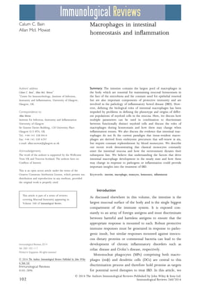
  • 4. seem surprising that m/ constitutively produce this proto- typical pro-inflammatory cytokine, TNFa may have much wider effects. For instance, TNFa can regulate enterocyte growth and alter the permeability of the epithelial barrier (49). Furthermore, TNFa stimulates production of matrix metalloproteinases and other tissue remodeling enzymes in the intestinal mesenchymal cells that are central to regulat- ing epithelial cell function (50). Given their high expression of MHCII and their ability to take up orally administered protein antigens (51–53) and bacteria (37), intestinal m/ may also serve as antigen-presenting cells, interacting with and influencing the differentiation of CD4+ T cells (Fig. 1). It has also been suggested that m/ may play a selective role in initiating the differentiation of FoxP3+ regulatory T cells in the intes- tine (54). However, as m/ do not migrate to the MLN (14) and the intestinal mucosa is essentially devoid of naive CD4+ T cells (55), they are unlikely to be involved in the initial priming of naive CD4+ T cells. Furthermore, LP m/ are extremely poor at activating naive CD4+ T cells in vitro (14). Thus if mucosal m/ do present antigens to CD4+ T cells, it seems more likely that this involves lymphocytes Commensal bacteria Ly6Chi blood monocyte (Ly6ChiCX3CR1int CD115+CCR2+) IL10 Elimination of invading bacteria MyD88/CX3CR1- dependent sampling of luminal contents IL10-dependent secondary expansion of Treg CCR2-dependent replenishment by Ly6Chi blood monocytes Resident M CX3CR1hi F4/80hi CD11b+ MHCIIhi CD64+ Mono/M Intermediate CX3CR1int Ly6C– MHCII+ CD64+ Maintenance of epithelial barrier integrity Regulatory T cells Engulfment of epithelial cell/ apoptotic material Newly extravasated monocyte CX3CR1int Ly6C+ MHCII– CD64lo Maturing monocyte CX3CR1int Ly6C+ MHCII+CD64+ PGE2 IL10 Epithelial barrier damage Pathogenic Bacteria Aberrant responses against commensal bacteria PGE2 Regulation of TNF & ROS production by neutrophils Neutrophil TNF , IL12, IL23, IL6, IL1 Maintenance of effector T cells Th1/Th17 T cell TGF CCL2 CCL7 ? Effector monocytes CX3CR1int Ly6C+ MHCII+ CD64+ CCL2, CCL3, CCL11 Recruitment of innate immune effector cells Pro-inflammatory M CX3CR1int Ly6C– MHCII+ CD64+ IL10 Retention of anti- inflammatory signature by CX3CRhiresident M Eosinophils TNF Homeostasis Inflammation CSF1 Fig. 1. Under steady state conditions, Ly6Chi monocytes constitutively enter the intestinal mucosa and differentiate into mature CX3CR1hi F4/80+ macrophages (m/) through a series of short-lived CX3CR1int intermediaries. These CX3CR1hi m/ are positioned immediately beneath the epithelial barrier and contribute to its integrity by secreting PGE2 which stimulates proliferation of epithelial progenitors. This positioning coupled with their high phagocytic capacity means they are poised to capture and destroy any invading commensals or pathogens, as well as clearing apoptotic or senescent cells. They may also be able to directly sample the luminal contents by the extending processes between the cells of the intestinal epithelial barrier. CX3CR1hi m/ also produce interleukin-10 (IL-10) constitutively, which facilitates secondary expansion of regulatory T cells in the mucosa and may also condition newly arrived monocytes. Regulatory T cells may also contribute to the conditioning of newly extravasated monocytes through their production of TGFb. When homeostasis is perturbed by inflammation or infection, Ly6Chi monocytes and their CX3CR1int derivatives accumulate in large numbers and display enhanced pro-inflammatory characteristics. They produce pro-inflammatory cytokines which may support the maintenance of other effector cells such as IFNc producing and IFNc+ IL-17 double producing T cells. They also orchestrate the recruitment of other innate effector cells such as eosinophils through secretion of inflammatory chemokines. Importantly, during inflammation, CX3CR1hi m/ retain their anti-inflammatory signature, e.g. IL-10 production. Elicited classical monocytes may also play a regulatory role by controlling the production of TNFa and ROS by neutrophils. © 2014 The Authors Immunological Reviews Published by John Wiley & Sons Ltd. Immunological Reviews 260/2014 105 Bain & Mowat Á Roles of intestinal macrophages
  • 5. that have returned to the mucosa after being first primed in the MLN. Consistent with this idea, Hadis et al. (56) have recently shown that m/ facilitate the secondary expansion and maintenance of antigen-specific FoxP3+ Treg in the intestinal mucosa through their production of IL-10 (Fig. 1). The importance of this local fine tuning of T-cell responses by m/ is highlighted by the loss of FoxP3 expression and suppressive capacity that occurs when T cells are rendered unresponsive to IL-10 through deletion of the IL-10R (57). Further evidence that intestinal m/ may con- tribute to the generation of homeostatic Treg comes from work showing that mice lacking TRAF6 expression in CD11c+ MP develop a spontaneous Th2 cell-mediated enter- itis. This is associated with a defective ability of mucosal MP to induce FoxP3+ T cells in vitro and reduced numbers of Treg in the LP (58). Although interpreted as reflecting a role for ‘DCs’, the relevant myeloid cells were defined only as CD11c+ and as we have noted, most of these cells in the normal LP are in fact m/. Further work is needed to define the precise cellular basis of TRAF6-mediated generation of Treg in the LP. Intestinal m/ may also contribute to the maintenance of other T cells in the mucosa. For instance, the production of IL1b in response to TLR stimulation may be relatively pre- served in intestinal m/ and this has been reported to help support the development of Th17 cells in the steady state intestine (59). It remains to be determined whether these effects of intestinal m/ on T cells require cognate interac- tions via peptide-MHC complexes and the TCR, or whether it involves non-specific factors such as polarizing cytokines alone, as has been suggested for the generation of Th17 responses by mucosal CD103+ CD11b+ DCs (60). Recent studies have proposed a further way in which mucosal m/ may contribute to indirectly to T-cell priming, through Connexin 43-mediated transfer of soluble antigen obtained from the lumen to neighboring CD103+ DCs (52). This cooperative process has been suggested to be important for the development of oral tolerance and if confirmed, these findings could indicate an important new role for m/ in the steady state intestine. Exactly how m/ in the LP acquire luminal antigen remains to be established with certainty. One possibility is that they may extend transepithelial dendrites (TED) across the epithelium to capture soluble antigen in the lumen (Fig. 1). Originally thought to be derived from DCs on the basis of the expression of MHCII and CD11c (61, 62), it now seems clear that these TED originate from CX3CR1+ m/. Their formation requires CX3CR1 (61, 63) and indeed, the induction of oral tolerance to protein antigens is impaired in CX3CR1-deficient mice (52, 56), consistent with a requirement for CX3CR1-mediated uptake of luminal antigen in this process. However, the physiological signifi- cance of such TED remains unclear. Not only did the origi- nal reports disagree on which part of the small intestine they were present in and if they were dependent on TLR signaling (61, 62), but more recent studies have failed to find TED anywhere in the steady state intestine (64). Fur- thermore, earlier studies on the uptake of soluble protein from the intestinal lumen reported that this process was not dependent on the CX3CL1–CX3CR1 axis (14). Finally, as we have noted, other work has also proposed that the defect in oral tolerance in CX3CR1-deficient mice reflects an absence of IL-10 production by intestinal m/ (56). If and how LP m/ contribute to the uptake of soluble antigen and then help shape subsequent immune responses clearly warrants further investigation. Despite their phagocytic activity and morphological appearance, there is similar uncertainty about whether LP m/ play a role in the capture of and induction of immunity or tolerance to local bacteria. Although it is well established that CD103+ DCs can present bacterial antigen to naive T cells in the MLN (53), initial reports indicated that the TED formed by MP in the mucosa could capture Salmonella organ- isms in the lumen (61, 62). Furthermore, a very recent study showing that CX3CR1+ cells may transport Salmonella in lymph to the MLN was interpreted as showing a role for migratory m/ in this process (65). However, this phenom- enon was only revealed after depletion of the normal micro- biota with antibiotics and the study did not exclude the possibility that the antigen loaded CX3CR1+ MP belonged to the population of bona fide CX3CR1 expressing DC that we and others have observed in intestinal lymph (13, 14). Again, these findings reveal how much is still to be under- stood about how m/ contribute to the induction and shap- ing of mucosal immune responses. Ontogeny of intestinal macrophages in the steady state The mononuclear phagocyte system Tissue m/ are traditionally viewed as part of the mononu- clear phagocyte system (MPS). First described in 1972 (66), the concept of the MPS proposes that all tissue m/ are the terminally differentiated progeny of blood mono- cytes, which constitutively enter tissues under steady state conditions, and are themselves replaced by rapidly dividing © 2014 The Authors Immunological Reviews Published by John Wiley & Sons Ltd. 106 Immunological Reviews 260/2014 Bain & Mowat Á Roles of intestinal macrophages
  • 6. precursors in the bone marrow (BM). Originating from the common myeloid progenitor, which also generates DCs and granulocytes, a common macrophage and dendritic cell progenitor gives rise to both committed DC precursors (pre-DCs) and to monocytes (67, 68), with the latter pass- ing through a recently identified common monocyte pro- genitor (69). The development of monocytes via these processes is dependent on the transcription factor PU.1 and the growth factors CSF1 (M-CSF), CSF2 (GM-CSF), and IL-3 (69). It is now known that the monocyte pool is heterogeneous and, in mice, CSF1R+ (CD115) monocyte subsets can be distinguished by their expression of Ly6C/Gr1 and the chemokine receptors CCR2 and CX3CR1 (70). The majority of blood monocytes expresses high levels of Ly6C/Gr-1, CCR2 and CD62L, but low levels of CX3CR1 (so-called Ly6Chi monocytes). The entry of Ly6Chi monocytes into the circulation from the BM is dependent on the CCL2-CCR2 chemokine axis and as a result, mice with genetic deletion of either of these molecules have markedly fewer circulating monocytes (71). Although initially referred to as ‘inflamma- tory’ monocytes because of an apparent preference to migrate to inflamed tissues, these Ly6Chi monocytes are now classified as ‘classical’ monocytes, as they fulfill all the roles previously attributed to generic monocytes in the MPS. In human, an analogous population of classical monocytes is identified as CD14hi CD16À and these also express CCR2 (72). A smaller population of murine monocytes expressing lower levels of Ly6C and CCR2, but higher amounts of CX3CR1 (Ly6Clo monocytes) were initially proposed to be the precursors of steady state tissue m/ (70). However, lit- tle direct evidence exists to support this idea, and more recently it has become clear that the primary function of Ly6Clo monocytes is to act as phagocytes in the blood- stream, patrolling and maintaining the vasculature (73, 74). As such, they are now commonly referred to as ‘patrolling’ or non-classical monocytes (75), and there is accumulating evidence that there is a developmental relationship between classical and non-classical blood monocytes, with the latter being the CSF1R-dependent progeny of the former (20, 76). The equivalents of these non-classical Ly6Clo monocytes in human are identified as CD14lo CD16+ , and these express relatively high levels of MHCII (70). Whether they exert similar patrolling functions to their murine counterparts is not known, although gene expression analysis has revealed many parallels between the monocyte subsets in mouse and human (72). Macrophage development independent of the MPS The validity of the MPS has been questioned recently, with multiple reports from independent groups proposing that the majority of tissue m/ exists independently of blood monocytes (77–79). Using a variety of techniques including fate mapping, parabiosis, and analysis of proliferative capac- ity, it has been shown that epidermal Langerhans cells (79, 80), lung alveolar m/ (20, 81), liver Kupffer cells (20, 77), microglia of the CNS (20, 77, 82, 83), and m/ of the peritoneal cavity (78) derive from embryonic precursors that seed tissues early in development and maintain them- selves through in situ proliferation. Although there is debate about whether these precursors originate in the mesen- chyme of the yolk sac, or from the fetal liver, an assumption has come about that all tissue m/ populations may be derived from these early progenitors (84). Our recent work shows that the intestine is an exception to this paradigm, as m/ in the adult steady state mucosa require continuous replenishment by Ly6Chi blood mono- cytes (12). Although earlier studies had shown that Ly6Chi monocytes could enter the colonic mucosa following intense depletion of resident MP (9), it was unclear whether such a mechanism operated in the unmanipulated mucosa and if the progeny represented m/ or DCs (10, 85). Using an adoptive transfer approach, we could demonstrate that Ly6Chi monocytes constantly enter the normal colonic mucosa and mature into CD64+ F4/80hi CX3CR1hi MHCII+ cells (12, 16), indistinguishable from endogenous resident m/ (Fig. 1). The development of mature m/ involves a local differentiation process, in which a series of intermedi- aries lose Ly6C and upregulate F4/80, CX3CR1, CD163, and CD206, as well as acquiring CD11c and MHCII expression (12), features that account for their previous misclassifica- tion as DCs (9, 10). Full maturation appears to take around 5 days and as well as adopting the phenotypic signature of mature colonic m/, the differentiating monocytes begin to acquire the characteristic functional signature of intestinal m/. Thus they become increasingly phagocytic and begin to produce IL-10, as well as developing resistance to TLR stimulation (12). There are multiple lines of evidence to support the idea that monocyte recruitment is the principal mechanism of replenishment of intestinal m/. Firstly, m/ in the adult mucosa have very poor proliferative capacity (12, 86), sug- gesting in situ self-renewal plays little or no role under nor- mal physiological conditions. Secondly, mice in which the CCL2–CCR2 axis has been disrupted by genetic deletion of © 2014 The Authors Immunological Reviews Published by John Wiley & Sons Ltd. Immunological Reviews 260/2014 107 Bain & Mowat Á Roles of intestinal macrophages
  • 7. either molecule have significantly fewer colonic m/ (12, 87). Next, the intestinal m/ pool derives almost exclusively from WT BM in WT:CCR2À/À mixed BM chimeric mice (16). Finally, cells matching the phenotype of intestinal m/ are completely eliminated by DT-mediated depletion in CCR2-DTR mice (88). This requirement for the CCL2-CCR2 axis does not simply reflect its role in BM egress of mono- cytes, as CCR2-sufficient Ly6Chi monocytes fail to enter the CCL2-deficient gut mucosa in WT-CCL2À/À parabiotic mice (87), demonstrating that the CCL2-CCR2 axis is needed for entry of monocytes into the intestinal LP. Although the source of CCL2 in the intestine is unclear, CX3CR1hi resi- dent m/ in the normal mucosa express high levels of the CCR2 ligands CCL2 and CCL7 (87, 89) and therefore they may control their own replenishment. This mechanism has been shown to occur in the mouse uterus, where CSF1R- signaling results in production of CCR2 ligands by uterine m/ (90). Intestinal m/ are all labeled in MacGreen mice in which GFP is expressed under control of the CSF1R pro- moter (91, 92) and thus it is intriguing to speculate that rather than being required for their local proliferation/sur- vival, CSF1R-signalling in gut m/ may be required to elicit monocyte recruitment to the mucosa, as has been suggested in other tissues (90, 93). As CCR2- and CCL2-deficient mice are not completely devoid of intestinal m/ (12, 87), there may be other mechanisms of replenishment in addition to the CCL2- CCR2 axis. This is unlikely to involve non-classical Ly6Clo monocytes, as these fail to migrate into non-lymphoid tis- sues when adoptively transferred (9, 12), and as discussed above, they seem to have a distinct effector role patrolling the vasculature (73). In addition, although CX3CR1-defi- cient mice have markedly fewer circulating Ly6Clo mono- cytes due to impaired survival (63, 94), the intestinal m/ compartment of these mice remains unaffected in size (56). In vitro studies have also implicated TGFb and IL-8 in the recruitment of monocytes to the intestinal mucosa (27, 86), but the role of these and other mediators has not been addressed in vivo. Why the gut mucosa should need continuous recruitment and differentiation of Ly6Chi monocytes is not completely understood. An obvious possibility is that this is required to monitor the constant exposure to the commensal microbiota and other environmental agents found at barrier surfaces. Although the abundance of differentiating monocytes is reduced in the dermis of germ free (GF) mice (16), differ- ent studies mice have reached conflicting conclusions on the size of colonic m/ compartment in GF mice (11, 95, 96). Furthermore, whether GF conditions or antibiotic treatment alter monocyte recruitment to the gut mucosa and/or their subsequent differentiation has never been formally exam- ined. An analogous monocyte differentiation process seems to exist in human intestine. We recently showed that CD14hi monocytes appear to differentiate into mature m/ through a series of intermediaries which involves downregulation of CD14 and CD11c, and upregulation of MHCII, CD209 and CD163 (12). Together these findings indicate that classical monocytes are the principal source of the resident intestinal m/ pool in mouse and human. Monocyte education in the mucosa The environmental cues that shape monocyte differentiation in the intestinal mucosa remain to be identified with cer- tainty, and it is likely that multiple factors are responsible for the imprinting of the phenotypic and functional charac- teristics adopted by maturing monocytes (Fig. 1). These fac- tors are likely to be specific to the intestinal mucosa, as monocytes do not acquire the same characteristics after entering other mucosal sites such as the lung (97). Multiple growth factors are responsible for the maturation of cells of the MPS, including flt3L, CSF1, and CSF2. As noted above, intestinal m/ do not expand in response to exogenous flt3L and remain equally abundant in flt3L-deficient mice (16, authors’ unpublished data). Similarly, although some m/ such as lung alveolar m/ have been shown to depend on the CSF2R for their development (78, 98), it is unlikely that this applies to intestinal m/, as cells matching their pheno- type are derived equally from CSF2R-deficient BM in WT: CSF2R-deficient BM chimeras (99). The development of intestinal m/ requires signaling through the CSF1R and as a result, they are derived almost exclusively from WT BM in WT:CSF1RÀ/À mixed BM chimeric mice (19). There are currently two identified ligands of the CSF1R: CSF1 and IL-34. To date, only microglia and Langerhans cells of the epidermis have been reported to depend on IL-34 and in these cases, this appears to be the driver of in situ self- renewal (100, 101), making it seem unlikely that IL-34 will play a role in intestinal m/ homeostasis. However this has never been examined directly. It is likely that CSF1 is the predominant ligand required in the gut as evidenced by a marked reduction in intestinal m/ in osteopetrotic Csf1op/op mice, which have a mutation in the gene encoding CSF1 (102). It also remains to be determined at which stage of intestinal m/ development CSF1R signalling is involved and specifically whether it is important simply for the generation © 2014 The Authors Immunological Reviews Published by John Wiley & Sons Ltd. 108 Immunological Reviews 260/2014 Bain & Mowat Á Roles of intestinal macrophages
  • 8. of progenitors, or also for the differentiation of monocytes that enter the mucosa. The loss of Ly6C and upregulation of F4/80 that occurs during the transition of Ly6Chi to Ly6Clo monocytes in blood is dependent on CSF1-CSF1R interac- tions (18, 20), as is the development of monocyte-derived m/ in other tissues (18, 19). However, it seems unlikely that CSF1R signalling alone can account for all aspects of intestinal m/ development given the fact that adoptively transferred monocytes generate entirely distinctive m/ in this tissue. Thus, the local environment of the intestinal mucosa must play the definitive role in local m/ develop- ment. A distinctive feature of intestinal m/ is their expression of MHCII at levels even greater than those seen on mucosal DCs. Indeed, acquisition of MHCII is one of the first phenotypic changes to occur during monocyte maturation in the intes- tine, although its functional significance remains unclear (12). This does not appear to be a default property of mono- cyte maturation, as it is not a universal feature of other mono- cyte-derived tissue m/ (90, 103) or of the blood Ly6Clo monocytes, which mature from Ly6Chi monocytes (97). IFNcR signaling is known to induce MHCII on m/ (104), but MHCII expression by intestinal m/ is normal in IFNcR-defi- cient mice (our unpublished observations). It also appears to be independent of CSF2, IL-10, and lymphocytes or their products (our unpublished observations). Recently, the Ran- dolph laboratory has suggested that signals from vascular endothelial cells may instruct upregulation of MHCII by extravasating monocytes (97). However, whether this occurs as monocytes enter the gut mucosa remains unclear. As noted above, acquisition of the CX3CR1hi phenotype appears to be associated with the development of an anti- inflammatory signature in fully differentiated intestinal m/. Given that the majority of m/ in other tissues lack CX3CR1, it is clear that the local environment of the gut must instruct developing monocytes to upregulate this molecule. The factor responsible has not been identified, although TGFb is a clear candidate, as it is known to induce CX3CR1 expression by mi- croglia in the brain (105). There is accumulating evidence that CX3CR1 itself may play a role in controlling the differen- tiation and function of intestinal m/. Firstly, as discussed above, the CX3CL1–CX3CR1 axis has been shown to control the ability of CX3CR1+ MPs to extend cellular protrusions into the intestinal lumen (61, 63, 106). Although this remains an area of contention, it is conceivable that the expression of CX3CL1 by enterocytes (63) could influence the anatomical positioning of m/ under the epithelial layer. However to date, it has not been reported that CX3CR1-deficient m/ inhabit different anatomical locales than their WT counter- parts. Next, the CX3CL1–CX3CR1 axis has been shown to be required for optimal production of IL-10 by intestinal m/ (56) and finally, CX3CR1-deficiency has been reported to lead to decreased numbers of intestinal m/ (107, 108) as well as altering susceptibility to chemically induced colitis. However, given that CX3CR1-deficient mice have been shown both to be protected (11, 63) and more susceptible to experimental colitis (107), further investigation of the CX3CR1–CX3CL1 axis is required to elucidate its involvement in the control of m/ function in the intestine. The IL-10–IL-10R axis plays a crucial role in conditioning the behavior of m/ in the mucosa. One such effect is the upregulation of scavenger receptors such as CD206 and CD163, which is reduced in IL-10-deficient mice (96, 109). Even more marked is the requirement for IL-10 in the hyp- oresponsiveness of intestinal m/ to pro-inflammatory stim- uli (44) (see below). The cellular source of the IL-10 remains unclear. Although m/ themselves produce IL-10 constitutively and this could act in an autocrine manner, selective knockout of IL-10 in LysM+ cells appears not to lead to spontaneous IBD in mice (110), suggesting other cells are more important. This is likely to be IL-10-produc- ing FoxP3+ CD4+ T cells, as these are abundant in the nor- mal intestine and IBD occurs after specific deletion of IL-10 in these cells (111, 112). The induction of IL-10 in matur- ing m/ may be a response to colonization of the intestinal tract by the commensal microbiota, as colonic m/ from mice reared in GF conditions produce less IL-10 (95, 96). However given that this failure to upregulate IL-10 produc- tion cannot be recapitulated in MyD88À/À mice (95), the mechanisms underlying this phenomenon require further investigation. Interestingly, the production of protective IL-10 by CD4+ T cells is also dependent on the microbiota (113). While it has been reported that IL-10 may be impor- tant for more general aspects of maturation of newly recruited monocytes in the peritoneum, including upregula- tion of MHCII (114), our work on inflamed intestine sug- gests that the lack of MHCII may reflect the failure of monocytes to differentiate fully due to the enhanced inflam- mation found in the absence of IL-10, rather than a direct effect of IL-10 (see below). Mechanisms of inflammatory anergy in intestinal macrophages Hyporesponsiveness to activation via TLRs and other stim- uli is a cardinal feature of resident intestinal m/. Early work suggested that this was the result of a failure to © 2014 The Authors Immunological Reviews Published by John Wiley & Sons Ltd. Immunological Reviews 260/2014 109 Bain & Mowat Á Roles of intestinal macrophages
  • 9. express pattern recognition receptors. However, it is now clear that gut-resident m/ in mice and human express a full range of TLR (12, 27), and it is now believed that regulation of adapter molecules downstream of the TLR may instead be responsible for the hyporesponsiveness. For instance, signaling molecules such as CD14, MyD88, TRAF-6, MD2, TRIF, and IRAK1 appear to be downregulated in mature intestinal m/ (27, 44, 89). In parallel, mechanisms that inhibit TLR signaling and/or NF- jB activation (e.g. IRAK-M and IkBNS) appear to overex- pressed in gut-resident m/ (27, 89, 109), suggesting that molecules that propagate TLR signaling are targeted rather than the TLR expression itself. At least some of these inhibitory processes may be driven by IL-10 (109). In mice, the hyporesponsiveness develops progressively as monocytes mature in the mucosa and it cor- relates with increasing IL-10 production (12). As a result, colonic m/ from IL-10À/À mice or mice with LysM-medi- ated deletion of the IL-10R signaling molecule STAT3 dis- play exaggerated pro-inflammatory responses to bacterial products (95, 96, 109, 115), and the animals develop spon- taneous colitis (116, 117). Importantly, IL-10R deficiency is also associated with an early onset form of severe IBD in human (118, 119). Thus, it is clear that IL-10 is one of the most important factors ensuring m/ quiescence in the face of environmental stimuli. However, other mechanisms are likely to be involved. For instance, TGFb has been shown to render blood monocytes hyporesponsive to a variety TLR ligands (27) and active TGFb is abundant in the steady state LP (120). The spontaneous, microbiota-dependent enteritis that occurs in mice lacking expression of TRAF6 in CD11c+ MP could also reflect defective TGFbR-dependent signaling in m/, as TRAF6 and its downstream partner TAK1 are crit- ical components in this pathway (58). The control of m/ activity may also involve dedicated inhibitory receptors including SIRPa and CD200R1. Indeed, the CD200–CD200R1 axis appears to play an important role in regulating alveolar m/ activity (121). However, our recent work suggests that the same regulatory loop is dis- pensable in the intestinal mucosa, with CD200R1-deficient colonic m/ showing no signs of dysregulation (8). Further- more, deletion of CD200R1 or its only ligand, CD200 does not render mice more susceptible to DSS-induced colitis, as would be expected if m/ activity was not controlled appro- priately (8). Although resident m/ express high levels of SIRPa, whether this plays any role in controlling intestinal m/ activity through interaction with its ligand CD47 has never been formally tested. The development of the hyporesponsive state appears to involve recognition of microbial signals, as colonic MP from GF mice respond robustly to stimulation with the TLR4 ago- nist LPS (96). However, it must be noted that in this study m/ were identified only as CD11b+ CD11cÀ cells, which will include many cell types and exclude the majority of res- ident m/ which express CD11c. Therefore, the full impact of the microbiota on the behavior of resident colonic m/ remains to be elucidated. Together, these findings demonstrate that fully responsive Ly6Chi monocytes continuously enter the steady state intesti- nal mucosa as a surveillance measure. In the absence of any threat, the monocytes adopt an anti-inflammatory phenotype which is imprinted by local factors in the mucosa. The nat- ure and mechanisms of action of these mediators remain to be defined fully. Monocytes and macrophages in intestinal inflammation It is well known that the composition of the human intesti- nal m/ pool changes considerably when there is perturba- tion of homeostasis. For instance, in the mucosa of Crohn’s disease and ulcerative colitis patients, there is an accumula- tion of pro-inflammatory macrophages. These can be identi- fied by their high expression of CD14, in contrast to the CD14lo/À population of resident m/ (12, 24, 122, 123). The ability of these cells to produce large amounts of medi- ators such as IL1, IL6, TNFa, reactive oxygen intermediaries and nitric oxide makes them quite distinct from the m/ found in healthy intestine and has led to interest in the idea of targeting the monocyte-macrophage lineage for therapeu- tic purposes. For these reasons, it has become important to establish whether the inflammatory cells represent newly arrived monocyte-derived cells, or are resident cells that have altered their behavior in the presence of inflammation. Mouse models of intestinal inflammation There is similar infiltration of pro-inflammatory monocytes and m/ in animal models of intestinal inflammation such as colitis induced by administration of DSS (12, 89), by the adoptive transfer of naive CD4+ T cells into lymphopenic hosts (16, 30), or after infection with Helicobacter hepaticus coupled with neutralization of IL-10 (our unpublished observations). In all these cases, the infiltrate is characterized by a reversal in the ratio of CX3CR1hi and CX3CR1int cells, caused by the accumulation of Ly6Chi monocytes and their CX3CR1int progeny (Fig. 1). The infiltrating CX3CR1int cells display typical pro-inflammatory characteristics, including © 2014 The Authors Immunological Reviews Published by John Wiley & Sons Ltd. 110 Immunological Reviews 260/2014 Bain & Mowat Á Roles of intestinal macrophages
  • 10. the production of TNFa, IL-6, IL-1b, IL-12, IL-23, and expression of iNOS. In contrast, the remaining CX3CR1hi m/ retain their anti-inflammatory signature, continuing to produce high levels of IL-10 and being TLR hyporesponsive, suggesting that resident m/ do not turn rogue during inflammation (12, 16, 30, 89). Not unexpectedly, CCR2 is required for the entry of pro-inflammatory cells into the inflamed colonic mucosa, with considerably fewer CCR2À/À cells entering the mucosa when co-transferred with their WT counterparts (89, 124). A pathological role for these elicited Ly6Chi monocytes is evidenced by amelioration of colitis in monocytopenic CCR2-deficient mice (124) and in mice depleted of CCR2-expressing cells (89). Furthermore, TNFa production by these cells appears to be central to dis- ease progression, as the severity of DSS-induced colitis is reduced in mice in which Ly6Chi monocytes are deficient in TNFa production (9). On the basis of our studies of DSS colitis, we have proposed that the normal monocyte differ- entiation process is arrested during inflammation, resulting in the retention of cells that remain responsive to TLR stim- ulation and produce copious amounts of pro-inflammatory cytokines and chemokines (12, 30, 89). Why these pro- cesses are disrupted in inflammation remains to be deter- mined, but presumably reflects alterations in the local microenvironment, leading to defects in the conditioning mechanisms that normally specify maturation of monocytes into anti-inflammatory m/. Clearly however, the resident m/ themselves cannot be influenced by these changes, indi- cating these are likely to be terminally differentiated cells and raising the possibility that homeostasis may be restored relatively quickly if the inflammatory environment can be modified. Although Ly6Chi monocytes and their immediate progeny appear to be detrimental during sterile models of intestinal inflammation (12, 124), they are also indispensable for the eradication of certain enteric pathogens. This is consistent with findings that elicited CX3CR1int cells, many of which are likely to be derivatives of Ly6Chi monocytes, have been shown to take up Salmonella organisms as efficiently as resi- dent CX3CR1hi m/ (37). During the acute ileitis caused by oral inoculation of certain mouse strains with the protozoan parasite Toxoplasma gondii, there is accumulation of Ly6Chi monocytes, which display the same behavior as those arriv- ing into the DSS-inflamed colon, including the production of the pro-inflammatory cytokines TNFa and IL-12, and reactive nitrogen species (125). The protective role of these monocytes is demonstrated by the fact that both CCR2- and CCL2-deficient mice succumb to lethal toxoplasmosis, and that the adoptive transfer of CCR2-competent Ly6Chi mono- cytes can rescue this lethality (125). Interestingly, the CCL3–CCR1 chemokine axis driven by innate lymphoid cells is also involved in the recruitment of pro-inflammatory Ly6Chi monocytes and protective immunity in this model of infection, indicating that CCR2 may not be the only mecha- nism responsible for accumulation of monocytes during inflammation (126). However, no defects in steady state intestinal m/ have been reported in mice lacking CCR1, perhaps suggesting distinct roles for individual chemokine receptors under different conditions. Ly6Chi monocytes have also been implicated in the pro- tective immune response to Citrobacter rodentium, a mouse model of enteropathogenic and enterohaemorrhagic E. coli infection in human, with delayed clearance in CCL2- and CCR2-deficient mice (127). However, recent elegant work has questioned this conclusion, by showing that ablation of the entire monocyte-macrophage compartment in mice with LysM-driven expression of DTR by CSF1R-expressing cells has no effect on susceptibility to C. rodentium infection (128). In contrast, the robust Th17 response needed to clear this organism (127) is absolutely dependent on classical migra- tory DC expressing zbtb46 (21). Interestingly however, monocyte-derived macrophages may make an additional contribution to the adaptive immune response under these conditions, by producing IL12, which maintains IFNc+ and IFNc+ IL17+ effector T cells in the mucosa (128). Thus, whereas resident m/ maintain regulatory T cells via IL-10 production, recently elicited monocytes may maintain effec- tor T cells through the production of pro-inflammatory cytokines (Fig. 1). Contrasting with their role as inflammatory effector cells, recent studies suggest that Ly6Chi monocytes may have a wider ability to regulate other aspects of intestinal immunity (Fig. 1). For instance, in the DSS colitis model, Ly6Chi monocytes mediate the recruitment of eosinophils through their production of CCL11 (eotaxin) (129). In T. gondii- induced ileitis, elicited Ly6Chi monocytes may actually pre- vent immunopathology by inhibiting the ability of local neutrophils to produce tissue damaging TNFa and ROI. This PGE2-dependent process involves production of IL-10 by monocytes (40) and may go some way to explain earlier observations of enhanced neutrophil accumulation in the mucosa of T. gondii infected CCR2-deficient animals (125). These results support other reports that monocytes are not intrinsically inflammatory in nature, but are highly plastic cells that coordinate the first stages of tissue repair in response to damage, infection, or inflammation (130). © 2014 The Authors Immunological Reviews Published by John Wiley & Sons Ltd. Immunological Reviews 260/2014 111 Bain & Mowat Á Roles of intestinal macrophages
  • 11. Furthermore, the regulatory properties of elicited Ly6Chi monocytes are diminished in GF mice, underlining how the local microenvironment determines the fate of monocytes in the mucosa (40). M/ have also been implicated in Th2 cell-mediated resis- tance to intestinal helminth infections (131). Their numbers are expanded during infection by organisms such as Trichuris muris, Nippostrongylus brasiliensis, Heligmosomoides polygyrus, Brugia malayi, and Litomosiodes sigmodontis (132–134), and under these conditions, ‘alternative’ activation of m/ by the Th2 prod- ucts IL-4 and IL-13 leads to the production anti-parasitic mediators such as arginase and RELM-a (133, 135). How- ever, the exact role of m/ in these infections is uncertain. The expulsion of these parasites is compromised in mice with m/ depletion secondary to absence of CCL2 (132) or administration of clodronate-containing liposomes (136, 137). Alternatively activated m/ are also partly responsible for the increased smooth muscle contractility which contrib- utes to parasite expulsion (136). However, the protective effects of m/ do not require them to produce arginase-1 (138) or even to be polarized via the IL-4 receptor (139). Thus, m/ may play an indirect role in protective immunity, perhaps by producing mediators that recruit other effector cells such as eosinophils and mast cells. Alternatively, they may be involved in the anti-inflammatory and tissue repair processes that are crucial components of the host response to multicellular pathogens and that are important properties of mediators such as arginase (140). The origin of the alternatively activated macrophages in helminth infection is also uncertain. Although the require- ment for CCL2 in parasite expulsion could indicate they are derived from recruited Ly6Chi monocytes as in the models of inflammation discussed above, this has never been inves- tigated directly. In addition, recent studies of macrophage expansion during helminth infection of the peritoneal and pleural cavities suggest that this may be driven by IL4- and CSF-1-dependent proliferation of the pre-existing resident population (141). The relative roles of in situ expansion of m/ and monocyte recruitment during intestinal helminth infection could generate important insights into the plastic- ity and functional properties of mucosal macrophages. Monocytes and macrophages in human IBD As discussed above, CD14hi MP accumulate in the mucosa of IBD patients (12, 24), and CD14hi cells in human are the equivalent of Ly6Chi monocytes in the mouse. As in mice, the CD14hi cells that appear in human intestine display enhanced production of TNFa, IL-1b, and IL-6 (123) and respiratory burst activity (46). They also retain their respon- siveness to microbial products (24, 122). Using the transfer of radiolabelled autologous blood monocytes, it has been demonstrated that the CD14hi cells in the inflamed mucosa derive from circulating monocytes (142), likely in response to elevated levels of the monocyte chemoattractants CCL2 and CCL4 in the inflamed mucosa (39). In addition to their own pro-inflammatory effects, elicited monocytes in human have also been shown to orchestrate the recruitment of other innate effector cells, such as CCR3+ eosinophils through their production of CCL11 (eotaxin-1) (25, 129). Furthermore, CD68+ m/ in the inflamed mucosa have enhanced expression of CD40 (143), which may facilitate their local interaction with effector T cells, analogous to elicited monocytes in mice. Thus, monocytes and m/ appear to behave similarly in the inflamed intestine of humans and mice. Furthermore, our findings that analogous processes may govern m/ differentiation in the two species intestine which seem to follow analogous processes in both steady state and in inflammation, suggest animal models remain a valuable means of exploring these processes. A role for elicited monocytes in T-cell priming? An area of controversy in myeloid cell biology is whether monocytes can generate APCs that can contribute to the ini- tiation of adaptive immune responses. Under the influence of IFNc, monocytes in inflamed tissues acquire MHCII and are proposed to migrate to draining LN in a CCR7-depen- dent manner, where they may prime naive CD4+ T cells. By their ability to produce large amounts of cytokines, these monocyte-derived ‘DCs’ (mo-DCs) may complement and shape the generation of effector T cells by conventional, flt3L-dependent DCs (144). It has been suggested that Ly6Chi monocytes entering the inflamed mucosa may behave in this manner, acquiring migratory capacity and transiting to the draining MLN (89, 95). In support of this idea, Zigmond et al. (89) reported that a population of zbtb46+ CD11b+ Ly6CÀ DCs in the colonic mucosa was depleted by administration of anti-CCR2 antibody and con- cluded that these were derived from Ly6Chi CCR2+ mono- cytes (89). However, it is clear that some bona fide mature CD11b+ DCs can express CCR2 (21, 145), and pre-DC themselves may express CCR2 (21), suggesting that in vivo depletion of CCR2+ cells may not be specific to monocytes and their progeny. Importantly, in our hands, CCR2+ CD11b+ DCs are derived from committed DC © 2014 The Authors Immunological Reviews Published by John Wiley & Sons Ltd. 112 Immunological Reviews 260/2014 Bain & Mowat Á Roles of intestinal macrophages
  • 12. precursors and Ly6Chi monocytes cannot repopulate any DC population in either steady state or inflamed intestine (12). Monocytes and m/ also cannot be found in the pseudo- afferent intestinal lymph of mice with colitis (authors’ unpublished observations), and although monocyte-derived MHCII+ APC do accumulate in the MLN during experimen- tal colitis, the vast majority of these arrive via the blood- stream in a CCR7-independent manner (16, 144). Thus, while it seems likely that inflammatory monocytes may help shape effector T-cell responses in the mucosa itself, it remains unclear whether they can also act as migratory APCs and initiate T-cell responses in the MLN. Monocytes/macrophages during resolution of inflammation Following the clearance of an infectious or inflammatory agent, the intestine must restore homeostasis so that chronic inflammation does not ensue. This is accompanied by major changes in the m/ pool. During the resolution of colitis in mice, the expanded population of CX3CR1int cells returns to normal size, paralleled by a reduction in granulocyte numbers (89). The mechanisms responsible for the contraction in inflammatory cells is unclear, although it seems likely that most elicited CX3CR1int cells are cleared by apoptosis, as has been shown for the mono- cyte-derived cells recruited to the inflamed peritoneum (146). On the other hand, given that Ly6Chi monocytes replenish CX3CR1hi macrophages in the steady state, it is also conceivable that some of the elicited Ly6Chi cells may convert into anti-inflammatory ‘resident’ m/ once the inflammation begins to resolve, although this has not been shown directly. Even if this does not occur, we have already noted that the resident population of CX3CR1hi m/ appears to persist during mucosal inflammation and irrespective of their origin, these anti-inflammatory cells may play an active role in resolution of tissue damage (Fig. 1). This is supported by the evidence discussed above that deletion of m/ or MyD88 signaling in myeloid cells leads to increased susceptibility to experimental colitis and impaired tissue repair (42, 43). Furthermore there is delayed resolution of DSS-induced colitis in CD68- dnTGFbRII mice which lack TGFb signaling on mature m/ (38). This is suggested to result in part from reduced IL- 10 and enhanced IL-33 production by mucosal m/. The ability of anti-TNFa antibodies to promote mucosal healing in patients with IBD also correlates with the generation of ‘regulatory’ m/ expressing the mannose receptor and able to suppress T-cell proliferation in vitro (147). Whether the promotion of tissue repair by mucosal m/ reflects the m/-derived mediators, such as arginase-1 and PGE-2 that can influence mesenchyme function or epithelial stem cell renewal directly, or if downstream effects on other immune cells are involved remains to be determined. Concluding remarks There have been significant advances recently in our under- standing of how m/ contribute to the maintenance of homeostasis in the intestine, largely due to improvements in methods for identifying them more precisely. Furthermore, we and others have established approaches which help explore the origins and developmental stages of intestinal m/. In particular, our findings show that the current para- digm that tissue-resident m/ are derived from fetal precur- sors that self-renew in situ, does not apply to the intestine. Rather, the mucosal m/ compartment requires continuous replenishment by blood monocytes that are Ly6Chi in mice and CD14hi in human and once considered to be inflamma- tory in nature. Under steady state conditions, these mono- cytes differentiate locally into anti-inflammatory m/ that express scavenger receptors, MHCII, are highly phagocytic and hyporesponsive to pro-inflammatory stimuli, but pro- duce large amounts of IL-10. This process is dictated by cues received from the immediate environment and ensures that mucosal m/ can ingest and degrade dying tissue cells and any commensal bacteria that penetrate across the epithe- lial barrier without provoking inflammation. They can also help maintain epithelial integrity and contribute to physio- logical tissue remodeling. Interruption of the mechanisms that promote the development and/or function of anti- inflammatory m/ leads to inflammation in response to luminal materials such as the microbiota. During inflamma- tion, or in response to invading pathogens, the normal pat- tern of monocyte differentiation is disrupted, leading to the accumulation of potent pro-inflammatory effector mono- cyte/m/. Although these clearly play important roles in tis- sue pathology and protective immunity, they also may be needed for controlling harmful neutrophil activity and for generating the inert m/ required for resolution of inflam- mation and tissue repair. These properties underline the plasticity of this population of ‘classical’ monocytes and understanding the local factors that determine their various fates after arrival in the mucosa under different conditions could hold promise as new therapeutic targets for the treat- ment of IBD. © 2014 The Authors Immunological Reviews Published by John Wiley & Sons Ltd. Immunological Reviews 260/2014 113 Bain & Mowat Á Roles of intestinal macrophages
  • 13. References 1. Lee SH, Starkey PM, Gordon S. Quantitative analysis of total macrophage content in adult mouse tissues. Immunochemical studies with monoclonal antibody F4/80. J Exp Med 1985;161:475–489. 2. Hume DA, Perry VH, Gordon S. The mononuclear phagocyte system of the mouse defined by immunohistochemical localisation of antigen F4/80: macrophages associated with epithelia. Anat Rec 1984;210:503–512. 3. Bain CC, Mowat AM. Intestinal macrophages – specialised adaptation to a unique environment. Eur J Immunol 2011;41:2494–2498. 4. Racine R, Chatterjee M, Winslow GM. CD11c expression identifies a population of extrafollicular antigen-specific splenic plasmablasts responsible for CD4 T-independent antibody responses during intracellular bacterial infection. J Immunol 2008;181:1375–1385. 5. Carlens J, Wahl B, Ballmaier M, Bulfone-Paus S, Forster R, Pabst O. Common gamma-chain-dependent signals confer selective survival of eosinophils in the murine small intestine. J Immunol 2009;183:5600–5607. 6. Uematsu S, et al. Regulation of humoral and cellular gut immunity by lamina propria dendritic cells expressing Toll-like receptor 5. Nat Immunol 2008;9:769–776. 7. McGarry MP, Stewart CC. Murine eosinophil granulocytes bind the murine macrophage- monocyte specific monoclonal antibody F4/80. J Leukoc Biol 1991;50:471–478. 8. Bain CC, Mowat AM. CD200 receptor and macrophage function in the intestine. Immunobiology 2012;217:643–651. 9. Varol C, et al. Intestinal lamina propria dendritic cell subsets have different origin and functions. Immunity 2009;31:502–512. 10. Bogunovic M, et al. Origin of the lamina propria dendritic cell network. Immunity 2009;31:513– 525. 11. Niess JH, Adler G. Enteric flora expands gut lamina propria CX3CR1+ dendritic cells supporting inflammatory immune responses under normal and inflammatory conditions. J Immunol 2010;184:2026–2037. 12. Bain CC, et al. Resident and pro-inflammatory macrophages in the colon represent alternative context-dependent fates of the same Ly6Chi monocyte precursors. Mucosal Immunol 2013;6:498–510. 13. Cerovic V, et al. Intestinal CD103(-) dendritic cells migrate in lymph and prime effector T cells. Mucosal Immunol 2013;6:104–113. 14. Schulz O, et al. Intestinal CD103+, but not CX3CR1+, antigen sampling cells migrate in lymph and serve classical dendritic cell functions. J Exp Med 2009;206:3101–3114. 15. Wumesh KC, et al. L-Myc expression by dendritic cells is required for optimal T-cell priming. Nature 2014;507:243–247. 16. Tamoutounour S, et al. CD64 distinguishes macrophages from dendritic cells in the gut and reveals the Th1-inducing role of mesenteric lymph node macrophages during colitis. Eur J Immunol 2012;42:3150–3166. 17. Gautier EL, et al. Gene-expression profiles and transcriptional regulatory pathways that underlie the identity and diversity of mouse tissue macrophages. Nat Immunol 2012;13:1118– 1128. 18. MacDonald KP, et al. An antibody against the colony-stimulating factor 1 receptor depletes the resident subset of monocytes and tissue- and tumor-associated macrophages but does not inhibit inflammation. Blood 2010;116:3955– 3963. 19. Schlitzer A, et al. IRF4 transcription factor-dependent CD11b+ dendritic cells in human and mouse control mucosal IL-17 cytokine responses. Immunity 2013;38:970–983. 20. Yona S, et al. Fate mapping reveals origins and dynamics of monocytes and tissue macrophages under homeostasis. Immunity 2013;38:79–91. 21. Satpathy AT, et al. Notch2-dependent classical dendritic cells orchestrate intestinal immunity to attaching-and-effacing bacterial pathogens. Nat Immunol 2013;14:937–948. 22. Schraml BU, et al. Genetic tracing via DNGR-1 expression history defines dendritic cells as a hematopoietic lineage. Cell 2013;154:843–858. 23. Miller JC, et al. Deciphering the transcriptional network of the dendritic cell lineage. Nat Immunol 2013;13:888–899. 24. Kamada N, et al. Unique CD14 intestinal macrophages contribute to the pathogenesis of Crohn disease via IL-23/IFN-gamma axis. J Clin Invest 2008;118:2269–2280. 25. Lampinen M, Waddell A, Ahrens R, Carlson M, Hogan SP. CD14+CD33+ myeloid cell-CCL11-eosinophil signature in ulcerative colitis. J Leukoc Biol 2013;94:1061–1070. 26. Rogler G, et al. Isolation and phenotypic characterization of colonic macrophages. Clin Exp Immunol 1998;112:205–215. 27. Smythies LE, et al. Inflammation anergy in human intestinal macrophages is due to Smad-induced IkappaBalpha expression and NF-kappaB inactivation. J Biol Chem 2010;285:19593–19604. 28. Mann ER, et al. Intestinal dendritic cells: their role in intestinal inflammation, manipulation by the gut microbiota and differences between mice and men. Immunol Lett 2013;150:30–40. 29. Mantovani A, Sica A, Locati M. Macrophage polarization comes of age. Immunity 2005;23:344–346. 30. Weber B, Saurer L, Schenk M, Dickgreber N, Mueller C. CX3CR1 defines functionally distinct intestinal mononuclear phagocyte subsets which maintain their respective functions during homeostatic and inflammatory conditions. Eur J Immunol 2011;41:773–779. 31. Mosser DM, Edwards JP. Exploring the full spectrum of macrophage activation. Nat Rev 2008;8:958–969. 32. Biswas SK, Mantovani A. Macrophage plasticity and interaction with lymphocyte subsets: cancer as a paradigm. Nat Immunol 2010;11:889–896. 33. Mikkelsen HB, Rumessen JJ. Characterization of macrophage-like cells in the external layers of human small and large intestine. Cell Tissue Res 1992;270:273–279. 34. Tajima T, et al. EP2 and EP4 receptors on muscularis resident macrophages mediate LPS-induced intestinal dysmotility via iNOS upregulation through cAMP/ERK signals. Am J Physiol Gastrointest Liver Physiol 2012;302: G524–G534. 35. Nagashima R, Maeda K, Imai Y, Takahashi T. Lamina propria macrophages in the human gastrointestinal mucosa: their distribution, immunohistological phenotype, and function. J Histochem Cytochem 1996;44: 721–731. 36. Denning TL, et al. Functional specializations of intestinal dendritic cell and macrophage subsets that control Th17 and regulatory T cell responses are dependent on the T cell/APC ratio, source of mouse strain, and regional localization. J Immunol 2011;187:733–747. 37. Muller AJ, et al. Salmonella gut invasion involves TTSS-2-dependent epithelial traversal, basolateral exit, and uptake by epithelium-sampling lamina propria phagocytes. Cell Host Microbe 2012;11:19–32. 38. Rani R, Smulian AG, Greaves DR, Hogan SP, Herbert DR. TGF-beta limits IL-33 production and promotes the resolution of colitis through regulation of macrophage function. Eur J Immunol 2011;41:2000–2009. 39. Smith PD, Smythies LE, Shen R, Greenwell-Wild T, Gliozzi M, Wahl SM. Intestinal macrophages and response to microbial encroachment. Mucosal Immunol 2010;4:31–42. 40. Grainger JR, et al. Inflammatory monocytes regulate pathologic responses to commensals during acute gastrointestinal infection. Nat Med 2013;19:713–721. 41. Pull SL, Doherty JM, Mills JC, Gordon JI, Stappenbeck TS. Activated macrophages are an adaptive element of the colonic epithelial progenitor niche necessary for regenerative responses to injury. Proc Natl Acad Sci USA 2005;102:99–104. 42. Qualls JE, Kaplan AM, Van Rooijen N, Cohen DA. Suppression of experimental colitis by intestinal mononuclear phagocytes. J Leukoc Biol 2006;80:802–815. 43. Malvin NP, Seno H, Stappenbeck TS. Colonic epithelial response to injury requires Myd88 signaling in myeloid cells. Mucosal Immunol 2012;5:194–206. 44. Smythies LE, et al. Human intestinal macrophages display profound inflammatory anergy despite avid phagocytic and bacteriocidal activity. J Clin Invest 2005;115:66–75. 45. N’Diaye E-N, et al. TREM-2 (triggering receptor expressed on myeloid cells 2) is a phagocytic receptor for bacteria. J Cell Biol 2009;184:215– 223. 46. Rugtveit J, Haraldsen G, Høgasen AK, Bakka A, Brandtzaeg P, Scott H. Respiratory burst of intestinal macrophages in inflammatory bowel © 2014 The Authors Immunological Reviews Published by John Wiley Sons Ltd. 114 Immunological Reviews 260/2014 Bain Mowat Á Roles of intestinal macrophages
  • 14. disease is mainly caused by CD14+L1+ monocyte derived cells. Gut 1995;37:367–373. 47. Roberts PJ, Riley GP, Morgan K, Miller R, Hunter JO, Middleton SJ. The physiological expression of inducible nitric oxide synthase (iNOS) in the human colon. J Clin Pathol 2001;54:293–297. 48. Hedl M, Li J, Cho JH, Abraham C. Chronic stimulation of Nod2 mediates tolerance to bacterial products. Proc Natl Acad Sci USA 2007;104:19440–19445. 49. Ma TY, et al. TNF-alpha-induced increase in intestinal epithelial tight junction permeability requires NF-kappa B activation. Am J Physiol Gastrointest Liver Physiol 2004;286:G367–G376. 50. Pender SL, Quinn JJ, Sanderson IR, MacDonald TT. Butyrate upregulates stromelysin-1 production by intestinal mesenchymal cells. Am J Physiol Gastrointest Liver Physiol 2000;279: G918–G924. 51. Chang S-Y, et al. Circulatory antigen processing by mucosal dendritic cells controls CD8(+) T cell activation. Immunity 2013;38:153–165. 52. Mazzini E, Massimiliano L, Penna G, Rescigno M. Oral tolerance can be established via gap junction transfer of fed antigens from CX3CR1(+) macrophages to CD103(+) dendritic cells. Immunity 2014;40:248–261. 53. Farache J, et al. Luminal bacteria recruit CD103+ dendritic cells into the intestinal epithelium to sample bacterial antigens for presentation. Immunity 2013;38:581–595. 54. Denning TL, Wang YC, Patel SR, Williams IR, Pulendran B. Lamina propria macrophages and dendritic cells differentially induce regulatory and interleukin 17-producing T cell responses. Nat Immunol 2007;8:1086–1094. 55. MacDonald TT, Pender SL. Lamina propria T cells. Chem Immunol 1998;71:103–117. 56. Hadis U, et al. Intestinal tolerance requires gut homing and expansion of FoxP3+ regulatory T cells in the lamina propria. Immunity 2011;34:237–246. 57. Murai M, et al. Interleukin 10 acts on regulatory T cells to maintain expression of the transcription factor Foxp3 and suppressive function in mice with colitis. Nat Immunol 2009;10:1178– 1184. 58. Han D, et al. Dendritic cell expression of the signaling molecule TRAF6 is critical for gut microbiota-dependent immune tolerance. Immunity 2013;38:1211–1222. 59. Shaw MH, Kamada N, Kim Y-G, Nu~nez G. Microbiota-induced IL-1b, but not IL-6, is critical for the development of steady-state TH17 cells in the intestine. J Exp Med 2012;209:251– 258. 60. Welty NE, Staley C, Ghilardi N, Sadowsky MJ, Igyarto BZ, Kaplan DH. Intestinal lamina propria dendritic cells maintain T cell homeostasis but do not affect commensalism. J Exp Med 2013;210:2011–2024. 61. Niess JH, et al. CX3CR1-mediated dendritic cell access to the intestinal lumen and bacterial clearance. Science 2005;307:254–258. 62. Chieppa M, Rescigno M, Huang AY, Germain RN. Dynamic imaging of dendritic cell extension into the small bowel lumen in response to epithelial cell TLR engagement. J Exp Med 2006;203:2841–2852. 63. Kim KW, et al. In vivo structure/function and expression analysis of the CX3C chemokine fractalkine. Blood 2011;118:e156–e167. 64. McDole JR, et al. Goblet cells deliver luminal antigen to CD103+ dendritic cells in the small intestine. Nature 2012;483:345–349. 65. Diehl GE, et al. Microbiota restricts trafficking of bacteria to mesenteric lymph nodes by CX(3)CR1 (hi) cells. Nature 2013;494:116–120. 66. van Furth R, Cohn ZA, Hirsch JG, Humphrey JH, Spector WG, Langevoort HL. The mononuclear phagocyte system: a new classification of macrophages, monocytes, and their precursor cells. Bull World Health Organ 1972;46:845– 852. 67. Fogg DK, et al. A clonogenic bone marrow progenitor specific for macrophages and dendritic cells. Science 2006;311:83–87. 68. Auffray C, et al. CX3CR1+ CD115+ CD135+ common macrophage/DC precursors and the role of CX3CR1 in their response to inflammation. J Exp Med 2009;206:595–606. 69. Hettinger J, et al. Origin of monocytes and macrophages in a committed progenitor. Nat Immunol 2013;14:821–830. 70. Geissmann F, Jung S, Littman DR. Blood monocytes consist of two principal subsets with distinct migratory properties. Immunity 2003;19:71–82. 71. Serbina NV, Pamer EG. Monocyte emigration from bone marrow during bacterial infection requires signals mediated by chemokine receptor CCR2. Nat Immunol 2006;7:311–317. 72. Ingersoll MA, et al. Comparison of gene expression profiles between human and mouse monocyte subsets. Blood 2010;115:e10–e19. 73. Auffray C, et al. Monitoring of blood vessels and tissues by a population of monocytes with patrolling behavior. Science 2007;317:666–670. 74. Carlin LM, et al. Nr4a1-dependent Ly6C(low) monocytes monitor endothelial cells and orchestrate their disposal. Cell 2013;153:362– 375. 75. Ziegler-Heitbrock L, et al. Nomenclature of monocytes and dendritic cells in blood. Blood 2010;116:e74–e80. 76. Sunderkotter C, et al. Subpopulations of mouse blood monocytes differ in maturation stage and inflammatory response. J Immunol 2004;172:4410–4417. 77. Schulz C, et al. A lineage of myeloid cells independent of Myb and hematopoietic stem cells. Science 2012;336:86–90. 78. Hashimoto D, et al. Tissue-resident macrophages self-maintain locally throughout adult life with minimal contribution from circulating monocytes. Immunity 2013;38:792–804. 79. Hoeffel G, et al. Adult Langerhans cells derive predominantly from embryonic fetal liver monocytes with a minor contribution of yolk sac-derived macrophages. J Exp Med 2012;209:1167–1181. 80. Chorro L, et al. Langerhans cell (LC) proliferation mediates neonatal development, homeostasis, and inflammation-associated expansion of the epidermal LC network. J Exp Med 2009;206:3089–3100. 81. Tarling JD, Lin HS, Hsu S. Self-renewal of pulmonary alveolar macrophages: evidence from radiation chimera studies. J Leukoc Biol 1987;42:443–446. 82. Ajami B, Bennett JL, Krieger C, Tetzlaff W, Rossi FM. Local self-renewal can sustain CNS microglia maintenance and function throughout adult life. Nat Neurosci 2007;10:1538–1543. 83. Kierdorf K, et al. Microglia emerge from erythromyeloid precursors via Pu.1- and Irf8-dependent pathways. Nat Neurosci 2013;16:273–280. 84. Satpathy AT, Wu X, Albring JC, Murphy KM. Re (de)fining the dendritic cell lineage. Nat Immunol 2012;13:1145–1154. 85. Bogunovic M, Mortha A, Muller PA, Merad M. Mononuclear phagocyte diversity in the intestine. Immunol Res 2012;54:37–49. 86. Smythies LE, et al. Mucosal IL-8 and TGF-beta recruit blood monocytes: evidence for cross-talk between the lamina propria stroma and myeloid cells. J Leukoc Biol 2006;80:492–499. 87. Takada Y, et al. Monocyte chemoattractant protein-1 contributes to gut homeostasis and intestinal inflammation by composition of IL-10-producing regulatory macrophage subset. J Immunol 2010;184:2671–2676. 88. Kinnebrew MA, et al. Interleukin 23 production by intestinal CD103(+)CD11b(+) dendritic cells in response to bacterial flagellin enhances mucosal innate immune defense. Immunity 2012;36:276–287. 89. Zigmond E, et al. Ly6C hi monocytes in the inflamed colon give rise to proinflammatory effector cells and migratory antigen-presenting cells. Immunity 2012;37:1076–1090. 90. Tagliani E, Shi C, Nancy P, Tay CS, Pamer EG, Erlebacher A. Coordinate regulation of tissue macrophage and dendritic cell population dynam- ics by CSF-1. J Exp Med 2011;208:1901–1916. 91. Sasmono RT, et al. A macrophage colony-stimulating factor receptor-green fluorescent protein transgene is expressed throughout the mononuclear phagocyte system of the mouse. Blood 2003;101:1155–1163. 92. Bradford BM, Sester DP, Hume DA, Mabbott NA. Defining the anatomical localisation of subsets of the murine mononuclear phagocyte system using integrin alpha X (Itgax, CD11c) and colony stimulating factor 1 receptor (Csf1r, CD115) expression fails to discriminate dendritic cells from macrophages. Immunobiology 2011;216:1228–1237. 93. Pierce JH, et al. Macrophage-colony-stimulating factor (CSF-1) induces proliferation, chemotaxis, and reversible monocytic differentiation in myeloid progenitor cells transfected with the human c-fms/CSF-1 receptor cDNA. Proc Natl Acad Sci USA 1990;87:5613–5617. 94. Landsman L, et al. CX3CR1 is required for monocyte homeostasis and atherogenesis by promoting cell survival. Blood 2009;113:963– 972. 95. Rivollier A, He J, Kole A, Valatas V, Kelsall BL. Inflammation switches the differentiation © 2014 The Authors Immunological Reviews Published by John Wiley Sons Ltd. Immunological Reviews 260/2014 115 Bain Mowat Á Roles of intestinal macrophages
  • 15. program of Ly6Chi monocytes from antiinflammatory macrophages to inflammatory dendritic cells in the colon. J Exp Med 2012;209:139–155. 96. Ueda Y, et al. Commensal microbiota induce LPS hyporesponsiveness in colonic macrophages via the production of IL-10. Int Immunol 2010;22:953–962. 97. Jakubzick C, et al. Minimal differentiation of classical monocytes as they survey steady-state tissues and transport antigen to lymph nodes. Immunity 2013;39:599–610. 98. Guilliams M, et al. Alveolar macrophages develop from fetal monocytes that differentiate into long-lived cells in the first week of life via GM-CSF. J Exp Med 2013;210:1977–1992. 99. Greter M, et al. GM-CSF controls nonlymphoid tissue dendritic cell homeostasis but is dispensable for the differentiation of inflammatory dendritic cells. Immunity 2012;36:1031–1046. 100. Wang Y, et al. IL-34 is a tissue-restricted ligand of CSF1R required for the development of Langerhans cells and microglia. Nat Immunol 2012;13:753–760. 101. Greter M, et al. Stroma-derived interleukin-34 controls the development and maintenance of langerhans cells and the maintenance of microglia. Immunity 2012;37:1050–1060. 102. Ryan GR, et al. Rescue of the colony-stimulating factor 1 (CSF-1)-nullizygous mouse (Csf1(op)/ Csf1(op)) phenotype with a CSF-1 transgene and identification of sites of local CSF-1 synthesis. Blood 2001;98:74–84. 103. Tamoutounour S, et al. Origins and functional specialization of macrophages and of conventional and monocyte-derived dendritic cells in mouse skin. Immunity 2013;39:925– 938. 104. Steimle V, Siegrist CA, Mottet A, Lisowska-Grospierre B, Mach B. Regulation of MHC class II expression by interferon-gamma mediated by the transactivator gene CIITA. Science 1994;265:106–109. 105. Chen S, Luo D, Streit WJ, Harrison JK. TGF-beta1 upregulates CX3CR1 expression and inhibits fractalkine-stimulated signaling in rat microglia. J Neuroimmunol 2002;133:46–55. 106. Rescigno M, et al. Dendritic cells express tight junction proteins and penetrate gut epithelial monolayers to sample bacteria. Nat Immunol 2001;2:361–367. 107. Medina-Contreras O, et al. CX3CR1 regulates intestinal macrophage homeostasis, bacterial translocation, and colitogenic Th17 responses in mice. J Clin Invest 2011;121:4787–4795. 108. Hochheiser K, et al. Exclusive CX3CR1 dependence of kidney DCs impacts glomerulonephritis progression. J Clin Invest 2013;123:4242–4254. 109. Hirotani T, et al. The nuclear IkappaB protein IkappaBNS selectively inhibits lipopolysaccharide-induced IL-6 production in macrophages of the colonic lamina propria. J Immunol 2005;174:3650–3657. 110. Schwarz T, et al. T cell-derived IL-10 determines leishmaniasis disease outcome and is suppressed by a dendritic cell based vaccine. PLoS Pathog 2013;9:e1003476. 111. Rubtsov YP, et al. Regulatory T cell-derived interleukin-10 limits inflammation at environmental interfaces. Immunity 2008;28:546–558. 112. Roers A, et al. T cell-specific inactivation of the interleukin 10 gene in mice results in enhanced T cell responses but normal innate responses to lipopolysaccharide or skin irritation. J Exp Med 2004;200:1289–1297. 113. Pils MC, et al. Commensal gut flora reduces susceptibility to experimentally induced colitis via T-cell-derived interleukin-10. Inflamm Bowel Dis 2011;17:2038–2046. 114. Nguyen H-H, Tran B-T, Muller W, Jack RS. IL-10 acts as a developmental switch guiding monocyte differentiation to macrophages during a murine peritoneal infection. J Immunol 2012;189:3112– 3120. 115. Hoshi N, et al. MyD88 signalling in colonic mononuclear phagocytes drives colitis in IL-10-deficient mice. Nat Commun 2012;3:1120. 116. Takeda K, et al. Enhanced Th1 activity and development of chronic enterocolitis in mice devoid of Stat3 in macrophages and neutrophils. Immunity 1999;10:39–49. 117. Kuhn R, Lohler J, Rennick D, Rajewsky K, Muller W. Interleukin-10-deficient mice develop chronic enterocolitis. Cell 1993;75:263–274. 118. Glocker EO, et al. Inflammatory bowel disease and mutations affecting the interleukin-10 receptor. N Engl J Med 2009;361:2033–2045. 119. Murugan D, et al. Very early onset inflammatory bowel disease associated with aberrant trafficking of IL-10R1 and cure by T cell replete haploidentical. Bone Marrow Transplantation. J Clin Immunol 2014;34:331–339. 120. Maheshwari A, et al. TGF-beta2 suppresses macrophage cytokine production and mucosal inflammatory responses in the developing intestine. Gastroenterology 2011;140:242–253. 121. Snelgrove RJ, et al. A critical function for CD200 in lung immune homeostasis and the severity of influenza infection. Nat Immunol 2008;9:1074– 1083. 122. Thiesen S, et al. CD14hiHLA-DRdim macrophages, with a resemblance to classical blood monocytes, dominate inflamed mucosa in Crohn’s disease. J Leukoc Biol 2013;95:531–541. 123. Rugtveit J, Nilsen EM, Bakka A, Carlsen H, Brandtzaeg P, Scott H. Cytokine profiles differ in newly recruited and resident subsets of mucosal macrophages from inflammatory bowel disease. Gastroenterology 1997;112:1493–1505. 124. Platt AM, Bain CC, Bordon Y, Sester DP, Mowat AM. An independent subset of TLR expressing CCR2-dependent macrophages promotes colonic inflammation. J Immunol 2010;184:6843–6854. 125. Dunay IR, et al. Gr1(+) inflammatory monocytes are required for mucosal resistance to the pathogen Toxoplasma gondii. Immunity 2008;29:306–317. 126. Schulthess J, et al. Interleukin-15-dependent NKp46+ innate lymphoid cells control intestinal inflammation by recruiting inflammatory monocytes. Immunity 2012;37:108–121. 127. Kim YG, et al. The Nod2 sensor promotes intestinal pathogen eradication via the chemokine CCL2-dependent recruitment of inflammatory monocytes. Immunity 2011;34:769–780. 128. Schreiber HA, et al. Intestinal monocytes and macrophages are required for T cell polarization in response to Citrobacter rodentium. J Exp Med 2013;210:2025–2039. 129. Waddell A, et al. Colonic eosinophilic inflammation in experimental colitis is mediated by Ly6C(high) CCR2(+) inflammatory monocyte/macrophage-derived CCL11. J Immunol 2011;186:5993–6003. 130. Arnold L, et al. Inflammatory monocytes recruited after skeletal muscle injury switch into antiinflammatory macrophages to support myogenesis. J Exp Med 2007;204:1057–1069. 131. Kreider T, Anthony RM, Urban JF, Gause WC. Alternatively activated macrophages in helminth infections. Curr Opin Immunol 2007;19:448–453. 132. de Schoolmeester ML, Little MC, Rollins BJ, Else KJ. Absence of CC chemokine ligand 2 results in an altered Th1/Th2 cytokine balance and failure to expel Trichuris muris infection. J Immunol 2003;170:4693–4700. 133. Filbey KJ, et al. Innate and adaptive type 2 immune cell responses in genetically controlled resistance to intestinal helminth infection. Immunol Cell Biol 2014. doi: 10.1038/icb.2013. 109 [Epub ahead of print]. 134. Schmidt S, et al. Nippostrongylus-induced intestinal hypercontractility requires IL-4 receptor alpha-responsiveness by T cells in mice. PLoS ONE 2012;7:e52211. 135. Reyes JL, Terrazas LI. The divergent roles of alternatively activated macrophages in helminthic infections. Parasite Immunol 2007;29:609–619. 136. Zhao A, et al. Th2 cytokine-induced alterations in intestinal smooth muscle function depend on alternatively activated macrophages. Gastroenterology 2008;135:217–225 e1. 137. Anthony RM, et al. Memory T(H)2 cells induce alternatively activated macrophages to mediate protection against nematode parasites. Nat Med 2006;12:955–960. 138. Bowcutt R, et al. Arginase-1-expressing macrophages are dispensable for resistance to infection with the gastrointestinal helminth Trichuris muris. Parasite Immunol 2011;33:411–420. 139. Herbert DR, et al. Alternative macrophage activation is essential for survival during schistosomiasis and downmodulates T helper 1 responses and immunopathology. Immunity 2004;20:623–635. 140. Herbert DR, et al. Arginase I suppresses IL-12/ IL-23p40-driven intestinal inflammation during acute schistosomiasis. J Immunol 2010;184:6438–6446. 141. Jenkins SJ, et al. Local macrophage proliferation, rather than recruitment from the blood, is a signature of TH2 inflammation. Science 2011;332:1284–1288. 142. Grimm MC, Pullman WE, Bennett GM, Sullivan PJ, Pavli P, Doe WF. Direct evidence of monocyte recruitment to inflammatory bowel disease mucosa. J Gastroenterol Hepatol 1995;10:387–395. © 2014 The Authors Immunological Reviews Published by John Wiley Sons Ltd. 116 Immunological Reviews 260/2014 Bain Mowat Á Roles of intestinal macrophages
  • 16. 143. Carlsen HS, Yamanaka T, Scott H, Rugtveit J, Brandtzaeg P. The proportion of CD40+ mucosal macrophages is increased in inflammatory bowel disease whereas CD40 ligand (CD154)+ T cells are relatively decreased, suggesting differential modulation of these costimulatory molecules in human gut lamina propria. Inflamm Bowel Dis 2006;12:1013–1024. 144. Siddiqui KR, Laffont S, Powrie F. E-cadherin marks a subset of inflammatory dendritic cells that promote T cell-mediated colitis. Immunity 2010;32:557–567. 145. Lewis KL, et al. Notch2 receptor signaling controls functional differentiation of dendritic cells in the spleen and intestine. Immunity 2011;35:780–791. 146. Gautier EL, Ivanov S, Lesnik P, Randolph GJ. Local apoptosis mediates clearance of macrophages from resolving inflammation in mice. Blood 2013;122:2714–2722. 147. Vos ACW, et al. Regulatory macrophages induced by infliximab are involved in healing in vivo and in vitro. Inflamm Bowel Dis 2012;18:401– 408. © 2014 The Authors Immunological Reviews Published by John Wiley Sons Ltd. Immunological Reviews 260/2014 117 Bain Mowat Á Roles of intestinal macrophages