Umbilical cord mesenchymal stem cells (UC-MSCs) show potential advantages over mesenchymal stem cells (MSCs) from other sources for regenerative medicine applications. UC-MSCs display higher proliferation rates and expression of embryonic genes compared to adult MSCs. Transcriptomic analyses indicate UC-MSCs express genes related to development of multiple tissues including bone, liver, cardiovascular and neural systems. While UC-MSCs can differentiate into cell types of multiple lineages, their therapeutic impact is thought to be mainly due to their paracrine effects and immunomodulatory properties. UC-MSCs could have advantages for treating autoimmune and neurodegenerative diseases.

![(e.g., trypsine, hayluronidase) and were performed with
or without the dissection of the umbilical cord into small
pieces and with or without removing the blood vessels.8
Recently, Han et al. have suggested that using 0.2% colla-
genase II at 37°C for a digestion of 16–20 h is an effective
and simple enzyme digestion method.7
Other groups have
found that enzymatic digestion can alter cell population
and function and, thus, have developed explant approaches
without using any enzyme and taking advantage of the
ability of MSCs to migrate from the tissue to adhere on the
plastic.8–10
Hua et al. have, very recently, compared three
explant and three enzymatic methods with regard to time of
primary culture, cell number, cell morphology, immune
phenotype, and differentiation potential of WJ-MSCs. They
have shown that the 10 mm-size tissue explant method was
the optimal protocol for the isolation of MSCs.11
Morphology and proliferation capacity
WJ-MSCs cultured in vitro shared a similar fibro-blastoid
shaped morphology to BM, amniotic fluid (AF), or teeth and
periodontal ligament (PDL)-MSC.12,13
The proliferation capacity of cells is important regarding
their application potential in cell therapy and tissue engi-
neering. WJ-MSCs proliferation capacity seems to be dif-
ferent from other sources MSCs. Indeed, for instance, Yu
et al. have shown that over a period of 7 days after seeding,
WJ-MSCs grew much faster than PDL-MSC and had a cell
doubling time of 22.23h against 27.51 h for PDL-MSC.14
Compared with BM-MSCs, WJ-MSCs grow much faster for
the early passages and have a cell doubling time (24h) almost
twice shorter than BM-MSCs (40h) over the 1st passage.
These observations were confirmed by Abu Kasim et al.
showing that WJ-MSCs and dental pulp-mesenchymal stem
cells (DP-MSCs) were highly proliferative as compared with
BM and adipose tissue (AT)-MSCs.15
Furthermore, WJ-MSCs
have a greater ability to form colony-forming unit-fibroblasts
colonies in vitro than BM-MSCs, and their formation’s fre-
quency depends on seeding cell density.4,16
Other studies focusing on DP-MSCs showed that cells
from both sources (WJ and teeth) initially grew slowly but
their proliferation rates were increased after the first sub-
culture.17
However, WJ-MSCs growth is influenced by the
number of culture passages in vitro, as amplifying these
cells until passage 10 will result in a slower cell growth
compared with the same cell culture at passage 5.18
A very recent study has evaluated the proliferation ki-
netics and phenotypic characteristics of MSCs derived
from WJ and AT during prolonged in vitro expansion and
found that WJ-MSCs were isolated with a high efficiency
and bore a substantially increased proliferation capac-
ity; whereas AT-MSCs exhibited a reduced proliferation
potential showing typical signs of senescence at an early
stage.19
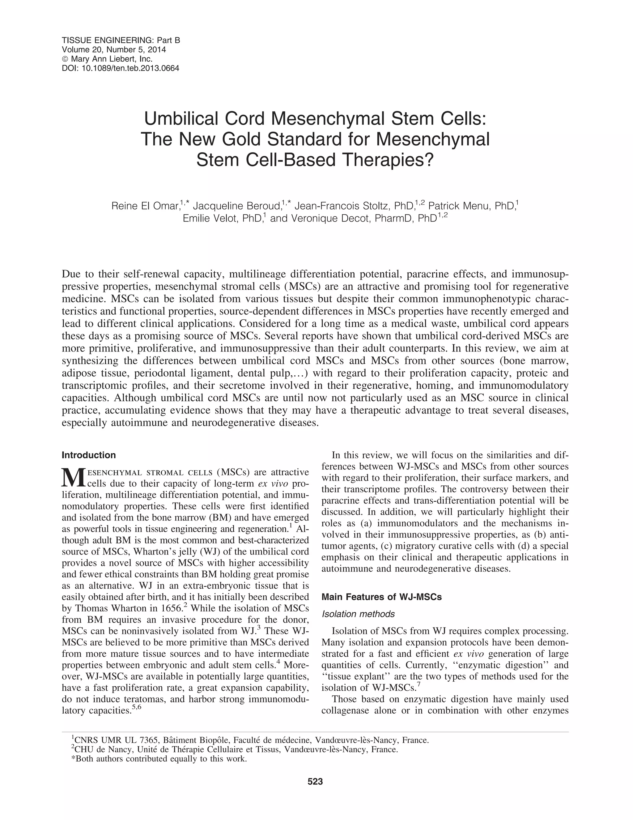
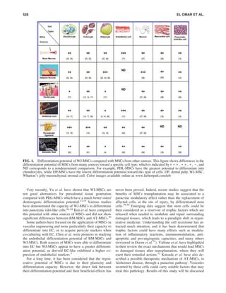
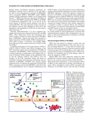
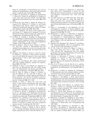
Marker expression at protein level
A large number of studies have analyzed the surface
markers of WJ-MSCs and compared their expression pro-
files with other sources of MSCs such as BM, teeth, or AF.
Table 1. Phenotypic Profile of WJ-MSCs Compared with MSCs from Other Sources
WJ-MSCs markers Compared with [] References
Positive: CD29, CD105, HLA-ABC, Oct-4,
Gata-4, Cx43, a-actin, cTnt
[AF-MSCs]
Similar marker expression except Oct-4: *25% for
WJ-MSC vs. *51% for AF MSC
18
Negative: CD34, HLA-DR
Positive: CD44, CD13, CD56, CD61, CD73,
CD105, CD90, CD166, CD29, HLA-ABC,
CD59
[dental pulp of milk and adult wisdom teeth-derived
MSCs]
Similar marker expression
17
Negative: HLA-DR
Positive: CD73, CD105, CD90, [PDL-MSCs]
Similar marker expression
14
Negative: CD34, HLA-DR, CD45, CD19,
CD11b
Positive: CD68 [promyelocytic cell line (HL-60): known to express
CD68]
Similar level expression
20
Negative: CD34, CD45, CD163
Positive: CD13, CD29, CD44, CD105,
CD106, CD73, CD166, HLA-ABC, CD90
[Bone marrow MSCs]
Similar marker expression except:
CD106: WJ BM
HLA-ABC: WJ BM
13,16,21
Negative: CD14, CD34, CD38, CD45 CD31,
HLA-DR
Positive: CD105, CD146, CD73, CD90 [human MSCs from: tibial plateau (TP), trabecular
bone, iliac crest (IC), BM, and WJ umbilical cord]
Similar level expression for all markers except CD46
(twice more expressed for IC than for WJ and TP)
22
Negative: CD14, CD34, CD31, CD45, CD3
Positive: CD44, CD73, CD105, CD90,
CD106, CD29, vimentin, laminin, Oct-4,
Nanog,
[Adult and fetal bone marrow (aBM-MSCs and fBM-
MSCs) and adipose tissue-derived MSC (AT-
MSCs)]
Similar marker expression except Oct-4 and Nanog
expressed only by BM-MSCs and WJ-MSCs
23
Negative: CD34, CD14, CD45, CD31, vWF
AF, amniotic fluid; AT, adipose tissue; BM, bone marrow; IC, iliac crest; MSCs, mesenchymal stromal cells; PDL, periodontal ligament;
TP, tibial plateau; WJ, Wharton’s jelly.
524 EL OMAR ET AL.](https://image.slidesharecdn.com/owwwokr5ro6ptl9u7ijg-signature-85efc1506f77855ccfc68f95a3434bbcf0e3b91fa903a87572f3046efae682e8-poli-201201232116/85/Cord-tissuegoldstandardarticle-2-320.jpg)
![which are required for a direct adhesion of MSCs to T
cells, are critical for subsequent MSC-mediated immuno-
suppression, and are inducible by the parallel presence of
interferon-gamma (IFN-g) and inflammatory cytokines.72
Another possible mechanism underlying the BM-MSC-
mediated suppression of T cells is to prevent their entry into
the S phase of the cell cycle by mediating irreversible
G0/G1 phase arrest through the inhibition of cyclin D2 ex-
pression.69,73
Similarly, it has been shown that the addition
of DP-MSCs to phytohemagglutinin-stimulated T cells
mediated an inhibition of their response.74
Increased ex-
pression of immunodulatory soluble factors (hepatocyte
growth factor [HGF]-b1, ICAM-1, IL-6, IL-10, trans-
forming growth factor-b1 [TGF-b1], VCAM1, and vas-
cular endothelial growth factor (VEGF)) secreted by
human DP-MSCs was detected in a co-culture system with
decreased expression levels of some pro-inflammatory
cytokines and increased levels of some anti-inflammatory
ones. Induction of Treg markers by human DP-MSCs
was also demonstrated.75
A very recent study has examined
the in vivo and in vitro immunomodulatory effects of
human supernumerary tooth-derived mesenchymal stem
cells (SNT-MSCs). It has been shown that, in in vitro co-
cultures, these cells suppressed the viability of T cells and
also the differentiation of Th17 cells. In vivo transplanta-
tion of SNT-MSCs in systemic lupus erythematosus model
MRL/Ipr mice suppressed increased levels of peripheral
Th17 cells and IL-17 as well as ex-vivo differentiation of
Th17 cells.76
Fetal MSCs have been reported to have similar inhib-
itory effects on T-lymphocytes. It has been shown that
mitogen-induced T-cell proliferation in an allogeneic
model transplant, as well as in a xenograft model, was
effectively suppressed by WJ-MSCs with levels compa-
rable to BM-MSCs immunosuppression.77
In addition,
IFN-g and/or tumor necrosis factor-alpha (TNF-a) pro-
duced by activated T cells stimulate the production of
indoleamine 2,3-dioxygenase (IDO) by MSCs, which, in
turn, inhibited T-cell proliferation.78
Tipnis et al. have
reported that the expression of B7-H1, a negative regu-
lator of T-cell activation constitutively expressed by WJ-
MSCs, is increased after IFN-g treatment. In addition,
IFN-g treatment induced IDO secretion by WJ-MSCs,
which inhibited T-cell proliferation.79
These results were
confirmed very recently by Manochantr et al. showing
that MSCs from amnion, placenta, and WJ can potentially
substitute BM-MSCs in several therapeutic applications.
Indeed, these cells inhibited alloreactive T-lymphocytes
in the mixed lymphocyte reaction in a similar degree as
BM-MSCs.80
MSCs and B cells: The reasearch on T-cell immunosup-
pression mediated by MSCs has attracted most of the at-
tention in clinical applications and has been widely studied.
However, B cells and humoral immune responses are more
and more known as important mediators of chronic allograft
rejection. Indeed, data about the influence of MSCs on B
cells growth, differentiation, and production of immuno-
globulins (Ig) are still scarce and controversial.81
B cells play an essential role in adaptive immunity. These
cells develop in the BM strictly after a close interaction
between B-cell progenitors and stromal cells that produce
cytokines which are capable of supporting B-cell survival
and proliferation.82
They are directly responsible for the
humoral immune response via the secretion of antibodies
against pathogenic or foreign antigens. A subset of B lin-
eage differentiates into memory B cells, which can medi-
ate a rapid response on secondary exposure to that same
antigen.
Corcione et al. demonstrated that BM-MSCs inhibited the
proliferation of B cells and significantly decreased the
production of IgM, IgG, and IgA83
; the same effect has been
reported by Che et al. showing that UC-MSCs significantly
suppressed the proliferation, differentiation, and immuno-
globulin secretion of B cells in vitro.84
To understand the
results of Che et al., it is essential to know that ‘‘B-
lymphcote-induced matruration protein-1’’ (Blimp-1), ‘‘X-
box binding protein-1’’ (Xbp-1), ‘‘B-cell lymphoma-6’’
(Bcl-6) and ‘‘paired box gene-5’’ (PAX-5) are known as the
main regulators of B-cell differentiation to immunoglobulin-
secreting cells. PAX-5 and Bcl-6 are required to keep B-cell
phenotypes. Blimp-1 inhibits the expression of both PAX-5
and Bcl-6 in order to let B cells differentiate. BCR signaling
involves the MAPK signaling pathway and increases the
transcriptional activity that is mediated by the transcription
factor activator protein-1 (AP-1), which leads to Blimp-1
expression. Che et al. have shown a suppression of Blimp-1
expression and an induction of PAX-5 in the co-cultures of
UC-MSCs and B-cells. Thay have also found that Akt and
p38 MAPK were inhibited by WJ-MSCs.84
However, these results have been contradicted by other
groups. Rasmusson et al. have shown an increase of B-cell
immunoglobulin secretion when co-cultured with BM-
MSCs; this effect varied depending on the type of stimulus
used to trigger B cells.85
Likewise, Traggiai et al. have
reported that BM-MSCs could promote B-cell expansion
and differentiation after treatment with an agonist of Toll-
like receptor 9.86
A recent study has demonstrated that UC-
MSCs promoted proliferation and differentiation of B cells
both in vitro and in vivo partially through prostaglandin E2
(PGE2) axis.82
Contradictions in the effects of MSCs on B cells could be
associated to the differences in the B-cell source, the manner
of their purification and stimulation, the culture conditions,
and many other factors. However, the microenvironment
plays a decisive role in determining the role that the MSCs
will play.
Innate response:
MSCs and NK cells: NK cells are major effector cells of
innate immunity, because they lack antigen-specific cell
surface receptors.87
They mediate antibody-dependent cel-
lular cytotoxicity as well as ‘‘spontaneous’’ killing of in-
fected or transformed cells through the release of perforin
and granzyme from cytotoxic granules.88
MSCs and NK cells have been shown to interact in vitro.
The outcome of this interaction may depend on the state of
NK-cell activation and/or the cytokines present in the cul-
ture medium. IFN-g-activated MSC-escaped NK cells me-
diated lysis through the induction of HLA-E and NK
inhibitory ligands.89,90
Previous studies have indicated that
cytokine-induced proliferation of NK cells leads to the
up-regulation of HLA class I on MSCs.90
In response to this
up-regulation, HLA class I molecules, including human
leukocyte antigen-G5 (HLA-G5), expressed by MSCs, bind
530 EL OMAR ET AL.](https://image.slidesharecdn.com/owwwokr5ro6ptl9u7ijg-signature-85efc1506f77855ccfc68f95a3434bbcf0e3b91fa903a87572f3046efae682e8-poli-201201232116/85/Cord-tissuegoldstandardarticle-8-320.jpg)
![However, technical and ethical obstacles have limited the
application of this therapy.167
Several groups have been interested in determining the
effect of MSCs on the CNS. Ribeiro et al. have shown
that AT-MSCs and WJ-MSCs are able to release trophic/
neuroregulatory factors that could improve the metabolic
viability of hippocampal neurons in vitro. These two types
of MSCs do not have similar functionality, however, be-
cause their secretomes act differently on cell viability and
on the densities of hippocampal neurons. Indeed, AT-MSCs
require exogenous factors such as bFGF to be added in the
primary cultures of hippocampal neurons in order to influ-
ence the metabolic viability and neuronal cell densities;
whereas WJ-MSCs are able to promote neuronal survival
without the addition of exogenous factors.168
Weiss et al. have characterized WJ-MSCs and compared
them with MSCs derived from other sources. In their study,
they tested the therapeutic effects of WJ-MSCs in parkin-
sonian rats. Their initial results demonstrated that WJ-MSCs
express growth factors and angiogenic factors, suggesting
that they may be useful for the treatment of neurodegener-
ative diseases. Indeed, the characterization of WJ-MSCs
reveals that they produce glial cell line-derived neurotrophic
factor located in the cytoplasm. WJ-MSCs also express
nestin, a marker of primitive neural stem cells.4
After neural
induction of WJ-MSCs, the expression of nestin was lower
in differentiated cells than in undifferentiated cells; whereas
the expression of tyrosine hydroxylase (a mature neural
marker of catecholaminergic neurons) was greater in dif-
ferentiated cells. Moreover, it has been shown that WJ-
MSCs express some genes encoding for proteins with a
neurotrophic effect: CNTF (ciliary neurotrophic factor),
VEGF, FGF20, and TRKC (neurotrophic tyrosine kinase).
In addition, when they are transplanted into parkinsonian
rats, WJ-MSCs can partially reverse the parkinsonian behav-
ioral phenotype.27
Yan et al. have managed to differentiate
WJ-MSCs into neural-like cells in vitro and have subse-
quently tested the therapeutic potential of differentiated
cells by implanting them into the striatum and substantia
nigra of methyl-4-phenyl-1, 2, 3, 6-tetrahydropyridine
(MPTP) lesioned hemi-parkinsonian rhesus monkeys. PD
monkeys transplanted with the induced cells showed an
improvement in behavioral measures. Furthermore, patho-
logical and immunohistochemical data have indicated
the presence of neuronal-like cells in the right brain hemi-
sphere of PD monkeys, suggesting that they may be dopa-
minergic neurons.167
Nearly identical results were obtained
when WJ-MSCs were replaced by AT-MSCs.169
The ben-
eficial effect of BM-MSCs on a parkinsonian rat model was
shown by Ye et al.170
Multiple Sclerosis. Multiple sclerosis (MS) is a progressive
neurodegenerative disorder of the CNS that is characterized by
chronic inflammation, demyelination, and neuronal damage.
Currently, there is no medical cure for MS, mainly owing to an
incomplete understanding of its pathophysiology.171
Recently, Payne et al. have assessed the therapeutic ef-
ficiency of BM-MSCs, AT-MSCs, and WJ-MSCs against
MS, using recombinant myelin oligodendrocyte glycopro-
tein (rMOG)-induced experimental autoimmune enceph-
alomyelitis (EAE), a model of MS in which both T- and
B-cells contribute to the disease pathogenesis. They have
demonstrated that BM-MSCs exerted more potent immu-
nomodulatory effects in vitro compared with AT-MSCs and
WJ-MSCs. Unexpectedly, however, BM-MSCs did not im-
pact the disease course, although the transplantation of AT-
MSCs ameliorated clinical signs in two animal models of
EAE. Furthermore, only AT-MSCs and WJ-MSCs ex-
pressed integrin-a4 (CD49d); BM-MSCs, which may not be
able to adhere to VCAM1, a critical step in the extravasation
of cells into the CNS during EAE, did not express integrin-
a4.172
In their recent work, Liu et al. showed that WJ-MSCs
could potentially play a therapeutic role in MS and could be
an alternative to BM-MSCs, which have been extensively
studied with regard to the treatment of MS.173–175
Thus, it is
believed that these cells could restore behavioral functions
and attenuate the histopathological deficits of EAE mice
over the long term (50 days).176
These results confirm those
of Liang et al., who transplanted WJ-MSCs to a patient with
refractory progressive MS and subsequently observed sta-
bilization of the disease.177
Alzheimer’s disease. Alzheimer’s disease (AD) is a pro-
gressive and fatal neurodegenerative disorder that is char-
acterized by a loss of memory and a deterioration of
cognitive ability. Cumulative evidence supports the hy-
pothesis that the accumulation of amyloid-b peptide (Ab) in
the brain and oxidative stress play critical roles in AD
pathogenesis.
Very recently, Liang et al. have attempted to differentiate
WJ-MSCs into cholinergic-like neurons. Cholinergic neu-
rons are neurons of the autonomic nervous system and are
one of the causes of cognitive disorders such as AD. To
induce differentiation, Liang et al. used a neural stem cell
conditioned medium supplemented with bone morphoge-
netic protein 4 (BMP4) and fibroblast growth factors 8
(FGF8). First, they observed morphological changes of WJ-
MSCs after culture in conditioned medium. These cells,
which under normal conditions have a bipolar spindle-like
morphology, changed into a bulbous shape with thin ex-
tensions touching each other to a certain extent after 16 days
of differentiation. These observations already suggest a
structural organization into axons. Moreover, they con-
firmed their hypothesis by showing an expression of cho-
linergic neuron markers, including choline acetyltransferase
and NF by immunofluorescence and RT-PCR 20 days after
the beginning of WJ-MSC induction. These in-vitro results
demonstrate that WJ-MSCs can be induced into cholinergic-
like neurons, which suggests that WJ-MSCs may be a very
good candidate for the treatment of AD.178
Patients suffer-
ing from AD show a decrease in the expression and activity
levels of neprilysin (neural endopeptidase [NEP]), which is
one of the several proteases involved in the proteolysis of
Ab. Thus, NEP has been intensively studied as a potential
therapeutic target for AD. Since MSCs have the ability to
synthesize vesicles (generated from the membrane), which
also have a real therapeutic potential, Katsuda et al. have
studied vesicles derived from AT-MSCs and identified their
effects in co-cultures with cells over-producing Ab. They
observed a decrease in the amount of Ab in the presence of
AT-MSC vesicles. This phenomenon is explained by the
initial expression on AT-MSC membranes of the NEP that is
later present as an active form in the vesicles. Furthermore,
they observed that AT-MSCs expressed NEP at a higher
536 EL OMAR ET AL.](https://image.slidesharecdn.com/owwwokr5ro6ptl9u7ijg-signature-85efc1506f77855ccfc68f95a3434bbcf0e3b91fa903a87572f3046efae682e8-poli-201201232116/85/Cord-tissuegoldstandardarticle-14-320.jpg)
![Cell Biol Int 2013 [Epub ahead of print]; DOI: 10.1002/
cbin.10188.
12. Kim, S.S., Kwon, D.W., Im, I., Kim, Y.D., Hwang, D.S.,
Holliday, L.S., Donatelli, R.E., Son, W.S., and Jun, E.S.
Differentiation and characteristics of undifferentiated
mesenchymal stem cells originating from adult premolar
periodontal ligaments. Korean J Orthod 42, 307, 2012.
13. Hsieh, J.Y., Fu, Y.S., Chang, S.J., Tsuang, Y.H., and
Wang, H.W. Functional module analysis reveals differ-
ential osteogenic and stemness potentials in human mes-
enchymal stem cells from bone marrow and Wharton’s
jelly of umbilical cord. Stem Cells Dev 19, 1895, 2010.
14. Yu, S., Long, J., Yu, J., Du, J., Ma, P., Ma, Y., Yang, D.,
and Fan, Z. Analysis of differentiation potentials and gene
expression profiles of mesenchymal stem cells derived
from periodontal ligament and Wharton’s jelly of the
umbilical cord. Cells Tissues Organs 197, 209, 2013.
15. Abu Kasim, N.H., Govindasamy, V., Gnanasegaran, N.,
Musa, S., Pradeep, P.J., Srijaya, T.C., and Aziz, Z.A.
Unique molecular signatures influencing the biological
function and fate of post-natal stem cells isolated from
different sources. J Tissue Eng Regen Med 2012 [Epub
ahead of print]; DOI: 10.1002/term.
16. Lu, L.L., Liu, Y.J., Yang, S.G., Zhao, Q.J., Wang, X.,
Gong, W., Han, Z.B., Xu, Z.S., Lu, Y.X., Liu, D., Chen,
Z.Z., and Han, Z.C. Isolation and characterization of
human umbilical cord mesenchymal stem cells with
hematopoiesis-supportive function and other potentials.
Haematologica 91, 1017, 2006.
17. Chen, H.C., Lee, Y.S., Sieber, M., Lu, H.T., Wei, P.C.,
Wang, C.N., Peng, H.H., Chao, A.S., Cheng, P.J., Chang,
S.D., Chen, S.J., and Wang, T.H. MicroRNA and mes-
senger RNA analyses of mesenchymal stem cells derived
from teeth and the Wharton jelly of umbilical cord. Stem
Cells Dev 21, 911, 2012.
18. Bai, J., Hu, Y., Wang, Y.R., Liu, L.F., Chen, J., Su, S.P.,
and Wang, Y. Comparison of human amniotic fluid-
derived and umbilical cord Wharton’s Jelly-derived mes-
enchymal stromal cells: characterization and myocardial
differentiation capacity. J Geriatr Cardiol 9, 166, 2012.
19. Christodoulou, I., Kolisis, F.N., Papaevangeliou, D., and
Zoumpourlis, V. Comparative evaluation of human mes-
enchymal stem cells of fetal (Wharton’s jelly) and adult
(adipose tissue) origin during prolonged in vitro expan-
sion: considerations for cytotherapy. Stem Cells Int 2013,
246134, 2013.
20. La Rocca, G., Anzalone, R., and Farina, F. The expression
of CD68 in human umbilical cord mesenchymal stem
cells: new evidences of presence in non-myeloid cell
types. Scand J Immunol 70, 161, 2009.
21. Malgieri, A., Kantzari, E., Patrizi, M.P., and Gambardella,
S. Bone marrow and umbilical cord blood human mes-
enchymal stem cells: state of the art. Int J Clin Exp Med 3,
248, 2010.
22. Torreggiani, E., Lisignoli, G., Manferdini, C., Lambertini,
E., Penolazzi, L., Vecchiatini, R., Gabusi, E., Chieco, P.,
Facchini, A., Gambari, R., and Piva, R. Role of slug
transcription factor in human mesenchymal stem cells. J
Cell Mol Med 16, 740, 2012.
23. Zhang, Z.Y., Teoh, S.H., Chong, M.S., Schantz, J.T., Fisk,
N.M., Choolani, M.A., and Chan, J. Superior osteogenic
capacity for bone tissue engineering of fetal compared
with perinatal and adult mesenchymal stem cells. Stem
Cells 27, 126, 2009.
24. Salehinejad, P., Alitheen, N.B., Ali, A.M., Omar, A.R.,
Mohit, M., Janzamin, E., Samani, F.S., Torshizi, Z., and
Nematollahi-Mahani, S.N. Comparison of different
methods for the isolation of mesenchymal stem cells from
human umbilical cord Wharton’s jelly. In Vitro Cell Dev
Biol Anim 48, 75, 2012.
25. Nekanti, U., Rao, V.B., Bahirvani, A.G., Jan, M., Totey,
S., and Ta, M. Long-term expansion and pluripotent
marker array analysis of Wharton’s jelly-derived mesen-
chymal stem cells. Stem Cells Dev 19, 117, 2010.
26. Fong, C.Y., Subramanian, A., Gauthaman, K., Venugopal,
J., Biswas, A., Ramakrishna, S., and Bongso, A. Human
umbilical cord Wharton’s jelly stem cells undergo en-
hanced chondrogenic differentiation when grown on na-
nofibrous scaffolds and in a sequential two-stage culture
medium environment. Stem Cell Rev 8, 195, 2012.
27. Weiss, M.L., Medicetty, S., Bledsoe, A.R., Rachakatla,
R.S., Choi, M., Merchav, S., Luo, Y., Rao, M.S., Vela-
galeti, G., and Troyer, D. Human umbilical cord matrix
stem cells: preliminary characterization and effect of
transplantation in a rodent model of Parkinson’s disease.
Stem Cells 24, 781, 2006.
28. De Kock, J., Najar, M., Bolleyn, J., Al Battah, F.,
Rodrigues, R.M., Buyl, K., Raicevic, G., Govaere, O.,
Branson, S., Meganathan, K., Gaspar, J.A., Roskams, T.,
Sachinidis, A., Lagneaux, L., Vanhaecke, T., and Rogiers,
V. Mesoderm-derived stem cells: the link between the
transcriptome and their differentiation potential. Stem
Cells Dev 21, 3309, 2012.
29. La Rocca, G., Anzalone, R., Corrao, S., Magno, F., Loria,
T., Lo Iacono, M., Di Stefano, A., Giannuzzi, P., Marasa,
L., Cappello, F., Zummo, G., and Farina, F. Isolation and
characterization of Oct-4 + /HLA-G + mesenchymal stem
cells from human umbilical cord matrix: differentiation
potential and detection of new markers. Histochem Cell
Biol 131, 267, 2009.
30. Zhang, J., Chen, G.H., Wang, Y.W., Zhao, J., Duan, H.F.,
Liao, L.M., Zhang, X.Z., Chen, Y.D., and Chen, H. Hy-
drogen peroxide preconditioning enhances the therapeutic
efficacy of Wharton’s Jelly mesenchymal stem cells after
myocardial infarction. Chin Med J (Engl) 125, 3472,
2012.
31. Corrao, S., La Rocca, G., Lo Iacono, M., Zummo, G.,
Gerbino, A., Farina, F., and Anzalone, R. New frontiers in
regenerative medicine in cardiology: the potential of
Wharton’s jelly mesenchymal stem cells. Curr Stem Cell
Res Ther 8, 39, 2013.
32. Hoganson, D.M., Meppelink, A.M., Hinkel, C.J., Gold-
man, S.M., Liu, X.H., Nunley, R.M., Gaut, J.P., and Va-
canti, J.P. Differentiation of human bone marrow
mesenchymal stem cells on decellularized extracellular
matrix materials. J Biomed Mater Res A 2013 [Epub
ahead of print]; DOI: 10.1002/jbm.a.34941.
33. Park, J.S., Shim, M.S., Shim, S.H., Yang, H.N., Jeon,
S.Y., Woo, D.G., Lee, D.R., Yoon, T.K., and Park, K.H.
Chondrogenic potential of stem cells derived from amni-
otic fluid, adipose tissue, or bone marrow encapsulated in
fibrin gels containing TGF-beta3. Biomaterials 32, 8139,
2011.
34. Vishnubalaji, R., Al-Nbaheen, M., Kadalmani, B., Al-
dahmash, A., and Ramesh, T. Comparative investigation
of the differentiation capability of bone-marrow- and ad-
ipose-derived mesenchymal stem cells by qualitative and
quantitative analysis. Cell Tissue Res 347, 419, 2013.
538 EL OMAR ET AL.](https://image.slidesharecdn.com/owwwokr5ro6ptl9u7ijg-signature-85efc1506f77855ccfc68f95a3434bbcf0e3b91fa903a87572f3046efae682e8-poli-201201232116/85/Cord-tissuegoldstandardarticle-16-320.jpg)
![35. Arminan, A., Gandia, C., Bartual, M., Garcia-Verdugo,
J.M., Lledo, E., Mirabet, V., Llop, M., Barea, J., Montero,
J.A., and Sepulveda, P. Cardiac differentiation is driven
by NKX2.5 and GATA4 nuclear translocation in tissue-
specific mesenchymal stem cells. Stem Cells Dev 18, 907,
2009.
36. Hu, L., Hu, J., Zhao, J., Liu, J., Ouyang, W., Yang, C.,
Gong, N., Du, L., Khanal, A., and Chen, L. Side-by-side
comparison of the biological characteristics of human
umbilical cord and adipose tissue-derived mesenchymal
stem cells. Biomed Res Int 2013, 438243, 2013.
37. Osathanon, T., Sawangmake, C., Nowwarote, N., and
Pavasant, P. Neurogenic differentiation of human dental
pulp stem cells using different induction protocols. Oral
Dis 2013 [Epub ahead of print]; DOI: 10.1111/odi.12119.
38. Kadar, K., Kiraly, M., Porcsalmy, B., Molnar, B., Racz,
G.Z., Blazsek, J., Kallo, K., Szabo, E.L., Gera, I., Gerber,
G., and Varga, G. Differentiation potential of stem cells
from human dental origin—promise for tissue engineer-
ing. J Physiol Pharmacol 60 Suppl 7, 167, 2009.
39. Carnevale, G., Riccio, M., Pisciotta, A., Beretti, F., Mar-
aldi, T., Zavatti, M., Cavallini, G.M., La Sala, G.B.,
Ferrari, A., and De Pol, A. In vitro differentiation into
insulin-producing beta-cells of stem cells isolated from
human amniotic fluid and dental pulp. Dig Liver Dis 45,
669, 2013.
40. Martinez, C., Rath, S., Van Gulden, S., Pelaez, D., Al-
fonso, A., Fernandez, N., Kos, L., Cheung, H., and Ra-
maswamy, S. Periodontal ligament cells cultured under
steady-flow environments demonstrate potential for use in
heart valve tissue engineering. Tissue Eng Part A 19, 458,
2013.
41. de Lara Janz, F., Favero, G.M., Bohatch, M.S., Jr., Aguiar
Debes, A., and Bydlowski, S.P. Simvastatin induces os-
teogenic differentiation in human amniotic fluid mesen-
chymal stem cells (AFMSC). Fundam Clin Pharmacol
2012 [Epub ahead of print]; DOI: 10.1111/fcp.12006.
42. Benavides, O.M., Petsche, J.J., Moise, K.J., Jr., Johnson,
A., and Jacot, J.G. Evaluation of endothelial cells differ-
entiated from amniotic fluid-derived stem cells. Tissue
Eng Part A 18, 1123, 2012.
43. Hartmann, K., Raabe, O., Wenisch, S., and Arnhold, S.
Amniotic fluid derived stem cells give rise to neuron-like
cells without a further differentiation potential into retina-
like cells. Am J Stem Cells 2, 108, 2013.
44. Baksh, D., Yao, R., and Tuan, R.S. Comparison of pro-
liferative and multilineage differentiation potential of
human mesenchymal stem cells derived from umbilical
cord and bone marrow. Stem Cells 25, 1384, 2007.
45. Jo, C.H., Yoon, P.W., Kim, H., Kang, K.S., and Yoon,
K.S. Comparative evaluation of in vivo osteogenic dif-
ferentiation of fetal and adult mesenchymal stem cell in
rat critical-sized femoral defect model. Cell Tissue Res
353, 41, 2013.
46. Kim, S.H., Abbasi, F., Lamendola, C., Reaven, G.M., and
McLaughlin, T. Glucose-stimulated insulin secretion in
gastric bypass patients with hypoglycemic syndrome: no
evidence for inappropriate pancreatic beta-cell function.
Obes Surg 20, 1110, 2012.
47. Tsai, P.J., Wang, H.S., Shyr, Y.M., Weng, Z.C., Tai, L.C.,
Shyu, J.F., and Chen, T.H. Transplantation of insulin-
producing cells from umbilical cord mesenchymal stem
cells for the treatment of streptozotocin-induced diabetic
rats. J Biomed Sci 19, 47, 2012.
48. Wang, H.S., Shyu, J.F., Shen, W.S., Hsu, H.C., Chi, T.C.,
Chen, C.P., Huang, S.W., Shyr, Y.M., Tang, K.T., and
Chen, T.H. Transplantation of insulin-producing cells
derived from umbilical cord stromal mesenchymal stem
cells to treat NOD mice. Cell Transplant 20, 455, 2011.
49. Bianco, P., Cao, X., Frenette, P.S., Mao, J.J., Robey, P.G.,
Simmons, P.J., and Wang, C.Y. The meaning, the sense
and the significance: translating the science of mesen-
chymal stem cells into medicine. Nat Med 19, 35, 2013.
50. Bollini, S., Gentili, C., Tasso, R., Anderson, K.S., and
Cancedda, R. The regenerative role of the fetal and adult
stem cell secretome. J Clin Med 2, 302, 2013.
51. Doorn, J., Moll, G., Le Blanc, K., van Blitterswijk, C., and
de Boer, J. Therapeutic applications of mesenchymal
stromal cells: paracrine effects and potential improve-
ments. Tissue Eng Part B Rev 18, 101, 2011.
52. Fernandez Vallone, V.B., Romaniuk, M.A., Choi, H.,
Labovsky, V., Otaegui, J., and Chasseing, N.A. Me-
senchymal stem cells and their use in therapy: what has
been achieved? Differentiation 85, 1, 2013.
53. Pourrajab, F., Forouzannia, S.K., and Tabatabaee, S.A.
Molecular characteristics of bone marrow mesenchymal
stem cells, source of regenerative medicine. Int J Cardiol
163, 125, 2013.
54. Stubbendorff, M., Deuse, T., Hua, X., Phan, T.T., Bie-
back, K., Atkinson, K., Eiermann, T.H., Velden, J.,
Schroeder, C., Reichenspurner, H., Robbins, R.C., Volk,
H.D., and Schrepfer, S. Immunological properties of
extraembryonic human mesenchymal stromal cells de-
rived from gestational tissue. Stem Cells Dev 22, 2619,
2013.
55. Patel, D.M., Shah, J., and Srivastava, A.S. Therapeutic
potential of mesenchymal stem cells in regenerative
medicine. Stem Cells Int 2013, 496218, 2013.
56. Hass, R., Kasper, C., Bohm, S., and Jacobs, R. Different
populations and sources of human mesenchymal stem
cells (MSC): a comparison of adult and neonatal tissue-
derived MSC. Cell Commun Signal 9, 12, 2011.
57. Ryan, J.M., Barry, F.P., Murphy, J.M., and Mahon, B.P.
Mesenchymal stem cells avoid allogeneic rejection. J In-
flamm (Lond) 2, 8, 2005.
58. De Miguel, M.P., Fuentes-Julian, S., Blazquez-Martinez,
A., Pascual, C.Y., Aller, M.A., Arias, J., and Arnalich-
Montiel, F. Immunosuppressive properties of mesenchy-
mal stem cells: advances and applications. Curr Mol Med
12, 574, 2012.
59. Yagi, H., Soto-Gutierrez, A., Parekkadan, B., Kitagawa,
Y., Tompkins, R.G., Kobayashi, N., and Yarmush, M.L.
Mesenchymal stem cells: Mechanisms of immuno-
modulation and homing. Cell Transplant 19, 667, 2010.
60. English, K., French, A., and Wood, K.J. Mesenchymal
stromal cells: facilitators of successful transplantation?
Cell Stem Cell 7, 431, 2010.
61. Uccelli, A., Moretta, L., and Pistoia, V. Mesenchymal
stem cells in health and disease. Nat Rev Immunol 8, 726,
2008.
62. Maitra, B., Szekely, E., Gjini, K., Laughlin, M.J., Dennis,
J., Haynesworth, S.E., and Koc, O.N. Human mesenchy-
mal stem cells support unrelated donor hematopoietic
stem cells and suppress T-cell activation. Bone Marrow
Transplant 33, 597, 2004.
63. Le Blanc, K., Frassoni, F., Ball, L., Locatelli, F., Roelofs,
H., Lewis, I., Lanino, E., Sundberg, B., Bernardo, M.E.,
Remberger, M., Dini, G., Egeler, R.M., Bacigalupo, A.,
IN SEARCH OF A NEW SOURCE OF MESENCHYMAL STEM CELLS 539](https://image.slidesharecdn.com/owwwokr5ro6ptl9u7ijg-signature-85efc1506f77855ccfc68f95a3434bbcf0e3b91fa903a87572f3046efae682e8-poli-201201232116/85/Cord-tissuegoldstandardarticle-17-320.jpg)
![inhibit IL-2-induced NK-cell proliferation. Blood 107,
1484, 2006.
91. Shiroishi, M., Tsumoto, K., Amano, K., Shirakihara, Y.,
Colonna, M., Braud, V.M., Allan, D.S., Makadzange, A.,
Rowland-Jones, S., Willcox, B., Jones, E.Y., van der
Merwe, P.A., Kumagai, I., and Maenaka, K. Human in-
hibitory receptors Ig-like transcript 2 (ILT2) and ILT4
compete with CD8 for MHC class I binding and bind
preferentially to HLA-G. Proc Natl Acad Sci U S A 100,
8856, 2003.
92. Favier, B., Lemaoult, J., Lesport, E., and Carosella, E.D.
ILT2/HLA-G interaction impairs NK-cell functions
through the inhibition of the late but not the early events
of the NK-cell activating synapse. FASEB J 24, 689,
2010.
93. Spaggiari, G.M., Capobianco, A., Abdelrazik, H., Bec-
chetti, F., Mingari, M.C., and Moretta, L. Mesenchymal
stem cells inhibit natural killer-cell proliferation, cytotox-
icity, and cytokine production: role of indoleamine 2,3-
dioxygenase and prostaglandin E2. Blood 111, 1327, 2008.
94. Zhao, Y., Cao, D., and Chen, W. [Regulation of mesen-
chymal stem cells derived from umbilical cord on natural
killer cells-mediated cytotoxicity against dendritic cells].
Nan Fang Yi Ke Da Xue Xue Bao 33, 121, 2013.
95. Ribeiro, A., Laranjeira, P., Mendes, S., Velada, I., Leite,
C., Andrade, P., Santos, F., Henriques, A., Graos, M.,
Cardoso, C.M., Martinho, A., Pais, M., da Silva, C.,
Cabral, J., Trindade, H., and Paiva, A. Mesenchymal stem
cells from umbilical cord matrix, adipose tissue and bone
marrow exhibit different capability to suppress peripheral
blood B, natural killer and T cells. Stem Cell Res Ther 4,
125, 2013.
96. Nauta, A.J., Kruisselbrink, A.B., Lurvink, E., Willemze,
R., and Fibbe, W.E. Mesenchymal stem cells inhibit gen-
eration and function of both CD34 + -derived and mono-
cyte-derived dendritic cells. J Immunol 177, 2080, 2006.
97. Spaggiari, G.M., Abdelrazik, H., Becchetti, F., and Mor-
etta, L. MSCs inhibit monocyte-derived DC maturation
and function by selectively interfering with the generation
of immature DCs: central role of MSC-derived prosta-
glandin E2. Blood 113, 6576, 2009.
98. Beyth, S., Borovsky, Z., Mevorach, D., Liebergall, M.,
Gazit, Z., Aslan, H., Galun, E., and Rachmilewitz, J.
Human mesenchymal stem cells alter antigen-presenting
cell maturation and induce T-cell unresponsiveness. Blood
105, 2214, 2005.
99. Saeidi, M., Masoud, A., Shakiba, Y., Hadjati, J., Mo-
hyeddin Bonab, M., Nicknam, M.H., Latifpour, M., and
Nikbin, B. Immunomodulatory effects of human umbilical
cord Wharton’s jelly-derived mesenchymal stem cells on
differentiation, maturation and endocytosis of monocyte-
derived dendritic cells. Iran J Allergy Asthma Immunol
12, 37, 2013.
100. Vija, L., Farge, D., Gautier, J.F., Vexiau, P., Dumitrache,
C., Bourgarit, A., Verrecchia, F., and Larghero, J. Me-
senchymal stem cells: stem cell therapy perspectives for
type 1 diabetes. Diabetes Metab 35, 85, 2009.
101. Hass, R., and Otte, A. Mesenchymal stem cells as all-
round supporters in a normal and neoplastic microenvi-
ronment. Cell Commun Signal 10, 13, 2012.
102. Casiraghi, F., Noris, M., and Remuzzi, G. Immuno-
modulatory effects of mesenchymal stromal cells in solid
organ transplantation. Curr Opin Organ Transplant 15,
731, 2010.
103. Ghannam, S., Bouffi, C., Djouad, F., Jorgensen, C., and
Noel, D. Immunosuppression by mesenchymal stem cells:
mechanisms and clinical applications. Stem Cell Res Ther
1, 2, 2010.
104. Meisel, R., Zibert, A., Laryea, M., Gobel, U., Daubener,
W., and Dilloo, D. Human bone marrow stromal cells
inhibit allogeneic T-cell responses by indoleamine 2,3-
dioxygenase-mediated tryptophan degradation. Blood
103, 4619, 2004.
105. Prasanna, S.J., and Jahnavi, V.S. Wharton’s jelly mes-
enchymal stem cells as off-the-shelf cellular therapeutics:
a closer look into their regenerative and immunomodu-
latory properties. Open Tissue Eng Regen Med J 4, 28,
2011.
106. Than, N.G., Romero, R., Erez, O., Weckle, A., Tarca,
A.L., Hotra, J., Abbas, A., Han, Y.M., Kim, S.S., Kusa-
novic, J.P., Gotsch, F., Hou, Z., Santolaya-Forgas, J.,
Benirschke, K., Papp, Z., Grossman, L.I., Goodman, M.,
and Wildman, D.E. Emergence of hormonal and redox
regulation of galectin-1 in placental mammals: implica-
tion in maternal-fetal immune tolerance. Proc Natl Acad
Sci U S A 105, 15819, 2008.
107. Carosella, E.D., Moreau, P., Le Maoult, J., Le Discorde,
M., Dausset, J., and Rouas-Freiss, N. HLA-G molecules:
from maternal-fetal tolerance to tissue acceptance. Adv
Immunol 81, 199, 2003.
108. Vogiagis, D., and Salamonsen, L.A. Review: the role of
leukaemia inhibitory factor in the establishment of preg-
nancy. J Endocrinol 160, 181, 1999.
109. Najar, M., Raicevic, G., Boufker, H.I., Fayyad-Kazan, H.,
De Bruyn, C., Meuleman, N., Bron, D., Toungouz, M.,
and Lagneaux, L. Adipose-tissue-derived and Wharton’s
jelly-derived mesenchymal stromal cells suppress lym-
phocyte responses by secreting leukemia inhibitory factor.
Tissue Eng Part A 16, 3537, 2010.
110. Prasanna, S.J., Gopalakrishnan, D., Shankar, S.R., and
Vasandan, A.B. Pro-inflammatory cytokines, IFNgamma
and TNFalpha, influence immune properties of human
bone marrow and Wharton jelly mesenchymal stem cells
differentially. PLoS One 5, e9016, 2010.
111. Selmani, Z., Naji, A., Gaiffe, E., Obert, L., Tiberghien, P.,
Rouas-Freiss, N., Carosella, E.D., and Deschaseaux, F.
HLA-G is a crucial immunosuppressive molecule secreted
by adult human mesenchymal stem cells. Transplantation
87, S62, 2009.
112. Lee, J.M., Jung, J., Lee, H.J., Jeong, S.J., Cho, K.J.,
Hwang, S.G., and Kim, G.J. Comparison of immuno-
modulatory effects of placenta mesenchymal stem cells
with bone marrow and adipose mesenchymal stem cells.
Int Immunopharmacol 13, 219, 2012.
113. Selmani, Z., Naji, A., Zidi, I., Favier, B., Gaiffe, E., Obert,
L., Borg, C., Saas, P., Tiberghien, P., Rouas-Freiss, N.,
Carosella, E.D., and Deschaseaux, F. Human leukocyte
antigen-G5 secretion by human mesenchymal stem cells is
required to suppress T lymphocyte and natural killer
function and to induce CD4 + CD25highFOXP3 + regu-
latory T cells. Stem Cells 26, 212, 2008.
114. La Rocca, G., Corrao, S., Lo Iacono, M., Corsello, T.,
Farina, F., and Anzalone, R. Novel immunomodulatory
makers expressed by human WJ-MSC: and updated
review in regenerative and reparative medicine. Open
Tissue Eng Regen Med J 5, 50, 2012.
115. Marquez-Curtis, L.A., and Janowska-Wieczorek, A. En-
hancing the migration ability of mesenchymal stromal
IN SEARCH OF A NEW SOURCE OF MESENCHYMAL STEM CELLS 541](https://image.slidesharecdn.com/owwwokr5ro6ptl9u7ijg-signature-85efc1506f77855ccfc68f95a3434bbcf0e3b91fa903a87572f3046efae682e8-poli-201201232116/85/Cord-tissuegoldstandardarticle-19-320.jpg)
![cells by targeting the SDF-1/CXCR4 Axis. Biomed Res
Int 2013, 15, 2013.
116. Balasubramanian, S., Venugopal, P., Sundarraj, S., Za-
karia, Z., Majumdar, A.S., and Ta, M. Comparison of
chemokine and receptor gene expression between Whar-
ton’s jelly and bone marrow-derived mesenchymal stro-
mal cells. Cytotherapy 14, 26, 2012.
117. Sohni, A., and Verfaillie, C.M. Mesenchymal stem cells
migration homing and tracking. Stem Cells Int 2013,
130763, 2013.
118. Ries, C., Egea, V., Karow, M., Kolb, H., Jochum, M., and
Neth, P. MMP-2, MT1-MMP, and TIMP-2 are essential
for the invasive capacity of human mesenchymal stem
cells: differential regulation by inflammatory cytokines.
Blood 109, 4055, 2007.
119. Chute, J.P. Stem cell homing. Curr Opin Hematol 13, 399,
2006.
120. Nourshargh, S., and Marelli-Berg, F.M. Transmigration
through venular walls: a key regulator of leukocyte
phenotype and function. Trends Immunol 26, 157,
2005.
121. Mishima, Y., and Lotz, M. Chemotaxis of human articular
chondrocytes and mesenchymal stem cells. J Orthop Res
26, 1407, 2008.
122. Chamberlain, G., Fox, J., Ashton, B., and Middleton, J.
Concise review: mesenchymal stem cells: their phenotype,
differentiation capacity, immunological features, and po-
tential for homing. Stem Cells 25, 2739, 2007.
123. Kalluri, R. Basement membranes: structure, assembly and
role in tumour angiogenesis. Nat Rev Cancer 3, 422, 2003.
124. Birkedal-Hansen, H., Moore, W.G., Bodden, M.K.,
Windsor, L.J., Birkedal-Hansen, B., DeCarlo, A., and
Engler, J.A. Matrix metalloproteinases: a review. Crit Rev
Oral Biol Med 4, 197, 1993.
125. Mandal, M., Mandal, A., Das, S., Chakraborti, T., and
Sajal, C. Clinical implications of matrix metalloprotei-
nases. Mol Cell Biochem 252, 305, 2003.
126. Lipari, L., Mauro, A., Tortorici, S., Burruano, F., Leone,
A., Spatola, G.F., Gerbino, A., Buscemi, M., and Tete, S.
Immunohistochemical and transcriptional expression of
the matrix metalloproteinases MMP-2 and MMP-9 in
normal and pathological human oral mucosa. J Biol Regul
Homeost Agents 23, 259, 2009.
127. Schneider, R.K., Puellen, A., Kramann, R., Raupach, K.,
Bornemann, J., Knuechel, R., Perez-Bouza, A., and Neuss,
S. The osteogenic differentiation of adult bone marrow
and perinatal umbilical mesenchymal stem cells and ma-
trix remodelling in three-dimensional collagen scaffolds.
Biomaterials 31, 467, 2010.
128. Mauro, A., Buscemi, M., and Gerbino, A. Immuno-
histochemical and transcriptional expression of matrix
metalloproteinases in full-term human umbilical cord and
human umbilical vein endothelial cells. J Mol Histol 41,
367, 2010.
129. Stagg, J. Mesenchymal stem cells in cancer. Stem Cell
Rev 4, 119, 2008.
130. Ljujic, B., Milovanovic, M., Volarevic, V., Murray, B.,
Bugarski, D., Przyborski, S., Arsenijevic, N., Lukic, M.L.,
and Stojkovic, M. Human mesenchymal stem cells cre-
ating an immunosuppressive environment and promote
breast cancer in mice. Sci Rep 3, 2298, 2013.
131. Torsvik, A., and Bjerkvig, R. Mesenchymal stem cell
signaling in cancer progression. Cancer Treat Rev 39, 180,
2013.
132. Mandel, K., Yang, Y., Schambach, A., Glage, S., Otte,
A., and Hass, R. Mesenchymal stem cells (MSC) directly
interact with breast cancer cells and promote tumor
cell growth in vitro and in vivo. Stem Cells Dev 22,
3114, 2013.
133. Studeny, M., Marini, F.C., Champlin, R.E., Zompetta, C.,
Fidler, I.J., and Andreeff, M. Bone marrow-derived mes-
enchymal stem cells as vehicles for interferon-beta de-
livery into tumors. Cancer Res 62, 3603, 2002.
134. Studeny, M., Marini, F.C., Dembinski, J.L., Zompetta, C.,
Cabreira-Hansen, M., Bekele, B.N., Champlin, R.E., and
Andreeff, M. Mesenchymal stem cells: potential precur-
sors for tumor stroma and targeted-delivery vehicles for
anticancer agents. J Natl Cancer Inst 96, 1593, 2004.
135. Mishra, P.J., Humeniuk, R., Medina, D.J., Alexe, G.,
Mesirov, J.P., Ganesan, S., Glod, J.W., and Banerjee, D.
Carcinoma-associated fibroblast-like differentiation of
human mesenchymal stem cells. Cancer Res 68, 4331,
2008.
136. Spaeth, E.L., Dembinski, J.L., Sasser, A.K., Watson, K.,
Klopp, A., Hall, B., Andreeff, M., and Marini, F. Me-
senchymal stem cell transition to tumor-associated fibro-
blasts contributes to fibrovascular network expansion and
tumor progression. PLoS One 4, e4992, 2009.
137. Shinagawa, K., Kitadai, Y., Tanaka, M., Sumida, T.,
Kodama, M., Higashi, Y., Tanaka, S., Yasui, W., and
Chayama, K. Mesenchymal stem cells enhance growth
and metastasis of colon cancer. Int J Cancer 127, 2323,
2010.
138. Patel, S.A., Meyer, J.R., Greco, S.J., Corcoran, K.E.,
Bryan, M., and Rameshwar, P. Mesenchymal stem cells
protect breast cancer cells through regulatory T cells: role
of mesenchymal stem cell-derived TGF-beta. J Immunol
184, 5885, 2010.
139. Amariglio, N., Hirshberg, A., Scheithauer, B.W., Cohen,
Y., Loewenthal, R., Trakhtenbrot, L., Paz, N., Koren-
Michowitz, M., Waldman, D., Leider-Trejo, L., Toren, A.,
Constantini, S., and Rechavi, G. Donor-derived brain tu-
mor following neural stem cell transplantation in an ataxia
telangiectasia patient. PLoS Med 6, e1000029, 2009.
140. Zhang, T., Lee, Y.W., Rui, Y.F., Cheng, T.Y., Jiang, X.H.,
and Li, G. Bone marrow-derived mesenchymal stem cells
promote growth and angiogenesis of breast and prostate
tumors. Stem Cell Res Ther 4, 70, 2013.
141. Luo, J., Ok Lee, S., Liang, L., Huang, C.K., Li, L., Wen,
S., and Chang, C. Infiltrating bone marrow mesenchymal
stem cells increase prostate cancer stem cell population
and metastatic ability via secreting cytokines to suppress
androgen receptor signaling. Oncogene 2013 [Epub ahead
of print]; DOI: 10.1038/onc.2013.233.
142. Karnoub, A.E., Dash, A.B., Vo, A.P., Sullivan, A.,
Brooks, M.W., Bell, G.W., Richardson, A.L., Polyak, K.,
Tubo, R., and Weinberg, R.A. Mesenchymal stem cells
within tumour stroma promote breast cancer metastasis.
Nature 449, 557, 2007.
143. Ferrand, J., Noel, D., Lehours, P., Prochazkova-Carlotti,
M., Chambonnier, L., Menard, A., Megraud, F., and
Varon, C. Human bone marrow-derived stem cells acquire
epithelial characteristics through fusion with gastrointes-
tinal epithelial cells. PLoS One 6, e19569, 2011.
144. Akimoto, K., Kimura, K., Nagano, M., Takano, S., To’a
Salazar, G., Yamashita, T., and Ohneda, O. Umbilical
cord blood-derived mesenchymal stem cells inhibit, but
adipose tissue-derived mesenchymal stem cells promote,
542 EL OMAR ET AL.](https://image.slidesharecdn.com/owwwokr5ro6ptl9u7ijg-signature-85efc1506f77855ccfc68f95a3434bbcf0e3b91fa903a87572f3046efae682e8-poli-201201232116/85/Cord-tissuegoldstandardarticle-20-320.jpg)
![This article has been cited by:
1. NN Gonçalves, CE Ambrósio, JA Piedrahita. 2014. Stem Cells and Regenerative Medicine in Domestic and Companion Animals:
A Multispecies Perspective. Reproduction in Domestic Animals 49:10.1111/rda.2014.49.issue-s4, 2-10. [CrossRef]](https://image.slidesharecdn.com/owwwokr5ro6ptl9u7ijg-signature-85efc1506f77855ccfc68f95a3434bbcf0e3b91fa903a87572f3046efae682e8-poli-201201232116/85/Cord-tissuegoldstandardarticle-23-320.jpg)


