Download to read offline
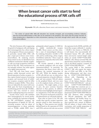

This document summarizes recent research on how breast cancer cells evade detection and killing by natural killer (NK) cells. It discusses three main mechanisms: 1) Breast cancer patients have NK cells with decreased activity and altered receptor expression, induced by tumor-produced TGFβ1. 2) Breast cancer cells modulate their immunogenicity by expressing ligands for inhibitory NK receptors and decreasing ligands for activating receptors. They also release immunosuppressive molecules like TGFβ1. 3) Breast cancer cells influence the terminal maturation of NK cells, increasing poorly differentiated and non-cytotoxic NK subsets in patients' blood and tumors. This may explain the poor cytotoxic functions observed in patients and highlights NK cell plasticity.