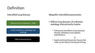
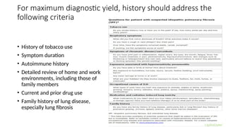
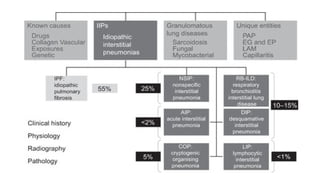
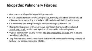
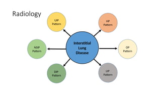
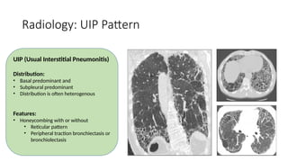
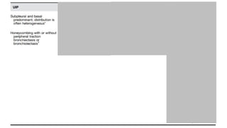
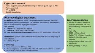
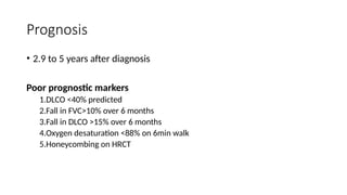
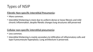
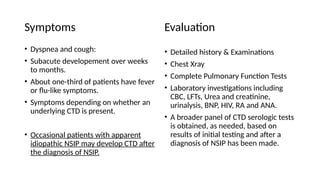
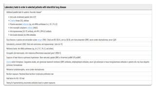
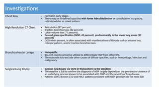
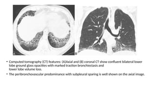
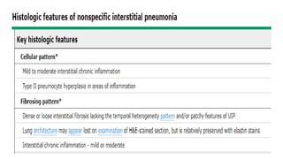
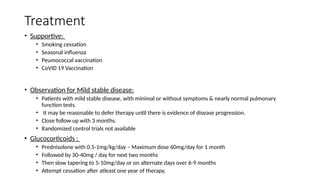
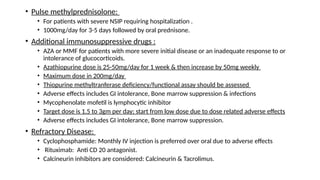
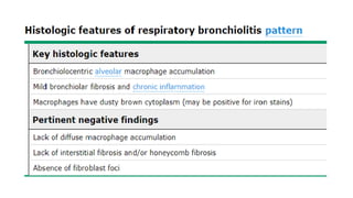
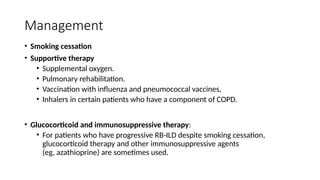
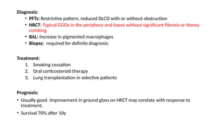
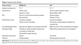
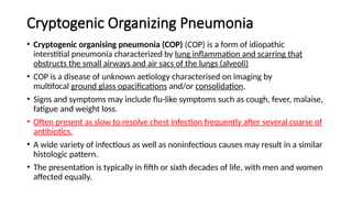
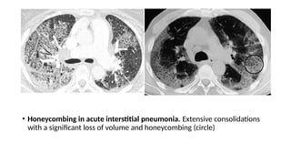
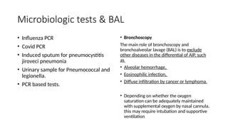
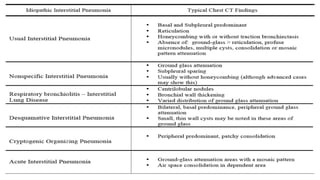
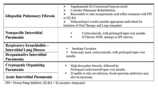
Idiopathic Interstitial
Pneumonia
Kaleemullah
SIUT Hospital Karachi
Definition
Interstitial Lung Disease Idiopathic interstitial pneumonias
• Diffuse lung diseases of unknown
aetiology that primarily involve
• Pulmonary interstitium (area between
alveolar epithelium and capillary
endothelium)
&
• Septal and bronchovascular tissues that
make up the fibrous framework of lung.
Diverse Group of Disorders ( >200)
Similar Symptoms, physiology and
radiology
Difficult Nomenclature
Symptoms and Signs
• Usually nonspecific.
• Persistent non productive cough
• Dyspnea on exertion are typical, with
variable onset and progression.
Common signs include
• Tachypnea.
• Reduced chest expansion.
• Bibasilar end-inspiratory crackles
• Digital clubbing
For maximum diagnostic yield, history should address the
following criteria
• History of tobacco use
• Symptom duration
• Autoimmune history
• Detailed review of home and work
environments, including those of
family members
• Current and prior drug use
• Family history of lung disease,
especially lung fibrosis
Investigations
Chest xray • Typically abnormal.
• But findings are not specific enough to differentiate between the various types.
Pulmonary
Function tests
• Results are restrictive, with reduced lung volumes and diffusion capacity.
• Hypoxemia is common during exercise and may be present at rest
HRCT • Distinguishes airspace from interstitial disease, is the most useful test and is always done.
• It provides assessment of the etiology, extent, and distribution of disease, and is more likely to
detect underlying or coexisting disease (eg, occult mediastinal adenopathy, cancer, emphysema).
Laboratory tests • Done for patients who have clinical features suggesting a connective tissue disorder, vasculitis, or
environmental exposure.
• ANA, ENA, RA factor and other more specific serologic tests for connective tissue diseases (eg,
RNP, SSA, SSB, scl70, Jo-1).
Transbronchial
lung biopsy
• Differentiate certain interstitial lung diseases, such as sarcoidosis and hypersensitivity
pneumonitis, but the biopsy does not yield enough tissue to diagnose the IIPs.
Bronchoalveolar
lavage
• Helps narrow the differential diagnosis in some patients and can provide information about
disease progression and response to therapy.
Surgical lung
biopsy
• Needed to confirm the diagnosis when the history and HRCT are nondiagnostic.
• Biopsy of multiple sites with a video-assisted thoracoscopic surgery (VATS) procedure is preferred
Idiopathic Pulmonary Fibrosis
• Most common idiopathic interstitial pneumonia
• IPF is a specific form of chronic, progressive, fibrosing interstitial pneumonia of
unknown cause, occurring primarily in older adults and limited to the lungs.
• It is defined by the histopathologic and/or radiologic pattern of UIP.
• Most patients present with progressive exertional shortness of breath and
chronic dry cough usually over a period of 24 months before diagnosis .
• Physical examination usually reveals fine end-inspiratory crackles and in severe
cases finger clubbing.
• Lung function tests show a restrictive pattern with decreased diffusing capacity of
the lungs for carbon monoxide (DLCO).
Pathology
• The exact etiology of the development of IPF is unknown.
• The characteristic feature involves abnormal fibroblast activation and subsequent
deposition of extracellular matrix by these cells.
• The current theory on the etiology of IPF suggests that recurrent injury to the
alveolar epithelium triggers a cascade of signaling by the immune system, leading
to fibroblast formation.
• Additional mechanisms contributing to IPF may include impaired DNA repair and
abnormal folding and processing of surfactant proteins.
• Honeycombing, particularly if it involves more than 5% of the lung volume, is an
almost 100% specific finding.
Genetic Mutations
• IPF involves several genes crucial in lung development and the response to
environmental injury, including WNT, TGF, Notch, and sonic hedgehog (SHH).
• Although most cases of IPF are idiopathic, some genetic factors that contribute to
familial cases have been identified.
• Mutations in the telomerase genes (such as TERT), surfactant genes (such
as SFTPA2), and mucin genes (such as MUC5B) have been known to lead to
pulmonary fibrosis.
• Hermansky-Pudlak syndrome is another rare autosomal recessive condition with
defects in lysosome-related organelles that lead to albinism, platelet defects, and
pulmonary fibrosis in many of the affected individuals.
Epidemiology
• Usually presents after the fifth or sixth decade of life
• More common in males
• In Europe, estimated IPF prevalence rates range from 0.33 to 2.51
• In North America, estimated prevalence rates range from 2.40 to 2.98 per 10,000.
• Incidence= 14.6 cases per 100,000 person-years
• Prevalence of 58.7 cases per 100,000 individuals.
• The prevalence of IPF typically rises with age, with the majority of patients aged
50 or older and up to 85% being more than the age of 70.
• However, the familial type of IPF tends to occur at a younger age, although it is
much less common, accounting for only 3.7% of cases.
• The reported incidence of IPF ranges between 2.3 and 5.3 cases per 100,000
people per year.[
Diagnosis of IPF
Radiology
Interstitial
Lung
Disease
NSIP
Pattern
UIP
Pattern
DIP
Pattern
LIP
Pattern
HP
Pattern
OP
Pattern
Radiology: UIP Pattern
UIP (Usual Interstitial Pneumonitis)
Distribution:
• Basal predominant and
• Subpleural predominant
• Distribution is often heterogenous
Features:
• Honeycombing with or without
• Reticular pattern
• Peripheral traction bronchiectasis or
bronchiolectasis
Histopathology Patterns
**IPF is the likely diagnosis when any of the following features are present:
• Moderate-to-severe traction bronchiectasis/bronchiolectasis (defined as mild traction bronchiectasis/bronchiolectasis in four or
more lobes including the lingual as a lobe, or moderate to severe traction bronchiectasis in two or more lobes) in a man over age 50
years or in a woman overage 60 years
• Extensive (30%) reticulation on HRCT and an age 70 years
• Increased neutrophils and/or absence of lymphocytosis in BAL fluid
• Multidisciplinary discussion reaches a confident diagnosis of IPF.
***Indeterminate for IPF
• Without an adequate biopsy is unlikely to be IPF
• With an adequate biopsy may be reclassified to a more specific diagnosis after multidisciplinary discussion and/or additional
consultation
Differential Diagnosis
UIP pattern of ILD can be seen in idiopathic pulmonary fibrosis or secondary to
underlying systemic diseases. These would include:
• Connective tissue disorders
• Rheumatoid arthritis
• Systemic sclerosis (scleroderma)
• Polymyositis/Dermatomyositis
• Mixed connective tissue disease
• Asbestosis
• Fibrotic hypersensitivity pneumonitis
• Radiation
• Medications/Drug toxicity: Amiodarone lung
Management of IPF
Supportive treatment
• Home oxygen if resting PaO2 <55 mmHg or <60mmHg with signs of PHT.
• Pulmonary rehab
• PPI for GERD
• Opioids for palliation
Pharmacological treatment:
Pirfenidone (Antifibrotic, inhibit collagen synthesis and reduce fibroblast
proliferation) used in patients with FVC between 50% and 80% predicted but
should be discontinued in progressive disease.
Take with food
i. Days 1-7: 267 mg PO TID (801 mg/day)
ii. Days 8-14: 534 mg PO TID (1602 mg/day)
iii. Day 15 and thereafter (maintenance): 801 mg PO TID; not to exceed 2403 mg/day
Nintedanib (Tyrosine kinase inhibitor) associated with reduced frequency of
exacerbations.
iv. 150 mg PO q12hr
Monitoring:
• Serial measurements of FVC and DLCO
• Disease progression is characterized by fall in FVC >10% or more within 6-12 months
• Or fall in DLCO >15% or more within 6-12 months
Lung Transplantation
• Ideally all patients should be
referred with UIP in HRCT or
Histopathology irrespective of
VC.
• Age <65y
• DLCO <40% predicted
• Fall in FVC>10% over 6 months
• Fall in DLCO >15% over 6
months
• Oxygen desaturation <88% on
6min walk
• Honeycombing on HRCT
Prognosis
• 2.9 to 5 years after diagnosis
Poor prognostic markers
1.DLCO <40% predicted
2.Fall in FVC>10% over 6 months
3.Fall in DLCO >15% over 6 months
4.Oxygen desaturation <88% on 6min walk
5.Honeycombing on HRCT
Nonspecific interstitial pneumonia
• 2nd
most common IIP after IPF
• NSIP is a chronic interstitial pneumonia that is characterized by a homogeneous
appearance of dense or loose interstitial fibrosis with mild to moderate chronic
interstitial inflammation.
• It is "nonspecific" in that it lacks the histopathologic features that characterize
usual interstitial pneumonia (UIP), desquamative interstitial pneumonia (DIP),
respiratory bronchiolitis-associated interstitial lung disease (RB-ILD), or acute
interstitial pneumonia (AIP).
• Usually occurs at the age 40-50 years.
• It may be common in Caucasian-European populations
• More common in women but idiopathic NSIP is similar in both genders
Types of NSIP
Fibrotic Non-specific Interstitial Pneumonia:
• More common.
• Interstitial thickening is more due to uniform dense or loose fibrosis and mild
chronic inflammation, despite fibrotic changes lung structures still preserved
Cellular non-specific interstitial pneumonia:
• Less common.
• Interstitial thickening is mainly secondary to infiltration of inflammatory cells and
type II pneumocyte hyperplasia. Lung architecture is preserved.
Causes of NSIP
• Connective Tissue Disease
• Polymyositis-dermatomyositis: NSIP > UIP
• Rheumatoid Arthritis : UIP > NSIP
• Sjogren’s syndrome : NSIP > UIP
• Systemic sclerosis : NSIP > UIP
• ANCA associated vasculitis : UIP > NSIP
• Interstitial pneumonia with Autoimmune
features
• Hypersensitivity pneumonitis
• Drug Induced NSIP
• Flecainide
• Methotrexate
• Nitrofurantoin
• Chlorambucil
• Carmustine
• Statins
• HIV associated NSIP
• Others
• Immunoglobulin G4 (IgG4)-
related systemic disease
• Graft versus Host Disease
• Familial interstitial pneumonia
Symptoms
• Dyspnea and cough:
• Subacute developement over weeks
to months.
• About one-third of patients have fever
or flu-like symptoms.
• Symptoms depending on whether an
underlying CTD is present.
• Occasional patients with apparent
idiopathic NSIP may develop CTD after
the diagnosis of NSIP.
Evaluation
• Detailed history & Examinations
• Chest Xray
• Complete Pulmonary Function Tests
• Laboratory investigations including
CBC, LFTs, Urea and creatinine,
urinalysis, BNP, HIV, RA and ANA.
• A broader panel of CTD serologic tests
is obtained, as needed, based on
results of initial testing and after a
diagnosis of NSIP has been made.
Investigations
Chest Xray • Normal in early stages
• There may be ill-defined opacities with lower lobe distribution or consolidation in a patchy,
reticulonodular or mixed pattern.
High Resolution CT Chest • Reticulation (87 percent),
• Traction bronchiectasis (82 percent),
• Lobar volume loss (77 percent),
• Ground glass opacification (GGO, 42 percent), predominantly in the lower lung zones (92
percent)
• GGO when present, is often associated with manifestations of fibrosis such as volume loss,
reticular pattern, and/or traction bronchiectasis.
Bronchoalveolar Lavage • Nonspecific.
• BAL cell counts cannot be utilized to differentiate NSIP from other IIPs.
• The main role is to exclude other causes of diffuse opacities, such as hemorrhage, infection and
malignancy.
Surgical Lung Biopsy • Surgical lung biopsy via VATS or thoracotomy is the standard.
• The need for a SLB to confirm the diagnosis of NSIP largely depends on the presence or absence of
an underlying process known to be associated with NSIP and the severity of lung disease.
• Patients with a known CTD and HRCT pattern consistent with NSIP generally do not need SLB
• Computed tomography (CT) features: (A)Axial and (B) coronal CT show confluent bilateral lower
lobe ground glass opacities with marked traction bronchiectasis and
lower lobe volume loss.
• The peribronchovascular predominance with subpleural sparing is well shown on the axial image.
Bilateral, peribronchovascular
ground glass attenuation
Traction bronchiectasis
A relative subpleural sparing
No honeycombing
Differential Diagnosis
• Idiopathic pulmonary fibrosis (UIP/IPF) is the IIP that has the greatest overlap with NSIP
• Diferentiated on the basis of onset, disease progression, underlying etiology, radiological &
histopathological features
Other IIPs
• Certain imaging features help to differentiate hypersensitivity pneumonitis (HP) from NSIP
• High resolution computed tomography (HRCT) findings of centrilobular nodules, mosaic air-
trapping, and upper lung zone distribution suggest HP rather than NSIP
Hypersensitivity pneumonitis
• IgG4-related systemic disease have interstitial lung disease.
• While some features are similar to NSIP
• Histopathology demonstrates infiltration of the lung interstitium with IgG4-positive plasma
cells.
Immunoglobulin G4 (IgG4)-
related systemic disease
• Familial interstitial pneumonia exhibit UIP pattern > NSIP
• HRCT characteristics of FIP: Diffuse craniocaudal distribution of opacities, whether reticular or
mixed reticular-ground glass. while UIP/IPF and NSIP are typically basilar predominant
Familial Interstitial pneumonia
• Rare genetic defect associated with oculocutaneous albinism, abnormal platelets, and fibrotic
ILD
• some patients have features more typical of NSIP, such as onset at an earlier age (eg, thirties)
reticular and ground glass opacities on HRCT, and on histopathology, temporal homogeneity,
sparse mononuclear cell infiltration, and diffuse collagen deposition
Hermans Pudlak Syndrome
Treatment
• Supportive:
• Smoking cessation
• Seasonal influenza
• Peumococcal vaccination
• CoVID 19 Vaccination
• Observation for Mild stable disease:
• Patients with mild stable disease, with minimal or without symptoms & nearly normal pulmonary
function tests.
• It may be reasonable to defer therapy until there is evidence of disease progression.
• Close follow up with 3 months.
• Randomized control trials not available
• Glucocorticoids :
• Prednisolone with 0.5-1mg/kg/day – Maximum dose 60mg/day for 1 month
• Followed by 30-40mg / day for next two months
• Then slow tapering to 5-10mg/day or on alternate days over 6-9 months
• Attempt cessation after atleast one year of therapy.
• Pulse methylprednisolone:
• For patients with severe NSIP requiring hospitalization .
• 1000mg/day for 3-5 days followed by oral prednisone.
• Additional immunosuppressive drugs :
• AZA or MMF for patients with more severe initial disease or an inadequate response to or
intolerance of glucocorticoids.
• Azathiopurine dose is 25-50mg/day for 1 week & then increase by 50mg weekly
• Maximum dose in 200mg/day
• Thiopurine methyltranferase deficiency/functional assay should be assessed
• Adverse effects includes GI intolerance, Bone marrow suppression & infections
• Mycophenolate mofetil is lymphocytic inhibitor
• Target dose is 1.5 to 3gm per day; start from low dose due to dose related adverse effects
• Adverse effects includes GI intolerance, Bone marrow suppression.
• Refractory Disease:
• Cyclophosphamide: Monthly IV injection is preferred over oral due to adverse effects
• Rituximab: Anti CD 20 antagonist.
• Calcineurin inhibitors are considered: Calcineurin & Tacrolimus.
Monitoring
• The response to therapy should be
assessed at one month, and then at
three to six month intervals, or sooner,
if the patient has more severe initial
disease or reports worsening
symptoms.
• Development of extrapulmonary
symptoms suggestive of CTD
• PFTs (eg, spirometry, DLCO, oximetry,
six-minute walk test)
• Interval high resolution computed
tomography (HRCT) is performed when
changes in the clinical
assessment and/or PFTs suggest
worsening disease or an intercurrent
process.
Prognosis
• In general, NSIP carries a much more
favourable prognosis.
• 5 years survival rate for cellular NSIP is
90%.
• 5 years survival in the fibrotic subtype
45-50%.
• Cellular NSIP shows an even better
response to corticosteroids and
carries a substantially better prognosis
than the fibrotic type
Respiratory bronchiolitis- ILD
• Respiratory (or "smoker's") bronchiolitis (RB) is a well-recognized pathological lesion
found in the lungs of many cigarette smokers that is generally not associated with
respiratory symptoms.
• The key features of RB are the pathologic findings of tan-pigmented macrophages (also
known as smokers' macrophages) in the respiratory bronchioles.
• Typically associated with heavy smoking (usually of 30 pack-years or more)
• often seen in young middle-aged patients (30-40 years of age).
Clinical features : subacute symptoms of cough and dyspnea.
• Pneumothorax, hemoptysis, and acute respiratory failure are less common initial
manifestations.
• Fine, bibasilar end-inspiratory crackles are common findings on chest examination.
Digital clubbing is un usual.
Diagnosis
The diagnosis of RB-ILD requires:
• A history of current or recent (within the past six months) cigarette smoking
• Clinical manifestations of cough or dyspnea, accompanied by crackles on chest
examination
• PFT findings of a restrictive or mixed obstructive and restrictive pattern.
• HRCT manifestations consistent with RB-ILD, especially diffuse or patchy GGO in a mosaic
pattern or fine nodules and air trapping.
• BAL findings of RB-ILD are nonspecific.
• TBB is obtained during bronchoscopy when technically feasible. As with BAL, the main
purpose is to exclude other diseases.
• Lung biopsy shows “bronchiolocentric pattern with tightly packed pigmented
macrophages” PAS positive.
• SLB is not always necessary in RB-ILD, a trial of smoking cessation may precede lung
biopsy.
• (A)Axial and (B) coronal computed tomography (CT) reconstructions in heavy cigarette smoker
show moderately extensive ground-glass opacities and centrilobular nodules (circles).
Management
• Smoking cessation
• Supportive therapy
• Supplemental oxygen.
• Pulmonary rehabilitation.
• Vaccination with influenza and pneumococcal vaccines,
• Inhalers in certain patients who have a component of COPD.
• Glucocorticoid and immunosuppressive therapy:
• For patients who have progressive RB-ILD despite smoking cessation,
glucocorticoid therapy and other immunosuppressive agents
(eg, azathioprine) are sometimes used.
Desquamative Interstitial Pneumonia
• Extensive form of RB-ILD
• Seen in smokers (60-87% cases)
• Onset of breathlessness and cough over weeks to months is typical.
• Clubbing common.
• In 20% of cases, it can also occur in the setting of CTD, Viral infections,
Drug/toxin exposure.
• Occurs in young patients typically in their 30s to 50s years of age with male
predominance 2:1.
Diagnosis:
• PFTs: Restrictive pattern, reduced DLCO with or without obstruction
• HRCT: Typical GGOs in the periphery and bases without significant fibrosis or Honey
combing
• BAL: Increase in pigmented macrophages
• Biopsy: required for definite diagnosis:
Treatment:
1. Smoking cessation
2. Oral corticosteroid therapy
3. Lung transplantation in selective patients
Prognosis:
• Usually good. Improvement in ground glass on HRCT may corelate with response to
treatment.
• Survival 70% after 10y
Cryptogenic Organizing Pneumonia
• Cryptogenic organising pneumonia (COP) (COP) is a form of idiopathic
interstitial pneumonia characterized by lung inflammation and scarring that
obstructs the small airways and air sacs of the lungs (alveoli)
• COP is a disease of unknown aetiology characterised on imaging by
multifocal ground glass opacifications and/or consolidation.
• Signs and symptoms may include flu-like symptoms such as cough, fever, malaise,
fatigue and weight loss.
• Often present as slow to resolve chest infection frequently after several coarse of
antibiotics.
• A wide variety of infectious as well as noninfectious causes may result in a similar
histologic pattern.
• The presentation is typically in fifth or sixth decades of life, with men and women
affected equally.
Causes of organizing pneumonia
• Cryptogenic OP
• OP secondary to:
• Infections(pneumonia, lung abscess, bronchiectasis)
• Drug reaction or radiotherapy
• Connective tissue disease(myositis, RA, Sjogrens)
• IBD
• Haematological malignancy
• Post bone marrow transplant
• Lung malignancy or airway obstruction
• Pulmonary infarction
Investigations
• Raised ESR, CRP and neutrophilia.
• PFTs: Mild to moderate restrictive pattern.
• Mild hypoxemia is common.
• BAL: lymphocytosis, neutrophilia and
eosinophilia.
CXR:
• Consolidations: Unilateral or Bilateral
patchy consolidations; usually
peripheral, subpleural &
peribronchovascular.
• Nodules: .
Foci of granulation tissue up
to 1 cm. May mimic neoplasm if >5 cm
in size
• CT Chest :
• Patchy consolidation with a predominantly subpleural and/or peribronchial
distribution
• Small, ill-defined peribronchial or peribronchiolar nodules
• Large nodules or masses.
• Bronchial wall thickening or dilatation in the abnormal lung regions
• Perilobular pattern with ill-defined linear opacities that are thicker than
the thickened interlobular septa
• Ground glass opacity or crazy paving
• The reverse halo sign (atoll sign) is considered to be highly specific, although
only seen in ~20% of patients with COP.
• Transbronchial lung biopsy The histologic features of organizing pneumonia can be
seen as a minor finding in other interstitial lung diseases, so reliance on small
transbronchial biopsies increases the likelihood of missing the central diagnosis.
• Gold standard is Surgical lung biopsy.
(A) peripheral consolidation and air bronchograms, (B)bronchocentric
distribution
© Consolidation, with more central ground-glass opacity, corresponding to the
reversed halo sign
Treatment
• Mild stable disease: Observation and reassessment at 8 to 12 weeks interval.
• Persistent or gradually worsening disease: Initial oral prednisolone 1-1.5mg/kg
daily for 3 months, slowly weaning the dose over 6 to 12 months.
• Fulminant disease use pulsed IV Methylprednisolone 1000mg on 3 consecutive
days followed by maintenance 0.5 to 1mg/kg/day of prednisolone.
• Azathioprine or cyclophosphamide may be considered in minimal response to
steroids.
Acute interstitial pneumonia
• Acute interstitial pneumonitis (AIP), also known as Hamman-Rich syndrome is a
rare and fulminant form of diffuse lung injury.
• AIP is similar in presentation to the ARDS and probably represents a subset of
cases of idiopathic ARDS.
• Typically affects middle-aged adults (mean ~ 50 years).
• Equal frequency in men and women.
• Symptoms onset is rapid; less then 2-3 weeks following a viral URTI
• Common initial symptoms include myalgia, arthralgia, pyrexia, chills, and malaise.
• Severe exertional dyspnoea develops over a matter of days to weeks
• The majority will have hypoxemia at presentation and most will require intubation
and mechanical ventilation within a few days.
• Widespread crackles on examination.
Evaluation
• Similar to that for acute respiratory distress syndrome.
• A plasma brain natriuretic peptide level and Echo to rule out Cardiogenic
Pulmonary Edema
• Autoimmune profile to exclude an acute exacerbation of previously undiagnosed
CTD such as poly/dermatomyosis
• ABGs often with a PaO2/FiO2 200 mmHg or less.
• CXR: B/L ground glass infiltrates sparing the costophrenic angles with air
bronchograms
• CT Chest:
• Early stage: Diffuse B/L GGOs
• Later stage: B/L consolidative lung opacities, esp: at lung bases & bronchial
dilations and architectural distortion e honey combing
• Honeycombing in acute interstitial pneumonia. Extensive consolidations
with a significant loss of volume and honeycombing (circle)
Microbiologic tests & BAL
• Influenza PCR
• Covid PCR
• Induced sputum for pneumocystitis
jiroveci pneumonia
• Urinary sample for Pneumococcal and
legionella.
• PCR based tests.
• Bronchoscopy
The main role of bronchoscopy and
bronchoalveolar lavage (BAL) is to exclude
other diseases in the differential of AIP, such
as
• Alveolar hemorrhage,
• Eosinophilic infection,
• Diffuse infiltration by cancer or lymphoma.
• Depending on whether the oxygen
saturation can be adequately maintained
with supplemental oxygen by nasal cannula,
this may require intubation and supportive
ventilation
Diagnosis
The diagnosis of acute interstitial
pneumonia (AIP) is based upon two
findings:
1. The presence of a clinical syndrome of
idiopathic acute respiratory distress
syndrome (ARDS)
&
2. Pathologic confirmation of diffuse
alveolar damage (DAD).
• Thus, after an initial clinical, laboratory,
and microbiologic evaluation, an open or
thoracoscopic lung biopsy is necessary to
confirm the diagnosis.
Treatment & Prognosis
• Mechanical ventilation and
corticosteroid therapy.
• I/V Methylpred 750mg to 1g on 3
consecuetive days followed by
maintenance therapy with 0.5 to
1mg/kg/day of prednisolone.
• Even despite mechanical ventilation, it
often carries a grave prognosis with >
70% mortality at 6 months.
Summary
Rare IIPs
• Idiopathic Lymphoid Interstitial Pneumonia
• LIP is generally associated with other clinical conditions, such as
collagen vascular disease and malignant lymphoma.
• As a rare condition, idiopathic LIP has newly been categorized within
the rare IIPs.
• LIP is rarely idiopathic; rather, it is usually a component of a
lymphoproliferative disorder, such as lymphoma, multicentric
Castleman’s disease, or IgG4-related disease, or a pulmonary
manifestation of connective tissue disease, such as Sjögren’s disease.
Epidemiology
• LIP can occur at any age. However, most of the patients are adults
with a mean age of 50 years.
• If a child presents with LIP, this can be indicative of AIDS.
• There is a recognised female predilection most likely attributable to
the fact that LIP occurs in patients with autoimmune disease such as
Sjogren syndrome, which is by far more common in women.
• Clinical presentation
• Cough (71 percent of cases) and dyspnea (61 percent), slowly progressive
over months and in rare cases several years (mean duration before
diagnosis = 19 months).
• Less frequently, patients may have systemic symptoms such as fever, night
sweat, arthralgia, and weight loss.
• Examination of the chest reveals crackles in most patients.
• If the disease progresses to the end-stage respiratory failure cyanosis and
clubbing may develop.
• Hypertrophy of the salivary glands observed in 20% of patients.
Radiographic features
• Plain radiograph
• Features can be non-specific, but may include:
• basilar reticular opacities
• nodular densities
• ground glass opacities
• consolidative opacities
• CT
• The following features may be seen with LIP on HRCT
• Mid to lower lobe predominance
• thickening of bronchovascular bundles
• interstitial thickening along lymph channels 2
• small but variable sized pulmonary nodules (can be centrilobular or subpleural,
and are often ill-defined)
• ground-glass changes
• scattered thin-walled cysts
• usually deep within the lung parenchyma
• typically abut vessels (i.e. is perivascular or subpleural)
• mediastinal lymphadenopathy
• A chest x-ray (A) shows
bilateral non-specific diffuse
interstitial coarsening and a
cyst in the right lower lobe
(arrow).
• A coronal reconstruction of a
CT scan (B) confirms the
presence of cysts in the right
lower lobe and left upper
lobe (arrows).
• An axial CT scan (C) shows
extensive bibasilar cystic
changes (dashed arrow)
and interstitial thickening
(arrow) with changes in the
left pleural space (arrowhead)
following wedge biopsy.
• A diagnosis of LIP was
confirmed by biopsy.
Histology
• As the clinical features, radiographic appearance, and BAL findings are not specific
for LIP, a definitive diagnosis requires a surgical lung biopsy.
• In LIP, there is a marked stromal infiltration by lymphoid cells, with significant
infiltration of the alveolar septa.
• The distribution of the disease is uniform. The main infiltrating cells are
lymphocytes, without atypia, plasma cells, and histiocytes. Eosinophils and
neutrophils are rare.
• Large lymphoid follicles with germinal centers are frequently present.
• Features suggesting malignant transformation include:
• lymphangitic or bronchovascular distribution,
• monoclonality,
• hilar or pleural involvement,
• bronchial wall infiltration,
• and loss of germinal center architecture.
• Once the diagnosis of LIP is ascertained, the specific cause or disease
association should be identified
• Serum protein electrophoresis (SPEP)
• Serum immunoglobulin (Ig)G4 level (consistent with IgG4-related disease if
≥2 times the upper limit of normal)
• HIV immunoassay
• Antinuclear antibody (ANA)
• Anti-Ro/SSA and anti-La/SSB antibodies
• Rheumatoid factor
Treatment and prognosis
• The natural history is variable, from near-complete resolution to
progressive disease. More than 30% of patients will develop the end-
stage disease and honeycombing despite treatment.
• Transformation to lymphoma can occur, particularly in a patient with
monoclonal gammopathy or hypogammaglobulinemia.
• Corticosteroids have been successfully trialed.
• Unclassifiable IIP
• Cases eventually diagnosed as unclassifiable IIP (UCIP) may include those with inadequate clinical,
radiologic, or pathologic data and those in which there is a major discordance between the clinical,
radiologic, and pathologic findings.
• The latter discordance may be due to therapeutic effects, an unusual variant of a recognized entity
that is not adequately characterized by the current American Thoracic Society/European Respiratory
Society (ATS/ERS) classification, or the presence of multiple radiologic and/or pathologic patterns in
one patient with idiopathic disease.
• According to the ATS/ERS, cases with autoimmune features, such as lung-dominant connective
tissue disease, are now classified as IPAF. Whether these cases should be treated as a subgroup of
connective tissue disease or IIPs is not clear. There are no established criteria to separate such cases
because of a shortage of concrete evidence. Some IPAF cases may also fulfill the criteria of UCIP.
• Further prospective studies are needed to determine the optimal approach to UCIP, but this
confusing category would benefit from a reorganization.

Idiopathic Interstitial Pneumonia by Kaleemullah

  • 1.
  • 2.
    Definition Interstitial Lung DiseaseIdiopathic interstitial pneumonias • Diffuse lung diseases of unknown aetiology that primarily involve • Pulmonary interstitium (area between alveolar epithelium and capillary endothelium) & • Septal and bronchovascular tissues that make up the fibrous framework of lung. Diverse Group of Disorders ( >200) Similar Symptoms, physiology and radiology Difficult Nomenclature
  • 3.
    Symptoms and Signs •Usually nonspecific. • Persistent non productive cough • Dyspnea on exertion are typical, with variable onset and progression. Common signs include • Tachypnea. • Reduced chest expansion. • Bibasilar end-inspiratory crackles • Digital clubbing
  • 4.
    For maximum diagnosticyield, history should address the following criteria • History of tobacco use • Symptom duration • Autoimmune history • Detailed review of home and work environments, including those of family members • Current and prior drug use • Family history of lung disease, especially lung fibrosis
  • 5.
    Investigations Chest xray •Typically abnormal. • But findings are not specific enough to differentiate between the various types. Pulmonary Function tests • Results are restrictive, with reduced lung volumes and diffusion capacity. • Hypoxemia is common during exercise and may be present at rest HRCT • Distinguishes airspace from interstitial disease, is the most useful test and is always done. • It provides assessment of the etiology, extent, and distribution of disease, and is more likely to detect underlying or coexisting disease (eg, occult mediastinal adenopathy, cancer, emphysema). Laboratory tests • Done for patients who have clinical features suggesting a connective tissue disorder, vasculitis, or environmental exposure. • ANA, ENA, RA factor and other more specific serologic tests for connective tissue diseases (eg, RNP, SSA, SSB, scl70, Jo-1). Transbronchial lung biopsy • Differentiate certain interstitial lung diseases, such as sarcoidosis and hypersensitivity pneumonitis, but the biopsy does not yield enough tissue to diagnose the IIPs. Bronchoalveolar lavage • Helps narrow the differential diagnosis in some patients and can provide information about disease progression and response to therapy. Surgical lung biopsy • Needed to confirm the diagnosis when the history and HRCT are nondiagnostic. • Biopsy of multiple sites with a video-assisted thoracoscopic surgery (VATS) procedure is preferred
  • 9.
    Idiopathic Pulmonary Fibrosis •Most common idiopathic interstitial pneumonia • IPF is a specific form of chronic, progressive, fibrosing interstitial pneumonia of unknown cause, occurring primarily in older adults and limited to the lungs. • It is defined by the histopathologic and/or radiologic pattern of UIP. • Most patients present with progressive exertional shortness of breath and chronic dry cough usually over a period of 24 months before diagnosis . • Physical examination usually reveals fine end-inspiratory crackles and in severe cases finger clubbing. • Lung function tests show a restrictive pattern with decreased diffusing capacity of the lungs for carbon monoxide (DLCO).
  • 10.
    Pathology • The exactetiology of the development of IPF is unknown. • The characteristic feature involves abnormal fibroblast activation and subsequent deposition of extracellular matrix by these cells. • The current theory on the etiology of IPF suggests that recurrent injury to the alveolar epithelium triggers a cascade of signaling by the immune system, leading to fibroblast formation. • Additional mechanisms contributing to IPF may include impaired DNA repair and abnormal folding and processing of surfactant proteins. • Honeycombing, particularly if it involves more than 5% of the lung volume, is an almost 100% specific finding.
  • 11.
    Genetic Mutations • IPFinvolves several genes crucial in lung development and the response to environmental injury, including WNT, TGF, Notch, and sonic hedgehog (SHH). • Although most cases of IPF are idiopathic, some genetic factors that contribute to familial cases have been identified. • Mutations in the telomerase genes (such as TERT), surfactant genes (such as SFTPA2), and mucin genes (such as MUC5B) have been known to lead to pulmonary fibrosis. • Hermansky-Pudlak syndrome is another rare autosomal recessive condition with defects in lysosome-related organelles that lead to albinism, platelet defects, and pulmonary fibrosis in many of the affected individuals.
  • 12.
    Epidemiology • Usually presentsafter the fifth or sixth decade of life • More common in males • In Europe, estimated IPF prevalence rates range from 0.33 to 2.51 • In North America, estimated prevalence rates range from 2.40 to 2.98 per 10,000. • Incidence= 14.6 cases per 100,000 person-years • Prevalence of 58.7 cases per 100,000 individuals. • The prevalence of IPF typically rises with age, with the majority of patients aged 50 or older and up to 85% being more than the age of 70. • However, the familial type of IPF tends to occur at a younger age, although it is much less common, accounting for only 3.7% of cases. • The reported incidence of IPF ranges between 2.3 and 5.3 cases per 100,000 people per year.[
  • 13.
  • 14.
  • 15.
    Radiology: UIP Pattern UIP(Usual Interstitial Pneumonitis) Distribution: • Basal predominant and • Subpleural predominant • Distribution is often heterogenous Features: • Honeycombing with or without • Reticular pattern • Peripheral traction bronchiectasis or bronchiolectasis
  • 23.
  • 24.
    **IPF is thelikely diagnosis when any of the following features are present: • Moderate-to-severe traction bronchiectasis/bronchiolectasis (defined as mild traction bronchiectasis/bronchiolectasis in four or more lobes including the lingual as a lobe, or moderate to severe traction bronchiectasis in two or more lobes) in a man over age 50 years or in a woman overage 60 years • Extensive (30%) reticulation on HRCT and an age 70 years • Increased neutrophils and/or absence of lymphocytosis in BAL fluid • Multidisciplinary discussion reaches a confident diagnosis of IPF. ***Indeterminate for IPF • Without an adequate biopsy is unlikely to be IPF • With an adequate biopsy may be reclassified to a more specific diagnosis after multidisciplinary discussion and/or additional consultation
  • 26.
    Differential Diagnosis UIP patternof ILD can be seen in idiopathic pulmonary fibrosis or secondary to underlying systemic diseases. These would include: • Connective tissue disorders • Rheumatoid arthritis • Systemic sclerosis (scleroderma) • Polymyositis/Dermatomyositis • Mixed connective tissue disease • Asbestosis • Fibrotic hypersensitivity pneumonitis • Radiation • Medications/Drug toxicity: Amiodarone lung
  • 27.
  • 28.
    Supportive treatment • Homeoxygen if resting PaO2 <55 mmHg or <60mmHg with signs of PHT. • Pulmonary rehab • PPI for GERD • Opioids for palliation Pharmacological treatment: Pirfenidone (Antifibrotic, inhibit collagen synthesis and reduce fibroblast proliferation) used in patients with FVC between 50% and 80% predicted but should be discontinued in progressive disease. Take with food i. Days 1-7: 267 mg PO TID (801 mg/day) ii. Days 8-14: 534 mg PO TID (1602 mg/day) iii. Day 15 and thereafter (maintenance): 801 mg PO TID; not to exceed 2403 mg/day Nintedanib (Tyrosine kinase inhibitor) associated with reduced frequency of exacerbations. iv. 150 mg PO q12hr Monitoring: • Serial measurements of FVC and DLCO • Disease progression is characterized by fall in FVC >10% or more within 6-12 months • Or fall in DLCO >15% or more within 6-12 months Lung Transplantation • Ideally all patients should be referred with UIP in HRCT or Histopathology irrespective of VC. • Age <65y • DLCO <40% predicted • Fall in FVC>10% over 6 months • Fall in DLCO >15% over 6 months • Oxygen desaturation <88% on 6min walk • Honeycombing on HRCT
  • 29.
    Prognosis • 2.9 to5 years after diagnosis Poor prognostic markers 1.DLCO <40% predicted 2.Fall in FVC>10% over 6 months 3.Fall in DLCO >15% over 6 months 4.Oxygen desaturation <88% on 6min walk 5.Honeycombing on HRCT
  • 30.
    Nonspecific interstitial pneumonia •2nd most common IIP after IPF • NSIP is a chronic interstitial pneumonia that is characterized by a homogeneous appearance of dense or loose interstitial fibrosis with mild to moderate chronic interstitial inflammation. • It is "nonspecific" in that it lacks the histopathologic features that characterize usual interstitial pneumonia (UIP), desquamative interstitial pneumonia (DIP), respiratory bronchiolitis-associated interstitial lung disease (RB-ILD), or acute interstitial pneumonia (AIP). • Usually occurs at the age 40-50 years. • It may be common in Caucasian-European populations • More common in women but idiopathic NSIP is similar in both genders
  • 31.
    Types of NSIP FibroticNon-specific Interstitial Pneumonia: • More common. • Interstitial thickening is more due to uniform dense or loose fibrosis and mild chronic inflammation, despite fibrotic changes lung structures still preserved Cellular non-specific interstitial pneumonia: • Less common. • Interstitial thickening is mainly secondary to infiltration of inflammatory cells and type II pneumocyte hyperplasia. Lung architecture is preserved.
  • 32.
    Causes of NSIP •Connective Tissue Disease • Polymyositis-dermatomyositis: NSIP > UIP • Rheumatoid Arthritis : UIP > NSIP • Sjogren’s syndrome : NSIP > UIP • Systemic sclerosis : NSIP > UIP • ANCA associated vasculitis : UIP > NSIP • Interstitial pneumonia with Autoimmune features • Hypersensitivity pneumonitis • Drug Induced NSIP • Flecainide • Methotrexate • Nitrofurantoin • Chlorambucil • Carmustine • Statins • HIV associated NSIP • Others • Immunoglobulin G4 (IgG4)- related systemic disease • Graft versus Host Disease • Familial interstitial pneumonia
  • 33.
    Symptoms • Dyspnea andcough: • Subacute developement over weeks to months. • About one-third of patients have fever or flu-like symptoms. • Symptoms depending on whether an underlying CTD is present. • Occasional patients with apparent idiopathic NSIP may develop CTD after the diagnosis of NSIP. Evaluation • Detailed history & Examinations • Chest Xray • Complete Pulmonary Function Tests • Laboratory investigations including CBC, LFTs, Urea and creatinine, urinalysis, BNP, HIV, RA and ANA. • A broader panel of CTD serologic tests is obtained, as needed, based on results of initial testing and after a diagnosis of NSIP has been made.
  • 35.
    Investigations Chest Xray •Normal in early stages • There may be ill-defined opacities with lower lobe distribution or consolidation in a patchy, reticulonodular or mixed pattern. High Resolution CT Chest • Reticulation (87 percent), • Traction bronchiectasis (82 percent), • Lobar volume loss (77 percent), • Ground glass opacification (GGO, 42 percent), predominantly in the lower lung zones (92 percent) • GGO when present, is often associated with manifestations of fibrosis such as volume loss, reticular pattern, and/or traction bronchiectasis. Bronchoalveolar Lavage • Nonspecific. • BAL cell counts cannot be utilized to differentiate NSIP from other IIPs. • The main role is to exclude other causes of diffuse opacities, such as hemorrhage, infection and malignancy. Surgical Lung Biopsy • Surgical lung biopsy via VATS or thoracotomy is the standard. • The need for a SLB to confirm the diagnosis of NSIP largely depends on the presence or absence of an underlying process known to be associated with NSIP and the severity of lung disease. • Patients with a known CTD and HRCT pattern consistent with NSIP generally do not need SLB
  • 36.
    • Computed tomography(CT) features: (A)Axial and (B) coronal CT show confluent bilateral lower lobe ground glass opacities with marked traction bronchiectasis and lower lobe volume loss. • The peribronchovascular predominance with subpleural sparing is well shown on the axial image.
  • 37.
    Bilateral, peribronchovascular ground glassattenuation Traction bronchiectasis A relative subpleural sparing No honeycombing
  • 39.
    Differential Diagnosis • Idiopathicpulmonary fibrosis (UIP/IPF) is the IIP that has the greatest overlap with NSIP • Diferentiated on the basis of onset, disease progression, underlying etiology, radiological & histopathological features Other IIPs • Certain imaging features help to differentiate hypersensitivity pneumonitis (HP) from NSIP • High resolution computed tomography (HRCT) findings of centrilobular nodules, mosaic air- trapping, and upper lung zone distribution suggest HP rather than NSIP Hypersensitivity pneumonitis • IgG4-related systemic disease have interstitial lung disease. • While some features are similar to NSIP • Histopathology demonstrates infiltration of the lung interstitium with IgG4-positive plasma cells. Immunoglobulin G4 (IgG4)- related systemic disease • Familial interstitial pneumonia exhibit UIP pattern > NSIP • HRCT characteristics of FIP: Diffuse craniocaudal distribution of opacities, whether reticular or mixed reticular-ground glass. while UIP/IPF and NSIP are typically basilar predominant Familial Interstitial pneumonia • Rare genetic defect associated with oculocutaneous albinism, abnormal platelets, and fibrotic ILD • some patients have features more typical of NSIP, such as onset at an earlier age (eg, thirties) reticular and ground glass opacities on HRCT, and on histopathology, temporal homogeneity, sparse mononuclear cell infiltration, and diffuse collagen deposition Hermans Pudlak Syndrome
  • 40.
    Treatment • Supportive: • Smokingcessation • Seasonal influenza • Peumococcal vaccination • CoVID 19 Vaccination • Observation for Mild stable disease: • Patients with mild stable disease, with minimal or without symptoms & nearly normal pulmonary function tests. • It may be reasonable to defer therapy until there is evidence of disease progression. • Close follow up with 3 months. • Randomized control trials not available • Glucocorticoids : • Prednisolone with 0.5-1mg/kg/day – Maximum dose 60mg/day for 1 month • Followed by 30-40mg / day for next two months • Then slow tapering to 5-10mg/day or on alternate days over 6-9 months • Attempt cessation after atleast one year of therapy.
  • 41.
    • Pulse methylprednisolone: •For patients with severe NSIP requiring hospitalization . • 1000mg/day for 3-5 days followed by oral prednisone. • Additional immunosuppressive drugs : • AZA or MMF for patients with more severe initial disease or an inadequate response to or intolerance of glucocorticoids. • Azathiopurine dose is 25-50mg/day for 1 week & then increase by 50mg weekly • Maximum dose in 200mg/day • Thiopurine methyltranferase deficiency/functional assay should be assessed • Adverse effects includes GI intolerance, Bone marrow suppression & infections • Mycophenolate mofetil is lymphocytic inhibitor • Target dose is 1.5 to 3gm per day; start from low dose due to dose related adverse effects • Adverse effects includes GI intolerance, Bone marrow suppression. • Refractory Disease: • Cyclophosphamide: Monthly IV injection is preferred over oral due to adverse effects • Rituximab: Anti CD 20 antagonist. • Calcineurin inhibitors are considered: Calcineurin & Tacrolimus.
  • 42.
    Monitoring • The responseto therapy should be assessed at one month, and then at three to six month intervals, or sooner, if the patient has more severe initial disease or reports worsening symptoms. • Development of extrapulmonary symptoms suggestive of CTD • PFTs (eg, spirometry, DLCO, oximetry, six-minute walk test) • Interval high resolution computed tomography (HRCT) is performed when changes in the clinical assessment and/or PFTs suggest worsening disease or an intercurrent process. Prognosis • In general, NSIP carries a much more favourable prognosis. • 5 years survival rate for cellular NSIP is 90%. • 5 years survival in the fibrotic subtype 45-50%. • Cellular NSIP shows an even better response to corticosteroids and carries a substantially better prognosis than the fibrotic type
  • 43.
    Respiratory bronchiolitis- ILD •Respiratory (or "smoker's") bronchiolitis (RB) is a well-recognized pathological lesion found in the lungs of many cigarette smokers that is generally not associated with respiratory symptoms. • The key features of RB are the pathologic findings of tan-pigmented macrophages (also known as smokers' macrophages) in the respiratory bronchioles. • Typically associated with heavy smoking (usually of 30 pack-years or more) • often seen in young middle-aged patients (30-40 years of age). Clinical features : subacute symptoms of cough and dyspnea. • Pneumothorax, hemoptysis, and acute respiratory failure are less common initial manifestations. • Fine, bibasilar end-inspiratory crackles are common findings on chest examination. Digital clubbing is un usual.
  • 44.
    Diagnosis The diagnosis ofRB-ILD requires: • A history of current or recent (within the past six months) cigarette smoking • Clinical manifestations of cough or dyspnea, accompanied by crackles on chest examination • PFT findings of a restrictive or mixed obstructive and restrictive pattern. • HRCT manifestations consistent with RB-ILD, especially diffuse or patchy GGO in a mosaic pattern or fine nodules and air trapping. • BAL findings of RB-ILD are nonspecific. • TBB is obtained during bronchoscopy when technically feasible. As with BAL, the main purpose is to exclude other diseases. • Lung biopsy shows “bronchiolocentric pattern with tightly packed pigmented macrophages” PAS positive. • SLB is not always necessary in RB-ILD, a trial of smoking cessation may precede lung biopsy.
  • 45.
    • (A)Axial and(B) coronal computed tomography (CT) reconstructions in heavy cigarette smoker show moderately extensive ground-glass opacities and centrilobular nodules (circles).
  • 47.
    Management • Smoking cessation •Supportive therapy • Supplemental oxygen. • Pulmonary rehabilitation. • Vaccination with influenza and pneumococcal vaccines, • Inhalers in certain patients who have a component of COPD. • Glucocorticoid and immunosuppressive therapy: • For patients who have progressive RB-ILD despite smoking cessation, glucocorticoid therapy and other immunosuppressive agents (eg, azathioprine) are sometimes used.
  • 48.
    Desquamative Interstitial Pneumonia •Extensive form of RB-ILD • Seen in smokers (60-87% cases) • Onset of breathlessness and cough over weeks to months is typical. • Clubbing common. • In 20% of cases, it can also occur in the setting of CTD, Viral infections, Drug/toxin exposure. • Occurs in young patients typically in their 30s to 50s years of age with male predominance 2:1.
  • 49.
    Diagnosis: • PFTs: Restrictivepattern, reduced DLCO with or without obstruction • HRCT: Typical GGOs in the periphery and bases without significant fibrosis or Honey combing • BAL: Increase in pigmented macrophages • Biopsy: required for definite diagnosis: Treatment: 1. Smoking cessation 2. Oral corticosteroid therapy 3. Lung transplantation in selective patients Prognosis: • Usually good. Improvement in ground glass on HRCT may corelate with response to treatment. • Survival 70% after 10y
  • 51.
    Cryptogenic Organizing Pneumonia •Cryptogenic organising pneumonia (COP) (COP) is a form of idiopathic interstitial pneumonia characterized by lung inflammation and scarring that obstructs the small airways and air sacs of the lungs (alveoli) • COP is a disease of unknown aetiology characterised on imaging by multifocal ground glass opacifications and/or consolidation. • Signs and symptoms may include flu-like symptoms such as cough, fever, malaise, fatigue and weight loss. • Often present as slow to resolve chest infection frequently after several coarse of antibiotics. • A wide variety of infectious as well as noninfectious causes may result in a similar histologic pattern. • The presentation is typically in fifth or sixth decades of life, with men and women affected equally.
  • 52.
    Causes of organizingpneumonia • Cryptogenic OP • OP secondary to: • Infections(pneumonia, lung abscess, bronchiectasis) • Drug reaction or radiotherapy • Connective tissue disease(myositis, RA, Sjogrens) • IBD • Haematological malignancy • Post bone marrow transplant • Lung malignancy or airway obstruction • Pulmonary infarction
  • 53.
    Investigations • Raised ESR,CRP and neutrophilia. • PFTs: Mild to moderate restrictive pattern. • Mild hypoxemia is common. • BAL: lymphocytosis, neutrophilia and eosinophilia. CXR: • Consolidations: Unilateral or Bilateral patchy consolidations; usually peripheral, subpleural & peribronchovascular. • Nodules: . Foci of granulation tissue up to 1 cm. May mimic neoplasm if >5 cm in size
  • 54.
    • CT Chest: • Patchy consolidation with a predominantly subpleural and/or peribronchial distribution • Small, ill-defined peribronchial or peribronchiolar nodules • Large nodules or masses. • Bronchial wall thickening or dilatation in the abnormal lung regions • Perilobular pattern with ill-defined linear opacities that are thicker than the thickened interlobular septa • Ground glass opacity or crazy paving • The reverse halo sign (atoll sign) is considered to be highly specific, although only seen in ~20% of patients with COP. • Transbronchial lung biopsy The histologic features of organizing pneumonia can be seen as a minor finding in other interstitial lung diseases, so reliance on small transbronchial biopsies increases the likelihood of missing the central diagnosis. • Gold standard is Surgical lung biopsy.
  • 55.
    (A) peripheral consolidationand air bronchograms, (B)bronchocentric distribution © Consolidation, with more central ground-glass opacity, corresponding to the reversed halo sign
  • 56.
    Treatment • Mild stabledisease: Observation and reassessment at 8 to 12 weeks interval. • Persistent or gradually worsening disease: Initial oral prednisolone 1-1.5mg/kg daily for 3 months, slowly weaning the dose over 6 to 12 months. • Fulminant disease use pulsed IV Methylprednisolone 1000mg on 3 consecutive days followed by maintenance 0.5 to 1mg/kg/day of prednisolone. • Azathioprine or cyclophosphamide may be considered in minimal response to steroids.
  • 57.
    Acute interstitial pneumonia •Acute interstitial pneumonitis (AIP), also known as Hamman-Rich syndrome is a rare and fulminant form of diffuse lung injury. • AIP is similar in presentation to the ARDS and probably represents a subset of cases of idiopathic ARDS. • Typically affects middle-aged adults (mean ~ 50 years). • Equal frequency in men and women. • Symptoms onset is rapid; less then 2-3 weeks following a viral URTI • Common initial symptoms include myalgia, arthralgia, pyrexia, chills, and malaise. • Severe exertional dyspnoea develops over a matter of days to weeks • The majority will have hypoxemia at presentation and most will require intubation and mechanical ventilation within a few days. • Widespread crackles on examination.
  • 58.
    Evaluation • Similar tothat for acute respiratory distress syndrome. • A plasma brain natriuretic peptide level and Echo to rule out Cardiogenic Pulmonary Edema • Autoimmune profile to exclude an acute exacerbation of previously undiagnosed CTD such as poly/dermatomyosis • ABGs often with a PaO2/FiO2 200 mmHg or less. • CXR: B/L ground glass infiltrates sparing the costophrenic angles with air bronchograms • CT Chest: • Early stage: Diffuse B/L GGOs • Later stage: B/L consolidative lung opacities, esp: at lung bases & bronchial dilations and architectural distortion e honey combing
  • 60.
    • Honeycombing inacute interstitial pneumonia. Extensive consolidations with a significant loss of volume and honeycombing (circle)
  • 61.
    Microbiologic tests &BAL • Influenza PCR • Covid PCR • Induced sputum for pneumocystitis jiroveci pneumonia • Urinary sample for Pneumococcal and legionella. • PCR based tests. • Bronchoscopy The main role of bronchoscopy and bronchoalveolar lavage (BAL) is to exclude other diseases in the differential of AIP, such as • Alveolar hemorrhage, • Eosinophilic infection, • Diffuse infiltration by cancer or lymphoma. • Depending on whether the oxygen saturation can be adequately maintained with supplemental oxygen by nasal cannula, this may require intubation and supportive ventilation
  • 62.
    Diagnosis The diagnosis ofacute interstitial pneumonia (AIP) is based upon two findings: 1. The presence of a clinical syndrome of idiopathic acute respiratory distress syndrome (ARDS) & 2. Pathologic confirmation of diffuse alveolar damage (DAD). • Thus, after an initial clinical, laboratory, and microbiologic evaluation, an open or thoracoscopic lung biopsy is necessary to confirm the diagnosis. Treatment & Prognosis • Mechanical ventilation and corticosteroid therapy. • I/V Methylpred 750mg to 1g on 3 consecuetive days followed by maintenance therapy with 0.5 to 1mg/kg/day of prednisolone. • Even despite mechanical ventilation, it often carries a grave prognosis with > 70% mortality at 6 months.
  • 63.
  • 73.
    Rare IIPs • IdiopathicLymphoid Interstitial Pneumonia • LIP is generally associated with other clinical conditions, such as collagen vascular disease and malignant lymphoma. • As a rare condition, idiopathic LIP has newly been categorized within the rare IIPs. • LIP is rarely idiopathic; rather, it is usually a component of a lymphoproliferative disorder, such as lymphoma, multicentric Castleman’s disease, or IgG4-related disease, or a pulmonary manifestation of connective tissue disease, such as Sjögren’s disease.
  • 74.
    Epidemiology • LIP canoccur at any age. However, most of the patients are adults with a mean age of 50 years. • If a child presents with LIP, this can be indicative of AIDS. • There is a recognised female predilection most likely attributable to the fact that LIP occurs in patients with autoimmune disease such as Sjogren syndrome, which is by far more common in women.
  • 75.
    • Clinical presentation •Cough (71 percent of cases) and dyspnea (61 percent), slowly progressive over months and in rare cases several years (mean duration before diagnosis = 19 months). • Less frequently, patients may have systemic symptoms such as fever, night sweat, arthralgia, and weight loss. • Examination of the chest reveals crackles in most patients. • If the disease progresses to the end-stage respiratory failure cyanosis and clubbing may develop. • Hypertrophy of the salivary glands observed in 20% of patients.
  • 76.
    Radiographic features • Plainradiograph • Features can be non-specific, but may include: • basilar reticular opacities • nodular densities • ground glass opacities • consolidative opacities
  • 77.
    • CT • Thefollowing features may be seen with LIP on HRCT • Mid to lower lobe predominance • thickening of bronchovascular bundles • interstitial thickening along lymph channels 2 • small but variable sized pulmonary nodules (can be centrilobular or subpleural, and are often ill-defined) • ground-glass changes • scattered thin-walled cysts • usually deep within the lung parenchyma • typically abut vessels (i.e. is perivascular or subpleural) • mediastinal lymphadenopathy
  • 78.
    • A chestx-ray (A) shows bilateral non-specific diffuse interstitial coarsening and a cyst in the right lower lobe (arrow). • A coronal reconstruction of a CT scan (B) confirms the presence of cysts in the right lower lobe and left upper lobe (arrows). • An axial CT scan (C) shows extensive bibasilar cystic changes (dashed arrow) and interstitial thickening (arrow) with changes in the left pleural space (arrowhead) following wedge biopsy. • A diagnosis of LIP was confirmed by biopsy.
  • 79.
    Histology • As theclinical features, radiographic appearance, and BAL findings are not specific for LIP, a definitive diagnosis requires a surgical lung biopsy. • In LIP, there is a marked stromal infiltration by lymphoid cells, with significant infiltration of the alveolar septa. • The distribution of the disease is uniform. The main infiltrating cells are lymphocytes, without atypia, plasma cells, and histiocytes. Eosinophils and neutrophils are rare. • Large lymphoid follicles with germinal centers are frequently present.
  • 80.
    • Features suggestingmalignant transformation include: • lymphangitic or bronchovascular distribution, • monoclonality, • hilar or pleural involvement, • bronchial wall infiltration, • and loss of germinal center architecture.
  • 81.
    • Once thediagnosis of LIP is ascertained, the specific cause or disease association should be identified • Serum protein electrophoresis (SPEP) • Serum immunoglobulin (Ig)G4 level (consistent with IgG4-related disease if ≥2 times the upper limit of normal) • HIV immunoassay • Antinuclear antibody (ANA) • Anti-Ro/SSA and anti-La/SSB antibodies • Rheumatoid factor
  • 82.
    Treatment and prognosis •The natural history is variable, from near-complete resolution to progressive disease. More than 30% of patients will develop the end- stage disease and honeycombing despite treatment. • Transformation to lymphoma can occur, particularly in a patient with monoclonal gammopathy or hypogammaglobulinemia. • Corticosteroids have been successfully trialed.
  • 83.
    • Unclassifiable IIP •Cases eventually diagnosed as unclassifiable IIP (UCIP) may include those with inadequate clinical, radiologic, or pathologic data and those in which there is a major discordance between the clinical, radiologic, and pathologic findings. • The latter discordance may be due to therapeutic effects, an unusual variant of a recognized entity that is not adequately characterized by the current American Thoracic Society/European Respiratory Society (ATS/ERS) classification, or the presence of multiple radiologic and/or pathologic patterns in one patient with idiopathic disease. • According to the ATS/ERS, cases with autoimmune features, such as lung-dominant connective tissue disease, are now classified as IPAF. Whether these cases should be treated as a subgroup of connective tissue disease or IIPs is not clear. There are no established criteria to separate such cases because of a shortage of concrete evidence. Some IPAF cases may also fulfill the criteria of UCIP. • Further prospective studies are needed to determine the optimal approach to UCIP, but this confusing category would benefit from a reorganization.

Editor's Notes

  • #42 For patients who relapse when prednisone is tapered or discontinued, low-dose prednisone can be maintained for a longer period. Or, if relapse occurs at higher prednisone doses, another immunosuppressive agent can be added as a glucocorticoid-sparing agent. Consider pcp prophylaxis in such pts,(20 mg steroid for >1 month) plus its gonna b difficult to differentiate worsening nsip from intercurrent pcp
  • #44 development of pulmonary hypertension, thromboembolic disease, infection, and other comorbid diseases (eg, heart failure, muscle weakness). SE if on immunosuppressive agents.
  • #45 2. a patchy submucosal and peribronchiolar infiltrate of lymphocytes and histiocytes, and peribronchiolar fibrosis that extends into contiguous alveolar walls.  3. The distinction between RB and RB-ILD is largely based on the clinical evidence of interstitial lung disease.
  • #48 The classic lesion in RB-ILD is the accumulation of tan-pigmented macrophages (also known as smokers' macrophages) within the lumens of first and second order respiratory bronchioles. The main feature that distinguishes DIP from RB-ILD is that DIP affects the lung in a more uniform and diffuse manner, lacking the bronchiolocentric distribution seen in RB-ILD, but there is considerable clinical, radiologic, and histologic overlap between these entities.  
  • #63 findings are similar to those seen in acute respiratory distress syndrome (ARDS) i.e. diffuse, bilateral, air-space opacification. bilateral, patchy, symmetric areas of ground glass attenuation airspace consolidation, septal thickening, and traction bronchiectasis.
  • #65 Most patients with AIP have a nonproductive cough, so sputum cultures are usually not available. Bronchoalveolar lavage and bronchial brushing are generally performed to obtain respiratory tract samples for immunofluorescence and culture.
  • #79  hepatosplenomegaly, lymphadenopathy, parotid gland enlargement, and arthritis depending on underlying disease involvement.
  • #81 These findings are similar to those of nonspecific interstitial pneumonia (NSIP) [24,34]. Lung cysts occur in 68 to 82 percent of patients with LIP (image 1), whereas they are rare in NSIP and pulmonary lymphoma, and can be helpful in distinguishing LIP from these entities