SlideShare a Scribd company logo
1 of 8
Download to read offline
Please cite this article in press as: Edholm, E.-S., et al., Insights into the function of IgD. Dev. Comp. Immunol. (2011), doi:10.1016/j.dci.2011.03.002
ARTICLE IN PRESS
GModel
DCI-1584; No.of Pages8
Developmental and Comparative Immunology xxx (2011) xxx–xxx
Contents lists available at ScienceDirect
Developmental and Comparative Immunology
journal homepage: www.elsevier.com/locate/dci
Insights into the function of IgD
Eva-Stina Edholm, Eva Bengten, Melanie Wilson∗
University of Mississippi Medical Center, 2500 North State Street, Jackson, MS 39216, United States
a r t i c l e i n f o
Article history:
Received 6 December 2010
Received in revised form 2 February 2011
Accepted 6 March 2011
Available online xxx
Keywords:
Teleosts
Immunoglobulin
IgD
Alternative splicing
B cells
a b s t r a c t
IgD, previously thought to be a recent addition to the immunoglobulin classes, has long been consid-
ered an enigmatic molecule. For example, it was debated if IgD had a specific function other than as an
antigen receptor co-expressed with IgM on naive B cells and if it had an important role in mammalian
immunity. However, during the past decade extensive sequencing of vertebrate genomes has shown that
IgD homologs are present in all vertebrate taxa, except for birds. Moreover, recent functional studies
indicate that IgD likely performs a unique role in vertebrate immune responses. The goal of this review is
to summarize the IgD gene organization and structural data, which demonstrate that IgD has an ancient
origin, and discuss the findings in catfish and humans that provide insight into the possible function of
this elusive immunoglobulin isotype.
© 2011 Elsevier Ltd. All rights reserved.
1. Introduction
It is well known that teleost B cells share many similarities with
mammalian B cells, including immunoglobulin (Ig) gene rearrange-
ments, allelic exclusion, production of membrane Ig and secreted
Ig forms (reviewed in Bengten et al., 2000, 2006a; Flajnik and
Kasahara, 2010; Miller et al., 1994; Solem and Stenvik, 2006; Warr,
1995) and the use of B cell receptor (BCR) accessory proteins CD79a
and CD79b for Ig signal transduction (Edholm et al., 2010; Sahoo
et al., 2008). Also, it is established that IgM is the predominant
Ig isotype found in teleost serum, while the other Ig isotypes, for
example, IgD in channel catfish, Ictalurus punctatus (Bengten et al.,
2002; Edholm et al., 2010) and IgT in rainbow trout, Oncorhynchus
mykiss (Zhang et al., 2010), are found in lesser amounts. In most
teleost, serum IgM is expressed as a tetramer, although IgM
monomers have been described in giant grouper, Epinephelus itaira
(Clem, 1971) and sheepshead, Archosargus probatocephalus (Lobb
and Clem, 1981). In contrast, serum IgT is expressed as a monomer
in rainbow trout serum, and a tetramer in gut mucous (Zhang et al.,
2010). At present serum IgD has only been described in catfish by
using Western blot analyses and whether it exists as a monomer
has yet to be determined. Currently, IgT has been identified at
the cDNA/DNA level in zebrafish Danio rerio (IgZ; Danilova et al.,
2005), common carp, Cyprinus carpio (Savan et al., 2005a), rainbow
trout (Hansen et al., 2005), Japanese pufferfish, Takifugu rubripes
(Savan et al., 2005b), grass carp, Ctenopharyngodon idella (Xiao et al.,
2010), three-spined stickleback, Gasterosteus aculeatus (Gambon-
∗ Corresponding author. Tel.: +1 601 984 1719; fax: +1 601 984 1708.
E-mail address: mwilson@umc.edu (M. Wilson).
Deza et al., 2010), and Atlantic salmon, Salmo salar (Tadiso et al.,
2010). However, PCR and genomic sequencing studies have failed
to isolate a catfish IgT gene homolog. Also, since IgT transcripts
were not found among the ∼500,000 catfish expressed sequence
tags in the NCBI database it is likely that the issue of catfish IgT will
only be resolved once the sequencing and annotation of the catfish
IgH locus (IGH) is completed. Currently, the only immunoglobulins
described in catfish are IgM and IgD and catfish IgM, like teleost
IgM in general, has been well studied and shown to be a structural
and functional homolog of mammalian IgM (Bengten et al., 2000,
2006b; Warr, 1995). In contrast, much less is known about teleost
IgD function. Hence, this review will focus on IgD and since IgD
has often been described as the most enigmatic vertebrate Ig, the
structure and function of teleost IgD is discussed in the context of
what we know concerning mammalian (and other vertebrate) IgD
structure and function.
IgD was first discovered in human serum as a myeloma pro-
tein in 1965 and then in a companion study was shown to be
present in normal serum (Rowe and Fahey, 1965a,b). Later it was
identified on the surface of B cells (Van Boxel et al., 1972). Yet a
unique function, in addition to initiating BCR signal transduction
in mature naive B cells, was not identified until recently (Chen
et al., 2009), and the significance or function of IgD has been a
subject of debate (reviewed in Geisberger et al., 2006). For exam-
ple, it was hypothesized that since anti-Ig␮ treatment of mouse
immature B cells resulted in clonal anergy or clonal deletion, the
signaling through IgD in mature B cells would result in a quali-
tative different signal from that of IgM (Nossal et al., 1979; Pike
et al., 1982). Support for this notion came from studies which used
anti-Ig␮ or anti-Ig␦ antibodies to crosslink IgM and IgD on the
surface of murine or human leukemia cell lines and these exper-
0145-305X/$ – see front matter © 2011 Elsevier Ltd. All rights reserved.
doi:10.1016/j.dci.2011.03.002
Please cite this article in press as: Edholm, E.-S., et al., Insights into the function of IgD. Dev. Comp. Immunol. (2011), doi:10.1016/j.dci.2011.03.002
ARTICLE IN PRESS
GModel
DCI-1584; No.of Pages8
2 E.-S. Edholm et al. / Developmental and Comparative Immunology xxx (2011) xxx–xxx
iments demonstrated that anti-IgM treatment resulted in growth
inhibition as indicated by decreased DNA synthesis, while anti-IgD
antibodies did not affect proliferation (Ales-Martinez et al., 1988;
Mongini et al., 1989). However, in the 1990s, studies using trans-
genic (knockout) mice that were germline deficient for either IgD
or IgM demonstrated that IgM or IgD, each by itself was capable of
functioning as a BCR and inducing normal B cell immune responses
(Lutz et al., 1998; Nitschke et al., 1993; Roes and Rajewsky, 1993).
Briefly, transgenic mice lacking IgM, expressed only IgD on pre-
and immature B cells, which not only matured normally, but also
populated peripheral tissues in a manner similar to that observed
in normal control mice that expressed both IgM and IgD. These IgM
knockout mice also mounted a fairly normal antibody response
even though antibody affinity maturation was delayed. Similar
results were also found in the studies using IgD knockout mice.
Thus, it was concluded that while IgD appears not to be required
for a normal B cell response, clearly IgD could substitute for IgM in
regards to both the B cell maturation process and immune function,
at least in mice. More recently, studies in human and catfish have
provided evidence indicating that IgD, in addition to functioning as
an Ag-binding receptor, is involved in immune responses to certain
pathogens and plays a role as a mediator of innate immunity (Chen
et al., 2009).
2. IgD is found in most major taxa of jawed vertebrates and
displays remarkable structural plasticity between species
Before the identification of an IgD homolog in catfish, IgD was
believed to represent a recently evolved Ig exclusive to primates
and rodents (Wilson et al., 1997). In fact it was only recently appre-
ciated that IgD is also found in a wide variety of other vertebrates,
including both mammals and ectotherms. Thus, it appears that IgD
and its orthologs represent an ancient Ig class, most likely as old as
IgM. The catfish IgD homolog was originally identified as a cDNA,
which consisted of a rearranged VH region (VDJ) spliced to a C␮1
domain, followed by seven novel C␦ domains, a transmembrane
(TM) region, and a short cytoplasmic tail (CYT) consisting of five
amino acids, K-V-K-I-A (Wilson et al., 1997). The finding of such
a long IgD-like transcript was surprising since human and mouse
IgD have three and two C␦ domains, respectively. A second catfish
IgD cDNA was also identified in the same cDNA library screen. This
cDNA was a partial transcript and it lacked the TM region and ended
in a predicted secreted tail (Bengten et al., 2002; Wilson et al., 1997).
Overall, the features supporting the classification of these novel
cDNAs as IgD included: (1) sequence similarities to mammalian Ig␦
as demonstrated by phylogenetic analysis, (2) location of the catfish
Ig␦ gene immediately 3 of the Ig␮ gene in the IGH chain locus, (3)
co-expression with Ig␮ in the catfish clonal 3B11 B cell line and (4)
the apparent expression of Ig␦ from a long primary transcript con-
taining a rearranged VDJ and Ig␮ and Ig␦ genes (Wilson et al., 1997).
This presence of the C␮1 domain in all catfish membrane Ig␦ tran-
scripts indicated that catfish IgD, like mammalian IgD, is produced
by alternative splicing of mRNA and not by mechanisms involving
classical isotype switching by chromosomal recombination. More-
over, the finding that Ig exons of different isotypes (IgM and IgD),
can combine was novel and later it was observed to occur in all
teleost IgD cDNAs sequenced to date (Fig. 1; Gambon-Deza et al.,
2010; Hirono et al., 2003; Hordvik, 2002; Hordvik et al., 1999; Saha
et al., 2004; Stenvik and Jorgensen, 2000). This inclusion of C␮1 in
catfish Ig␦ transcripts was hypothesized to be necessary since it
would permit the covalent association of Ig␦ H chains with catfish
IgL chains, i.e., the C␮1 domain provides the appropriately located
cysteine required for IgH–IgL chain association. The inclusion of
C␮1 or a C␮1-like sequence in Ig␦ transcripts is also observed in
artiodactyls. For example, the nucleotide identities of the C␦1 exon
in cow, Bos taurus; sheep, Ovis aries; and pig, Sus scrofa, range from
95.4% to 98.7% when compared to their respective C␮1 exon of each
species and in the pig the first CH exon in an IgD transcript can either
be a C␦1 or C␮1 (Zhao and Hammarstrom, 2003; Zhao et al., 2002).
Also, IgD has been identified in other mammalian species such as,
dogs (Rogers et al., 2006), horses (Wagner, 2006) and giant pandas
(Zhao et al., 2007). As for the non-mammalian taxa, IgD genes and
cDNAs have been described in the leopard gecko (Eublepharis mac-
ularius; Gambon-Deza and Espinel, 2008), green anole lizard (Anolis
carolinensis; Wei et al., 2009), Chinese soft-shelled turtle (Pelodis-
cus sinensis; Xu et al., 2009), Xenopus tropicalis (Ohta and Flajnik,
2006) and most recently in the platypus (Ornithorhynchus anatinus;
Zhao et al., 2009). Notably, phylogenetic analyses demonstrate that
shark IgW is an ortholog of IgD, which further illustrates the ancient
origin of this isotype (Ohta and Flajnik, 2006). IgW was originally
identified as IgNARC in the nurse shark (Ginglymostoma cirratum;
Greenberg et al., 1996), IgW in the sandbar shark (Carcharhinus
plumbeus; Berstein et al., 1996), and IgX in the clearnose skate (Raja
eglanteria; Anderson et al., 1999), reviewed by Flajnik and Rumfelt
(2000). However, it appears, at least from the present available data,
that cartilaginous fish belonging to the subclass of holocephali may
lack an IgD ortholog, since our searches of the existing database for
elephant shark (Callorhinchus milii) and a previous study of spotted
ratfish (Hydrolagus colliei; Rast et al., 1998) immunoglobulins failed
to identify any IgD gene or cDNA sequences. In addition, IgD is not
present in the galliforms (chicken, Gallus gallus; Zhao et al., 2000),
anseriforms (duck, Anas platyrhynchos; Lundqvist et al., 2001) and
in some mammals such as the rabbit and opossum (Oryctolagus
cuniculus; Lanning et al., 2003; Monodelphis domestica; Wang et al.,
2009; reviewed in Sun et al., 2010).
Here, it is also important to emphasize that structural variations
of IgD occur between different vertebrates. In placental mammals,
the Ig␦ genes and the encoded IgD proteins are structurally highly
conserved (Rogers et al., 2006; White et al., 1985; Zhao et al., 2007).
For example, all mammalian IgD genes currently sequenced are
located directly 3 of the Ig␮ gene and with the exception of rodents
all consists of eight exons. Exon 1 encodes the first Ig domain (C␦1)
and exons 2 (H1) and 3 (H2) encode the extended hinge region that
is characteristic of most mammalian IgD proteins. The second and
third Ig domains (C␦2 and C␦3) are encoded by exons 4 and 5; exon
6 encodes the hydrophilic secreted tail (␦Sec) and exons 7 and 8
encode the membrane tail (␦TM1 and ␦TM2). Mouse and rat IgD
genes consist of six exons and encode for shorter IgD forms since
these species lack the exons encoding H2 and C␦2. Also, while two
hinge regions are present in the pig and giant panda IgD genes,
these species only utilize their first hinge exon (H1) resulting in a
less extended IgD hinge in these species (supplemental Fig. 1; Zhao
et al., 2003, 2007).
In contrast, while the IgD genes of ectothermic vertebrates vary
in their number of C␦ exons, C␦ exon duplications, and the number
of IgD gene copies, the size of the expressed IgD in these species is
relatively large. The number of unique C␦ domains in these differ-
ent species ranges from 6 to 11, and some species have two exons
that encode their TM and others have only a single TM exon. Also,
some teleost express IgD transcripts that have blocks of repeated
C␦ domains and all teleost IgD molecules examined are chimeric
since they include C␮1. None of the cold-blooded vertebrate IgD
genes sequenced to date have individual exons that would encode
a hinge, i.e., the C regions are composed of Ig domains. Perhaps, the
lack of a hinge in the IgD in these species is compensated by the
length of the IgD H chain that may allow for some flexibility.
Briefly and beginning with the teleosts, in the catfish the IGH
locus spans ∼1 Mbp and contains multiple internal duplications and
transpositions. It contains three IgD genes termed IGHD1, IGHD2
and IGHD3 and each is individually linked to either a functional
Ig␮ gene or an Ig␮ pseudogene (Bengten et al., 2006a,b, 2002).
Please cite this article in press as: Edholm, E.-S., et al., Insights into the function of IgD. Dev. Comp. Immunol. (2011), doi:10.1016/j.dci.2011.03.002
ARTICLE IN PRESS
GModel
DCI-1584; No.of Pages8
E.-S. Edholm et al. / Developmental and Comparative Immunology xxx (2011) xxx–xxx 3
Fig. 1. Schematic representations of different vertebrate IgD molecules. Comparison of IgD molecules in different species. The C␦ domains are dark grey, black indicates
inclusion of C␮1, dark hatched grey indicates inclusion of C␮1-like domain, light grey indicates IgL C domains, and white boxes indicate V domains. Hinge regions are
indicated by angles and are hatched; the predicted inter-chain disulphide bonds are also shown.
IGHD1 is located directly 3 of the functional C␮ gene and consists
of 14 exons, with the exons encoding C␦2-C␦3-C␦4 repeated three
times in tandem: C␦1-(C␦2-C␦3-C␦4)3-C␦5-C␦6-C␦7-TM. This gene
encodes the chimeric membrane form of IgD and notably mem-
brane IgD transcripts do not contain exon duplications. Similar IgD
gene organizations with varying numbers of C␦2-C␦3-C␦4 block
duplications also occur in Atlantic salmon, Atlantic halibut, grass
carp, fugu and zebrafish (Hordvik, 2002; Hordvik et al., 1999; Saha
et al., 2004; Xiao et al., 2010). The other two catfish IgD genes
are found ∼500 kbp 5 of IGHD1 and are linked with their cor-
responding pseudo C␮ genes. The IGHD2 gene is truncated and
consists of intact C␦1 and C␦2 exons and a C␦3 exon that con-
tains a two-nucleotide deletion creating a frame shift. Currently,
it is unknown if this gene is functional since IGHD2 transcripts
have not been found, however there is a polyadenylation site 3
of the C␦3. IGHD3, is almost identical to IGHD1 except its termi-
nal exon encodes the secreted IgD tail (␦Sec). Thus, in catfish, one
gene encodes the IgD membrane form and a second gene encodes
secreted IgD and both are associated with functional enhancers.
In comparison, different types of IgD gene duplications occur in
pufferfish, stickleback, Atlantic salmon and Atlantic cod, Gadus
morhua. As in the catfish, the pufferfish membrane and secreted
forms of IgD are encoded by separate genes; the gene encoding
the putative secreted Ig form is linked to a separate array of D
and JH genes found 5 of the D and JH gene array linked to the
Ig␮ and membrane Ig␦ genes (Aparicio et al., 2002). Also, later in
a second study, which focused on membrane IgD, it was shown
that the pufferfish membrane IgD gene lacks the tandem C␦2-
C␦3-C␦4 duplication(s) found in ictalurids and salmonids. Instead
the pufferfish gene has a longer tandem duplication of C␦1-C␦2-
C␦3-C␦4-C␦5-C␦6 and the resulting cDNA transcript consisted of
VDJ-C␮1-(C␦1-C␦2-C␦3-C␦4-C␦5-C␦6)2-C␦7-TM (Fig. 2; Saha et al.,
2004). In the stickleback the IGH locus contains three arrays of VHn-
D-JH-C␨-D3-J4-C␮-C␦ in tandem (Bao et al., 2010). The three IgD
genes do not contain any exon duplications and are ∼90% identi-
cal at the amino acid level to each other. However, the C␦5 exon
in IGHD1 contains a nucleotide deletion. Thus, in stickleback, while
there are three sets of IGH genes, there is no evidence of a functional
difference between the IgD genes. In the salmonids, as a conse-
quence of the tetraploid ancestry, there are two IGH loci on separate
chromosomes. For example, in the Atlantic salmon each of the two
IGH loci, have multiple duplications of the IgT gene, but only one set
of the IgM and IgD genes (Yasuike et al., 2010). The corresponding
C␦ exons of the two IgD genes in the IGHA and IGHB loci are very
similar to each other and an alignment of C␦ translated exons from
Ig␦A and Ig␦1B gives an identity index of 97% (Hordvik, 2002). The
main difference is in the number of C␦2-C␦3-C␦4 tandem duplica-
tions. In Ig␦A there are three C␦2-C␦3-C␦4 tandem duplications and
in Ig␦B there are four (Yasuike et al., 2010). To date, the Atlantic cod
IgD gene appears to be the one exception to the general teleost IgD
structural pattern. It is much shorter and lacks C␦3, C␦4, C␦5, and
C␦6 exons. Instead the Atlantic cod gene contains a tandem C␦1-C␦2
duplication separated by an intron containing a short exon termed
␦y. The second C␦1-C␦2 is followed by C␦7 and two TM exons and
Please cite this article in press as: Edholm, E.-S., et al., Insights into the function of IgD. Dev. Comp. Immunol. (2011), doi:10.1016/j.dci.2011.03.002
ARTICLE IN PRESS
GModel
DCI-1584; No.of Pages8
4 E.-S. Edholm et al. / Developmental and Comparative Immunology xxx (2011) xxx–xxx
Fig. 2. Comparison of IgD gene organizations in various teleost. Schematics show only the genomic organization of the C␮ and C␦ genes. Boxes indicate the C␮ and C␦ exons,
which are labeled and color-coded. The color-coding of the different exons is based on their phylogenetic relatedness as demonstrated in supplemental Fig. 2. The tandem
exon duplications within individual IgD genes are outlined. Drawing is not to scale.
full-length transcripts consist of VDJ-C␮1-C␦1-C␦2-␦y-C␦1-C␦2-
C␦7-TM1-TM2 (Stenvik and Jorgensen, 2000). Here, it is important
to emphasize that when individual C␦ domains from different
teleost species are compared, it is clear that respective domains
are homologous, i.e., D1 domains cluster together, D2 domains
cluster together, etc. (supplemental Fig. 2). Also, previous phyloge-
netic analyses of different teleost C␦ domains suggested that exons
C␦5-C␦6-C␦7 arose from a gene duplication of exons C␦2-C␦3-C␦4
(Gambon-Deza et al., 2010; Hordvik et al., 1999) and teleost C␦1,
C␦5 and C␦6 domains clearly are related to human C␦1, C␦2 and C␦3
domains, respectively (Hordvik et al., 1999; Wilson et al., 1997).
IgD in amphibians and reptiles is similar in length to most of
the teleost IgD orthologs. Xenopus IgD transcripts consist of a rear-
ranged VDJ, eight C␦ domains and a TM; the Xenopus Ig␦ gene
contains eight C␦ exons, followed by two TM exons (Ohta and
Flajnik, 2006; Zhao et al., 2006). As for the reptiles, the IgD gene in
the green anole lizard is single copy and consists of 11 C␦ domains
followed by two TM exons (Wei et al., 2009; and supplemental Fig.
1). Currently, the only transcripts sequenced for the green anole
were obtained by nested PCR and they consists of VDJ-C␦1-C␦2-
C␦3-C␦4-TM1-TM2. In contrast, the leopard gecko has two IgD
genes that are quite different (Gambon-Deza and Espinel, 2008).
The first IgD gene is directly 3 of the IgM gene and contains 11 C␦
exons and one TM exon and a membrane IgD transcript shows that
the TM1 exon splices into a cryptic splice within the C␦7 domain.
The second IgD gene, termed IgD2, is located ∼20 kb 3 of the first
IgD gene. It contains only seven Ig domains and is predicted to be
the result of a partial duplication of the first IgD gene since the
first four and seventh exons resemble C␦ exons and exons 3 and
4 resemble IgA-like exons. Transcripts originating from this gene
have not been published.
Interestingly, all cold-blooded vertebrates, except for elasmo-
branchs, lack a typical mammalian IgD secreted exon that is usually
found 5 of the TM exons (Fig. 2 and supplemental Fig. 1) and tran-
scripts encoding a secreted IgD form are quite rare in these species.
For teleosts, these transcripts arise by two different mechanisms:
(1) the existence of a separate gene encoding a secreted IgD form
as in the catfish and pufferfish or (2) alternative splicing of a mem-
brane IgD gene as in the Atlantic salmon. As discussed above and
in Section 3, catfish secreted IgD is encoded by the IGHD3 gene
and pufferfish have a secreted IgD gene upstream of the IgM gene.
Importantly, IgD can be detected in catfish serum by using the
anti-IgDsec 2E5 monoclonal Ab (Bengten et al., 2002). As deter-
mined by western blot analyses two forms of secreted IgD of ∼130
and ∼180 kDa in size is routinely observed in sera from individual
catfish and the difference in size may be due to either the inclu-
sion or exclusion of the duplicated C␦2-C␦3-C␦4 domains (Fig. 3;
Edholm et al., 2010). Based on densitometry of Coomassie blue-
stained gels the concentration of secreted IgD in catfish serum is
∼0.04 mg/ml (Bengten et al., 2002). The evidence for an Atlantic
salmon putative secreted IgD form comes from RT-PCR studies,
which identified splice variants where the TM2 exon is spliced
directly to C␦6. This splicing generates a transcript ending in a 14
amino acid tail (Hordvik, 2002). For reptiles, only the leopard gecko
has been shown to have a secreted IgD form, which was identified
as a cDNA transcript that ends with the C␦11 exon and contains 24
additional nucleotides that are spliced out of the membrane tran-
scripts. This results in an IgD secreted form with an eight amino
acid tail (Gambon-Deza and Espinel, 2008). In contrast, different
secreted IgD (IgW or IgNARC) forms are more abundantly expressed
in the elasmobranch species: nurse shark, horned shark, Heterodon-
tus francisci, clearnose skate, bounded houndshark, Triakis scyllium
(Anderson et al., 1999; Honda et al., 2010; Rumfelt et al., 2004).
Likewise, short and long forms of IgD are found in the sarcoptery-
giian dipnoi, as represented by the African lungfish, Protopterus
annectens (Ota et al., 2003). The IgD genes in the elasmobranchs
contain six C␦ exons and alternative splicing produces both long
and short forms of secreted and membrane IgD. Specifically, long
Please cite this article in press as: Edholm, E.-S., et al., Insights into the function of IgD. Dev. Comp. Immunol. (2011), doi:10.1016/j.dci.2011.03.002
ARTICLE IN PRESS
GModel
DCI-1584; No.of Pages8
E.-S. Edholm et al. / Developmental and Comparative Immunology xxx (2011) xxx–xxx 5
Fig. 3. Catfish secreted IgD varies in size. Serum from eight different outbred catfish was visualized by Western blot analysis using anti-IgDsec mAb 2E5 followed by goat
anti-mouse Ig (H + L) HRP. The different size variants of ∼130 and 180 kDa may be due to the inclusion or exclusion of a repeated block of C␦2, C␦3, and C␦4 exons found
within the IGH3 locus that encodes the secreted IgD form. A schematic illustrating the different secreted IgD proteins is shown at left; C␦ domains are in grey and the secreted
tail is white. Molecular weight size markers in kDa are shown at right of serum panel.
secreted IgD transcripts contain six C␦ exons and short IgD tran-
scripts have only the first two C␦ exons. The short membrane IgD
transcripts also only contain the first two C␦ exons, while the long
membrane IgD transcripts contain the first four C␦ domains. Fur-
thermore, long IgD/IgW forms were identified in the nurse shark
by using immunoprecipitation protocols (Greenberg et al., 1996).
In the African lungfish membrane IgD forms have yet to be iden-
tified, but sequencing showed that secreted IgD transcripts either
contain seven C␦ exons or two C␦ exons. Finally, recent annotation
of the monotreme duck-billed platypus genome demonstrated that
the platypus IgD gene is located between the IgM and IgG1 genes
(Gambon-Deza et al., 2009; Zhao et al., 2009). It consists of 10 C␦
exons followed by two TM exons and to date a secreted exon has
not been found. Besides resembling the IgD expressed by teleosts,
amphibians and leopard gecko in its length, phylogenetic analy-
ses and amino acid comparisons show that all of the platypus C␦
domains have a corresponding domain in reptile IgD. Also a domain
by domain comparison demonstrated that platypus C␦1, C␦6 and
C␦7 domains are homologous to the mammals C␦1, C␦2 and C␦3
(Zhao et al., 2009).
Taken together the available IgD sequencing data indicate, in
marked contrast to IgM, that IgD genes have undergone numerous
alterations involving both exon duplications and deletions lead-
ing to different IgD structural forms in different species (Hordvik
et al., 1999; Ohta and Flajnik, 2006; Rogers et al., 2006; Stenvik and
Jorgensen, 2000; Tucker et al., 1980; Wagner et al., 2004; White
et al., 1985; Wilson et al., 1997; Zhao and Hammarstrom, 2003;
Zhao et al., 2002).
3. IgD-expressing B cells in humans and catfish
Most of what we know about IgD expression and function
comes from studies performed using human and mouse tissues
and cell lines, and knockout mice. Also, it was only after the initial
sequencing of IgD in catfish and in other teleosts, followed by the
widespread sequencing of different genomes, that IgD was found
in vertebrates other than primates and rodents. However, despite
the many publications demonstrating that IgD has a more ancient
origin than originally thought (see above), very few reports have
focused on the function and cellular expression of this isotype in
vertebrates, except for the studies performed in humans and mice.
Consequently, this section summarizes our recent characterization
studies of catfish IgD-bearing cell populations and how these relate
to human IgD-bearing cells.
In humans (and mice), IgM and IgD are expressed on the surface
of naive mature B cells by alternative splicing of a long primary
RNA transcript. These double positive B cells (IgM+/IgD+) make
up the majority of the peripheral B cells and upon Ag binding,
they down-regulate their IgD expression. In contrast, the major-
ity of long-lived memory B cells have class switched, undergone
somatic hypermutation and express either high affinity IgG, IgE,
or IgA (McHeyer-Williams, 2003). Interestingly, class switching is
also involved in generating a unique IgM−/IgD+ (IgD-only) B cell
population in humans, i.e., the cells use a cryptic switch region
found between the C␮ and C␦ genes (Chen et al., 2009). Human
IgD-only B cells while rare in peripheral blood (∼3.0%), have been
shown to make up a substantial population (20–25%) of the human
upper respiratory tract mucosal B cells and in the past 12 years,
several laboratories have been focusing on this unique IgD-only B
cell population (Chen et al., 2009; Kluin et al., 1995; Owens et al.,
1991; White et al., 1990; Yasui et al., 1989). Most IgD-only B cells
are found associated with tonsillar tissues. The IgD-only B cells are
larger, have more cytoplasm than conventional naive IgM+/IgD+ B
cells, and exhibit a centroblast morphology (Billian et al., 1996;
Liu et al., 1996). Notably, >90% of these IgD-only B cells utilize
IgL ␭ chains (Arpin et al., 1997) and sequence analyses showed
that these cells express highly mutated VH and VL regions. Also,
since the mutations are found scattered throughout the CDR and
FR regions it was proposed that the different V regions would prob-
ably be unable to bind Ag (Liu et al., 1996; Seifert et al., 2009;
Wilson et al., 1998; Zheng et al., 2005). Moreover, studies per-
formed by Chen et al. (2009) demonstrated that secreted IgD made
by the IgD-only B cells is bound to the surface of basophils (and
mast cells) by a calcium-mobilizing receptor. First, immunofluores-
cence staining showed that IgD is found on the surface and in the
cytoplasm of mucosal and circulating basophils, which indicates
IgD-bound immune complexes are internalized. Second, cross-
linking of basophil-bound IgD using an anti-IgD mAb triggered an
intracellular calcium flux and induced secretion of B cell activation
factors and cytokines, including BAFF (B cell-activating factor of TNF
family), APRIL (a proliferation-inducing TNF ligand), IL-3, and IL-4.
Third, IgD cross-linking induced basophils to secrete the antimicro-
bial factors cathelicidin, pentraxin-3, and ␤-defensin. In addition,
the authors showed that supernatants collected from IgD cross-
linked basophils inhibited the growth of the respiratory pathogens
Moraxella catarrhalis and Haemophilus infuenzae. However, even
though the putative IgD-binding receptor (Fc␦R) was not isolated,
the authors’ studies suggest that it is a high affinity receptor since
adding exogenous labeled IgD to cultured basophils did not lead
to an increase in surface IgD staining, indicating saturation of the
binding sites. Exogenous IgD could only be bound by the basophils
after they were stripped of their IgD and IgD binding by pre-
treated basophils was abolished if the “added” IgD was denatured
or if stripped basophils were treated with trypsin. Interestingly, in
this same study, patients with autoinflammatory syndromes were
shown to have more circulating and mucosal IgD-only B cells and
more mucosal “IgD-armed” basophils than healthy subjects, which
suggests that inflammation promotes the switch to IgD. Also, cross-
linking of IgD-armed basophils (from these patients) resulted in the
increased secretion of both IL-1 and TNF, which suggests that IgD
bound to the surface of basophils in the respiratory mucosa is able
to trigger an innate immune response (Chen et al., 2009).
As for catfish IgD, in the same study it was shown by using
flow cytometry that catfish express two types of IgD+ B cell popu-
Please cite this article in press as: Edholm, E.-S., et al., Insights into the function of IgD. Dev. Comp. Immunol. (2011), doi:10.1016/j.dci.2011.03.002
ARTICLE IN PRESS
GModel
DCI-1584; No.of Pages8
6 E.-S. Edholm et al. / Developmental and Comparative Immunology xxx (2011) xxx–xxx
Fig. 4. Catfish express three types of IgD+
-bearing B cells. The single IgM+
(IgM+
/IgD−
), double positive IgM+
/IgD+
, and IgD+
-only (IgM−
/IgD+
) B cells are depicted with their
membrane immunoglobulins associated with CD79 accessory molecules. IgM+
B cells produce secreted IgM tetramers and the IgD+
-only B-cells are the main producers of
“V-less” secreted IgD (ref). Immunoglobulin domains are represented as in Fig. 2: C␮, black; C␦, dark grey, CL, light grey; V, white. The CD79a and CD79b Ig domains are
stripped and Y marks the location of the two tyrosine residues in the immunoreceptor tyrosine-based activation motifs.
lations (see below) and a population of circulating granular cells
that are armed with exogenous IgD via a putative IgD-binding
receptor. Currently, the origin of these granular cells is unknown,
however at the message level these cells did not express message
for either membrane or secreted IgD, IgM, or TCR and they did
not react with an anti-catfish neutrophil mAb. However, these IgD-
granulocytes could not be isolated via direct selection techniques as
this invariably resulted in degranulation and granulocyte destruc-
tion. Recently, a more detailed characterization of catfish IgD-only
B cells was published by Edholm et al. (2010). Basically, catfish
express three different types of B cells: single positive IgM+/IgD− B
cells, double positive IgM+/IgD+ B cells and IgD-only B cells (Fig. 4).
The IgM+/IgD− and IgM+/IgD+ B cells are small agranular lympho-
cytes and are morphologically indistinguishable from each other,
while the IgD-only B cells resemble human IgD-only B cells. They
are large, exhibit a plasmablast morphology and have a higher cyto-
plasm to nucleus ratio as compared to catfish IgM+ B cells and in
contrast to mammalian IgD-only B cells, catfish IgD-only B cells
can represent as much as 60–80% of the total peripheral blood B
cells, depending on the individual fish. Notably, as shown by cell
sorting experiments, co-immunoprecipitations, and RT-PCR, cat-
fish IgD-only B cells exclusively utilize IgL ␴ chains, an IgL isotype
restricted to ectothermic vertebrates. Also, even though the cat-
fish IgM+/IgD− and IgM+/IgD+ B cell populations have the potential
to utilize any of the four different IgL chain isotypes (IgL F, IgL G,
IgL ␭ and IgL ␴) most IgM+ B cells utilize the IgL ␬ orthologs, F
and G, i.e., only 2–5% of IgM+ B cells isolated from peripheral blood
express IgL ␴ or IgL ␭ chains, isotypes known to have very limited
repertoires in catfish (Edholm et al., 2009). Currently, while the dif-
ferent roles of catfish IgM+/IgD+ and IgD-only B cells have not been
defined, we hypothesize that catfish membrane IgD functions as
a typical Ag binding receptor. Both cell types express message for
membrane IgD with functionally rearranged VDJ regions and the
B cell accessory signaling molecules CD79a and CD79b. Also, co-
immunoprecipitation experiments demonstrate that membrane
IgD associates with IgL chains, and requires association of CD79
molecules for B cell surface expression. Furthermore, no bias in the
V, D or J family usage of membrane IgD transcripts was observed in
the different IgD-expressing B cell types. In addition, anti-IgD cross-
linking in IgM+/IgD+ B cells induces a calcium flux, albeit it is slower
than the response induced by IgM cross-linking of IgM+/IgD− and
IgM+/IgD+ B cells (Edholm unpublished). Whether this is due to
Please cite this article in press as: Edholm, E.-S., et al., Insights into the function of IgD. Dev. Comp. Immunol. (2011), doi:10.1016/j.dci.2011.03.002
ARTICLE IN PRESS
GModel
DCI-1584; No.of Pages8
E.-S. Edholm et al. / Developmental and Comparative Immunology xxx (2011) xxx–xxx 7
lower levels of IgD expression on the surface of IgD+ B cells and/or
the nature of the anti-IgD mAb has not been determined.
Probably the most surprising observation from the catfish IgD+
B cell studies was the finding that secreted IgD transcripts isolated
from sorted IgD-only B cells lacked V regions, instead the C␦1 was
directly spliced to a functional leader sequence located ∼8 kb 5
of the C␦1 in the “IgD secreted form” IGHD3 gene (Edholm et al.,
2010). The isolation of catfish V-less secreted IgD transcripts is
intriguing especially when considering Chen et al. (2009) showed
that both M. catarrhalis and H. influenzae bound IgD secreted by
stimulated human IgD-only B cells, regardless of the IgD’s Ag speci-
ficity. Also, as previously demonstrated (Riesbeck and Nordstrom,
2006), this secreted IgD bound M. catarrhalis IgD-binding protein
(MID), which is also expressed by H. influenzae. MID is an ∼200 kDa
adhesin with two functional domains, one mediates adherence to
mucosal epithelial cells and the other binds to the C␦1 domain of
IgD and studies show that the purified protein activates IgD+ B
cells. Therefore, our working hypothesis for catfish IgD function
is that IgD-only B cells, and possibly a subset of IgM+/IgD+ B cells,
becomes activated by a certain “pathogen(s)” that can bind IgD.
Once activated, these cells secrete V-less IgD, which may function
as a pattern recognition molecule when bound to circulating
granulocytes. This may also explain the low and varying levels
of secreted IgD in catfish serum. In turn, the granulocytes upon
binding the “target pathogen” through the IgD Fc-region would
become activated. Here, it should be noted that the frequency
of IgD-only B cells and IgD-bearing granulocytes varies among
individual fish and the presence of these two populations do
not always correlate (Edholm et al., 2010 and unpublished).
Consequently, our studies are focused on identifying possible
“target pathogens” or IgD-only B cell activation triggering events
and isolating and determining the nature of the IgD-bearing
granulocytes. Additionally, another important question that needs
to be addressed is the relationship or lineage of the different IgD+
B cell populations. It may be that the catfish IgM+/IgD+ B cells
represent traditional naive B lymphocytes, which upon Ag acti-
vation differentiate into IgM producing plasma cells or memory
B cells, while the IgD-only B cells constitute a separate B cell
linage that expands in response to a yet unknown stimulus. Alter-
natively, it may be that the small population of IgM+/IgD+/IgL
␴+ B cells upon activation differentiate into IgD-only B
cells.
4. Conclusion
In conclusion and as noted by others (Flajnik and Kasahara,
2010; Ohta and Flajnik, 2006; Zhao et al., 2009) IgD repre-
sents an ancient Ig isotype that is found in all vertebrate taxa,
except for birds. Moreover, IgD has been shown to display
remarkable plasticity as illustrated by the occurrence of multiple
structural variants and splice forms in the different vertebrates.
Also the changes in the IgD genes that result in long chimeric
molecules in teleost as compared to shorter hinge containing
molecules in placental mammals indicates that IgD has been sub-
jected to species-specific adaptations. For example, the finding
of IgD-only B cells and the fact that secreted IgD can be bound
by granulocytes via an IgD-binding receptor suggests that IgD
plays a direct role in immune defense at least in these two
species.
Acknowledgements
This work was supported by grants from the U.S. Department of
Agriculture (2006-35204-16880), and UMMC IRSP (59908).
Appendix A. Supplementary data
Supplementary data associated with this article can be found, in
the online version, at doi:10.1016/j.dci.2011.03.002.
References
Ales-Martinez, J.E., Warner, G.L., Scott, D.W., 1988. Immunoglobulins D and M medi-
ate signals that are qualitatively different in B cells with an immature phenotype.
Proc. Natl. Acad. Sci. U.S.A. 85, 6919–6923.
Anderson, M.K., Strong, S.J., Litman, R.T., Luer, C.A., Amemiya, C.T., Rast, J.P., Litman,
G.W., 1999. A long form of the skate IgX gene exhibits a striking resemblance to
the new shark IgW and IgNARC genes. Immunogenetics 49, 56–67.
Aparicio, S., Chapman, J., Stupka, E., Putnam, N., Chia, J.M., Dehal, P., Christoffels, A.,
Rash, S., Hoon, S., Smit, A., Gelpke, M.D., Roach, J., Oh, T., Ho, I.Y., Wong, M., Detter,
C., Verhoef, F., Predki, P., Tay, A., Lucas, S., Richardson, P., Smith, S.F., Clark, M.S.,
Edwards, Y.J., Doggett, N., Zharkikh, A., Tavtigian, S.V., Pruss, D., Barnstead, M.,
Evans, C., Baden, H., Powell, J., Glusman, G., Rowen, L., Hood, L., Tan, Y.H., Elgar,
G., Hawkins, T., Venkatesh, B., Rokhsar, D., Brenner, S., 2002. Whole-genome
shotgun assembly and analysis of the genome of Fugu rubripes. Science 297,
1301–1310.
Arpin, C., de Bouteiller, O., Razanajaona, D., Briere, F., Banchereau, J., Lebecque,
S., Liu, Y.J., 1997. Human peripheral B cell development. sIgM−
IgD+
CD38+
hypermutated germinal center centroblasts preferentially express Ig lambda
light chain and have undergone mu-to-delta switch. Ann. N. Y. Acad. Sci. 815,
193–198.
Bao, Y., Wang, T., Guo, Y., Zhao, Z., Li, N., Zhao, Y., 2010. The immunoglobulin gene
loci in the teleost Gasterosteus aculeatus. Fish Shellfish Immunol. 28, 40–48.
Bengten, E., Clem, L.W., Miller, N.W., Warr, G.W., Wilson, M., 2006a. Channel cat-
fish immunoglobulins: repertoire and expression. Dev. Comp. Immunol. 30,
77–92.
Bengten, E., Quiniou, S., Hikima, J., Waldbieser, G., Warr, G.W., Miller, N.W., Wilson,
M., 2006b. Structure of the catfish IGH locus: analysis of the region including
the single functional IGHM gene. Immunogenetics 58, 831–844.
Bengten, E., Quiniou, S.M., Stuge, T.B., Katagiri, T., Miller, N.W., Clem, L.W., Warr,
G.W., Wilson, M., 2002. The IgH locus of the channel catfish, Ictalurus punctatus,
contains multiple constant region gene sequences: different genes encode heavy
chains of membrane and secreted IgD. J. Immunol. 169, 2488–2497.
Bengten, E., Wilson, M., Miller, N., Clem, L.W., Pilstrom, L., Warr, G.W., 2000.
Immunoglobulin isotypes: structure, function, and genetics. Curr. Top. Micro-
biol. Immunol. 248, 189–219.
Berstein, R.M., Schluter, S.F., Shen, S., Marchalonis, J.J., 1996. A new high molecular
weight immunoglobulin class from the carcharhine shark: implications for the
properties of the primordial immunoglobulin. Proc. Natl. Acad. Sci. U.S.A. 93,
3289–3293.
Billian, G., Bella, C., Mondiere, P., Defrance, T., 1996. Identification of a tonsil IgD+
B
cell subset with phenotypical and functional characteristics of germinal center
B cells. Eur. J. Immunol. 26, 1712–1719.
Chen, K., Xu, W., Wilson, M., He, B., Miller, N.W., Bengten, E., Edholm, E.S., Santini,
P.A., Rath, P., Chiu, A., Cattalini, M., Litzman, J., Huang, J.B.B., Meini, B., Riesbeck,
A., Cunningham-Rundles, K., Plebani, C., Cerutti, A.A., 2009. Immunoglobulin D
enhances immune surveillance by activating antimicrobial, proinflammatory
and B cell-stimulating programs in basophils. Nat. Immunol. 10, 889–898.
Clem, L.W., 1971. Phylogeny of immunoglobulin structure and function. IV.
Immunoglobulins of the giant grouper, Epinephelus itaira. J. Biol. Chem. 246,
9–15.
Danilova, N., Bussmann, J., Jekosch, K., Steiner, L.A., 2005. The immunoglobulin
heavy-chain locus in zebrafish: identification and expression of a previously
unknown isotype, immunoglobulin Z. Nat. Immunol. 6, 295–302.
Edholm, E.S., Bengten, E., Stafford, J.L., Sahoo, M., Taylor, E.B., Miller, N.W., Wilson, M.,
2010. Identification of two IgD+
B cell populations in channel catfish, Ictalurus
punctatus. J. Immunol. 185, 4082–4094.
Edholm, E.S., Wilson, M., Sahoo, M., Miller, N.W., Pilstrom, L., Wermenstam, N.E.,
Bengten, E., 2009. Identification of Igsigma and Iglambda in channel catfish,
Ictalurus punctatus, and Iglambda in Atlantic cod, Gadus morhua. Immunogenet-
ics 61, 353–370.
Flajnik, M., Rumfelt, L.L., 2000. The immune system of cartilaginous fish. Curr. Top.
Microbiol. Immunol. 248, 249–270.
Flajnik, M.F., Kasahara, M., 2010. Origin and evolution of the adaptive immune sys-
tem: genetic events and selective pressures. Nat. Rev. Genet. 11, 47–59.
Gambon-Deza, F., Espinel, C.S., 2008. IgD in the reptile leopard gecko. Mol. Immunol.
45, 3470–3476.
Gambon-Deza, F., Sanchez-Espinel, C., Magadan-Mompo, S., 2009. The immunoglob-
ulin heavy chain locus in the platypus (Ornithorhynchus anatinus). Mol. Immunol.
46, 2515–2523.
Gambon-Deza, F., Sanchez-Espinel, C., Magadan-Mompo, S., 2010. Presence of an
unique IgT on the IGH locus in three-spined stickleback fish (Gasterosteus aculea-
tus) and the very recent generation of a repertoire of VH genes. Dev. Comp.
Immunol. 34, 114–122.
Geisberger, R., Lamers, M., Achatz, G., 2006. The riddle of the dual expression of IgM
and IgD. Immunology 118, 429–437.
Greenberg, A.S., Hughes, A.L., Guo, J., Avila, D., McKinney, E.C., Flajnik, M.F., 1996.
A novel “chimeric” antibody class in cartilaginous fish: IgM may not be the
primordial immunoglobulin. Eur. J. Immunol. 26, 1123–1129.
Please cite this article in press as: Edholm, E.-S., et al., Insights into the function of IgD. Dev. Comp. Immunol. (2011), doi:10.1016/j.dci.2011.03.002
ARTICLE IN PRESS
GModel
DCI-1584; No.of Pages8
8 E.-S. Edholm et al. / Developmental and Comparative Immunology xxx (2011) xxx–xxx
Hansen, J.D., Landis, E.D., Phillips, R.B., 2005. Discovery of a unique Ig heavy-
chain isotype (IgT) in rainbow trout: implications for a distinctive B cell
developmental pathway in teleost fish. Proc. Natl. Acad. Sci. U.S.A. 102,
6919–6924.
Hirono, I., Nam, B.H., Enomoto, J., Uchino, K., Aoki, T., 2003. Cloning and character-
isation of a cDNA encoding Japanese flounder Paralichthys olivaceus IgD. Fish
Shellfish Immunol. 15, 63–70.
Honda, Y., Kondo, H., Caipang, C.M., Hirono, I., Aoki, T., 2010. cDNA cloning of the
immunoglobulin heavy chain genes in banded houndshark Triakis scyllium. Fish
Shellfish Immunol. 29, 854–861.
Hordvik, I., 2002. Identification of a novel immunoglobulin delta transcript and
comparative analysis of the genes encoding IgD in Atlantic salmon and Atlantic
halibut. Mol. Immunol. 39, 85–91.
Hordvik, I., Thevarajan, J., Samdal, I., Bastani, N., Krossoy, B., 1999. Molecular cloning
and phylogenetic analysis of the Atlantic salmon immunoglobulin D gene. Scand.
J. Immunol. 50, 202–210.
Kluin, P.M., Kayano, H., Zani, V.J., Kluin-Nelemans, H.C., Tucker, P.W., Satterwhite,
E., Dyer, M.J., 1995. IgD class switching: identification of a novel recombination
site in neoplastic and normal B cells. Eur. J. Immunol. 25, 3504–3508.
Lanning, D.K., Zhai, S.K., Knight, K.L., 2003. Analysis of the 3 Cmu region of the rabbit
Ig heavy chain locus. Gene 309, 135–144.
Liu, Y.J., de Bouteiller, O., Arpin, C., Briere, F., Galibert, L., Ho, S., Martinez-Valdez, H.,
Banchereau, J., Lebecque, S., 1996. Normal human IgD+
IgM−
germinal center B
cells can express up to 80 mutations in the variable region of their IgD transcripts.
Immunity 4, 603–613.
Lobb, C.J., Clem, L.W., 1981. Phylogeny of immunoglobulin structure and function-
XII. Secretory immunoglobulins in the bile of the marine teleost Archosargus
probatocephalus. Mol. Immunol. 18, 615–619.
Lundqvist, M.L., Middleton, D.L., Hazard, S., Warr, G.W., 2001. The immunoglobulin
heavy chain locus of the duck. Genomic organization and expression of D, J, and
C region genes. J. Biol. Chem. 276, 46729–46736.
Lutz, C., Ledermann, B., Kosco-Vilbois, M.H., Ochsenbein, A.F., Zinkernagel, R.M.,
Kohler, G., Brombacher, F., 1998. IgD can largely substitute for loss of IgM func-
tion in B cells. Nature 393, 797–801.
McHeyer-Williams, M., 2003. B-cell signaling mechanisms and activation. In: Paul,
W.E. (Ed.), Fundamental Immunology. , 5th edition, pp. 195–2285.
Miller, N.W., Rycyzyn, M.A., Wilson, M.R., Warr, G.W., Naftel, J.P., Clem, L.W., 1994.
Development and characterization of channel catfish long term B cell lines. J.
Immunol. 152, 2180–2189.
Mongini, P.K., Blessinger, C., Posnett, D.N., Rudich, S.M., 1989. Membrane IgD and
membrane IgM differ in capacity to transduce inhibitory signals within the same
human B cell clonal populations. J. Immunol. 143, 1565–1574.
Nitschke, L., Kosco, M.H., Kohler, G., Lamers, M.C., 1993. Immunoglobulin D-deficient
mice can mount normal immune responses to thymus-independent and -
dependent antigens. Proc. Natl. Acad. Sci. U.S.A. 90, 1887–1891.
Nossal, G.J., Pike, B.L., Battye, F.L., 1979. Mechanisms of clonal abortion tolerogenesis.
II. Clonal behaviour of immature B cells following exposure to anti-mu chain
antibody. Immunology 37, 203–215.
Ohta, Y., Flajnik, M., 2006. IgD, like IgM, is a primordial immunoglobulin class perpet-
uated in most jawed vertebrates. Proc. Natl. Acad. Sci. U.S.A. 103, 10723–10728.
Ota, T., Rast, J.P., Litman, G.W., Amemiya, C.T., 2003. Lineage-restricted retention of
a primitive immunoglobulin heavy chain isotype within the Dipnoi reveals an
evolutionary paradox. Proc. Natl. Acad. Sci. U.S.A. 100, 2501–2506.
Owens Jr., J.D., Finkelman, F.D., Mountz, J.D., Mushinski, J.F., 1991. Nonhomolo-
gous recombination at sites within the mouse JH-C delta locus accompanies
C mu deletion and switch to immunoglobulin D secretion. Mol. Cell. Biol. 11,
5660–5670.
Pike, B.L., Boyd, A.W., Nossal, G.J., 1982. Clonal anergy: the universally anergic B
lymphocyte. Proc. Natl. Acad. Sci. U.S.A. 79, 2013–2017.
Rast, J.P., Amemiya, C.T., Litman, R.T., Strong, S.J., Litman, G.W., 1998. Distinct pat-
terns of IgH structure and organization in a divergent lineage of chrondrichthyan
fishes. Immunogenetics 47, 234–245.
Riesbeck, K., Nordstrom, T., 2006. Structure and immunological action of the human
pathogen Moraxella catarrhalis IgD-binding protein. Crit. Rev. Immunol. 26,
353–376.
Roes, J., Rajewsky, K., 1993. Immunoglobulin D (IgD)-deficient mice reveal an auxil-
iary receptor function for IgD in antigen-mediated recruitment of B cells. J. Exp.
Med. 177, 45–55.
Rogers, K.A., Richardson, J.P., Scinicariello, F., Attanasio, R., 2006. Molecular charac-
terization of immunoglobulin D in mammals: immunoglobulin heavy constant
delta genes in dogs, chimpanzees and four old world monkey species. Immunol-
ogy 118, 88–100.
Rowe, D.S., Fahey, J.L., 1965a. A new class of human immunoglobulins I. A unique
myeloma protein. J. Exp. Med. 121, 171–184.
Rowe, D.S., Fahey, J.L., 1965b. A new class of human immunoglobulins. II. Normal
serum Igd. J. Exp. Med. 121, 185–199.
Rumfelt, L.L., Diaz, M., Lohr, R.L., Mochon, E., Flajnik, M.F., 2004. Unprecedented
multiplicity of Ig transmembrane and secretory mRNA forms in the cartilaginous
fish. J. Immunol. 173, 1129–1139.
Saha, N.R., Suetake, H., Kikuchi, K., Suzuki, Y., 2004. Fugu immunoglobulin D: a
highly unusual gene with unprecedented duplications in its constant region.
Immunogenetics 56, 438–447.
Sahoo, M., Edholm, E.S., Stafford, J.L., Bengten, E., Miller, N.W., Wilson, M., 2008. B
cell receptor accessory molecules in the channel catfish, Ictalurus punctatus. Dev.
Comp. Immunol. 32, 1385–1397.
Savan, R., Aman, A., Nakao, M., Watanuki, H., Sakai, M., 2005a. Discovery of a novel
immunoglobulin heavy chain gene chimera from common carp (Cyprinus carpio
L.). Immunogenetics 57, 458–463.
Savan, R., Aman, A., Sato, K., Yamaguchi, R., Sakai, M., 2005b. Discovery of a new class
of immunoglobulin heavy chain from fugu. Eur. J. Immunol. 35, 3320–3331.
Seifert, M., Steimle-Grauer, S.A., Goossens, T., Hansmann, M.L., Brauninger, A., Kup-
pers, R., 2009. A model for the development of human IgD-only B cells: genotypic
analyses suggest their generation in superantigen driven immune responses.
Mol. Immunol. 46, 630–639.
Solem, S.T., Stenvik, J., 2006. Antibody repertoire development in teleosts – a review
with emphasis on salmonids and Gadus morhua L. Dev. Comp. Immunol. 30,
57–76.
Stenvik, J., Jorgensen, T.O., 2000. Immunoglobulin D (IgD) of Atlantic cod has a unique
structure. Immunogenetics 51, 452–461.
Sun, Y., Wei, Z., Hammarstrom, L., Zhao, Y., 2010. The immunoglobulin delta gene in
jawed vertebrates: a comparative overview. Dev. Comp. Immunol..
Tadiso, T.M., Lie, K.K., Hordvik, I., 2010. Molecular cloning of IgT from Atlantic salmon,
and analysis of the relative expression of tau, mu and delta in different tissues.
Vet. Immunol. Immunopathol..
Tucker, P.W., Liu, C.P., Mushinski, J.F., Blattner, F.R., 1980. Mouse immunoglobulin
D: messenger RNA and genomic DNA sequences. Science 209, 1353–1360.
Van Boxel, J.A., Paul, W.E., Terry, W.D., Green, I., 1972. Communications. IgD-bearing
human lymphocytes. J. Immunol. 109, 648–651.
Wagner, B., 2006. Immunoglobulins and immunoglobulin genes of the horse. Dev.
Comp. Immunol. 30, 155–164.
Wagner, B., Miller, D.C., Lear, T.L., Antczak, D.F., 2004. The complete map of the Ig
heavy chain constant gene region reveals evidence for seven IgG isotypes and
for IgD in the horse. J. Immunol. 173, 3230–3242.
Wang, X., Olp, J.J., Miller, R.D., 2009. On the genomics of immunoglobulins in the gray,
short-tailed opossum Monodelphis domestica. Immunogenetics 61, 581–596.
Warr, G.W., 1995. The immunoglobulin genes of fish. Dev. Comp. Immunol. 19, 1–12.
Wei, Z., Wu, Q., Ren, L., Hu, X., Guo, Y., Warr, G.W., Hammarstrom, L., Li, N., Zhao, Y.,
2009. Expression of IgM, IgD, and IgY in a reptile, Anolis carolinensis. J. Immunol.
183, 3858–3864.
White, M.B., Shen, A.L., Word, C.J., Tucker, P.W., Blattner, F.R., 1985. Human
immunoglobulin D: genomic sequence of the delta heavy chain. Science 228,
733–737.
White, M.B., Word, C.J., Humphries, C.G., Blattner, F.R., Tucker, P.W., 1990.
Immunoglobulin D switching can occur through homologous recombination in
human B cells. Mol. Cell. Biol. 10, 3690–3699.
Wilson, M., Bengten, E., Miller, N.W., Clem, L.W., Du Pasquier, L., Warr, G.W., 1997. A
novel chimeric Ig heavy chain from a teleost fish shares similarities to IgD. Proc.
Natl. Acad. Sci. U.S.A. 94, 4593–4597.
Wilson, P.C., de Bouteiller, O., Liu, Y.J., Potter, K., Banchereau, J., Capra, J.D., Pas-
cual, V., 1998. Somatic hypermutation introduces insertions and deletions into
immunoglobulin V genes. J. Exp. Med. 187, 59–70.
Xiao, F.S., Wang, Y.P., Yan, W., Chang, M.X., Yao, W.J., Xu, Q.Q., Wang, X.X., Gao, Q., Nie,
P., 2010. Ig heavy chain genes and their locus in grass carp Ctenopharyngodon
idella. Fish Shellfish Immunol. 29, 594–599.
Xu, Z., Wang, G.L., Nie, P., 2009. IgM, IgD and IgY and their expression pattern in the
Chinese soft-shelled turtle Pelodiscus sinensis. Mol. Immunol. 46, 2124–2132.
Yasui, H., Akahori, Y., Hirano, M., Yamada, K., Kurosawa, Y., 1989. Class switch from
mu to delta is mediated by homologous recombination between sigma mu and
sigma mu sequences in human immunoglobulin gene loci. Eur. J. Immunol. 19,
1399–1403.
Yasuike, M., de Boer, J., von Schalburg, K.R., Cooper, G.A., McKinnel, L., Messmer,
A., So, S., Davidson, W.S., Koop, B.F., 2010. Evolution of duplicated IgH loci in
Atlantic salmon, Salmo salar. BMC Genomics 11, 486.
Zhang, Y.A., Salinas, I., Li, J., Parra, D., Bjork, S., Xu, Z., LaPatra, S.E., Bartholomew, J.,
Sunyer, J.O., 2010. IgT, a primitive immunoglobulin class specialized in mucosal
immunity. Nat. Immunol. 11, 827–835.
Zhao, Y., Cui, H., Whittington, C.M., Wei, Z., Zhang, X., Zhang, Z., Yu, L., Ren, L., Hu, X.,
Zhang, Y., Hellman, L., Belov, K., Li, N., Hammarstrom, L., 2009. Ornithorhynchus
anatinus (platypus) links the evolution of immunoglobulin genes in eutherian
mammals and nonmammalian tetrapods. J. Immunol. 183, 3285–3293.
Zhao, Y., Hammarstrom, L., 2003. Cloning of the complete rat immunoglobulin delta
gene: evolutionary implications. Immunology 108, 288–295.
Zhao, Y., Kacskovics, I., Pan, Q., Liberles, D.A., Geli, J., Davis, S.K., Rabbani, H.,
Hammarstrom, L., 2002. Artiodactyl IgD: the missing link. J. Immunol. 169,
4408–4416.
Zhao, Y., Pan-Hammarstrom, Q., Kacskovics, I., Hammarstrom, L., 2003. The porcine
Ig delta gene: unique chimeric splicing of the first constant region domain in its
heavy chain transcripts. J. Immunol. 171, 1312–1318.
Zhao, Y., Pan-Hammarstrom, Q., Yu, S., Wertz, N., Zhang, X., Li, N., Butler, J.E., Ham-
marstrom, L., 2006. Identification of IgF, a hinge-region-containing Ig class, and
IgD in Xenopus tropicalis. Proc. Natl. Acad. Sci. U.S.A. 103, 12087–12092.
Zhao, Y., Rabbani, H., Shimizu, A., Hammarstrom, L., 2000. Mapping of the chicken
immunoglobulin heavy-chain constant region gene locus reveals an inverted
alpha gene upstream of a condensed upsilon gene. Immunology 101, 348–353.
Zhao, Z., Zhao, Y., Pan-Hammarstrom, Q., Liu, W., Liu, Z., Li, N., Hammarstrom, L.,
2007. Physical mapping of the giant panda immunoglobulin heavy chain con-
stant region genes. Dev. Comp. Immunol. 31, 1034–1049.
Zheng, N.Y., Wilson, K., Jared, M., Wilson, P.C., 2005. Intricate targeting of
immunoglobulin somatic hypermutation maximizes the efficiency of affinity
maturation. J. Exp. Med. 201, 1467–1478.

More Related Content

What's hot

NJ Stem Cell Symposium 2011 Abstract
NJ Stem Cell Symposium 2011 AbstractNJ Stem Cell Symposium 2011 Abstract
NJ Stem Cell Symposium 2011 AbstractChristopher S Park
 
Genetics of gene expression primer
Genetics of gene expression primerGenetics of gene expression primer
Genetics of gene expression primerChris Cotsapas
 
The Yoyo Has Stopped: Reviewing the Evidence for a Low Basal Human Protein...
The Yoyo Has Stopped:  Reviewing the Evidence for a Low Basal Human Protein...The Yoyo Has Stopped:  Reviewing the Evidence for a Low Basal Human Protein...
The Yoyo Has Stopped: Reviewing the Evidence for a Low Basal Human Protein...Chris Southan
 
Shigella flexneri serotype 1c derived from serotype 1a by acquisition of gtrI...
Shigella flexneri serotype 1c derived from serotype 1a by acquisition of gtrI...Shigella flexneri serotype 1c derived from serotype 1a by acquisition of gtrI...
Shigella flexneri serotype 1c derived from serotype 1a by acquisition of gtrI...Swee Seong TANG
 
Genetic and Molecular Characterization of a Dental Pathogen Using a Genome-Wi...
Genetic and Molecular Characterization of a Dental Pathogen Using a Genome-Wi...Genetic and Molecular Characterization of a Dental Pathogen Using a Genome-Wi...
Genetic and Molecular Characterization of a Dental Pathogen Using a Genome-Wi...shabeel pn
 
Chitin gama delta linfocitos
Chitin gama delta linfocitosChitin gama delta linfocitos
Chitin gama delta linfocitosUFRJ
 
journal.pone.0113435
journal.pone.0113435journal.pone.0113435
journal.pone.0113435Eric Chiou
 
Gene expression
Gene expressionGene expression
Gene expressionAlbert
 
AmandaGallagherFinalPoster
AmandaGallagherFinalPosterAmandaGallagherFinalPoster
AmandaGallagherFinalPosterAmanda Gallagher
 

What's hot (20)

NJ Stem Cell Symposium 2011 Abstract
NJ Stem Cell Symposium 2011 AbstractNJ Stem Cell Symposium 2011 Abstract
NJ Stem Cell Symposium 2011 Abstract
 
FINAL (5)
FINAL (5)FINAL (5)
FINAL (5)
 
Genetics of gene expression primer
Genetics of gene expression primerGenetics of gene expression primer
Genetics of gene expression primer
 
Pros and Cons of the C3H HeJ versus the Humanized Mouse Model
Pros and Cons of the C3H HeJ versus the Humanized Mouse ModelPros and Cons of the C3H HeJ versus the Humanized Mouse Model
Pros and Cons of the C3H HeJ versus the Humanized Mouse Model
 
Fall project
Fall projectFall project
Fall project
 
Ablooglu, AJ (2014) JBC
Ablooglu, AJ (2014) JBCAblooglu, AJ (2014) JBC
Ablooglu, AJ (2014) JBC
 
The Yoyo Has Stopped: Reviewing the Evidence for a Low Basal Human Protein...
The Yoyo Has Stopped:  Reviewing the Evidence for a Low Basal Human Protein...The Yoyo Has Stopped:  Reviewing the Evidence for a Low Basal Human Protein...
The Yoyo Has Stopped: Reviewing the Evidence for a Low Basal Human Protein...
 
Bio outsource publications
Bio outsource publicationsBio outsource publications
Bio outsource publications
 
SURF Poster
SURF PosterSURF Poster
SURF Poster
 
HUMAIRA Tasnuva
HUMAIRA TasnuvaHUMAIRA Tasnuva
HUMAIRA Tasnuva
 
Poster FINAL
Poster FINALPoster FINAL
Poster FINAL
 
Shigella flexneri serotype 1c derived from serotype 1a by acquisition of gtrI...
Shigella flexneri serotype 1c derived from serotype 1a by acquisition of gtrI...Shigella flexneri serotype 1c derived from serotype 1a by acquisition of gtrI...
Shigella flexneri serotype 1c derived from serotype 1a by acquisition of gtrI...
 
Genetic and Molecular Characterization of a Dental Pathogen Using a Genome-Wi...
Genetic and Molecular Characterization of a Dental Pathogen Using a Genome-Wi...Genetic and Molecular Characterization of a Dental Pathogen Using a Genome-Wi...
Genetic and Molecular Characterization of a Dental Pathogen Using a Genome-Wi...
 
Chitin gama delta linfocitos
Chitin gama delta linfocitosChitin gama delta linfocitos
Chitin gama delta linfocitos
 
journal.pone.0113435
journal.pone.0113435journal.pone.0113435
journal.pone.0113435
 
nihms462541
nihms462541nihms462541
nihms462541
 
Biochemistry Poster
Biochemistry PosterBiochemistry Poster
Biochemistry Poster
 
Gene expression
Gene expressionGene expression
Gene expression
 
AmandaGallagherFinalPoster
AmandaGallagherFinalPosterAmandaGallagherFinalPoster
AmandaGallagherFinalPoster
 
17 ftp
17 ftp17 ftp
17 ftp
 

Viewers also liked

Ellen DeGeneres
Ellen DeGeneresEllen DeGeneres
Ellen DeGeneresLuna
 
All My Sons -Act 3
All My Sons -Act 3All My Sons -Act 3
All My Sons -Act 3Luna
 
Language: presentation of subject 2016
Language: presentation of subject 2016Language: presentation of subject 2016
Language: presentation of subject 2016Luna
 
Yalta Conference
Yalta ConferenceYalta Conference
Yalta ConferenceLuna
 
Face Analysis
Face AnalysisFace Analysis
Face AnalysisLuna
 
Computer Organization (Unit-1)
Computer Organization (Unit-1)Computer Organization (Unit-1)
Computer Organization (Unit-1)Harsh Pandya
 
The planners & The city planners
The planners & The city plannersThe planners & The city planners
The planners & The city plannersLuna
 
Whole genome sequencing of bacteria & analysis
Whole genome sequencing of bacteria & analysisWhole genome sequencing of bacteria & analysis
Whole genome sequencing of bacteria & analysisdrelamuruganvet
 
Video on the web: from trend to landmark
Video on the web: from trend to landmarkVideo on the web: from trend to landmark
Video on the web: from trend to landmarkG&P communication srl
 
Come valorizzare la reputazione del proprio brand sul web
 Come valorizzare la reputazione del proprio brand sul web Come valorizzare la reputazione del proprio brand sul web
Come valorizzare la reputazione del proprio brand sul webG&P communication srl
 
Le nuove abitudini di consumo in tempi di crisi
Le nuove abitudini di consumo in tempi di crisiLe nuove abitudini di consumo in tempi di crisi
Le nuove abitudini di consumo in tempi di crisiG&P communication srl
 
Internet e CRM per Cosmetique Active. Due nuove opportunità per creare una re...
Internet e CRM per Cosmetique Active. Due nuove opportunità per creare una re...Internet e CRM per Cosmetique Active. Due nuove opportunità per creare una re...
Internet e CRM per Cosmetique Active. Due nuove opportunità per creare una re...G&P communication srl
 
Nuovi modelli di comunicazione digitale: branded content e storytelling. Graz...
Nuovi modelli di comunicazione digitale: branded content e storytelling. Graz...Nuovi modelli di comunicazione digitale: branded content e storytelling. Graz...
Nuovi modelli di comunicazione digitale: branded content e storytelling. Graz...G&P communication srl
 
Web reputation, un patrimonio da mantenere ed accrescere
Web reputation, un patrimonio da mantenere ed accrescereWeb reputation, un patrimonio da mantenere ed accrescere
Web reputation, un patrimonio da mantenere ed accrescereG&P communication srl
 
Digital Signage: dal merchandising tradizionale alla comunicazione digitale
Digital Signage: dal merchandising tradizionale alla comunicazione digitaleDigital Signage: dal merchandising tradizionale alla comunicazione digitale
Digital Signage: dal merchandising tradizionale alla comunicazione digitaleG&P communication srl
 
DA 10 anni il piacere di piacerti è on line
DA 10 anni il piacere di piacerti è on lineDA 10 anni il piacere di piacerti è on line
DA 10 anni il piacere di piacerti è on lineG&P communication srl
 

Viewers also liked (17)

Ellen DeGeneres
Ellen DeGeneresEllen DeGeneres
Ellen DeGeneres
 
All My Sons -Act 3
All My Sons -Act 3All My Sons -Act 3
All My Sons -Act 3
 
Language: presentation of subject 2016
Language: presentation of subject 2016Language: presentation of subject 2016
Language: presentation of subject 2016
 
Yalta Conference
Yalta ConferenceYalta Conference
Yalta Conference
 
Face Analysis
Face AnalysisFace Analysis
Face Analysis
 
Computer Organization (Unit-1)
Computer Organization (Unit-1)Computer Organization (Unit-1)
Computer Organization (Unit-1)
 
Autoimmune diseases
Autoimmune diseasesAutoimmune diseases
Autoimmune diseases
 
The planners & The city planners
The planners & The city plannersThe planners & The city planners
The planners & The city planners
 
Whole genome sequencing of bacteria & analysis
Whole genome sequencing of bacteria & analysisWhole genome sequencing of bacteria & analysis
Whole genome sequencing of bacteria & analysis
 
Video on the web: from trend to landmark
Video on the web: from trend to landmarkVideo on the web: from trend to landmark
Video on the web: from trend to landmark
 
Come valorizzare la reputazione del proprio brand sul web
 Come valorizzare la reputazione del proprio brand sul web Come valorizzare la reputazione del proprio brand sul web
Come valorizzare la reputazione del proprio brand sul web
 
Le nuove abitudini di consumo in tempi di crisi
Le nuove abitudini di consumo in tempi di crisiLe nuove abitudini di consumo in tempi di crisi
Le nuove abitudini di consumo in tempi di crisi
 
Internet e CRM per Cosmetique Active. Due nuove opportunità per creare una re...
Internet e CRM per Cosmetique Active. Due nuove opportunità per creare una re...Internet e CRM per Cosmetique Active. Due nuove opportunità per creare una re...
Internet e CRM per Cosmetique Active. Due nuove opportunità per creare una re...
 
Nuovi modelli di comunicazione digitale: branded content e storytelling. Graz...
Nuovi modelli di comunicazione digitale: branded content e storytelling. Graz...Nuovi modelli di comunicazione digitale: branded content e storytelling. Graz...
Nuovi modelli di comunicazione digitale: branded content e storytelling. Graz...
 
Web reputation, un patrimonio da mantenere ed accrescere
Web reputation, un patrimonio da mantenere ed accrescereWeb reputation, un patrimonio da mantenere ed accrescere
Web reputation, un patrimonio da mantenere ed accrescere
 
Digital Signage: dal merchandising tradizionale alla comunicazione digitale
Digital Signage: dal merchandising tradizionale alla comunicazione digitaleDigital Signage: dal merchandising tradizionale alla comunicazione digitale
Digital Signage: dal merchandising tradizionale alla comunicazione digitale
 
DA 10 anni il piacere di piacerti è on line
DA 10 anni il piacere di piacerti è on lineDA 10 anni il piacere di piacerti è on line
DA 10 anni il piacere di piacerti è on line
 

Similar to Insights into the function of IgD

Immune system evolution new
Immune system evolution newImmune system evolution new
Immune system evolution newPrajakta Shinde
 
Immunoglobulin Microbiology.pdf
Immunoglobulin Microbiology.pdfImmunoglobulin Microbiology.pdf
Immunoglobulin Microbiology.pdfanisree2
 
aPATTERNS & PHENOTYPESA Novel Planar Polarity Gene Pep.docx
aPATTERNS & PHENOTYPESA Novel Planar Polarity Gene Pep.docxaPATTERNS & PHENOTYPESA Novel Planar Polarity Gene Pep.docx
aPATTERNS & PHENOTYPESA Novel Planar Polarity Gene Pep.docxrossskuddershamus
 
Lab Report Green Fluorescent Protein
Lab Report Green Fluorescent ProteinLab Report Green Fluorescent Protein
Lab Report Green Fluorescent ProteinTina Jordan
 
FiebigerPlatzer2015-IgE_FcERI_antigen_presentation_cancer
FiebigerPlatzer2015-IgE_FcERI_antigen_presentation_cancerFiebigerPlatzer2015-IgE_FcERI_antigen_presentation_cancer
FiebigerPlatzer2015-IgE_FcERI_antigen_presentation_cancerKristi Baker
 
Prevalence of Gene Polymorphisms in Intervertebral Disc Degenerative Diseases
Prevalence of Gene Polymorphisms in Intervertebral Disc Degenerative DiseasesPrevalence of Gene Polymorphisms in Intervertebral Disc Degenerative Diseases
Prevalence of Gene Polymorphisms in Intervertebral Disc Degenerative Diseasesijtsrd
 
Query Sequence And Analysis Of Verify3D
Query Sequence And Analysis Of Verify3DQuery Sequence And Analysis Of Verify3D
Query Sequence And Analysis Of Verify3DAmber Wheeler
 
In silico approach for viral mutations and sustainability of immunizations
In silico approach for viral mutations and sustainability of immunizationsIn silico approach for viral mutations and sustainability of immunizations
In silico approach for viral mutations and sustainability of immunizationsIJERA Editor
 
A RESEARCH ON GENOMIC IMPRINTINGGenomic imprinting is the epig.docx
A RESEARCH ON GENOMIC IMPRINTINGGenomic imprinting is the epig.docxA RESEARCH ON GENOMIC IMPRINTINGGenomic imprinting is the epig.docx
A RESEARCH ON GENOMIC IMPRINTINGGenomic imprinting is the epig.docxransayo
 

Similar to Insights into the function of IgD (20)

ADC - Dr. Papp
ADC - Dr. PappADC - Dr. Papp
ADC - Dr. Papp
 
Immune system evolution new
Immune system evolution newImmune system evolution new
Immune system evolution new
 
Ferretti et al., BBI 2016
Ferretti et al., BBI 2016Ferretti et al., BBI 2016
Ferretti et al., BBI 2016
 
Immunoglobulin Microbiology.pdf
Immunoglobulin Microbiology.pdfImmunoglobulin Microbiology.pdf
Immunoglobulin Microbiology.pdf
 
1041
10411041
1041
 
Pattemore 2015
Pattemore 2015Pattemore 2015
Pattemore 2015
 
aPATTERNS & PHENOTYPESA Novel Planar Polarity Gene Pep.docx
aPATTERNS & PHENOTYPESA Novel Planar Polarity Gene Pep.docxaPATTERNS & PHENOTYPESA Novel Planar Polarity Gene Pep.docx
aPATTERNS & PHENOTYPESA Novel Planar Polarity Gene Pep.docx
 
HLA-DRB1
HLA-DRB1HLA-DRB1
HLA-DRB1
 
Lab Report Green Fluorescent Protein
Lab Report Green Fluorescent ProteinLab Report Green Fluorescent Protein
Lab Report Green Fluorescent Protein
 
Biology of basophils & basophil activation test
Biology of basophils & basophil activation testBiology of basophils & basophil activation test
Biology of basophils & basophil activation test
 
Oliver_2014
Oliver_2014Oliver_2014
Oliver_2014
 
FiebigerPlatzer2015-IgE_FcERI_antigen_presentation_cancer
FiebigerPlatzer2015-IgE_FcERI_antigen_presentation_cancerFiebigerPlatzer2015-IgE_FcERI_antigen_presentation_cancer
FiebigerPlatzer2015-IgE_FcERI_antigen_presentation_cancer
 
Knockout mouse for allergy
Knockout mouse for allergyKnockout mouse for allergy
Knockout mouse for allergy
 
Minhas_GLI2_2015
Minhas_GLI2_2015Minhas_GLI2_2015
Minhas_GLI2_2015
 
Prevalence of Gene Polymorphisms in Intervertebral Disc Degenerative Diseases
Prevalence of Gene Polymorphisms in Intervertebral Disc Degenerative DiseasesPrevalence of Gene Polymorphisms in Intervertebral Disc Degenerative Diseases
Prevalence of Gene Polymorphisms in Intervertebral Disc Degenerative Diseases
 
Query Sequence And Analysis Of Verify3D
Query Sequence And Analysis Of Verify3DQuery Sequence And Analysis Of Verify3D
Query Sequence And Analysis Of Verify3D
 
In silico approach for viral mutations and sustainability of immunizations
In silico approach for viral mutations and sustainability of immunizationsIn silico approach for viral mutations and sustainability of immunizations
In silico approach for viral mutations and sustainability of immunizations
 
ISX9 - Karthik Gopalakrishnan
ISX9 - Karthik GopalakrishnanISX9 - Karthik Gopalakrishnan
ISX9 - Karthik Gopalakrishnan
 
Virador fast 102913
Virador fast 102913Virador fast 102913
Virador fast 102913
 
A RESEARCH ON GENOMIC IMPRINTINGGenomic imprinting is the epig.docx
A RESEARCH ON GENOMIC IMPRINTINGGenomic imprinting is the epig.docxA RESEARCH ON GENOMIC IMPRINTINGGenomic imprinting is the epig.docx
A RESEARCH ON GENOMIC IMPRINTINGGenomic imprinting is the epig.docx
 

Recently uploaded

18-04-UA_REPORT_MEDIALITERAСY_INDEX-DM_23-1-final-eng.pdf
18-04-UA_REPORT_MEDIALITERAСY_INDEX-DM_23-1-final-eng.pdf18-04-UA_REPORT_MEDIALITERAСY_INDEX-DM_23-1-final-eng.pdf
18-04-UA_REPORT_MEDIALITERAСY_INDEX-DM_23-1-final-eng.pdfssuser54595a
 
Solving Puzzles Benefits Everyone (English).pptx
Solving Puzzles Benefits Everyone (English).pptxSolving Puzzles Benefits Everyone (English).pptx
Solving Puzzles Benefits Everyone (English).pptxOH TEIK BIN
 
Presiding Officer Training module 2024 lok sabha elections
Presiding Officer Training module 2024 lok sabha electionsPresiding Officer Training module 2024 lok sabha elections
Presiding Officer Training module 2024 lok sabha electionsanshu789521
 
Employee wellbeing at the workplace.pptx
Employee wellbeing at the workplace.pptxEmployee wellbeing at the workplace.pptx
Employee wellbeing at the workplace.pptxNirmalaLoungPoorunde1
 
Final demo Grade 9 for demo Plan dessert.pptx
Final demo Grade 9 for demo Plan dessert.pptxFinal demo Grade 9 for demo Plan dessert.pptx
Final demo Grade 9 for demo Plan dessert.pptxAvyJaneVismanos
 
Introduction to AI in Higher Education_draft.pptx
Introduction to AI in Higher Education_draft.pptxIntroduction to AI in Higher Education_draft.pptx
Introduction to AI in Higher Education_draft.pptxpboyjonauth
 
call girls in Kamla Market (DELHI) 🔝 >༒9953330565🔝 genuine Escort Service 🔝✔️✔️
call girls in Kamla Market (DELHI) 🔝 >༒9953330565🔝 genuine Escort Service 🔝✔️✔️call girls in Kamla Market (DELHI) 🔝 >༒9953330565🔝 genuine Escort Service 🔝✔️✔️
call girls in Kamla Market (DELHI) 🔝 >༒9953330565🔝 genuine Escort Service 🔝✔️✔️9953056974 Low Rate Call Girls In Saket, Delhi NCR
 
A Critique of the Proposed National Education Policy Reform
A Critique of the Proposed National Education Policy ReformA Critique of the Proposed National Education Policy Reform
A Critique of the Proposed National Education Policy ReformChameera Dedduwage
 
CARE OF CHILD IN INCUBATOR..........pptx
CARE OF CHILD IN INCUBATOR..........pptxCARE OF CHILD IN INCUBATOR..........pptx
CARE OF CHILD IN INCUBATOR..........pptxGaneshChakor2
 
Science 7 - LAND and SEA BREEZE and its Characteristics
Science 7 - LAND and SEA BREEZE and its CharacteristicsScience 7 - LAND and SEA BREEZE and its Characteristics
Science 7 - LAND and SEA BREEZE and its CharacteristicsKarinaGenton
 
Introduction to ArtificiaI Intelligence in Higher Education
Introduction to ArtificiaI Intelligence in Higher EducationIntroduction to ArtificiaI Intelligence in Higher Education
Introduction to ArtificiaI Intelligence in Higher Educationpboyjonauth
 
“Oh GOSH! Reflecting on Hackteria's Collaborative Practices in a Global Do-It...
“Oh GOSH! Reflecting on Hackteria's Collaborative Practices in a Global Do-It...“Oh GOSH! Reflecting on Hackteria's Collaborative Practices in a Global Do-It...
“Oh GOSH! Reflecting on Hackteria's Collaborative Practices in a Global Do-It...Marc Dusseiller Dusjagr
 
Call Girls in Dwarka Mor Delhi Contact Us 9654467111
Call Girls in Dwarka Mor Delhi Contact Us 9654467111Call Girls in Dwarka Mor Delhi Contact Us 9654467111
Call Girls in Dwarka Mor Delhi Contact Us 9654467111Sapana Sha
 
BASLIQ CURRENT LOOKBOOK LOOKBOOK(1) (1).pdf
BASLIQ CURRENT LOOKBOOK  LOOKBOOK(1) (1).pdfBASLIQ CURRENT LOOKBOOK  LOOKBOOK(1) (1).pdf
BASLIQ CURRENT LOOKBOOK LOOKBOOK(1) (1).pdfSoniaTolstoy
 
SOCIAL AND HISTORICAL CONTEXT - LFTVD.pptx
SOCIAL AND HISTORICAL CONTEXT - LFTVD.pptxSOCIAL AND HISTORICAL CONTEXT - LFTVD.pptx
SOCIAL AND HISTORICAL CONTEXT - LFTVD.pptxiammrhaywood
 
Crayon Activity Handout For the Crayon A
Crayon Activity Handout For the Crayon ACrayon Activity Handout For the Crayon A
Crayon Activity Handout For the Crayon AUnboundStockton
 
Pharmacognosy Flower 3. Compositae 2023.pdf
Pharmacognosy Flower 3. Compositae 2023.pdfPharmacognosy Flower 3. Compositae 2023.pdf
Pharmacognosy Flower 3. Compositae 2023.pdfMahmoud M. Sallam
 
Biting mechanism of poisonous snakes.pdf
Biting mechanism of poisonous snakes.pdfBiting mechanism of poisonous snakes.pdf
Biting mechanism of poisonous snakes.pdfadityarao40181
 

Recently uploaded (20)

18-04-UA_REPORT_MEDIALITERAСY_INDEX-DM_23-1-final-eng.pdf
18-04-UA_REPORT_MEDIALITERAСY_INDEX-DM_23-1-final-eng.pdf18-04-UA_REPORT_MEDIALITERAСY_INDEX-DM_23-1-final-eng.pdf
18-04-UA_REPORT_MEDIALITERAСY_INDEX-DM_23-1-final-eng.pdf
 
Solving Puzzles Benefits Everyone (English).pptx
Solving Puzzles Benefits Everyone (English).pptxSolving Puzzles Benefits Everyone (English).pptx
Solving Puzzles Benefits Everyone (English).pptx
 
Presiding Officer Training module 2024 lok sabha elections
Presiding Officer Training module 2024 lok sabha electionsPresiding Officer Training module 2024 lok sabha elections
Presiding Officer Training module 2024 lok sabha elections
 
Employee wellbeing at the workplace.pptx
Employee wellbeing at the workplace.pptxEmployee wellbeing at the workplace.pptx
Employee wellbeing at the workplace.pptx
 
Final demo Grade 9 for demo Plan dessert.pptx
Final demo Grade 9 for demo Plan dessert.pptxFinal demo Grade 9 for demo Plan dessert.pptx
Final demo Grade 9 for demo Plan dessert.pptx
 
Introduction to AI in Higher Education_draft.pptx
Introduction to AI in Higher Education_draft.pptxIntroduction to AI in Higher Education_draft.pptx
Introduction to AI in Higher Education_draft.pptx
 
call girls in Kamla Market (DELHI) 🔝 >༒9953330565🔝 genuine Escort Service 🔝✔️✔️
call girls in Kamla Market (DELHI) 🔝 >༒9953330565🔝 genuine Escort Service 🔝✔️✔️call girls in Kamla Market (DELHI) 🔝 >༒9953330565🔝 genuine Escort Service 🔝✔️✔️
call girls in Kamla Market (DELHI) 🔝 >༒9953330565🔝 genuine Escort Service 🔝✔️✔️
 
A Critique of the Proposed National Education Policy Reform
A Critique of the Proposed National Education Policy ReformA Critique of the Proposed National Education Policy Reform
A Critique of the Proposed National Education Policy Reform
 
CARE OF CHILD IN INCUBATOR..........pptx
CARE OF CHILD IN INCUBATOR..........pptxCARE OF CHILD IN INCUBATOR..........pptx
CARE OF CHILD IN INCUBATOR..........pptx
 
Science 7 - LAND and SEA BREEZE and its Characteristics
Science 7 - LAND and SEA BREEZE and its CharacteristicsScience 7 - LAND and SEA BREEZE and its Characteristics
Science 7 - LAND and SEA BREEZE and its Characteristics
 
Introduction to ArtificiaI Intelligence in Higher Education
Introduction to ArtificiaI Intelligence in Higher EducationIntroduction to ArtificiaI Intelligence in Higher Education
Introduction to ArtificiaI Intelligence in Higher Education
 
“Oh GOSH! Reflecting on Hackteria's Collaborative Practices in a Global Do-It...
“Oh GOSH! Reflecting on Hackteria's Collaborative Practices in a Global Do-It...“Oh GOSH! Reflecting on Hackteria's Collaborative Practices in a Global Do-It...
“Oh GOSH! Reflecting on Hackteria's Collaborative Practices in a Global Do-It...
 
Call Girls in Dwarka Mor Delhi Contact Us 9654467111
Call Girls in Dwarka Mor Delhi Contact Us 9654467111Call Girls in Dwarka Mor Delhi Contact Us 9654467111
Call Girls in Dwarka Mor Delhi Contact Us 9654467111
 
BASLIQ CURRENT LOOKBOOK LOOKBOOK(1) (1).pdf
BASLIQ CURRENT LOOKBOOK  LOOKBOOK(1) (1).pdfBASLIQ CURRENT LOOKBOOK  LOOKBOOK(1) (1).pdf
BASLIQ CURRENT LOOKBOOK LOOKBOOK(1) (1).pdf
 
Model Call Girl in Bikash Puri Delhi reach out to us at 🔝9953056974🔝
Model Call Girl in Bikash Puri  Delhi reach out to us at 🔝9953056974🔝Model Call Girl in Bikash Puri  Delhi reach out to us at 🔝9953056974🔝
Model Call Girl in Bikash Puri Delhi reach out to us at 🔝9953056974🔝
 
Staff of Color (SOC) Retention Efforts DDSD
Staff of Color (SOC) Retention Efforts DDSDStaff of Color (SOC) Retention Efforts DDSD
Staff of Color (SOC) Retention Efforts DDSD
 
SOCIAL AND HISTORICAL CONTEXT - LFTVD.pptx
SOCIAL AND HISTORICAL CONTEXT - LFTVD.pptxSOCIAL AND HISTORICAL CONTEXT - LFTVD.pptx
SOCIAL AND HISTORICAL CONTEXT - LFTVD.pptx
 
Crayon Activity Handout For the Crayon A
Crayon Activity Handout For the Crayon ACrayon Activity Handout For the Crayon A
Crayon Activity Handout For the Crayon A
 
Pharmacognosy Flower 3. Compositae 2023.pdf
Pharmacognosy Flower 3. Compositae 2023.pdfPharmacognosy Flower 3. Compositae 2023.pdf
Pharmacognosy Flower 3. Compositae 2023.pdf
 
Biting mechanism of poisonous snakes.pdf
Biting mechanism of poisonous snakes.pdfBiting mechanism of poisonous snakes.pdf
Biting mechanism of poisonous snakes.pdf
 

Insights into the function of IgD

  • 1. Please cite this article in press as: Edholm, E.-S., et al., Insights into the function of IgD. Dev. Comp. Immunol. (2011), doi:10.1016/j.dci.2011.03.002 ARTICLE IN PRESS GModel DCI-1584; No.of Pages8 Developmental and Comparative Immunology xxx (2011) xxx–xxx Contents lists available at ScienceDirect Developmental and Comparative Immunology journal homepage: www.elsevier.com/locate/dci Insights into the function of IgD Eva-Stina Edholm, Eva Bengten, Melanie Wilson∗ University of Mississippi Medical Center, 2500 North State Street, Jackson, MS 39216, United States a r t i c l e i n f o Article history: Received 6 December 2010 Received in revised form 2 February 2011 Accepted 6 March 2011 Available online xxx Keywords: Teleosts Immunoglobulin IgD Alternative splicing B cells a b s t r a c t IgD, previously thought to be a recent addition to the immunoglobulin classes, has long been consid- ered an enigmatic molecule. For example, it was debated if IgD had a specific function other than as an antigen receptor co-expressed with IgM on naive B cells and if it had an important role in mammalian immunity. However, during the past decade extensive sequencing of vertebrate genomes has shown that IgD homologs are present in all vertebrate taxa, except for birds. Moreover, recent functional studies indicate that IgD likely performs a unique role in vertebrate immune responses. The goal of this review is to summarize the IgD gene organization and structural data, which demonstrate that IgD has an ancient origin, and discuss the findings in catfish and humans that provide insight into the possible function of this elusive immunoglobulin isotype. © 2011 Elsevier Ltd. All rights reserved. 1. Introduction It is well known that teleost B cells share many similarities with mammalian B cells, including immunoglobulin (Ig) gene rearrange- ments, allelic exclusion, production of membrane Ig and secreted Ig forms (reviewed in Bengten et al., 2000, 2006a; Flajnik and Kasahara, 2010; Miller et al., 1994; Solem and Stenvik, 2006; Warr, 1995) and the use of B cell receptor (BCR) accessory proteins CD79a and CD79b for Ig signal transduction (Edholm et al., 2010; Sahoo et al., 2008). Also, it is established that IgM is the predominant Ig isotype found in teleost serum, while the other Ig isotypes, for example, IgD in channel catfish, Ictalurus punctatus (Bengten et al., 2002; Edholm et al., 2010) and IgT in rainbow trout, Oncorhynchus mykiss (Zhang et al., 2010), are found in lesser amounts. In most teleost, serum IgM is expressed as a tetramer, although IgM monomers have been described in giant grouper, Epinephelus itaira (Clem, 1971) and sheepshead, Archosargus probatocephalus (Lobb and Clem, 1981). In contrast, serum IgT is expressed as a monomer in rainbow trout serum, and a tetramer in gut mucous (Zhang et al., 2010). At present serum IgD has only been described in catfish by using Western blot analyses and whether it exists as a monomer has yet to be determined. Currently, IgT has been identified at the cDNA/DNA level in zebrafish Danio rerio (IgZ; Danilova et al., 2005), common carp, Cyprinus carpio (Savan et al., 2005a), rainbow trout (Hansen et al., 2005), Japanese pufferfish, Takifugu rubripes (Savan et al., 2005b), grass carp, Ctenopharyngodon idella (Xiao et al., 2010), three-spined stickleback, Gasterosteus aculeatus (Gambon- ∗ Corresponding author. Tel.: +1 601 984 1719; fax: +1 601 984 1708. E-mail address: mwilson@umc.edu (M. Wilson). Deza et al., 2010), and Atlantic salmon, Salmo salar (Tadiso et al., 2010). However, PCR and genomic sequencing studies have failed to isolate a catfish IgT gene homolog. Also, since IgT transcripts were not found among the ∼500,000 catfish expressed sequence tags in the NCBI database it is likely that the issue of catfish IgT will only be resolved once the sequencing and annotation of the catfish IgH locus (IGH) is completed. Currently, the only immunoglobulins described in catfish are IgM and IgD and catfish IgM, like teleost IgM in general, has been well studied and shown to be a structural and functional homolog of mammalian IgM (Bengten et al., 2000, 2006b; Warr, 1995). In contrast, much less is known about teleost IgD function. Hence, this review will focus on IgD and since IgD has often been described as the most enigmatic vertebrate Ig, the structure and function of teleost IgD is discussed in the context of what we know concerning mammalian (and other vertebrate) IgD structure and function. IgD was first discovered in human serum as a myeloma pro- tein in 1965 and then in a companion study was shown to be present in normal serum (Rowe and Fahey, 1965a,b). Later it was identified on the surface of B cells (Van Boxel et al., 1972). Yet a unique function, in addition to initiating BCR signal transduction in mature naive B cells, was not identified until recently (Chen et al., 2009), and the significance or function of IgD has been a subject of debate (reviewed in Geisberger et al., 2006). For exam- ple, it was hypothesized that since anti-Ig␮ treatment of mouse immature B cells resulted in clonal anergy or clonal deletion, the signaling through IgD in mature B cells would result in a quali- tative different signal from that of IgM (Nossal et al., 1979; Pike et al., 1982). Support for this notion came from studies which used anti-Ig␮ or anti-Ig␦ antibodies to crosslink IgM and IgD on the surface of murine or human leukemia cell lines and these exper- 0145-305X/$ – see front matter © 2011 Elsevier Ltd. All rights reserved. doi:10.1016/j.dci.2011.03.002
  • 2. Please cite this article in press as: Edholm, E.-S., et al., Insights into the function of IgD. Dev. Comp. Immunol. (2011), doi:10.1016/j.dci.2011.03.002 ARTICLE IN PRESS GModel DCI-1584; No.of Pages8 2 E.-S. Edholm et al. / Developmental and Comparative Immunology xxx (2011) xxx–xxx iments demonstrated that anti-IgM treatment resulted in growth inhibition as indicated by decreased DNA synthesis, while anti-IgD antibodies did not affect proliferation (Ales-Martinez et al., 1988; Mongini et al., 1989). However, in the 1990s, studies using trans- genic (knockout) mice that were germline deficient for either IgD or IgM demonstrated that IgM or IgD, each by itself was capable of functioning as a BCR and inducing normal B cell immune responses (Lutz et al., 1998; Nitschke et al., 1993; Roes and Rajewsky, 1993). Briefly, transgenic mice lacking IgM, expressed only IgD on pre- and immature B cells, which not only matured normally, but also populated peripheral tissues in a manner similar to that observed in normal control mice that expressed both IgM and IgD. These IgM knockout mice also mounted a fairly normal antibody response even though antibody affinity maturation was delayed. Similar results were also found in the studies using IgD knockout mice. Thus, it was concluded that while IgD appears not to be required for a normal B cell response, clearly IgD could substitute for IgM in regards to both the B cell maturation process and immune function, at least in mice. More recently, studies in human and catfish have provided evidence indicating that IgD, in addition to functioning as an Ag-binding receptor, is involved in immune responses to certain pathogens and plays a role as a mediator of innate immunity (Chen et al., 2009). 2. IgD is found in most major taxa of jawed vertebrates and displays remarkable structural plasticity between species Before the identification of an IgD homolog in catfish, IgD was believed to represent a recently evolved Ig exclusive to primates and rodents (Wilson et al., 1997). In fact it was only recently appre- ciated that IgD is also found in a wide variety of other vertebrates, including both mammals and ectotherms. Thus, it appears that IgD and its orthologs represent an ancient Ig class, most likely as old as IgM. The catfish IgD homolog was originally identified as a cDNA, which consisted of a rearranged VH region (VDJ) spliced to a C␮1 domain, followed by seven novel C␦ domains, a transmembrane (TM) region, and a short cytoplasmic tail (CYT) consisting of five amino acids, K-V-K-I-A (Wilson et al., 1997). The finding of such a long IgD-like transcript was surprising since human and mouse IgD have three and two C␦ domains, respectively. A second catfish IgD cDNA was also identified in the same cDNA library screen. This cDNA was a partial transcript and it lacked the TM region and ended in a predicted secreted tail (Bengten et al., 2002; Wilson et al., 1997). Overall, the features supporting the classification of these novel cDNAs as IgD included: (1) sequence similarities to mammalian Ig␦ as demonstrated by phylogenetic analysis, (2) location of the catfish Ig␦ gene immediately 3 of the Ig␮ gene in the IGH chain locus, (3) co-expression with Ig␮ in the catfish clonal 3B11 B cell line and (4) the apparent expression of Ig␦ from a long primary transcript con- taining a rearranged VDJ and Ig␮ and Ig␦ genes (Wilson et al., 1997). This presence of the C␮1 domain in all catfish membrane Ig␦ tran- scripts indicated that catfish IgD, like mammalian IgD, is produced by alternative splicing of mRNA and not by mechanisms involving classical isotype switching by chromosomal recombination. More- over, the finding that Ig exons of different isotypes (IgM and IgD), can combine was novel and later it was observed to occur in all teleost IgD cDNAs sequenced to date (Fig. 1; Gambon-Deza et al., 2010; Hirono et al., 2003; Hordvik, 2002; Hordvik et al., 1999; Saha et al., 2004; Stenvik and Jorgensen, 2000). This inclusion of C␮1 in catfish Ig␦ transcripts was hypothesized to be necessary since it would permit the covalent association of Ig␦ H chains with catfish IgL chains, i.e., the C␮1 domain provides the appropriately located cysteine required for IgH–IgL chain association. The inclusion of C␮1 or a C␮1-like sequence in Ig␦ transcripts is also observed in artiodactyls. For example, the nucleotide identities of the C␦1 exon in cow, Bos taurus; sheep, Ovis aries; and pig, Sus scrofa, range from 95.4% to 98.7% when compared to their respective C␮1 exon of each species and in the pig the first CH exon in an IgD transcript can either be a C␦1 or C␮1 (Zhao and Hammarstrom, 2003; Zhao et al., 2002). Also, IgD has been identified in other mammalian species such as, dogs (Rogers et al., 2006), horses (Wagner, 2006) and giant pandas (Zhao et al., 2007). As for the non-mammalian taxa, IgD genes and cDNAs have been described in the leopard gecko (Eublepharis mac- ularius; Gambon-Deza and Espinel, 2008), green anole lizard (Anolis carolinensis; Wei et al., 2009), Chinese soft-shelled turtle (Pelodis- cus sinensis; Xu et al., 2009), Xenopus tropicalis (Ohta and Flajnik, 2006) and most recently in the platypus (Ornithorhynchus anatinus; Zhao et al., 2009). Notably, phylogenetic analyses demonstrate that shark IgW is an ortholog of IgD, which further illustrates the ancient origin of this isotype (Ohta and Flajnik, 2006). IgW was originally identified as IgNARC in the nurse shark (Ginglymostoma cirratum; Greenberg et al., 1996), IgW in the sandbar shark (Carcharhinus plumbeus; Berstein et al., 1996), and IgX in the clearnose skate (Raja eglanteria; Anderson et al., 1999), reviewed by Flajnik and Rumfelt (2000). However, it appears, at least from the present available data, that cartilaginous fish belonging to the subclass of holocephali may lack an IgD ortholog, since our searches of the existing database for elephant shark (Callorhinchus milii) and a previous study of spotted ratfish (Hydrolagus colliei; Rast et al., 1998) immunoglobulins failed to identify any IgD gene or cDNA sequences. In addition, IgD is not present in the galliforms (chicken, Gallus gallus; Zhao et al., 2000), anseriforms (duck, Anas platyrhynchos; Lundqvist et al., 2001) and in some mammals such as the rabbit and opossum (Oryctolagus cuniculus; Lanning et al., 2003; Monodelphis domestica; Wang et al., 2009; reviewed in Sun et al., 2010). Here, it is also important to emphasize that structural variations of IgD occur between different vertebrates. In placental mammals, the Ig␦ genes and the encoded IgD proteins are structurally highly conserved (Rogers et al., 2006; White et al., 1985; Zhao et al., 2007). For example, all mammalian IgD genes currently sequenced are located directly 3 of the Ig␮ gene and with the exception of rodents all consists of eight exons. Exon 1 encodes the first Ig domain (C␦1) and exons 2 (H1) and 3 (H2) encode the extended hinge region that is characteristic of most mammalian IgD proteins. The second and third Ig domains (C␦2 and C␦3) are encoded by exons 4 and 5; exon 6 encodes the hydrophilic secreted tail (␦Sec) and exons 7 and 8 encode the membrane tail (␦TM1 and ␦TM2). Mouse and rat IgD genes consist of six exons and encode for shorter IgD forms since these species lack the exons encoding H2 and C␦2. Also, while two hinge regions are present in the pig and giant panda IgD genes, these species only utilize their first hinge exon (H1) resulting in a less extended IgD hinge in these species (supplemental Fig. 1; Zhao et al., 2003, 2007). In contrast, while the IgD genes of ectothermic vertebrates vary in their number of C␦ exons, C␦ exon duplications, and the number of IgD gene copies, the size of the expressed IgD in these species is relatively large. The number of unique C␦ domains in these differ- ent species ranges from 6 to 11, and some species have two exons that encode their TM and others have only a single TM exon. Also, some teleost express IgD transcripts that have blocks of repeated C␦ domains and all teleost IgD molecules examined are chimeric since they include C␮1. None of the cold-blooded vertebrate IgD genes sequenced to date have individual exons that would encode a hinge, i.e., the C regions are composed of Ig domains. Perhaps, the lack of a hinge in the IgD in these species is compensated by the length of the IgD H chain that may allow for some flexibility. Briefly and beginning with the teleosts, in the catfish the IGH locus spans ∼1 Mbp and contains multiple internal duplications and transpositions. It contains three IgD genes termed IGHD1, IGHD2 and IGHD3 and each is individually linked to either a functional Ig␮ gene or an Ig␮ pseudogene (Bengten et al., 2006a,b, 2002).
  • 3. Please cite this article in press as: Edholm, E.-S., et al., Insights into the function of IgD. Dev. Comp. Immunol. (2011), doi:10.1016/j.dci.2011.03.002 ARTICLE IN PRESS GModel DCI-1584; No.of Pages8 E.-S. Edholm et al. / Developmental and Comparative Immunology xxx (2011) xxx–xxx 3 Fig. 1. Schematic representations of different vertebrate IgD molecules. Comparison of IgD molecules in different species. The C␦ domains are dark grey, black indicates inclusion of C␮1, dark hatched grey indicates inclusion of C␮1-like domain, light grey indicates IgL C domains, and white boxes indicate V domains. Hinge regions are indicated by angles and are hatched; the predicted inter-chain disulphide bonds are also shown. IGHD1 is located directly 3 of the functional C␮ gene and consists of 14 exons, with the exons encoding C␦2-C␦3-C␦4 repeated three times in tandem: C␦1-(C␦2-C␦3-C␦4)3-C␦5-C␦6-C␦7-TM. This gene encodes the chimeric membrane form of IgD and notably mem- brane IgD transcripts do not contain exon duplications. Similar IgD gene organizations with varying numbers of C␦2-C␦3-C␦4 block duplications also occur in Atlantic salmon, Atlantic halibut, grass carp, fugu and zebrafish (Hordvik, 2002; Hordvik et al., 1999; Saha et al., 2004; Xiao et al., 2010). The other two catfish IgD genes are found ∼500 kbp 5 of IGHD1 and are linked with their cor- responding pseudo C␮ genes. The IGHD2 gene is truncated and consists of intact C␦1 and C␦2 exons and a C␦3 exon that con- tains a two-nucleotide deletion creating a frame shift. Currently, it is unknown if this gene is functional since IGHD2 transcripts have not been found, however there is a polyadenylation site 3 of the C␦3. IGHD3, is almost identical to IGHD1 except its termi- nal exon encodes the secreted IgD tail (␦Sec). Thus, in catfish, one gene encodes the IgD membrane form and a second gene encodes secreted IgD and both are associated with functional enhancers. In comparison, different types of IgD gene duplications occur in pufferfish, stickleback, Atlantic salmon and Atlantic cod, Gadus morhua. As in the catfish, the pufferfish membrane and secreted forms of IgD are encoded by separate genes; the gene encoding the putative secreted Ig form is linked to a separate array of D and JH genes found 5 of the D and JH gene array linked to the Ig␮ and membrane Ig␦ genes (Aparicio et al., 2002). Also, later in a second study, which focused on membrane IgD, it was shown that the pufferfish membrane IgD gene lacks the tandem C␦2- C␦3-C␦4 duplication(s) found in ictalurids and salmonids. Instead the pufferfish gene has a longer tandem duplication of C␦1-C␦2- C␦3-C␦4-C␦5-C␦6 and the resulting cDNA transcript consisted of VDJ-C␮1-(C␦1-C␦2-C␦3-C␦4-C␦5-C␦6)2-C␦7-TM (Fig. 2; Saha et al., 2004). In the stickleback the IGH locus contains three arrays of VHn- D-JH-C␨-D3-J4-C␮-C␦ in tandem (Bao et al., 2010). The three IgD genes do not contain any exon duplications and are ∼90% identi- cal at the amino acid level to each other. However, the C␦5 exon in IGHD1 contains a nucleotide deletion. Thus, in stickleback, while there are three sets of IGH genes, there is no evidence of a functional difference between the IgD genes. In the salmonids, as a conse- quence of the tetraploid ancestry, there are two IGH loci on separate chromosomes. For example, in the Atlantic salmon each of the two IGH loci, have multiple duplications of the IgT gene, but only one set of the IgM and IgD genes (Yasuike et al., 2010). The corresponding C␦ exons of the two IgD genes in the IGHA and IGHB loci are very similar to each other and an alignment of C␦ translated exons from Ig␦A and Ig␦1B gives an identity index of 97% (Hordvik, 2002). The main difference is in the number of C␦2-C␦3-C␦4 tandem duplica- tions. In Ig␦A there are three C␦2-C␦3-C␦4 tandem duplications and in Ig␦B there are four (Yasuike et al., 2010). To date, the Atlantic cod IgD gene appears to be the one exception to the general teleost IgD structural pattern. It is much shorter and lacks C␦3, C␦4, C␦5, and C␦6 exons. Instead the Atlantic cod gene contains a tandem C␦1-C␦2 duplication separated by an intron containing a short exon termed ␦y. The second C␦1-C␦2 is followed by C␦7 and two TM exons and
  • 4. Please cite this article in press as: Edholm, E.-S., et al., Insights into the function of IgD. Dev. Comp. Immunol. (2011), doi:10.1016/j.dci.2011.03.002 ARTICLE IN PRESS GModel DCI-1584; No.of Pages8 4 E.-S. Edholm et al. / Developmental and Comparative Immunology xxx (2011) xxx–xxx Fig. 2. Comparison of IgD gene organizations in various teleost. Schematics show only the genomic organization of the C␮ and C␦ genes. Boxes indicate the C␮ and C␦ exons, which are labeled and color-coded. The color-coding of the different exons is based on their phylogenetic relatedness as demonstrated in supplemental Fig. 2. The tandem exon duplications within individual IgD genes are outlined. Drawing is not to scale. full-length transcripts consist of VDJ-C␮1-C␦1-C␦2-␦y-C␦1-C␦2- C␦7-TM1-TM2 (Stenvik and Jorgensen, 2000). Here, it is important to emphasize that when individual C␦ domains from different teleost species are compared, it is clear that respective domains are homologous, i.e., D1 domains cluster together, D2 domains cluster together, etc. (supplemental Fig. 2). Also, previous phyloge- netic analyses of different teleost C␦ domains suggested that exons C␦5-C␦6-C␦7 arose from a gene duplication of exons C␦2-C␦3-C␦4 (Gambon-Deza et al., 2010; Hordvik et al., 1999) and teleost C␦1, C␦5 and C␦6 domains clearly are related to human C␦1, C␦2 and C␦3 domains, respectively (Hordvik et al., 1999; Wilson et al., 1997). IgD in amphibians and reptiles is similar in length to most of the teleost IgD orthologs. Xenopus IgD transcripts consist of a rear- ranged VDJ, eight C␦ domains and a TM; the Xenopus Ig␦ gene contains eight C␦ exons, followed by two TM exons (Ohta and Flajnik, 2006; Zhao et al., 2006). As for the reptiles, the IgD gene in the green anole lizard is single copy and consists of 11 C␦ domains followed by two TM exons (Wei et al., 2009; and supplemental Fig. 1). Currently, the only transcripts sequenced for the green anole were obtained by nested PCR and they consists of VDJ-C␦1-C␦2- C␦3-C␦4-TM1-TM2. In contrast, the leopard gecko has two IgD genes that are quite different (Gambon-Deza and Espinel, 2008). The first IgD gene is directly 3 of the IgM gene and contains 11 C␦ exons and one TM exon and a membrane IgD transcript shows that the TM1 exon splices into a cryptic splice within the C␦7 domain. The second IgD gene, termed IgD2, is located ∼20 kb 3 of the first IgD gene. It contains only seven Ig domains and is predicted to be the result of a partial duplication of the first IgD gene since the first four and seventh exons resemble C␦ exons and exons 3 and 4 resemble IgA-like exons. Transcripts originating from this gene have not been published. Interestingly, all cold-blooded vertebrates, except for elasmo- branchs, lack a typical mammalian IgD secreted exon that is usually found 5 of the TM exons (Fig. 2 and supplemental Fig. 1) and tran- scripts encoding a secreted IgD form are quite rare in these species. For teleosts, these transcripts arise by two different mechanisms: (1) the existence of a separate gene encoding a secreted IgD form as in the catfish and pufferfish or (2) alternative splicing of a mem- brane IgD gene as in the Atlantic salmon. As discussed above and in Section 3, catfish secreted IgD is encoded by the IGHD3 gene and pufferfish have a secreted IgD gene upstream of the IgM gene. Importantly, IgD can be detected in catfish serum by using the anti-IgDsec 2E5 monoclonal Ab (Bengten et al., 2002). As deter- mined by western blot analyses two forms of secreted IgD of ∼130 and ∼180 kDa in size is routinely observed in sera from individual catfish and the difference in size may be due to either the inclu- sion or exclusion of the duplicated C␦2-C␦3-C␦4 domains (Fig. 3; Edholm et al., 2010). Based on densitometry of Coomassie blue- stained gels the concentration of secreted IgD in catfish serum is ∼0.04 mg/ml (Bengten et al., 2002). The evidence for an Atlantic salmon putative secreted IgD form comes from RT-PCR studies, which identified splice variants where the TM2 exon is spliced directly to C␦6. This splicing generates a transcript ending in a 14 amino acid tail (Hordvik, 2002). For reptiles, only the leopard gecko has been shown to have a secreted IgD form, which was identified as a cDNA transcript that ends with the C␦11 exon and contains 24 additional nucleotides that are spliced out of the membrane tran- scripts. This results in an IgD secreted form with an eight amino acid tail (Gambon-Deza and Espinel, 2008). In contrast, different secreted IgD (IgW or IgNARC) forms are more abundantly expressed in the elasmobranch species: nurse shark, horned shark, Heterodon- tus francisci, clearnose skate, bounded houndshark, Triakis scyllium (Anderson et al., 1999; Honda et al., 2010; Rumfelt et al., 2004). Likewise, short and long forms of IgD are found in the sarcoptery- giian dipnoi, as represented by the African lungfish, Protopterus annectens (Ota et al., 2003). The IgD genes in the elasmobranchs contain six C␦ exons and alternative splicing produces both long and short forms of secreted and membrane IgD. Specifically, long
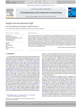
  • 5. Please cite this article in press as: Edholm, E.-S., et al., Insights into the function of IgD. Dev. Comp. Immunol. (2011), doi:10.1016/j.dci.2011.03.002 ARTICLE IN PRESS GModel DCI-1584; No.of Pages8 E.-S. Edholm et al. / Developmental and Comparative Immunology xxx (2011) xxx–xxx 5 Fig. 3. Catfish secreted IgD varies in size. Serum from eight different outbred catfish was visualized by Western blot analysis using anti-IgDsec mAb 2E5 followed by goat anti-mouse Ig (H + L) HRP. The different size variants of ∼130 and 180 kDa may be due to the inclusion or exclusion of a repeated block of C␦2, C␦3, and C␦4 exons found within the IGH3 locus that encodes the secreted IgD form. A schematic illustrating the different secreted IgD proteins is shown at left; C␦ domains are in grey and the secreted tail is white. Molecular weight size markers in kDa are shown at right of serum panel. secreted IgD transcripts contain six C␦ exons and short IgD tran- scripts have only the first two C␦ exons. The short membrane IgD transcripts also only contain the first two C␦ exons, while the long membrane IgD transcripts contain the first four C␦ domains. Fur- thermore, long IgD/IgW forms were identified in the nurse shark by using immunoprecipitation protocols (Greenberg et al., 1996). In the African lungfish membrane IgD forms have yet to be iden- tified, but sequencing showed that secreted IgD transcripts either contain seven C␦ exons or two C␦ exons. Finally, recent annotation of the monotreme duck-billed platypus genome demonstrated that the platypus IgD gene is located between the IgM and IgG1 genes (Gambon-Deza et al., 2009; Zhao et al., 2009). It consists of 10 C␦ exons followed by two TM exons and to date a secreted exon has not been found. Besides resembling the IgD expressed by teleosts, amphibians and leopard gecko in its length, phylogenetic analy- ses and amino acid comparisons show that all of the platypus C␦ domains have a corresponding domain in reptile IgD. Also a domain by domain comparison demonstrated that platypus C␦1, C␦6 and C␦7 domains are homologous to the mammals C␦1, C␦2 and C␦3 (Zhao et al., 2009). Taken together the available IgD sequencing data indicate, in marked contrast to IgM, that IgD genes have undergone numerous alterations involving both exon duplications and deletions lead- ing to different IgD structural forms in different species (Hordvik et al., 1999; Ohta and Flajnik, 2006; Rogers et al., 2006; Stenvik and Jorgensen, 2000; Tucker et al., 1980; Wagner et al., 2004; White et al., 1985; Wilson et al., 1997; Zhao and Hammarstrom, 2003; Zhao et al., 2002). 3. IgD-expressing B cells in humans and catfish Most of what we know about IgD expression and function comes from studies performed using human and mouse tissues and cell lines, and knockout mice. Also, it was only after the initial sequencing of IgD in catfish and in other teleosts, followed by the widespread sequencing of different genomes, that IgD was found in vertebrates other than primates and rodents. However, despite the many publications demonstrating that IgD has a more ancient origin than originally thought (see above), very few reports have focused on the function and cellular expression of this isotype in vertebrates, except for the studies performed in humans and mice. Consequently, this section summarizes our recent characterization studies of catfish IgD-bearing cell populations and how these relate to human IgD-bearing cells. In humans (and mice), IgM and IgD are expressed on the surface of naive mature B cells by alternative splicing of a long primary RNA transcript. These double positive B cells (IgM+/IgD+) make up the majority of the peripheral B cells and upon Ag binding, they down-regulate their IgD expression. In contrast, the major- ity of long-lived memory B cells have class switched, undergone somatic hypermutation and express either high affinity IgG, IgE, or IgA (McHeyer-Williams, 2003). Interestingly, class switching is also involved in generating a unique IgM−/IgD+ (IgD-only) B cell population in humans, i.e., the cells use a cryptic switch region found between the C␮ and C␦ genes (Chen et al., 2009). Human IgD-only B cells while rare in peripheral blood (∼3.0%), have been shown to make up a substantial population (20–25%) of the human upper respiratory tract mucosal B cells and in the past 12 years, several laboratories have been focusing on this unique IgD-only B cell population (Chen et al., 2009; Kluin et al., 1995; Owens et al., 1991; White et al., 1990; Yasui et al., 1989). Most IgD-only B cells are found associated with tonsillar tissues. The IgD-only B cells are larger, have more cytoplasm than conventional naive IgM+/IgD+ B cells, and exhibit a centroblast morphology (Billian et al., 1996; Liu et al., 1996). Notably, >90% of these IgD-only B cells utilize IgL ␭ chains (Arpin et al., 1997) and sequence analyses showed that these cells express highly mutated VH and VL regions. Also, since the mutations are found scattered throughout the CDR and FR regions it was proposed that the different V regions would prob- ably be unable to bind Ag (Liu et al., 1996; Seifert et al., 2009; Wilson et al., 1998; Zheng et al., 2005). Moreover, studies per- formed by Chen et al. (2009) demonstrated that secreted IgD made by the IgD-only B cells is bound to the surface of basophils (and mast cells) by a calcium-mobilizing receptor. First, immunofluores- cence staining showed that IgD is found on the surface and in the cytoplasm of mucosal and circulating basophils, which indicates IgD-bound immune complexes are internalized. Second, cross- linking of basophil-bound IgD using an anti-IgD mAb triggered an intracellular calcium flux and induced secretion of B cell activation factors and cytokines, including BAFF (B cell-activating factor of TNF family), APRIL (a proliferation-inducing TNF ligand), IL-3, and IL-4. Third, IgD cross-linking induced basophils to secrete the antimicro- bial factors cathelicidin, pentraxin-3, and ␤-defensin. In addition, the authors showed that supernatants collected from IgD cross- linked basophils inhibited the growth of the respiratory pathogens Moraxella catarrhalis and Haemophilus infuenzae. However, even though the putative IgD-binding receptor (Fc␦R) was not isolated, the authors’ studies suggest that it is a high affinity receptor since adding exogenous labeled IgD to cultured basophils did not lead to an increase in surface IgD staining, indicating saturation of the binding sites. Exogenous IgD could only be bound by the basophils after they were stripped of their IgD and IgD binding by pre- treated basophils was abolished if the “added” IgD was denatured or if stripped basophils were treated with trypsin. Interestingly, in this same study, patients with autoinflammatory syndromes were shown to have more circulating and mucosal IgD-only B cells and more mucosal “IgD-armed” basophils than healthy subjects, which suggests that inflammation promotes the switch to IgD. Also, cross- linking of IgD-armed basophils (from these patients) resulted in the increased secretion of both IL-1 and TNF, which suggests that IgD bound to the surface of basophils in the respiratory mucosa is able to trigger an innate immune response (Chen et al., 2009). As for catfish IgD, in the same study it was shown by using flow cytometry that catfish express two types of IgD+ B cell popu-
  • 6. Please cite this article in press as: Edholm, E.-S., et al., Insights into the function of IgD. Dev. Comp. Immunol. (2011), doi:10.1016/j.dci.2011.03.002 ARTICLE IN PRESS GModel DCI-1584; No.of Pages8 6 E.-S. Edholm et al. / Developmental and Comparative Immunology xxx (2011) xxx–xxx Fig. 4. Catfish express three types of IgD+ -bearing B cells. The single IgM+ (IgM+ /IgD− ), double positive IgM+ /IgD+ , and IgD+ -only (IgM− /IgD+ ) B cells are depicted with their membrane immunoglobulins associated with CD79 accessory molecules. IgM+ B cells produce secreted IgM tetramers and the IgD+ -only B-cells are the main producers of “V-less” secreted IgD (ref). Immunoglobulin domains are represented as in Fig. 2: C␮, black; C␦, dark grey, CL, light grey; V, white. The CD79a and CD79b Ig domains are stripped and Y marks the location of the two tyrosine residues in the immunoreceptor tyrosine-based activation motifs. lations (see below) and a population of circulating granular cells that are armed with exogenous IgD via a putative IgD-binding receptor. Currently, the origin of these granular cells is unknown, however at the message level these cells did not express message for either membrane or secreted IgD, IgM, or TCR and they did not react with an anti-catfish neutrophil mAb. However, these IgD- granulocytes could not be isolated via direct selection techniques as this invariably resulted in degranulation and granulocyte destruc- tion. Recently, a more detailed characterization of catfish IgD-only B cells was published by Edholm et al. (2010). Basically, catfish express three different types of B cells: single positive IgM+/IgD− B cells, double positive IgM+/IgD+ B cells and IgD-only B cells (Fig. 4). The IgM+/IgD− and IgM+/IgD+ B cells are small agranular lympho- cytes and are morphologically indistinguishable from each other, while the IgD-only B cells resemble human IgD-only B cells. They are large, exhibit a plasmablast morphology and have a higher cyto- plasm to nucleus ratio as compared to catfish IgM+ B cells and in contrast to mammalian IgD-only B cells, catfish IgD-only B cells can represent as much as 60–80% of the total peripheral blood B cells, depending on the individual fish. Notably, as shown by cell sorting experiments, co-immunoprecipitations, and RT-PCR, cat- fish IgD-only B cells exclusively utilize IgL ␴ chains, an IgL isotype restricted to ectothermic vertebrates. Also, even though the cat- fish IgM+/IgD− and IgM+/IgD+ B cell populations have the potential to utilize any of the four different IgL chain isotypes (IgL F, IgL G, IgL ␭ and IgL ␴) most IgM+ B cells utilize the IgL ␬ orthologs, F and G, i.e., only 2–5% of IgM+ B cells isolated from peripheral blood express IgL ␴ or IgL ␭ chains, isotypes known to have very limited repertoires in catfish (Edholm et al., 2009). Currently, while the dif- ferent roles of catfish IgM+/IgD+ and IgD-only B cells have not been defined, we hypothesize that catfish membrane IgD functions as a typical Ag binding receptor. Both cell types express message for membrane IgD with functionally rearranged VDJ regions and the B cell accessory signaling molecules CD79a and CD79b. Also, co- immunoprecipitation experiments demonstrate that membrane IgD associates with IgL chains, and requires association of CD79 molecules for B cell surface expression. Furthermore, no bias in the V, D or J family usage of membrane IgD transcripts was observed in the different IgD-expressing B cell types. In addition, anti-IgD cross- linking in IgM+/IgD+ B cells induces a calcium flux, albeit it is slower than the response induced by IgM cross-linking of IgM+/IgD− and IgM+/IgD+ B cells (Edholm unpublished). Whether this is due to
  • 7. Please cite this article in press as: Edholm, E.-S., et al., Insights into the function of IgD. Dev. Comp. Immunol. (2011), doi:10.1016/j.dci.2011.03.002 ARTICLE IN PRESS GModel DCI-1584; No.of Pages8 E.-S. Edholm et al. / Developmental and Comparative Immunology xxx (2011) xxx–xxx 7 lower levels of IgD expression on the surface of IgD+ B cells and/or the nature of the anti-IgD mAb has not been determined. Probably the most surprising observation from the catfish IgD+ B cell studies was the finding that secreted IgD transcripts isolated from sorted IgD-only B cells lacked V regions, instead the C␦1 was directly spliced to a functional leader sequence located ∼8 kb 5 of the C␦1 in the “IgD secreted form” IGHD3 gene (Edholm et al., 2010). The isolation of catfish V-less secreted IgD transcripts is intriguing especially when considering Chen et al. (2009) showed that both M. catarrhalis and H. influenzae bound IgD secreted by stimulated human IgD-only B cells, regardless of the IgD’s Ag speci- ficity. Also, as previously demonstrated (Riesbeck and Nordstrom, 2006), this secreted IgD bound M. catarrhalis IgD-binding protein (MID), which is also expressed by H. influenzae. MID is an ∼200 kDa adhesin with two functional domains, one mediates adherence to mucosal epithelial cells and the other binds to the C␦1 domain of IgD and studies show that the purified protein activates IgD+ B cells. Therefore, our working hypothesis for catfish IgD function is that IgD-only B cells, and possibly a subset of IgM+/IgD+ B cells, becomes activated by a certain “pathogen(s)” that can bind IgD. Once activated, these cells secrete V-less IgD, which may function as a pattern recognition molecule when bound to circulating granulocytes. This may also explain the low and varying levels of secreted IgD in catfish serum. In turn, the granulocytes upon binding the “target pathogen” through the IgD Fc-region would become activated. Here, it should be noted that the frequency of IgD-only B cells and IgD-bearing granulocytes varies among individual fish and the presence of these two populations do not always correlate (Edholm et al., 2010 and unpublished). Consequently, our studies are focused on identifying possible “target pathogens” or IgD-only B cell activation triggering events and isolating and determining the nature of the IgD-bearing granulocytes. Additionally, another important question that needs to be addressed is the relationship or lineage of the different IgD+ B cell populations. It may be that the catfish IgM+/IgD+ B cells represent traditional naive B lymphocytes, which upon Ag acti- vation differentiate into IgM producing plasma cells or memory B cells, while the IgD-only B cells constitute a separate B cell linage that expands in response to a yet unknown stimulus. Alter- natively, it may be that the small population of IgM+/IgD+/IgL ␴+ B cells upon activation differentiate into IgD-only B cells. 4. Conclusion In conclusion and as noted by others (Flajnik and Kasahara, 2010; Ohta and Flajnik, 2006; Zhao et al., 2009) IgD repre- sents an ancient Ig isotype that is found in all vertebrate taxa, except for birds. Moreover, IgD has been shown to display remarkable plasticity as illustrated by the occurrence of multiple structural variants and splice forms in the different vertebrates. Also the changes in the IgD genes that result in long chimeric molecules in teleost as compared to shorter hinge containing molecules in placental mammals indicates that IgD has been sub- jected to species-specific adaptations. For example, the finding of IgD-only B cells and the fact that secreted IgD can be bound by granulocytes via an IgD-binding receptor suggests that IgD plays a direct role in immune defense at least in these two species. Acknowledgements This work was supported by grants from the U.S. Department of Agriculture (2006-35204-16880), and UMMC IRSP (59908). Appendix A. Supplementary data Supplementary data associated with this article can be found, in the online version, at doi:10.1016/j.dci.2011.03.002. References Ales-Martinez, J.E., Warner, G.L., Scott, D.W., 1988. Immunoglobulins D and M medi- ate signals that are qualitatively different in B cells with an immature phenotype. Proc. Natl. Acad. Sci. U.S.A. 85, 6919–6923. Anderson, M.K., Strong, S.J., Litman, R.T., Luer, C.A., Amemiya, C.T., Rast, J.P., Litman, G.W., 1999. A long form of the skate IgX gene exhibits a striking resemblance to the new shark IgW and IgNARC genes. Immunogenetics 49, 56–67. Aparicio, S., Chapman, J., Stupka, E., Putnam, N., Chia, J.M., Dehal, P., Christoffels, A., Rash, S., Hoon, S., Smit, A., Gelpke, M.D., Roach, J., Oh, T., Ho, I.Y., Wong, M., Detter, C., Verhoef, F., Predki, P., Tay, A., Lucas, S., Richardson, P., Smith, S.F., Clark, M.S., Edwards, Y.J., Doggett, N., Zharkikh, A., Tavtigian, S.V., Pruss, D., Barnstead, M., Evans, C., Baden, H., Powell, J., Glusman, G., Rowen, L., Hood, L., Tan, Y.H., Elgar, G., Hawkins, T., Venkatesh, B., Rokhsar, D., Brenner, S., 2002. Whole-genome shotgun assembly and analysis of the genome of Fugu rubripes. Science 297, 1301–1310. Arpin, C., de Bouteiller, O., Razanajaona, D., Briere, F., Banchereau, J., Lebecque, S., Liu, Y.J., 1997. Human peripheral B cell development. sIgM− IgD+ CD38+ hypermutated germinal center centroblasts preferentially express Ig lambda light chain and have undergone mu-to-delta switch. Ann. N. Y. Acad. Sci. 815, 193–198. Bao, Y., Wang, T., Guo, Y., Zhao, Z., Li, N., Zhao, Y., 2010. The immunoglobulin gene loci in the teleost Gasterosteus aculeatus. Fish Shellfish Immunol. 28, 40–48. Bengten, E., Clem, L.W., Miller, N.W., Warr, G.W., Wilson, M., 2006a. Channel cat- fish immunoglobulins: repertoire and expression. Dev. Comp. Immunol. 30, 77–92. Bengten, E., Quiniou, S., Hikima, J., Waldbieser, G., Warr, G.W., Miller, N.W., Wilson, M., 2006b. Structure of the catfish IGH locus: analysis of the region including the single functional IGHM gene. Immunogenetics 58, 831–844. Bengten, E., Quiniou, S.M., Stuge, T.B., Katagiri, T., Miller, N.W., Clem, L.W., Warr, G.W., Wilson, M., 2002. The IgH locus of the channel catfish, Ictalurus punctatus, contains multiple constant region gene sequences: different genes encode heavy chains of membrane and secreted IgD. J. Immunol. 169, 2488–2497. Bengten, E., Wilson, M., Miller, N., Clem, L.W., Pilstrom, L., Warr, G.W., 2000. Immunoglobulin isotypes: structure, function, and genetics. Curr. Top. Micro- biol. Immunol. 248, 189–219. Berstein, R.M., Schluter, S.F., Shen, S., Marchalonis, J.J., 1996. A new high molecular weight immunoglobulin class from the carcharhine shark: implications for the properties of the primordial immunoglobulin. Proc. Natl. Acad. Sci. U.S.A. 93, 3289–3293. Billian, G., Bella, C., Mondiere, P., Defrance, T., 1996. Identification of a tonsil IgD+ B cell subset with phenotypical and functional characteristics of germinal center B cells. Eur. J. Immunol. 26, 1712–1719. Chen, K., Xu, W., Wilson, M., He, B., Miller, N.W., Bengten, E., Edholm, E.S., Santini, P.A., Rath, P., Chiu, A., Cattalini, M., Litzman, J., Huang, J.B.B., Meini, B., Riesbeck, A., Cunningham-Rundles, K., Plebani, C., Cerutti, A.A., 2009. Immunoglobulin D enhances immune surveillance by activating antimicrobial, proinflammatory and B cell-stimulating programs in basophils. Nat. Immunol. 10, 889–898. Clem, L.W., 1971. Phylogeny of immunoglobulin structure and function. IV. Immunoglobulins of the giant grouper, Epinephelus itaira. J. Biol. Chem. 246, 9–15. Danilova, N., Bussmann, J., Jekosch, K., Steiner, L.A., 2005. The immunoglobulin heavy-chain locus in zebrafish: identification and expression of a previously unknown isotype, immunoglobulin Z. Nat. Immunol. 6, 295–302. Edholm, E.S., Bengten, E., Stafford, J.L., Sahoo, M., Taylor, E.B., Miller, N.W., Wilson, M., 2010. Identification of two IgD+ B cell populations in channel catfish, Ictalurus punctatus. J. Immunol. 185, 4082–4094. Edholm, E.S., Wilson, M., Sahoo, M., Miller, N.W., Pilstrom, L., Wermenstam, N.E., Bengten, E., 2009. Identification of Igsigma and Iglambda in channel catfish, Ictalurus punctatus, and Iglambda in Atlantic cod, Gadus morhua. Immunogenet- ics 61, 353–370. Flajnik, M., Rumfelt, L.L., 2000. The immune system of cartilaginous fish. Curr. Top. Microbiol. Immunol. 248, 249–270. Flajnik, M.F., Kasahara, M., 2010. Origin and evolution of the adaptive immune sys- tem: genetic events and selective pressures. Nat. Rev. Genet. 11, 47–59. Gambon-Deza, F., Espinel, C.S., 2008. IgD in the reptile leopard gecko. Mol. Immunol. 45, 3470–3476. Gambon-Deza, F., Sanchez-Espinel, C., Magadan-Mompo, S., 2009. The immunoglob- ulin heavy chain locus in the platypus (Ornithorhynchus anatinus). Mol. Immunol. 46, 2515–2523. Gambon-Deza, F., Sanchez-Espinel, C., Magadan-Mompo, S., 2010. Presence of an unique IgT on the IGH locus in three-spined stickleback fish (Gasterosteus aculea- tus) and the very recent generation of a repertoire of VH genes. Dev. Comp. Immunol. 34, 114–122. Geisberger, R., Lamers, M., Achatz, G., 2006. The riddle of the dual expression of IgM and IgD. Immunology 118, 429–437. Greenberg, A.S., Hughes, A.L., Guo, J., Avila, D., McKinney, E.C., Flajnik, M.F., 1996. A novel “chimeric” antibody class in cartilaginous fish: IgM may not be the primordial immunoglobulin. Eur. J. Immunol. 26, 1123–1129.
  • 8. Please cite this article in press as: Edholm, E.-S., et al., Insights into the function of IgD. Dev. Comp. Immunol. (2011), doi:10.1016/j.dci.2011.03.002 ARTICLE IN PRESS GModel DCI-1584; No.of Pages8 8 E.-S. Edholm et al. / Developmental and Comparative Immunology xxx (2011) xxx–xxx Hansen, J.D., Landis, E.D., Phillips, R.B., 2005. Discovery of a unique Ig heavy- chain isotype (IgT) in rainbow trout: implications for a distinctive B cell developmental pathway in teleost fish. Proc. Natl. Acad. Sci. U.S.A. 102, 6919–6924. Hirono, I., Nam, B.H., Enomoto, J., Uchino, K., Aoki, T., 2003. Cloning and character- isation of a cDNA encoding Japanese flounder Paralichthys olivaceus IgD. Fish Shellfish Immunol. 15, 63–70. Honda, Y., Kondo, H., Caipang, C.M., Hirono, I., Aoki, T., 2010. cDNA cloning of the immunoglobulin heavy chain genes in banded houndshark Triakis scyllium. Fish Shellfish Immunol. 29, 854–861. Hordvik, I., 2002. Identification of a novel immunoglobulin delta transcript and comparative analysis of the genes encoding IgD in Atlantic salmon and Atlantic halibut. Mol. Immunol. 39, 85–91. Hordvik, I., Thevarajan, J., Samdal, I., Bastani, N., Krossoy, B., 1999. Molecular cloning and phylogenetic analysis of the Atlantic salmon immunoglobulin D gene. Scand. J. Immunol. 50, 202–210. Kluin, P.M., Kayano, H., Zani, V.J., Kluin-Nelemans, H.C., Tucker, P.W., Satterwhite, E., Dyer, M.J., 1995. IgD class switching: identification of a novel recombination site in neoplastic and normal B cells. Eur. J. Immunol. 25, 3504–3508. Lanning, D.K., Zhai, S.K., Knight, K.L., 2003. Analysis of the 3 Cmu region of the rabbit Ig heavy chain locus. Gene 309, 135–144. Liu, Y.J., de Bouteiller, O., Arpin, C., Briere, F., Galibert, L., Ho, S., Martinez-Valdez, H., Banchereau, J., Lebecque, S., 1996. Normal human IgD+ IgM− germinal center B cells can express up to 80 mutations in the variable region of their IgD transcripts. Immunity 4, 603–613. Lobb, C.J., Clem, L.W., 1981. Phylogeny of immunoglobulin structure and function- XII. Secretory immunoglobulins in the bile of the marine teleost Archosargus probatocephalus. Mol. Immunol. 18, 615–619. Lundqvist, M.L., Middleton, D.L., Hazard, S., Warr, G.W., 2001. The immunoglobulin heavy chain locus of the duck. Genomic organization and expression of D, J, and C region genes. J. Biol. Chem. 276, 46729–46736. Lutz, C., Ledermann, B., Kosco-Vilbois, M.H., Ochsenbein, A.F., Zinkernagel, R.M., Kohler, G., Brombacher, F., 1998. IgD can largely substitute for loss of IgM func- tion in B cells. Nature 393, 797–801. McHeyer-Williams, M., 2003. B-cell signaling mechanisms and activation. In: Paul, W.E. (Ed.), Fundamental Immunology. , 5th edition, pp. 195–2285. Miller, N.W., Rycyzyn, M.A., Wilson, M.R., Warr, G.W., Naftel, J.P., Clem, L.W., 1994. Development and characterization of channel catfish long term B cell lines. J. Immunol. 152, 2180–2189. Mongini, P.K., Blessinger, C., Posnett, D.N., Rudich, S.M., 1989. Membrane IgD and membrane IgM differ in capacity to transduce inhibitory signals within the same human B cell clonal populations. J. Immunol. 143, 1565–1574. Nitschke, L., Kosco, M.H., Kohler, G., Lamers, M.C., 1993. Immunoglobulin D-deficient mice can mount normal immune responses to thymus-independent and - dependent antigens. Proc. Natl. Acad. Sci. U.S.A. 90, 1887–1891. Nossal, G.J., Pike, B.L., Battye, F.L., 1979. Mechanisms of clonal abortion tolerogenesis. II. Clonal behaviour of immature B cells following exposure to anti-mu chain antibody. Immunology 37, 203–215. Ohta, Y., Flajnik, M., 2006. IgD, like IgM, is a primordial immunoglobulin class perpet- uated in most jawed vertebrates. Proc. Natl. Acad. Sci. U.S.A. 103, 10723–10728. Ota, T., Rast, J.P., Litman, G.W., Amemiya, C.T., 2003. Lineage-restricted retention of a primitive immunoglobulin heavy chain isotype within the Dipnoi reveals an evolutionary paradox. Proc. Natl. Acad. Sci. U.S.A. 100, 2501–2506. Owens Jr., J.D., Finkelman, F.D., Mountz, J.D., Mushinski, J.F., 1991. Nonhomolo- gous recombination at sites within the mouse JH-C delta locus accompanies C mu deletion and switch to immunoglobulin D secretion. Mol. Cell. Biol. 11, 5660–5670. Pike, B.L., Boyd, A.W., Nossal, G.J., 1982. Clonal anergy: the universally anergic B lymphocyte. Proc. Natl. Acad. Sci. U.S.A. 79, 2013–2017. Rast, J.P., Amemiya, C.T., Litman, R.T., Strong, S.J., Litman, G.W., 1998. Distinct pat- terns of IgH structure and organization in a divergent lineage of chrondrichthyan fishes. Immunogenetics 47, 234–245. Riesbeck, K., Nordstrom, T., 2006. Structure and immunological action of the human pathogen Moraxella catarrhalis IgD-binding protein. Crit. Rev. Immunol. 26, 353–376. Roes, J., Rajewsky, K., 1993. Immunoglobulin D (IgD)-deficient mice reveal an auxil- iary receptor function for IgD in antigen-mediated recruitment of B cells. J. Exp. Med. 177, 45–55. Rogers, K.A., Richardson, J.P., Scinicariello, F., Attanasio, R., 2006. Molecular charac- terization of immunoglobulin D in mammals: immunoglobulin heavy constant delta genes in dogs, chimpanzees and four old world monkey species. Immunol- ogy 118, 88–100. Rowe, D.S., Fahey, J.L., 1965a. A new class of human immunoglobulins I. A unique myeloma protein. J. Exp. Med. 121, 171–184. Rowe, D.S., Fahey, J.L., 1965b. A new class of human immunoglobulins. II. Normal serum Igd. J. Exp. Med. 121, 185–199. Rumfelt, L.L., Diaz, M., Lohr, R.L., Mochon, E., Flajnik, M.F., 2004. Unprecedented multiplicity of Ig transmembrane and secretory mRNA forms in the cartilaginous fish. J. Immunol. 173, 1129–1139. Saha, N.R., Suetake, H., Kikuchi, K., Suzuki, Y., 2004. Fugu immunoglobulin D: a highly unusual gene with unprecedented duplications in its constant region. Immunogenetics 56, 438–447. Sahoo, M., Edholm, E.S., Stafford, J.L., Bengten, E., Miller, N.W., Wilson, M., 2008. B cell receptor accessory molecules in the channel catfish, Ictalurus punctatus. Dev. Comp. Immunol. 32, 1385–1397. Savan, R., Aman, A., Nakao, M., Watanuki, H., Sakai, M., 2005a. Discovery of a novel immunoglobulin heavy chain gene chimera from common carp (Cyprinus carpio L.). Immunogenetics 57, 458–463. Savan, R., Aman, A., Sato, K., Yamaguchi, R., Sakai, M., 2005b. Discovery of a new class of immunoglobulin heavy chain from fugu. Eur. J. Immunol. 35, 3320–3331. Seifert, M., Steimle-Grauer, S.A., Goossens, T., Hansmann, M.L., Brauninger, A., Kup- pers, R., 2009. A model for the development of human IgD-only B cells: genotypic analyses suggest their generation in superantigen driven immune responses. Mol. Immunol. 46, 630–639. Solem, S.T., Stenvik, J., 2006. Antibody repertoire development in teleosts – a review with emphasis on salmonids and Gadus morhua L. Dev. Comp. Immunol. 30, 57–76. Stenvik, J., Jorgensen, T.O., 2000. Immunoglobulin D (IgD) of Atlantic cod has a unique structure. Immunogenetics 51, 452–461. Sun, Y., Wei, Z., Hammarstrom, L., Zhao, Y., 2010. The immunoglobulin delta gene in jawed vertebrates: a comparative overview. Dev. Comp. Immunol.. Tadiso, T.M., Lie, K.K., Hordvik, I., 2010. Molecular cloning of IgT from Atlantic salmon, and analysis of the relative expression of tau, mu and delta in different tissues. Vet. Immunol. Immunopathol.. Tucker, P.W., Liu, C.P., Mushinski, J.F., Blattner, F.R., 1980. Mouse immunoglobulin D: messenger RNA and genomic DNA sequences. Science 209, 1353–1360. Van Boxel, J.A., Paul, W.E., Terry, W.D., Green, I., 1972. Communications. IgD-bearing human lymphocytes. J. Immunol. 109, 648–651. Wagner, B., 2006. Immunoglobulins and immunoglobulin genes of the horse. Dev. Comp. Immunol. 30, 155–164. Wagner, B., Miller, D.C., Lear, T.L., Antczak, D.F., 2004. The complete map of the Ig heavy chain constant gene region reveals evidence for seven IgG isotypes and for IgD in the horse. J. Immunol. 173, 3230–3242. Wang, X., Olp, J.J., Miller, R.D., 2009. On the genomics of immunoglobulins in the gray, short-tailed opossum Monodelphis domestica. Immunogenetics 61, 581–596. Warr, G.W., 1995. The immunoglobulin genes of fish. Dev. Comp. Immunol. 19, 1–12. Wei, Z., Wu, Q., Ren, L., Hu, X., Guo, Y., Warr, G.W., Hammarstrom, L., Li, N., Zhao, Y., 2009. Expression of IgM, IgD, and IgY in a reptile, Anolis carolinensis. J. Immunol. 183, 3858–3864. White, M.B., Shen, A.L., Word, C.J., Tucker, P.W., Blattner, F.R., 1985. Human immunoglobulin D: genomic sequence of the delta heavy chain. Science 228, 733–737. White, M.B., Word, C.J., Humphries, C.G., Blattner, F.R., Tucker, P.W., 1990. Immunoglobulin D switching can occur through homologous recombination in human B cells. Mol. Cell. Biol. 10, 3690–3699. Wilson, M., Bengten, E., Miller, N.W., Clem, L.W., Du Pasquier, L., Warr, G.W., 1997. A novel chimeric Ig heavy chain from a teleost fish shares similarities to IgD. Proc. Natl. Acad. Sci. U.S.A. 94, 4593–4597. Wilson, P.C., de Bouteiller, O., Liu, Y.J., Potter, K., Banchereau, J., Capra, J.D., Pas- cual, V., 1998. Somatic hypermutation introduces insertions and deletions into immunoglobulin V genes. J. Exp. Med. 187, 59–70. Xiao, F.S., Wang, Y.P., Yan, W., Chang, M.X., Yao, W.J., Xu, Q.Q., Wang, X.X., Gao, Q., Nie, P., 2010. Ig heavy chain genes and their locus in grass carp Ctenopharyngodon idella. Fish Shellfish Immunol. 29, 594–599. Xu, Z., Wang, G.L., Nie, P., 2009. IgM, IgD and IgY and their expression pattern in the Chinese soft-shelled turtle Pelodiscus sinensis. Mol. Immunol. 46, 2124–2132. Yasui, H., Akahori, Y., Hirano, M., Yamada, K., Kurosawa, Y., 1989. Class switch from mu to delta is mediated by homologous recombination between sigma mu and sigma mu sequences in human immunoglobulin gene loci. Eur. J. Immunol. 19, 1399–1403. Yasuike, M., de Boer, J., von Schalburg, K.R., Cooper, G.A., McKinnel, L., Messmer, A., So, S., Davidson, W.S., Koop, B.F., 2010. Evolution of duplicated IgH loci in Atlantic salmon, Salmo salar. BMC Genomics 11, 486. Zhang, Y.A., Salinas, I., Li, J., Parra, D., Bjork, S., Xu, Z., LaPatra, S.E., Bartholomew, J., Sunyer, J.O., 2010. IgT, a primitive immunoglobulin class specialized in mucosal immunity. Nat. Immunol. 11, 827–835. Zhao, Y., Cui, H., Whittington, C.M., Wei, Z., Zhang, X., Zhang, Z., Yu, L., Ren, L., Hu, X., Zhang, Y., Hellman, L., Belov, K., Li, N., Hammarstrom, L., 2009. Ornithorhynchus anatinus (platypus) links the evolution of immunoglobulin genes in eutherian mammals and nonmammalian tetrapods. J. Immunol. 183, 3285–3293. Zhao, Y., Hammarstrom, L., 2003. Cloning of the complete rat immunoglobulin delta gene: evolutionary implications. Immunology 108, 288–295. Zhao, Y., Kacskovics, I., Pan, Q., Liberles, D.A., Geli, J., Davis, S.K., Rabbani, H., Hammarstrom, L., 2002. Artiodactyl IgD: the missing link. J. Immunol. 169, 4408–4416. Zhao, Y., Pan-Hammarstrom, Q., Kacskovics, I., Hammarstrom, L., 2003. The porcine Ig delta gene: unique chimeric splicing of the first constant region domain in its heavy chain transcripts. J. Immunol. 171, 1312–1318. Zhao, Y., Pan-Hammarstrom, Q., Yu, S., Wertz, N., Zhang, X., Li, N., Butler, J.E., Ham- marstrom, L., 2006. Identification of IgF, a hinge-region-containing Ig class, and IgD in Xenopus tropicalis. Proc. Natl. Acad. Sci. U.S.A. 103, 12087–12092. Zhao, Y., Rabbani, H., Shimizu, A., Hammarstrom, L., 2000. Mapping of the chicken immunoglobulin heavy-chain constant region gene locus reveals an inverted alpha gene upstream of a condensed upsilon gene. Immunology 101, 348–353. Zhao, Z., Zhao, Y., Pan-Hammarstrom, Q., Liu, W., Liu, Z., Li, N., Hammarstrom, L., 2007. Physical mapping of the giant panda immunoglobulin heavy chain con- stant region genes. Dev. Comp. Immunol. 31, 1034–1049. Zheng, N.Y., Wilson, K., Jared, M., Wilson, P.C., 2005. Intricate targeting of immunoglobulin somatic hypermutation maximizes the efficiency of affinity maturation. J. Exp. Med. 201, 1467–1478.