SlideShare a Scribd company logo
1 of 12
Download to read offline
a
RESEARCH ARTICLE
Cis-regulatory control of human GLI2 expression in the
developing neural tube and limb bud
Rashid Minhas,1
Stefan Pauls,2
Shahid Ali,1
Laura Doglio,2
Muhammad Ramzan Khan,3
Greg Elgar,2
and Amir Ali Abbasi1
*
1
National Center for Bioinformatics, Program of Comparative and Evolutionary Genomics, Faculty of Biological Sciences, Quaid-i-Azam University, Islamabad 45320,
Pakistan
2
Division of Systems Biology, MRC National Institute for Medical Research, The Ridgeway, Mill Hill, London NW7, 1AA, United Kingdom
3
National Institute for Genomics and Advanced Biotechnology, National Agricultural Research Center, Park Road, Islamabad, Pakistan
Background: GLI2, a zinc finger transcription factor, mediates Sonic hedgehog signaling, a critical pathway in vertebrate
embryogenesis. GLI2 has been implicated in diverse set of embryonic developmental processes, including patterning of cen-
tral nervous system and limbs. In humans, mutations in GLI2 are associated with several developmental defects, including hol-
oprosencephaly and polydactyly. Results: Here, we demonstrate in transient transgenic zebrafish assays, the potential of a
subset of tetrapod-teleost conserved non-coding elements (CNEs) residing within human GLI2 intronic intervals to induce
reporter gene expression at known regions of endogenous GLI2 transcription. The regulatory activities of these elements are
observed in several embryonic domains, including neural tube and pectoral fin. Moreover, our data reveal an overlapping
expression profile of duplicated copies of an enhancer during zebrafish evolution. Conclusions: Our data suggest that during
vertebrate history GLI2 acquired a high level of complexity in the genetic mechanisms regulating its expression during spatio-
temporal patterning of the central nervous system (CNS) and limbs. Developmental Dynamics 244:681–692, 2015. VC 2015 Wiley
Periodicals, Inc.
Key words: GLI2; gene regulation; comparative genomics; enhancer; CNE; vertebrate development
Submitted 29 September 2014; First Decision 29 January 2015; Accepted 16 February 2015; Published online 25 February 2015
Introduction
The Hedgehog family of signaling molecules forms a key regula-
tory network, conserved from vertebrates to invertebrates, con-
trolling multiple developmental processes (Sasaki H, 1999). The
vertebrate Gli1, Gli2, and Gli3 (glioma-associated oncogene 1, 2,
and 3) genes evolved by gene duplication events of a single
ancestral gene (Ruppert JM, 1998). The genomes of model inver-
tebrates such as Drosophila possess a single homolog of the ver-
tebrate GLI family, encoding a zinc finger containing
transcription factor Cubitus interruptus (Ci) by which all Hh sig-
naling is transduced (Alexandre et al., 1996; Aza-Blanc and
Kornberg, 1999). Additionally, in teleost, Gli2 underwent zebra-
fish specific duplication event producing two gene copies, namely
gli2a and gli2b (Ke et al., 2005; Abbasi et al., 2009). The GLI fam-
ily plays a critical role in vertebrate embryonic patterning, more
specifically in the central nervous system, the anterior-posterior
axis of the embryonic limb bud and in craniofacial structures and
internal organs (Ruppert JM, 1998). GLI1 is unique in comparison
to GLI2 and GLI3 because it only comprises a C-terminal tran-
scriptional activation domain, while bifunctional GLI2 and GLI3
possess a C-terminal activation and N-terminal repression
domain (Dai P, 1999; Sasaki H, 1999).
Mutational studies in mice verified that GLI2 and GLI3, which
are expressed widely in populations of cells responsive to Shh sig-
naling, are the key Shh mediators for embryogenesis. However,
GLI1 which evolved at a faster rate than its paralogs in tetrapods is
not obligatory for development (Mo et al., 1997; Ding et al., 1998;
Motoyama J, 1998; Abbasi et al., 2009). The patterning of the ven-
tral hindbrain, skeletal muscle formation and limb anterior-
posterior polarity is due to a redundant function of GLI2/GLI3, and
both are highly expressed during the process of gastrulation and
neurulation (Hui et al., 1994; Sasaki H, 1999; McDermott et al.,
2005; Magdaleno et al., 2006; Lebel et al., 2007; Bowers et al.,
2012). Moreover, studies in mice and frog suggest that during
development the GLI family acts in a combinatorial manner that is
context-dependent and species-specific (Ruiz i Altaba, 1998).
Mutations in human GLI2 have been linked with holoprosen-
cephaly, pituitary anomalies (hypopituitarism), congenital growth
hormone deficiency, cranial and midline facial deformities, and
abnormalities in limb development (pre-axial and post-axial pol-
ydactyly) (Roessler et al., 2003; Roessler et al., 2005; Bertolacini
et al., 2012; Franca et al., 2013). Gli2 also plays a crucial role in
DEVELOPMENTALDYNAMICS
This work was funded by Higher Education Comission Pakistan
grant (No. 20-2085/NRPU/R&/HEC/12/760) to A.A.A., and by MRC
core funding (U117597141) to G.E. R.M. holds a fellowship under
Indigenous PhD fellowships (Phase-II) of Higher Education Commis-
sion of Pakistan.
*Corresponding author: E-mail: abbasiam@qau.edu.pk, Tel Office: 192-
51-9064-4109, Tel Lab: 192-51-9064-4114, Cell: 192-334-8959-354
Article is online at: http://onlinelibrary.wiley.com/doi/10.1002/dvdy.
24266/abstract
VC 2015 Wiley Periodicals, Inc.
DEVELOPMENTAL DYNAMICS 244:681–692, 2015
DOI: 10.1002/DVDY.24266
681
carcinogenesis and transgenic mice studies suggest that over-
expressing Gli2 in cutaneous keratinocytes develops multiple
basal cell carcinoma, while the human hepatocellular carcinoma
cell lines and hepatocellular tissues show high levels of GLI2
(Grachtchouk M, 2000; Cheng et al., 2009). Other human tumors
associated with GLI2 are: medulloblastomas, prostate and breast
cancer (Fulda et al., 2002; Okano et al., 2003; Sicklick et al.,
2006). A tight temporal and spatial control of GLI2 expression is
thus indispensable for multiple patterning steps in different tis-
sues or organs.
Gene expression plays a significant role in the evolution of
vertebrate complexity and diversity in development (Carroll,
2008). Precise spatial and temporal expression of a gene is con-
trolled by cis-acting elements (enhancers, repressors and
silencers) that often reside hundreds of kilobases away or within
an intron of the gene they control (Lettice et al., 2003; Levine
and Tjian, 2003; Parveen et al., 2013). Each of these cis-acting
regulatory modules achieve an explicit function, such as the acti-
vation of its associated trans-dev gene in a specific cell and tissue
type or at a precise phase in development (Paparidis et al., 2007;
Lee et al., 2011; Pauls et al., 2012). A gene might thus contain
multiple cis-regulatory modules, each of which contributes, in a
somewhat combinatorial manner, to its overall spatial and tem-
poral regulation (Parveen et al., 2013). Conserved non-coding
DNA sequences from human to fish (evolutionary distance 450
Myr) clustered around developmental genes, are strong candi-
dates for putative enhancers harbouring binding sites for multiple
transcription factors (Woolfe et al., 2005; Pennacchio et al.,
2006; Abbasi et al., 2007). Furthermore, some mutations in cis-
acting regulatory regions can cause aberrant gene expression
leading to a number of developmental defects (Lettice et al.,
2003; Jeong et al., 2008; Ragvin et al., 2010).
GLI paralogs are critical for early embryonic development in
co-operation with Shh, triggering a sequence of reactions in
downstream target genes (Lee et al., 1997; Ding et al., 1998; Dai
P, 1999). The GLI family has a divergent, as well as an overlap-
ping, role in growth and patterning of vertebrate development,
owing to a mutual GLI code required for faithful transduction of
Hh signaling (Ruiz i Altaba, 1998). Studies have already deci-
phered GLI3-specific cis-acting regulatory sequences and how
they control spatio-temporal expression in vertebrate develop-
ment (Abbasi et al., 2007; Paparidis et al., 2007; Abbasi et al.,
2010; Abbasi et al., 2013). However, the cis-acting control of the
GLI2 gene remains unexplored.
As an initial attempt to elucidate the cis-regulatory underpin-
nings of human GLI2 expression, we have chosen tetrapod-teleost
evolutionarily conserved intronic regions of GLI2, for functional
assay in zebrafish. A stringent criteria of at least 50% conservation
over a 50bp window across all species, was employed to highlight
the tetrapod-teleost conserved regions, as shown by previous stud-
ies that a criteria of 50-70% is ideal to identify putative regulatory
elements (Woolfe et al., 2005; McEwen et al., 2006; Abbasi et al.,
2007; Woolfe and Elgar, 2008). Our results show that intronic
CNEs from the GLI2 gene act as tissue-specific enhancers and that
reporter gene expression induced by these elements correlates with
previously reported endogenous gli2 expression in zebrafish.
Highly specific reporter expression is mainly observed in the neu-
ral tube and pectoral fin. Our data reveal that tetrapod-teleost
CNEs, located in the introns of GLI2, mark critical components of
the cis-regulatory inventory for spatial and temporal deployment
of this key developmental gene.
Results
Identification of Tetrapod-teleost conserved non-
coding elements at the GLI2 locus by comparative
sequence analysis
A multi-species alignment of human GLI2 with orthologous
genomic sequences from mouse, chicken, fugu, and zebrafish,
revealed five anciently conserved non-coding elements embedded
exclusively in the intronic intervals of GLI2 gene (Fig. 1), maintain-
ing at least 50% identity over a 50bp window across all species.
Comparative analysis of 100kb upstream or downstream region of
the GLI2 gene did not detect any significant sequence similarity
from human to fish. CNE1 and CNE5 are present within intron 1,
while highly conserved CNE2 is present within intron 2, CNE3 is
located in intron 4, and CNE4 is positioned within intron 7. None
of the selected conserved non-coding elements overlap with exons
or non-protein coding RNAs. The details of conserved tetrapod/tele-
ost amplicons selected for functional analysis are described in Table
1. Moreover, highly conserved element CNE2 has two co-orthologs
in the zebrafish genome. These duplicated CNEs (dCNEs) are posi-
tioned within intron 2 of the zebrafish gli2a gene (Dr-gli2-CNE2a)
and downstream of the zebrafish gli2b gene (Dr-gli2-CNE2b).
Identification of transcription factor binding sites
(TFBSs) within GLI2-associated CNEs
Spatio-temporal expression of enhancers is due to the interaction
between CNEs and multiple co-operative transcription factors
(Parveen et al., 2013). To generate a list of candidate sites that
might be responsible for the cis-regulatory activity of GLI2-associ-
ated CNEs, tetrapod/teleost orthologous sequences were anlayzed.
These sequences were carefully screened with the MEME motif dis-
covery tool to identify conserved sequence motifs (for details see
Materials and Methods). These evolutionarily conserved sequence
motifs were then compared with position weight matrices (PWMs)
for known vertebrate transcription factors (TFs) (Mahony and
Benos, 2007). This identified putative TFBSs within each of the
functionally categorized GLI2-intronic enhancers. Many of these
transcription factors (Table 1) are key developmental regulators
and are known to be co-expressed with Gli2 during early embry-
onic development of limbs and CNS, as verified from the Mouse
Genome Informatics database (http://www.informatics.jax.org/).
In vivo characterization of CNEs using a co-injection
assay in transiently transfected zebrafish embryos
The CNEs identified through comparative genomics were next
tested in vivo using zebrafish as a model organism. CNEs were
co-injected with a GFP reporter into zebrafish embryos and then
monitored for enhancer activity at set time points, as described
previously (Woolfe et al., 2005). The reporter gene activity of all
five CNEs showed reproducible tissue-specific expression after 24
(day-2) and 48 hours (day-3) post fertilization (hpf) in multiple
independent transient transgenic assays. Percentage and catego-
ries of cell types that are positive of each CNE are represented as
a schematic (Fig. 2), and in tabular form (Table 2).
CNE1 activates GFP expression predominantly in muscle cells
(17%), forebrain (8%), midbrain (3%), hindbrain (19%) and spinal
cord (6%) at day two; whereas at day three of development
reporter expression is most frequently observed in hindbrain
DEVELOPMENTALDYNAMICS 682 MINHAS ET AL.
(45% of expressing embryos) (Fig. 3A and 3B). CNE2 induces GFP
expression exclusively in notochord (79%) at $26-33 hpf, as well
as at $48-56 hpf (Fig. 3C and 3D). CNE3 upregulates reporter
gene expression in otic vesicle (48%) (Fig. 3E), muscle cells
(52%), and hindbrain (11%) at $26-33 hpf, whereas at $48-56
hpf the main expression domains for CNE3 are hindbrain (66%)
(Fig. 3F), otic vesicle (25%), and muscle cells (25%). CNE4 directs
GFP expression specifically in the muscle cells at day two (93%)
and at day three (96%) (Fig. 3G and 3H). CNE5 drives GFP expres-
sion mainly in two major domains of the CNS, i.e. hindbrain
(23%) and spinal cord (28%) at $26-33 hpf, whereas at day three
CNE5 strongly enhances reporter expression in spinal cord (53%),
hindbrain (40%) and muscle cells (30%) (Fig. 3I and 3J).
Reproducible GFP expression of GLI2-associated CNEs
by Tol2 transgenesis
To validate the results from our co-injection assays, and to inves-
tigate the potential cis-regulatory function of GLI2-associated
CNEs in greater detail, we used a different strategy based on a
Tol2 vector with a c-fos minimal promoter. Tol2 based transgene-
sis has the advantage of stronger reporter expression with
reduced mosaicism due to efficient and stable integration in the
genome (Fisher et al., 2006; Kawakami, 2007). The injected
embryos were screened ($26-33 hpf and $48-56 hpf) for compa-
rable GFP expression with the co-injection assays. Moreover,
zebrafish embryos showing significant and consistent GFP
expression in those tissues which were not observed in co-
injection assay were also noted (Table 2).
With the exception of CNE1 and CNE4 (which were able to
induce reporter expression with co-injection assay), the remain-
der of the CNEs also upregulate GFP expression, driven by the c-
fos promoter, in the Tol2 based transgenic assay. Based on our
Tol2 results we categorized the human GLI2-associated CNEs
into two major categories, those inducing GFP expression pre-
dominantly in various domains of CNS (hindbrain and spinal
cord) and those which upregulate GFP expression in the develop-
ing zebrafish fin.
CNE2, CNE3, and CNE5 are central nervous system
specific enhancers
In our zebrafish co-injection assay, the major GFP expression
domain for CNE2 was notochord, whereas in the Tol2 based
transgenic assay significant and reproducible reporter gene
expression is detected not only in notochord (data not shown),
DEVELOPMENTALDYNAMICS
Fig. 1. Tetrapod-teleost conservation of the non-coding sequence across the GLI2 locusPair-wise sequence alignment of the genomic interval
containing the human GLI2 locus (ENSG00000074047) and 100kb flanking upstream and downstream sequence with orthologous counterparts of
tetrapod (mouse and chicken) and teleost fishes (fugu; gli2b and zebrafish; gli2a). These are shown as a VISTA (Visualization Tool for Alignment)
graphical output using MLAGAN algorithm, using human sequence as a baseline. The red arrow shows the length of the GLI2 gene (257kb) and
the direction of transcription. The 5 conserved non-coding elements highlighted in light blue color and indicated on top as red numbers, were
selected for functional assay. Criteria of alignment were 50bp length and 50% conservation cut-off. Conserved protein-coding exons (1-14), and
non-coding intronic sequences are depicted by blue and pink peaks respectively. The selected CNEs are conserved in mouse, chicken and fugu
while CNE2 (marked with an asterisk) is highly conserved and has two co-orthologs in zebrafish (details are given in main text). Y-axis indicates
percentage sequence identity and x-axis indicates the length of the sequences. kb, kilobase; Ex, exon; CNE, conserved non-coding elements.
CIS-REGULATORY CONTROL OF HUMAN GLI2 683
but also in hindbrain (60%) and spinal cord (70%) (Fig. 4A and
4D). CNE2 is unable to upregulate GFP expression in the develop-
ing hindbrain and spinal cord using co-injection assay, probably
due to high levels of mosaicism associated with this strategy
(Woolfe et al., 2005; Abbasi et al., 2007; Kawakami, 2007).
CNE3 and CNE5 drive reporter gene expression frequently in
hindbrain and spinal cord using the co-injection strategy. In the
Tol2 assay, CNE3 (63%) and CNE5 (58%) induced GFP expression
is detected in the hindbrain neurons (Fig. 4B and 4C) and spinal
cord (Fig. 4E and 4F). Reproducible GFP expression in the CNS
with both assays using independent promoters suggests that GLI2-
associated CNEs might play a role in neuronal development.
CNE2 and CNE5 govern GFP expression in the
developing pectoral fin
In co-injection experiments none of the GLI2-associated CNEs
are able to upregulate reporter gene expression in the developing
pectoral fin, probably due to the high levels of mosaicism com-
pared with the more penetrant Tol2 based strategy (Woolfe et al.,
2005; Abbasi et al., 2007). However, when tested in the Tol2
assay, CNE2 and CNE5 are able to drive robust and reproducible
GFP expression in the pectoral fin (Fig. 5A and 5B) at 48 hpf
(36% and 31% for CNE2 and CNE5 respectively).
Zebrafish co-orthologs of human GLI2 CNE2 appear to
drive overlapping expression in hindbrain and pectoral
fin with both the co-injection and Tol2 assays
Zebrafish co-orthologs (Dr-gli2_CNE2a and Dr-gli2_CNE2b) of
human GLI2-CNE2 share high sequence similarity within and
between both lineages (human and zebrafish) (Fig. 6A). To inves-
tigate the functional aspects of zebrafish duplicated enhancers
we amplified Dr-gli2_CNE2a and Dr-gli2_CNE2b of almost equal
length, i.e. 207 and 208bp respectively, from zebrafish genomic
DNA and injected them into zebrafish embryos using the co-
injection strategy. Dr-gli2_CNE2a and Dr-gli2_CNE2b drive over-
lapping GFP expression at 48 hpf in the primary neurons of the
hindbrain, 2.3% and 3% respectively (Fig. 6B, 6C, 7A and 7B).
To verify the co-injection results, Dr-gli2_CNE2a and Dr-
gli2_CNE2b were then injected in zebrafish embryos using the
Tol2 based transgenic assay. These dCNEs appear to drive over-
lapping GFP expression predominantly in the hindbrain region
(Fig. 7C and 7D, CNE2a, 33%; CNE2b, 25%). The results are in
concert with the co-injection assays for these duplicated zebra-
fish CNEs, as far as reporter expression in hindbrain is concerned.
In addition, both the dCNEs demonstrate robust and reproducible
reporter expression in the developing pectoral fin (CNE2a, 41%;
CNE2b, 45%) of zebrafish embryos (Fig. 7E and 7F).
Discussion
Evolutionary sequence comparison reveals candidate
GLI2 enhancers
Genomic comparisons of distantly related vertebrate species have
revealed many genomic intervals that have remained conserved
during vertebrate evolution (Abbasi et al., 2007). Some of these
sequences correspond to coding genes and non-coding RNAs,
however two thirds of them are unlikely to produce a functional
transcript (Dermitzakis et al., 2005). These sequences fall in the
DEVELOPMENTALDYNAMICS
TABLE1.Tetrapod-teleostconservednon-codingelements(CNEs)fromintronsofhumanGLI2selectedforfunctionalanalysisintransgeniczebrafish
assays
ElementRegion
Amplicon
CoordinatesChr2
Amplicon
Size
Conservation
Human-Fugu
50%;>50bpCo-injectionTol2ConservedPutativeTFBSs
CNE1Intron1121667550–121668542993bp72%(131bp)þ-Ttk,frem1,SMAD3,SOX10,Hmx3,Nkx2-5,En,Ftz,Lhx3,
Ptx1,GATA-3,GATA-2
CNE2Intron2121689507–121690037531bp67%(128bp)þþAML1,Pbx-1,HOXA4,CHX10,TCF11,OCT_4,NRSF,E2F,
TCF11,SREBP-1
CNE3Intron4121719881–121720730850bp74%(73bp)þþEBF,RORalpha1,STAT1,Foxa2,Ptx1,POU3F2,Apex1,
GATA-1,NF-kappaB
CNE4Intron7121730105–1217314021298bp67%(163bp)þ-STAT5B,POU5F1,Brn-2,Pax-5,CEBP,GATA-1,DMRT2,
Pbx,Alx-4,SOX17,CREB
CNE5Intron1121563302–121563959658bp75%(221bp)þþAIRE,En,RAP1,Pbx,TCF11,Nrf-2,HOXA7,SOX17,FOXO3,
Foxc1,TFII-I,MAZ
Location,coordinates(Ensembl70:Jan2013),ampliconsize,human-fuguconservationscoreofselectedsubsetofhuman-GLI2CNEs(functionallytestedinthisstudy)are
indicated.TheelementswhichinducedGFPexpressionusingco-injection(b-globinminimalpromoter)andTol2assay(c-fosminimalpromoter)inzebrafishembryosare
shownaspositive.Inaddition,thetablealsoprovidesinformationaboutthetranscriptionalfactorbindingsites(resultsfromTRANSFACdatabase).
684 MINHAS ET AL.
new category of elements, which are termed as conserved non-
coding elements (CNEs) (Woolfe et al., 2005). These elements are
experimentally characterized to harbour transcription factor bind-
ing sites, and are implicated in the control of gene expression
(Pennacchio et al., 2006). Therefore, comparative genomics based
strategies have emerged as a reliable methodology to predict
genomic intervals harboring transcriptional regulatory elements
even in the absence of knowledge about the specific characteristics
of individual cis-regulatory element (Wasserman et al., 2000).
Members of the GLI family (GLI1, GLI2, and GLI3) have vital
roles in many Shh-targeted developmental processes of the limb,
neural tube and many internal organs. Previously, comparative
analyses of human and fugu GLI3 locus has identified 11 GLI3-
associated CNEs distributed throughout the introns of GLI3 gene.
The regulatory potential of a subset of GLI3-associated CNEs was
in agreement with the reported Gli3 endogenous expression in
vertebrates (Abbasi et al., 2007; Abbasi et al., 2010; Abbasi et al.,
2013). Similarly, several studies indicate that GLI2 has essential
functions controlling multiple patterning steps in different tis-
sues/organs, and therefore a tight temporal and spatial control of
gene expression is indispensable. However, cis-regulatory under-
pinnings of the human GLI2 gene remain unknown. The identifi-
cation of cis-acting regulatory elements interacting with the
GLI2 promoter could facilitate the detection of factors controlling
the tissue-specific availability of GLI2 in trans in hedgehog target
cells. In turn, identification of transcription factors for spatial
and temporal control of GLI2 expression would greatly enhance
our understanding of the regulatory network that coordinates the
multitude of patterning events associated with the hedgehog sig-
naling pathway.
By employing multi-species sequence alignment we identified
an ancient (tetrapod-teleost conserved) non-coding architecture
within the introns of GLI2. To test possible enhancers of expres-
sion we selected human CNEs encompassing >50bp tracks with
more than 50% sequence similarity between tetrapod and teleost.
The selected subset of intra-GLI2 CNEs were BLAST against
human genome using UCSC and Ensembl genome browsers, to
verify whether these genomic intervals are unique to GLI2 gene,
or have a significant sequence similarity with the GLI2 paralogs
(GLI1 and GLI3). This analysis revealed that all the five CNEs
have significant hits against human GLI2 gene only, and no evi-
dence of overlap was found nearby or within the GLI1 and GLI3
genes. Similar to the previously identified GLI3-associated
enhancers (Abbasi et al., 2007), GLI2 CNEs are distributed across
almost the entire gene interval (Fig. 1), with two elements in
intron 1 and one in each of introns 2, 4, and 7.
GLI2-associated CNEs show tissue-specific regulatory
activity in vivo
In order to address the in vivo role of the GLI2-associated CNEs,
we used transient reporter gene expression in zebrafish embryos
with two different approaches: firstly, exploiting a co-injection
strategy using a minimal b-globin promoter, and secondly,
through direct cloning into a Tol2 vector with a c-fos promoter
(Woolfe et al., 2005; Abbasi et al., 2007; Pauls et al., 2012). These
DEVELOPMENTALDYNAMICS
Fig. 2. Schematic representation of GFP expression induced by GLI2-associated CNEs in zebrafish embryos at day-2 ($24 hpf) and day-3 ($48
hpf)The reporter gene was induced by individual GLI2-associated CNEs (indicated by name) and the GFP signal was noted at each stage.
Embryos on the left side represent day-2 data while the embryos on the right side representing the day-3 data. GFP-positive cells are marked
onto camera Lucida drawings of a zebrafish embryo on day-2 ($26-30 hpf) and day-3 ($48-54 hpf) of development. The outcomes from all
embryos with expression were overlaid to generate a compound representation of the GFP expression pattern. Categories of cell type that were
positive for a given CNE are color coded, with each dot representing a single GFP-expressing cell. Bar graphs show the percentage (y-axis) of
GFP positive embryos with expression in each domain 1-14. The percentage of GFP expressing embryos per CNE is indicated beneath each
schematic (EE¼ %). EE, expressing embryos; hpf, hours post fertilization.
CIS-REGULATORY CONTROL OF HUMAN GLI2 685
approaches, exploiting the transparency and rapid development
of zebrafish embryos, have shown their potential for functionally
testing enhancer elements among conserved non-coding regions
(Woolfe et al., 2005; Fisher et al., 2006). GFP up-regulation is
observed consistently in co-injection assay, however, it is a labo-
rious technique to inject and screen hundreds of embryos to gen-
erate a comprehensive view of the reporter expression pattern
(Woolfe et al., 2005). Moreover, failure of detecting reporter
expression with co-injection assay for some GLI2-associated
CNEs in fin, illuminates the fact that this strategy is not suitable
to detect the activity of limb-specific regulators. In comparison,
Tol2 transposon constructs injected with transposase mRNA inte-
grates in the genome of somatic cells with high efficiency and
tissue-specific GFP expression can be observed easily due to non-
mosaic reporter pattern. Thus, it is a more suitable strategy for
smaller domains like fin (Fisher et al., 2006; Birnbaum et al.,
2012). Our results with two different approaches indicate that
multiple evolutionarily conserved GLI2-associated human cis-
regulators control highly coordinated GFP reporter gene expres-
sion in transient transgenic zebrafish, mimicking a subset of the
known repertoire of endogenous gli2 expression (Table 2 and 3)
(Du and Dienhart, 2001; Ke et al., 2005; Thisse and Thisse, 2005;
Ke et al., 2008; Wang et al., 2013).
CNE2, CNE3, and CNE5 induce reporter expression
that coincides with known sites of GLI2 activity
in neural tube
Gli2 plays an activator role in the hindbrain and spinal cord pat-
terning; studies in Gli2À
/À
mice have shown diverse ventral pat-
terning defects in hindbrain and spinal cord with a severely
affected floor plate (FP) and interneurons (Ding et al., 1998; Lebel
et al., 2007). Our data shows that reporter expression driven by a
subset of identified GLI2 intronic enhancers (CNE2, CNE3, and
CNE5) is largely confined to the zebrafish hindbrain (Fig. 4A-4C)
and dorsal spinal cord neurons (Fig. 4D-4F), reflecting the com-
plex role of gli2 in neural tube development (Table 3). These in
vivo data revealed the overlapping contribution of CNE2, CNE3,
and CNE5 in the precise patterning of the neural tube. Further-
more, in silico analysis of CNE2, CNE3, and CNE5 predicts
human-fugu conserved binding sites for a number of develop-
mentally important transcription factors that are known to be co-
expressed with Gli2 during neural tube formation (Table 1).
Cis-regulatory control of GLI2 expression in
developing pectoral fin
The Gli2 expression patterns within the nascent limb bud are
highly dynamic and context dependent. Establishment of anterior-
posterior positional identities in the limb requires integration of
the spatial distribution, timing, and dosage of GLI2 expression
(Bowers et al., 2012). RNA in situ studies in mice embryos have
shown that at E10.5, Gli2 and Gli3 are broadly expressed in undif-
ferentiated mesenchyme of the emerging limb bud, except poste-
rior mesenchyme, where the genetic antagonism between Gli2 and
Shh leads to reduced expression of Gli2 (Mo et al., 1997; Bai and
Joyner, 2001). Gli2-
/-
mice show reduced ossification in various
bones in limbs including; stylopod (humerus, femur), zeugopod
(radius, ulna, tibia and fibula) as well as shortening of the autopod
(limb) (Mo et al., 1997). In Gli3 mutant mice, Gli2 is vital for the
patterning of anterior and posterior regions of the autopod (Bowers
et al., 2012). Moreover, studies in zebrafish showed that gli2a
expression in the fin bud appears at 30 hpf (Table 3) (Thisse and
Thisse, 2005), and by 36 hpf is expressed uniformly throughout the
mesenchyme of the developing pectoral fin (Karlstrom et al.,
2003). In addition, limb deformities like preaxial and postaxial pol-
ydactyly are well documented with GLI2 mutations (Roessler et al.,
2003; Roessler et al., 2005; Bertolacini et al., 2012).
Consistent with a role for gli2a in fin, we find that CNE2 and
CNE5 drive reproducible reporter expression in the developing
pectoral fin (Fig. 5A and 5B). The primary expansion of teleost
paired fins is strikingly parallel to that of tetrapod limb buds and
is controlled by a similar mechanism (Zhang et al., 2010). Evolu-
tion of cis-acting elements is proposed to be the key regulator in
the development and structural framework of the vertebrate fin/
limb skeleton. Shh and Gli family are the key signalling transduc-
tion pathways implicated in fin and limb morphologies (Mo et al.,
1997; Abbasi, 2011).
Overlapping activity of two independent enhancers in fin indi-
cates that GLI2 harbors multiple cis-regulatory modules required
for the normal development of limb. Furthermore, the roles of
CNE2 and CNE5 in the pectoral fin are reflected by the presence
of binding sites for numerous established transcription factors
which are known to be co-expressed with Gli2 in the developing
limb (Table 1). Spatial and temporal activity of these enhancers
needs to be investigated further in tetrapod model animals like
mice, to completely decipher the mechanism of Gli2 in anterior-
DEVELOPMENTALDYNAMICS
TABLE 2. Comparison of reporter gene expression induced by intra-GLI2 CNEs
Element Forebrain Midbrain Hindbrain Spinal cord Notochord Muscle cells Pectoral fin Otic vesicle
CNE1 þ þ þ þ - þ - -
CNE2 - - þ þ þ þ þ -
CNE3 - - þ þ - þ - þ
CNE4 - - - - - þ - -
CNE5 - - þ þ - þ þ -
Dr-gli2a_CNE2a - - þ þ - þ þ -
Dr-gli2b_CNE2b - - þ - - þ þ -
Comparison of CNEs mediated reporter gene expression in zebrafish embryonic domains. The “þ” sign indicates the domain
where GFP signal is detected, while “-” sign indicates those domains where GFP signal is not observed. CNE, Conserved non-
coding element; Dr, Danio rerio; GFP, Green fluorescent protein.
686 MINHAS ET AL.
DEVELOPMENTALDYNAMICS
Fig. 3. GLI2-associated CNEs upregulate GFP expression in live zebrafish embryos, using co-injection assaysIntronic GLI2-associated conserved
non-coding elements (CNE1-CNE5) mediate GFP expression pattern in live embryos at day-2 and day-3, fluorescent views (A-D), merged bright
field and fluorescence views (E-J). Embryos A, C, E, G and J are $26–33 hpf, while embryos B, D, F, H and I are $48–54 hpf. Orientation of the
embryos is anterior to left, dorsal to top, with lateral views. White arrowheads indicate GFP expressing cells. CNE1 drives GFP expression in mus-
cle fibers in the trunk region (A), and in the neurons of the midbrain, hindbrain (B). CNE2 expresses GFP in the developing notochord at day-2
($26–33 hpf) (C), as well as at day-3 ($48 hpf) (D). CNE3 drives GFP expression in the otic vesicle (E), and in the primary neurons of hindbrain (F).
CNE4 showed GFP signal in muscle fibers at day-2 and day-3 (G & H). GFP expression was observed in the neurons of hindbrain (I), spinal cord
and muscle fibers (J) by CNE5. hpf, hours post fertilization; mb, midbrain; hb, hindbrain; nc, notochord; sc, spinal cord; m, muscle; ov, otic
vesicle.
CIS-REGULATORY CONTROL OF HUMAN GLI2 687
DEVELOPMENTALDYNAMICS
Fig. 4. CNE2, CNE3, and CNE5 induced GFP expression was detected in various domains of CNS, using Tol2 based transgenic assaysPatterns
of GFP expression was observed by GLI2-associated CNEs in central nervous system. Images of live zebrafish embryos at $48-54 hpf, lateral
views, anterior to left, dorsal to top. Arrowheads and marked area point to GFP expressing cells. Using the Tol2 system, injected zebrafish
embryos demonstrate that CNE2, CNE3 and CNE5 are central nervous system enhancers; induce GFP expression in primary neurons of hindbrain
(A, B and C respectively) and spinal cord (D, E and F respectively). hb, hindbrain; sc, spinal cord.
Fig. 5. CNE2 and CNE5 are fin-specific enhancersImages of live zebrafish embryos at $48-54 hpf, lateral views, anterior to left, dorsal to top.
Arrowheads indicate GFP expressing cells. Using the Tol2 system, CNE2 (A) and CNE5 (B) induced GFP expression in the developing fin. f, fin.
688 MINHAS ET AL.
DEVELOPMENTALDYNAMICS
Fig. 6. Sequence alignment and schematic representation of GFP expression induced by dCNEs in co-injection assayClustalW alignment of
human GLI2-CNE2 and its co-orthologous sequences in zebrafish (A). The reporter gene was induced by Dr-gli2_CNE2a (B) and Dr-gli2_CNE2b
(C). GFP signal was noted at $24 and $48 hpf stage. The outcomes from all embryos with expression were overlaid to give a compound represen-
tation of the GFP expression pattern. The percentages of positive embryos per CNE are indicated. Dr, Danio rerio; EE, expressing embryos.
Fig. 7. Duplicated copies of zebrafish CNE2 (CNE2a and CNE2b) induce overlapping GFP expression in hindbrain and pectoral finGFP expres-
sion is presented in live embryos, by transiently transfected co-injection (A and B) and Tol2 transgenic assays (C, D, E, and F). All embryos were
at $48-54 hpf. Lateral views, anterior to the left, dorsal to the top. GFP expression is indicated by arrowheads and marked area in the following
tissue or cell types: hindbrain (A and C) and pectoral fin (E) by Dr-gli2_CNE2a; hindbrain (B and D) and pectoral fin (F) by Dr-gli2_CNE2b. hb, hind-
brain; f, fin.
CIS-REGULATORY CONTROL OF HUMAN GLI2 689
posterior polarity of the limb and to define the expression pattern
boundaries driven by GLI2-associated CNEs.
dCNEs have overlapping expression pattern in
hindbrain and pectoral fin
Comparative sequence analysis revealed two copies (co-ortho-
logs) of human CNE2 in zebrafish. One of the copies (CNE2a) was
positioned within intron 2 of the gli2a gene whilst the other copy
(CNE2b) resides downstream of the gli2b gene. CNE2a (119bp)
and CNE2b (117bp) share significant sequence similarity with
each other (75%), and also with human GLI2-CNE2 (117bp), i.e.
68% and 65% respectively (Fig. 6A). We analyzed each of these
elements independently in transgenic zebrafish and compared
reporter gene expression induced by these elements with each
other. Careful examination of resultant data shows apparently
overlapping reporter expression of CNE2a and CNE2b in zebrafish
hindbrain (Fig. 7A-7D) and developing fin (Fig. 7E and 7F),
wherein endogenous expression of gli2a/gli2b genes in a compli-
mentary manner has already been reported (Table 3) (Thisse and
Thisse, 2005; Ke et al., 2008). To decipher the reason for this
overlapping expression of the duplicated CNEs, and any indica-
tion of subfunctionalization involved in the process, more precise
assays need to be performed. These findings reflect that the com-
bined activity of gli2a/gli2b cis-regions is essential for the nor-
mal development of zebrafish hindbrain and fin.
Conclusions
In this study, we have used deep evolutionary constraint as an
indicator to pinpoint the cis-regulatory catalogue of the develop-
mentally important human GLI2 gene. Our data demonstrate that
like its evolutionary counterpart GLI3, the cis-regulatory reper-
toire of GLI2 resides within its intronic intervals and has
remained conserved since the divergence of the tetrapod-teleost
lineages. We have shown that this ancient catalogue of intronic
enhancers regulates reporter expression in a diverse set of embry-
onic domains where GLI2 is known to be expressed endoge-
nously. Furthermore, GLI2 enhancers depict considerable overlap
in their expression territories during zebrafish development. Elu-
cidation of the GLI2-associated cis-regulatory network offers a
novel perspective for understanding the genetic mechanisms by
which the downstream effectors of Hh signaling cascade might
be regulated during embryogenesis. In addition, these cis-regula-
tory sequences are strong candidates for mutational studies of
GLI2-associated human birth defects, which cannot be attributed
to any exonic mutation.
Experimental Procedures
Strategy to hunt the putative cis-acting regulatory
elements of GLI2 for functional analysis
The human GLI2 genomic sequence was obtained from the
Ensembl genome browser (http://www.ensembl.org) release-65
(GRCh37) with 100kb flanking region along with orthologous
sequences from mouse (NCBIM37), chicken (Galgal4), fugu
(Fugu4) and zebrafish (Zv9). Multi-species sequence comparisons
were performed using the MLAGAN alignment tool kit (Brudno
et al., 2003). Human sequence was used as a baseline and anno-
tated by exon/intron information available at Ensembl genome
browser. The MLAGAN alignment was visualized using VISTA
(Mayor et al., 2000). The conservation was measured using a 50
bp window and a cutoff score of 50% identity.
Identification of putative TFBSs
To identify putative conserved TFBSs for each CNE the ortholo-
gous sequences of terrestrial and non-terrestrial vertebrates were
retrieved from Ensembl genome database by BLASTN based simi-
larity search. Each of the GLI2-associated CNE with orthologous
sequences were analyzed using MEME motif discovery algorithm
(Bailey et al., 2009). MEME, is a PWM (position weight matrices)
based algorithm, which identifies over-represented motifs in the
DEVELOPMENTALDYNAMICS
TABLE 3. Reported endogenous expression pattern of Gli2 and gli2a/gli2b in vertebrates
Gene Endogenous expression Source
GLI2/gli2a Forebrain (Hui et al., 1994)
Midbrain (Magdaleno et al., 2006), (Ke et al., 2005)
Hindbrain (Ke et al., 2008), (Thisse and Thisse, 2005), (Hui et al., 1994)
Spinal cord (Sasaki H, 1999), (Lee et al., 1997)
Notochord (Ding et al., 1998)
Limb/Pectoral fin (Mo et al., 1997), (Karlstrom et al., 2003)
Muscle cells (Du and Dienhart, 2001)
Otic vesicle (Hui et al., 1994)
gli2b Forebrain (Thisse and Thisse, 2005)
Midbrain (Ke et al., 2005)
Hindbrain (Ke et al., 2008)
Spinal cord (Thisse and Thisse, 2005)
Notochord (Thisse and Thisse, 2005)
Pectoral fin Needs to be investigated
Muscle cells (Wang et al. 2013)
Otic vesicle (Ke et al., 2005)
This table provides information of already reported endogenous expression of Gli2 in mice and its co-orthologs (gli2a/gli2b) in zebra-
fish. The last column provides the source of literature that addresses the function and endogenous expression pattern of Gli2/gli2.
690 MINHAS ET AL.
query set. Considering the expected length of transcription fac-
tors, the criteria for minimum length was set from 6 to 12bp. The
identified motifs of each CNE were characterized further by using
the STAMP tool (Mahony and Benos, 2007) to determine known
transcription factors against TRANSFAC (v11.3) database (Matys
et al., 2003). Each of the specified transcription factors were then
chosen for endogenous gene expression (RNA in-situ hybridiza-
tion) studies using Mouse Genome Informatics database (http://
www.informatics.jax.org/).
Zebrafish transgenic assays
Genomic DNA of human and zebrafish was used to amplify the
conserved non-coding regions for zebrafish transgenic assays. The
reporter expression cassette consisting of EGFP under the control of
mouse b-globin minimal promoter, was also amplified from plasmid
vector by PCR (Thermo DNA Taq, Thermo Scientific) and purified
using PureLink PCR purification kit (Life Technologies). PCR puri-
fied product of CNEs (30ng/ml) and b-globin-GFP promoter-reporter
cassette (15ng/ml) were combined with 0.5% phenol red (Sigma),
used as a tracer dye as described previously (Woolfe et al., 2005).
The injection mix was injected into 1-2 cell stage of the zebrafish
embryos. The injected embryos were raised at 28.5

C in 1X embryo
medium containing 0.003% PTU to prevent pigmentation. The
zebrafish embryos were dechorionated manually by fine forceps at
day-2 and anaesthetized by Tricaine. The transgenic embryos were
screened for GFP signals under an inverted fluorescent microscope
(IX81, Olympus, Japan) using an FVII CCD monochrome digital
software. The schematics for location and tissue-specific expression
of each CNE was generated using Adobe Photoshop software.
To verify the expression of CNEs using Tol2 GFP system (Fisher
et al., 2006), the CNEs were first amplified with a final 10–30 min
extension step. The freshly amplified PCR ($500ng/ml) products
were cloned into pCR8/GW/TOPO vector (Life Technologies) to
make entry clones (Pauls et al., 2012). Orientation screening to
determine the sense strand was followed by a LR recombination
reaction between topo entry clone ($100 ng/ml) and destination
vector pGW_cfosEGFP ($100 ng/ml); Gateway LR Clonase II
enzyme (Life Technologies) was used. The destination clones con-
sisting of CNEs and minimal c-fos promoter were sequenced for
confirmation of positive orientation into the transposon con-
struct. The purified transposon construct (25ng/ml), 0.5ml trans-
posase RNA enzyme (175ng/ml), and 0.5ml phenol red stock, were
injected into one-cell stage zebrafish embryos. The transient
transgenic embryos were screened at day-2 and day-3 using a
Leica MZ 16F fluorescence stereomicroscope, and photographs
taken with a Leica DFC310FX camera.
Acknowledgments
We are grateful to MRC NIMR Biological Services (UK) for excel-
lent fish care. We also thank Joseph Grice and Dr. Boris Noyvert
for useful discussions during this project.
Competing interests
The authors declare that they have no competing interests.
Author contributions
A.A.A. and G.E. conceived the project and designed the experi-
ments. Computational analysis were performed by R.M. The wet
lab expermients were performed by R.M., S.P., L.D., S.A., and
M.R.K. The manuscript was written by A.A.A., R.M., and G.E.
References
Abbasi AA. 2011. Evolution of vertebrate appendicular structures:
Insight from genetic and palaeontological data. Dev Dyn 240:
1005–1016.
Abbasi AA, Goode DK, Amir S, Grzeschik KH. 2009. Evolution and
functional diversification of the GLI family of transcription factors
in vertebrates. Evol Bioinform Online 5:5–13.
Abbasi AA, Minhas R, Schmidt A, Koch S, Grzeschik KH. 2013.
Cis-regulatory underpinnings of human GLI3 expression in
embryonic craniofacial structures and internal organs. Dev
Growth Differ 55:699–709.
Abbasi AA, Paparidis Z, Malik S, Bangs F, Schmidt A, Koch S,
Lopez-Rios J, Grzeschik KH. 2010. Human intronic enhancers
control distinct sub-domains of Gli3 expression during mouse
CNS and limb development. BMC Dev Biol 10:44.
Abbasi AA, Paparidis Z, Malik S, Goode DK, Callaway H, Elgar G,
Grzeschik KH. 2007. Human GLI3 intragenic conserved non-coding
sequences are tissue-specific enhancers. PLoS One 2:e366.
Alexandre C, Jacinto A, Ingham PW. 1996. Transcriptional activa-
tion of hedgehog target genes in Drosophila is mediated directly
by the cubitus interruptus protein, a member of the GLI family of
zinc finger DNA-binding proteins. Genes Dev 10:2003–2013.
Aza-Blanc P, Kornberg T. 1999. Ci: a complex transducer of the
hedgehog signal. Trends in genetics: TIG 15:458–462.
Bai CB, Joyner AL. 2001. Gli1 can rescue the in vivo function of
Gli2. Development 128:5161–5172.
Bailey T, Boden M, Buske F, Frith M, Grant C, Clementi L, Ren J,
Li W, Noble W. 2009. MEME SUITE: tools for motif discovery
and searching. Nucleic acids research 37:8.
Bertolacini CD, Ribeiro-Bicudo LA, Petrin A, Richieri-Costa A,
Murray JC. 2012. Clinical findings in patients with GLI2 muta-
tions–phenotypic variability. Clin Genet 81:70–75.
Birnbaum R, Clowney E, Agamy O, Kim M, Zhao J, Yamanaka T,
Pappalardo Z, Clarke S, Wenger A, Nguyen L, Gurrieri F,
Everman D, Schwartz C, Birk O, Bejerano G, Lomvardas S,
Ahituv N. 2012. Coding exons function as tissue-specific
enhancers of nearby genes. Genome research 22:1059–1068.
Bowers M, Eng L, Lao Z, Turnbull RK, Bao X, Riedel E, Mackem S,
Joyner AL. 2012. Limb anterior-posterior polarity integrates activator
and repressor functions of GLI2 as well as GLI3. Dev Biol 370:110–124.
Brudno M, Do CB, Cooper GM, Kim MF, Davydov E, Green ED,
Sidow A, Batzoglou S. 2003. LAGAN and Multi-LAGAN: efficient
tools for large-scale multiple alignment of genomic DNA.
Genome Res 13:721–731.
Carroll SB. 2008. Evo-devo and an expanding evolutionary synthe-
sis: a genetic theory of morphological evolution. Cell 134:25–36.
Cheng WT, Xu K, Tian DY, Zhang ZG, Liu LJ, Chen Y. 2009. Role
of Hedgehog signaling pathway in proliferation and invasiveness
of hepatocellular carcinoma cells. Int J Oncol 34:829–836.
Dai P AH, Tanaka Y, Maekawa T, Nakafuku M, Ishii S. 1999. Sonic
Hedgehog-induced activation of the Gli1 promoter is mediated
by GLI3. J Biol Chem. 274.
Dermitzakis ET, Reymond A, Antonarakis SE. 2005. Conserved
non-genic sequences - an unexpected feature of mammalian
genomes. Nat Rev Genet 6:151–157.
Ding Q, Motoyama J, Gasca S, Mo R, Sasaki H, Rossant J, Hui CC.
1998. Diminished Sonic hedgehog signaling and lack of floor plate
differentiation in Gli2 mutant mice. Development 125:2533–2543.
Du SJ, Dienhart M. 2001. Gli2 mediation of hedgehog signals in
slow muscle induction in zebrafish. Differentiation 67:84–91.
Fisher S, Grice EA, Vinton RM, Bessling SL, Urasaki A, Kawakami
K, McCallion AS. 2006. Evaluating the biological relevance of
putative enhancers using Tol2 transposon-mediated transgenesis
in zebrafish. Nat Protoc 1:1297–1305.
Franca MM, Jorge AA, Carvalho LR, Costalonga EF, Otto AP,
Correa FA, Mendonca BB, Arnhold IJ. 2013. Relatively high fre-
quency of non-synonymous GLI2 variants in patients with con-
genital hypopituitarism without holoprosencephaly. Clin
Endocrinol (Oxf) 78:551–557.
DEVELOPMENTALDYNAMICS CIS-REGULATORY CONTROL OF HUMAN GLI2 691
Fulda S, Meyer E, Debatin KM. 2002. Inhibition of TRAIL-induced
apoptosis by Bcl-2 overexpression. Oncogene 21:2283–2294.
Grachtchouk M MR, Yu S, Zhang X, Sasaki H, Hui CC, Dlugosz
AA. 2000. Basal cell carcinomas in mice overexpressing Gli2 in
skin. Nat Genet. 24:216–217.
Hui CC, Slusarski D, Platt KA, Holmgren R, Joyner AL. 1994.
Expression of three mouse homologs of the Drosophila segment
polarity gene cubitus interruptus, Gli, Gli-2, and Gli-3, in ecto-
derm- and mesoderm-derived tissues suggests multiple roles
during postimplantation development. Dev Biol 162:402–413.
Jeong Y, Leskow FC, El-Jaick K, Roessler E, Muenke M, Yocum A,
Dubourg C, Li X, Geng X, Oliver G, Epstein DJ. 2008. Regulation
of a remote Shh forebrain enhancer by the Six3 homeoprotein.
Nat Genet 40:1348–1353.
Karlstrom RO, Tyurina OV, Kawakami A, Nishioka N, Talbot WS,
Sasaki H, Schier AF. 2003. Genetic analysis of zebrafish gli1 and
gli2 reveals divergent requirements for gli genes in vertebrate
development. Development 130:1549–1564.
Kawakami K. 2007. Tol2: a versatile gene transfer vector in verte-
brates. Genome Biol 8 Suppl 1:S7.
Ke Z, Emelyanov A, Lim SE, Korzh V, Gong Z. 2005. Expression of
a novel zebrafish zinc finger gene, gli2b, is affected in Hedgehog
and Notch signaling related mutants during embryonic develop-
ment. Dev Dyn 232:479–486.
Ke Z, Kondrichin I, Gong Z, Korzh V. 2008. Combined activity of
the two Gli2 genes of zebrafish play a major role in Hedgehog
signaling during zebrafish neurodevelopment. Mol Cell Neurosci
37:388–401.
Lebel M, Mo R, Shimamura K, Hui CC. 2007. Gli2 and Gli3 play
distinct roles in the dorsoventral patterning of the mouse hind-
brain. Dev Biol 302:345–355.
Lee AP, Brenner S, Venkatesh B. 2011. Mouse transgenesis identifies
conserved functional enhancers and cis-regulatory motif in the ver-
tebrate LIM homeobox gene Lhx2 locus. PLoS One 6:e20088.
Lee J, Platt KA, Censullo P, Ruiz i Altaba A. 1997. Gli1 is a target
of Sonic hedgehog that induces ventral neural tube develop-
ment. Development 124:2537–2552.
Lettice LA, Heaney SJ, Purdie LA, Li L, de Beer P, Oostra BA, Goode
D, Elgar G, Hill RE, de Graaff E. 2003. A long-range Shh enhancer
regulates expression in the developing limb and fin and is associ-
ated with preaxial polydactyly. Hum Mol Genet 12:1725–1735.
Levine M, Tjian R. 2003. Transcription regulation and animal diver-
sity. Nature 424:147–151.
Magdaleno S, Jensen P, Brumwell CL, Seal A, Lehman K, Asbury
A, Cheung T, Cornelius T, Batten DM, Eden C, Norland SM, Rice
DS, Dosooye N, Shakya S, Mehta P, Curran T. 2006. BGEM: an
in situ hybridization database of gene expression in the embry-
onic and adult mouse nervous system. PLoS Biol 4:e86.
Mahony S, Benos PV. 2007. STAMP: a web tool for exploring DNA-
binding motif similarities. Nucleic Acids Res 35:W253-258.
Matys V, Fricke E, Geffers R, Gossling E, Haubrock M, Hehl R,
Hornischer K, Karas D, Kel AE, Kel-Margoulis OV, Kloos DU,
Land S, Lewicki-Potapov B, Michael H, Munch R, Reuter I,
Rotert S, Saxel H, Scheer M, Thiele S, Wingender E. 2003.
TRANSFAC: transcriptional regulation, from patterns to profiles.
Nucleic Acids Res 31:374–378.
Mayor C, Brudno M, Schwartz JR, Poliakov A, Rubin EM, Frazer KA,
Pachter LS, Dubchak I. 2000. VISTA: visualizing global DNA sequence
alignments of arbitrary length. Bioinformatics 16:1046–1047.
McDermott A, Gustafsson M, Elsam T, Hui CC, Emerson CP, Jr.,
Borycki AG. 2005. Gli2 and Gli3 have redundant and context-
dependent function in skeletal muscle formation. Development
132:345–357.
McEwen GK, Woolfe A, Goode D, Vavouri T, Callaway H, Elgar G. 2006.
Ancient duplicated conserved noncoding elements in vertebrates: a
genomic and functional analysis. Genome Res 16:451–465.
Mo R, Freer AM, Zinyk DL, Crackower MA, Michaud J, Heng HH, Chik
KW, Shi XM, Tsui LC, Cheng SH, Joyner AL, Hui C. 1997. Specific
and redundant functions of Gli2 and Gli3 zinc finger genes in skeletal
patterning and development. Development 124:113–123.
Motoyama J LJ, Mo R, Ding Q, Post M, Hui CC. 1998. Essential
function of Gli2 and Gli3 in the formation of lung, trachea and
oesophagus. Nat Genet. 20:54–57.
Okano H, Shiraki K, Inoue H, Kawakita T, Yamanaka T, Deguchi M,
Sugimoto K, Sakai T, Ohmori S, Fujikawa K, Murata K, Nakano
T. 2003. Cellular FLICE/caspase-8-inhibitory protein as a princi-
pal regulator of cell death and survival in human hepatocellular
carcinoma. Lab Invest 83:1033–1043.
Paparidis Z, Abbasi AA, Malik S, Goode DK, Callaway H, Elgar G,
deGraaff E, Lopez-Rios J, Zeller R, Grzeschik KH. 2007. Ultra-
conserved non-coding sequence element controls a subset of
spatiotemporal GLI3 expression. Dev Growth Differ 49:543–553.
Parveen N, Masood A, Iftikhar N, Minhas BF, Minhas R, Nawaz U,
Abbasi AA. 2013. Comparative genomics using teleost fish helps
to systematically identify target gene bodies of functionally
defined human enhancers. BMC Genomics 14:122.
Pauls S, Smith SF, Elgar G. 2012. Lens development depends on a
pair of highly conserved Sox21 regulatory elements. Dev Biol
365:310–318.
Pennacchio LA, Ahituv N, Moses AM, Prabhakar S, Nobrega MA,
Shoukry M, Minovitsky S, Dubchak I, Holt A, Lewis KD,
Plajzer-Frick I, Akiyama J, De Val S, Afzal V, Black BL, Couronne
O, Eisen MB, Visel A, Rubin EM. 2006. In vivo enhancer analysis
of human conserved non-coding sequences. Nature 444:499–
502.
Ragvin A, Moro E, Fredman D, Navratilova P, Drivenes O,
Engstrom PG, Alonso ME, de la Calle Mustienes E, Gomez
Skarmeta JL, Tavares MJ, Casares F, Manzanares M, van
Heyningen V, Molven A, Njolstad PR, Argenton F, Lenhard B,
Becker TS. 2010. Long-range gene regulation links genomic type
2 diabetes and obesity risk regions to HHEX, SOX4, and IRX3.
Proc Natl Acad Sci U S A 107:775–780.
Roessler E, Du YZ, Mullor JL, Casas E, Allen WP, Gillessen-
Kaesbach G, Roeder ER, Ming JE, Ruiz i Altaba A, Muenke M.
2003. Loss-of-function mutations in the human GLI2 gene are
associated with pituitary anomalies and holoprosencephaly-like
features. Proc Natl Acad Sci U S A 100:13424–13429.
Roessler E, Ermilov AN, Grange DK, Wang A, Grachtchouk M,
Dlugosz AA, Muenke M. 2005. A previously unidentified amino-
terminal domain regulates transcriptional activity of wild-type and
disease-associated human GLI2. Hum Mol Genet 14:2181–2188.
Ruiz i Altaba A. 1998. Combinatorial Gli gene function in floor plate
and neuronal inductions by Sonic hedgehog. Development.
Ruppert JM KK, Wong AJ, Bigner SH, Kao FT, Law ML, Seuanez
HN, O’Brien SJ, Vogelstein B. 1998. The GLI-Kruppel family of
human genes. Mol Cell Biol.:3104–3113.
Sasaki H NY, Hui C, Nakafuku M, Kondoh H. 1999. Regulation of
Gli2 and Gli3 activities by an amino-terminal repression domain:
implication of Gli2 and Gli3 as primary mediators of Shh signal-
ing. Development. 126:3915–3924.
Sicklick JK, Li YX, Jayaraman A, Kannangai R, Qi Y, Vivekanandan
P, Ludlow JW, Owzar K, Chen W, Torbenson MS, Diehl AM.
2006. Dysregulation of the Hedgehog pathway in human hepato-
carcinogenesis. Carcinogenesis 27:748–757.
Thisse C, Thisse B. 2005. High Throughput Expression Analysis of
ZF-Models Consortium Clones. ZFIN Direct Data Submission
(http://zfin.org).
Wang X, Zhao Z, Muller J, Iyu A, Khng AJ, Guccione E, Ruan Y,
Ingham PW. 2013. Targeted inactivation and identification of tar-
gets of the Gli2a transcription factor in the zebrafish. Biol Open
2:1203–1213.
Wasserman WW, Palumbo M, Thompson W, Fickett JW, Lawrence
CE. 2000. Human-mouse genome comparisons to locate regula-
tory sites. Nat Genet 26:225–228.
Woolfe A, Elgar G. 2008. Organization of conserved elements near
key developmental regulators in vertebrate genomes. Adv Genet
61:307–338.
Woolfe A, Goodson M, Goode DK, Snell P, McEwen GK, Vavouri T,
Smith SF, North P, Callaway H, Kelly K, Walter K, Abnizova I,
Gilks W, Edwards YJ, Cooke JE, Elgar G. 2005. Highly con-
served non-coding sequences are associated with vertebrate
development. PLoS Biol 3:e7.
Zhang J, Wagh P, Guay D, Sanchez-Pulido L, Padhi BK, Korzh V,
Andrade-Navarro MA, Akimenko MA. 2010. Loss of fish
actinotrichia proteins and the fin-to-limb transition. Nature 466:
234–237.
DEVELOPMENTALDYNAMICS 692 MINHAS ET AL.

More Related Content

What's hot

Guidelines and techniques for iPSC
Guidelines and techniques for iPSCGuidelines and techniques for iPSC
Guidelines and techniques for iPSClihuaibei
 
Klf2 is an essential factor that sustains ground state pluripotency cell st...
Klf2 is an essential factor that sustains ground state pluripotency   cell st...Klf2 is an essential factor that sustains ground state pluripotency   cell st...
Klf2 is an essential factor that sustains ground state pluripotency cell st...Jia-Chi Yeo, PhD
 
Effect of Follicle Size on Oocyte Recovery and Maturation In Vitro in Iraqi S...
Effect of Follicle Size on Oocyte Recovery and Maturation In Vitro in Iraqi S...Effect of Follicle Size on Oocyte Recovery and Maturation In Vitro in Iraqi S...
Effect of Follicle Size on Oocyte Recovery and Maturation In Vitro in Iraqi S...Mohammed Muayad TA
 
Rhys Evans EWMA conference presentation
Rhys Evans EWMA conference presentationRhys Evans EWMA conference presentation
Rhys Evans EWMA conference presentationRhys Evans
 
Features of safety assessment studies to be considered in assessing thyroid e...
Features of safety assessment studies to be considered in assessing thyroid e...Features of safety assessment studies to be considered in assessing thyroid e...
Features of safety assessment studies to be considered in assessing thyroid e...Joseph Holson
 
Bio202 18 19_ageing
Bio202 18 19_ageingBio202 18 19_ageing
Bio202 18 19_ageingswasticcc
 
Review paper paola caballero
Review paper  paola caballeroReview paper  paola caballero
Review paper paola caballeropcaballero21
 
TiF1-gamma Plays an Essential Role in Murine Hematopoiesis and Regulates Tran...
TiF1-gamma Plays an Essential Role in Murine Hematopoiesis and Regulates Tran...TiF1-gamma Plays an Essential Role in Murine Hematopoiesis and Regulates Tran...
TiF1-gamma Plays an Essential Role in Murine Hematopoiesis and Regulates Tran...Joe Lee
 
FASEBJ-2015-277350v1-Varisco
FASEBJ-2015-277350v1-VariscoFASEBJ-2015-277350v1-Varisco
FASEBJ-2015-277350v1-VariscoMontell Brown
 
Leyva et al Chem&Biol 2010 (dragged)
Leyva et al Chem&Biol 2010 (dragged)Leyva et al Chem&Biol 2010 (dragged)
Leyva et al Chem&Biol 2010 (dragged)Hyunsun Park
 
Gnb5 delayed neuronal development-Journal_of_Neurochemistry
Gnb5 delayed neuronal development-Journal_of_NeurochemistryGnb5 delayed neuronal development-Journal_of_Neurochemistry
Gnb5 delayed neuronal development-Journal_of_NeurochemistryMritunjay Pandey
 
The Effects of Genetic Alteration on Reprogramming of Fibroblasts into Induc...
The Effects of Genetic Alteration on Reprogramming of  Fibroblasts into Induc...The Effects of Genetic Alteration on Reprogramming of  Fibroblasts into Induc...
The Effects of Genetic Alteration on Reprogramming of Fibroblasts into Induc...remedypublications2
 
Evolutionary genetics of hybrid maize
Evolutionary genetics of hybrid maizeEvolutionary genetics of hybrid maize
Evolutionary genetics of hybrid maizejrossibarra
 
Genes related to aging, obesity and myocardial infarction
Genes related to aging, obesity and myocardial infarctionGenes related to aging, obesity and myocardial infarction
Genes related to aging, obesity and myocardial infarctionThet Su Win
 

What's hot (19)

Guidelines and techniques for iPSC
Guidelines and techniques for iPSCGuidelines and techniques for iPSC
Guidelines and techniques for iPSC
 
Klf2 is an essential factor that sustains ground state pluripotency cell st...
Klf2 is an essential factor that sustains ground state pluripotency   cell st...Klf2 is an essential factor that sustains ground state pluripotency   cell st...
Klf2 is an essential factor that sustains ground state pluripotency cell st...
 
Effect of Follicle Size on Oocyte Recovery and Maturation In Vitro in Iraqi S...
Effect of Follicle Size on Oocyte Recovery and Maturation In Vitro in Iraqi S...Effect of Follicle Size on Oocyte Recovery and Maturation In Vitro in Iraqi S...
Effect of Follicle Size on Oocyte Recovery and Maturation In Vitro in Iraqi S...
 
Rhys Evans EWMA conference presentation
Rhys Evans EWMA conference presentationRhys Evans EWMA conference presentation
Rhys Evans EWMA conference presentation
 
Features of safety assessment studies to be considered in assessing thyroid e...
Features of safety assessment studies to be considered in assessing thyroid e...Features of safety assessment studies to be considered in assessing thyroid e...
Features of safety assessment studies to be considered in assessing thyroid e...
 
Bio202 18 19_ageing
Bio202 18 19_ageingBio202 18 19_ageing
Bio202 18 19_ageing
 
Review paper paola caballero
Review paper  paola caballeroReview paper  paola caballero
Review paper paola caballero
 
publication 3
publication 3publication 3
publication 3
 
TiF1-gamma Plays an Essential Role in Murine Hematopoiesis and Regulates Tran...
TiF1-gamma Plays an Essential Role in Murine Hematopoiesis and Regulates Tran...TiF1-gamma Plays an Essential Role in Murine Hematopoiesis and Regulates Tran...
TiF1-gamma Plays an Essential Role in Murine Hematopoiesis and Regulates Tran...
 
FASEBJ-2015-277350v1-Varisco
FASEBJ-2015-277350v1-VariscoFASEBJ-2015-277350v1-Varisco
FASEBJ-2015-277350v1-Varisco
 
Leyva et al Chem&Biol 2010 (dragged)
Leyva et al Chem&Biol 2010 (dragged)Leyva et al Chem&Biol 2010 (dragged)
Leyva et al Chem&Biol 2010 (dragged)
 
Gnb5 delayed neuronal development-Journal_of_Neurochemistry
Gnb5 delayed neuronal development-Journal_of_NeurochemistryGnb5 delayed neuronal development-Journal_of_Neurochemistry
Gnb5 delayed neuronal development-Journal_of_Neurochemistry
 
7477.full
7477.full7477.full
7477.full
 
The Effects of Genetic Alteration on Reprogramming of Fibroblasts into Induc...
The Effects of Genetic Alteration on Reprogramming of  Fibroblasts into Induc...The Effects of Genetic Alteration on Reprogramming of  Fibroblasts into Induc...
The Effects of Genetic Alteration on Reprogramming of Fibroblasts into Induc...
 
Holubcova et al
Holubcova et alHolubcova et al
Holubcova et al
 
Transgenesis in Transgenic Animals and Its Applications
Transgenesis in Transgenic Animals and Its ApplicationsTransgenesis in Transgenic Animals and Its Applications
Transgenesis in Transgenic Animals and Its Applications
 
Evolutionary genetics of hybrid maize
Evolutionary genetics of hybrid maizeEvolutionary genetics of hybrid maize
Evolutionary genetics of hybrid maize
 
Luis alberto velasquez cumplido curriculum
Luis alberto velasquez cumplido curriculumLuis alberto velasquez cumplido curriculum
Luis alberto velasquez cumplido curriculum
 
Genes related to aging, obesity and myocardial infarction
Genes related to aging, obesity and myocardial infarctionGenes related to aging, obesity and myocardial infarction
Genes related to aging, obesity and myocardial infarction
 

Viewers also liked

Viewers also liked (6)

Programa6
Programa6Programa6
Programa6
 
Doc1 uno
Doc1 uno Doc1 uno
Doc1 uno
 
My resume
My resumeMy resume
My resume
 
tgs Aspectos mas importantes
tgs Aspectos mas importantestgs Aspectos mas importantes
tgs Aspectos mas importantes
 
MENIKAH MENURUT ISLAM Dengan menikah kita bahagia
MENIKAH MENURUT ISLAM Dengan menikah kita bahagiaMENIKAH MENURUT ISLAM Dengan menikah kita bahagia
MENIKAH MENURUT ISLAM Dengan menikah kita bahagia
 
電子學I+隨堂講義 第2章(學生用本)
電子學I+隨堂講義 第2章(學生用本)電子學I+隨堂講義 第2章(學生用本)
電子學I+隨堂講義 第2章(學生用本)
 

Similar to Minhas_GLI2_2015

FGFR2 mutation spectrum
FGFR2 mutation spectrumFGFR2 mutation spectrum
FGFR2 mutation spectrumShih-hsin Kan
 
ZFN-Science-Rats
ZFN-Science-RatsZFN-Science-Rats
ZFN-Science-RatsGreg Davis
 
TIF1-gamma Controls Erythroid Cell Fate by Regulating Transcription Elongation
TIF1-gamma Controls Erythroid Cell Fate by Regulating Transcription ElongationTIF1-gamma Controls Erythroid Cell Fate by Regulating Transcription Elongation
TIF1-gamma Controls Erythroid Cell Fate by Regulating Transcription ElongationJoe Lee
 
Horizantal gene transfer in evolution of nematodes
Horizantal gene transfer in evolution of nematodesHorizantal gene transfer in evolution of nematodes
Horizantal gene transfer in evolution of nematodespriyank mhatre
 
A RESEARCH ON GENOMIC IMPRINTINGGenomic imprinting is the epig.docx
A RESEARCH ON GENOMIC IMPRINTINGGenomic imprinting is the epig.docxA RESEARCH ON GENOMIC IMPRINTINGGenomic imprinting is the epig.docx
A RESEARCH ON GENOMIC IMPRINTINGGenomic imprinting is the epig.docxransayo
 
Opinion_FOXP3_Frontiers in Genetics
Opinion_FOXP3_Frontiers in GeneticsOpinion_FOXP3_Frontiers in Genetics
Opinion_FOXP3_Frontiers in GeneticsSurekha Tippisetty
 
cloning. Second, it is sensitive. Activities canbe detected
cloning. Second, it is sensitive. Activities canbe detected cloning. Second, it is sensitive. Activities canbe detected
cloning. Second, it is sensitive. Activities canbe detected WilheminaRossi174
 
Chitin gama delta linfocitos
Chitin gama delta linfocitosChitin gama delta linfocitos
Chitin gama delta linfocitosUFRJ
 
Science-2015-Siklenka-science.aab2006
Science-2015-Siklenka-science.aab2006Science-2015-Siklenka-science.aab2006
Science-2015-Siklenka-science.aab2006Sarah Kimmins
 
Multiscale integrative data analytics in pharmacogenomics
Multiscale integrative data analytics in pharmacogenomicsMultiscale integrative data analytics in pharmacogenomics
Multiscale integrative data analytics in pharmacogenomicsDr. Gerry Higgins
 
Greig Cephalopolysyndactyly Syndrome
Greig Cephalopolysyndactyly SyndromeGreig Cephalopolysyndactyly Syndrome
Greig Cephalopolysyndactyly Syndromeucsfstudents
 
FOLLICULAR FLUID FACTORS IN OVULATION- DR.NITISH KULKARNI
FOLLICULAR FLUID FACTORS IN OVULATION- DR.NITISH KULKARNIFOLLICULAR FLUID FACTORS IN OVULATION- DR.NITISH KULKARNI
FOLLICULAR FLUID FACTORS IN OVULATION- DR.NITISH KULKARNINitish Kulkarni
 

Similar to Minhas_GLI2_2015 (20)

FGFR2 mutation spectrum
FGFR2 mutation spectrumFGFR2 mutation spectrum
FGFR2 mutation spectrum
 
ZFN-Science-Rats
ZFN-Science-RatsZFN-Science-Rats
ZFN-Science-Rats
 
TIF1-gamma Controls Erythroid Cell Fate by Regulating Transcription Elongation
TIF1-gamma Controls Erythroid Cell Fate by Regulating Transcription ElongationTIF1-gamma Controls Erythroid Cell Fate by Regulating Transcription Elongation
TIF1-gamma Controls Erythroid Cell Fate by Regulating Transcription Elongation
 
Horizantal gene transfer in evolution of nematodes
Horizantal gene transfer in evolution of nematodesHorizantal gene transfer in evolution of nematodes
Horizantal gene transfer in evolution of nematodes
 
A RESEARCH ON GENOMIC IMPRINTINGGenomic imprinting is the epig.docx
A RESEARCH ON GENOMIC IMPRINTINGGenomic imprinting is the epig.docxA RESEARCH ON GENOMIC IMPRINTINGGenomic imprinting is the epig.docx
A RESEARCH ON GENOMIC IMPRINTINGGenomic imprinting is the epig.docx
 
Opinion_FOXP3_Frontiers in Genetics
Opinion_FOXP3_Frontiers in GeneticsOpinion_FOXP3_Frontiers in Genetics
Opinion_FOXP3_Frontiers in Genetics
 
H2A Case Study
H2A Case StudyH2A Case Study
H2A Case Study
 
MBHD 2013 MEposter
MBHD 2013 MEposterMBHD 2013 MEposter
MBHD 2013 MEposter
 
d
dd
d
 
Gdf9 and bmp15
Gdf9 and bmp15Gdf9 and bmp15
Gdf9 and bmp15
 
PIIS089662731300319X
PIIS089662731300319XPIIS089662731300319X
PIIS089662731300319X
 
Heimbruch 2015
Heimbruch 2015Heimbruch 2015
Heimbruch 2015
 
Heimbruch 2015
Heimbruch 2015Heimbruch 2015
Heimbruch 2015
 
cloning. Second, it is sensitive. Activities canbe detected
cloning. Second, it is sensitive. Activities canbe detected cloning. Second, it is sensitive. Activities canbe detected
cloning. Second, it is sensitive. Activities canbe detected
 
Chitin gama delta linfocitos
Chitin gama delta linfocitosChitin gama delta linfocitos
Chitin gama delta linfocitos
 
Cdi gene in pollen germination
Cdi gene in pollen germinationCdi gene in pollen germination
Cdi gene in pollen germination
 
Science-2015-Siklenka-science.aab2006
Science-2015-Siklenka-science.aab2006Science-2015-Siklenka-science.aab2006
Science-2015-Siklenka-science.aab2006
 
Multiscale integrative data analytics in pharmacogenomics
Multiscale integrative data analytics in pharmacogenomicsMultiscale integrative data analytics in pharmacogenomics
Multiscale integrative data analytics in pharmacogenomics
 
Greig Cephalopolysyndactyly Syndrome
Greig Cephalopolysyndactyly SyndromeGreig Cephalopolysyndactyly Syndrome
Greig Cephalopolysyndactyly Syndrome
 
FOLLICULAR FLUID FACTORS IN OVULATION- DR.NITISH KULKARNI
FOLLICULAR FLUID FACTORS IN OVULATION- DR.NITISH KULKARNIFOLLICULAR FLUID FACTORS IN OVULATION- DR.NITISH KULKARNI
FOLLICULAR FLUID FACTORS IN OVULATION- DR.NITISH KULKARNI
 

Minhas_GLI2_2015

  • 1. a RESEARCH ARTICLE Cis-regulatory control of human GLI2 expression in the developing neural tube and limb bud Rashid Minhas,1 Stefan Pauls,2 Shahid Ali,1 Laura Doglio,2 Muhammad Ramzan Khan,3 Greg Elgar,2 and Amir Ali Abbasi1 * 1 National Center for Bioinformatics, Program of Comparative and Evolutionary Genomics, Faculty of Biological Sciences, Quaid-i-Azam University, Islamabad 45320, Pakistan 2 Division of Systems Biology, MRC National Institute for Medical Research, The Ridgeway, Mill Hill, London NW7, 1AA, United Kingdom 3 National Institute for Genomics and Advanced Biotechnology, National Agricultural Research Center, Park Road, Islamabad, Pakistan Background: GLI2, a zinc finger transcription factor, mediates Sonic hedgehog signaling, a critical pathway in vertebrate embryogenesis. GLI2 has been implicated in diverse set of embryonic developmental processes, including patterning of cen- tral nervous system and limbs. In humans, mutations in GLI2 are associated with several developmental defects, including hol- oprosencephaly and polydactyly. Results: Here, we demonstrate in transient transgenic zebrafish assays, the potential of a subset of tetrapod-teleost conserved non-coding elements (CNEs) residing within human GLI2 intronic intervals to induce reporter gene expression at known regions of endogenous GLI2 transcription. The regulatory activities of these elements are observed in several embryonic domains, including neural tube and pectoral fin. Moreover, our data reveal an overlapping expression profile of duplicated copies of an enhancer during zebrafish evolution. Conclusions: Our data suggest that during vertebrate history GLI2 acquired a high level of complexity in the genetic mechanisms regulating its expression during spatio- temporal patterning of the central nervous system (CNS) and limbs. Developmental Dynamics 244:681–692, 2015. VC 2015 Wiley Periodicals, Inc. Key words: GLI2; gene regulation; comparative genomics; enhancer; CNE; vertebrate development Submitted 29 September 2014; First Decision 29 January 2015; Accepted 16 February 2015; Published online 25 February 2015 Introduction The Hedgehog family of signaling molecules forms a key regula- tory network, conserved from vertebrates to invertebrates, con- trolling multiple developmental processes (Sasaki H, 1999). The vertebrate Gli1, Gli2, and Gli3 (glioma-associated oncogene 1, 2, and 3) genes evolved by gene duplication events of a single ancestral gene (Ruppert JM, 1998). The genomes of model inver- tebrates such as Drosophila possess a single homolog of the ver- tebrate GLI family, encoding a zinc finger containing transcription factor Cubitus interruptus (Ci) by which all Hh sig- naling is transduced (Alexandre et al., 1996; Aza-Blanc and Kornberg, 1999). Additionally, in teleost, Gli2 underwent zebra- fish specific duplication event producing two gene copies, namely gli2a and gli2b (Ke et al., 2005; Abbasi et al., 2009). The GLI fam- ily plays a critical role in vertebrate embryonic patterning, more specifically in the central nervous system, the anterior-posterior axis of the embryonic limb bud and in craniofacial structures and internal organs (Ruppert JM, 1998). GLI1 is unique in comparison to GLI2 and GLI3 because it only comprises a C-terminal tran- scriptional activation domain, while bifunctional GLI2 and GLI3 possess a C-terminal activation and N-terminal repression domain (Dai P, 1999; Sasaki H, 1999). Mutational studies in mice verified that GLI2 and GLI3, which are expressed widely in populations of cells responsive to Shh sig- naling, are the key Shh mediators for embryogenesis. However, GLI1 which evolved at a faster rate than its paralogs in tetrapods is not obligatory for development (Mo et al., 1997; Ding et al., 1998; Motoyama J, 1998; Abbasi et al., 2009). The patterning of the ven- tral hindbrain, skeletal muscle formation and limb anterior- posterior polarity is due to a redundant function of GLI2/GLI3, and both are highly expressed during the process of gastrulation and neurulation (Hui et al., 1994; Sasaki H, 1999; McDermott et al., 2005; Magdaleno et al., 2006; Lebel et al., 2007; Bowers et al., 2012). Moreover, studies in mice and frog suggest that during development the GLI family acts in a combinatorial manner that is context-dependent and species-specific (Ruiz i Altaba, 1998). Mutations in human GLI2 have been linked with holoprosen- cephaly, pituitary anomalies (hypopituitarism), congenital growth hormone deficiency, cranial and midline facial deformities, and abnormalities in limb development (pre-axial and post-axial pol- ydactyly) (Roessler et al., 2003; Roessler et al., 2005; Bertolacini et al., 2012; Franca et al., 2013). Gli2 also plays a crucial role in DEVELOPMENTALDYNAMICS This work was funded by Higher Education Comission Pakistan grant (No. 20-2085/NRPU/R&/HEC/12/760) to A.A.A., and by MRC core funding (U117597141) to G.E. R.M. holds a fellowship under Indigenous PhD fellowships (Phase-II) of Higher Education Commis- sion of Pakistan. *Corresponding author: E-mail: abbasiam@qau.edu.pk, Tel Office: 192- 51-9064-4109, Tel Lab: 192-51-9064-4114, Cell: 192-334-8959-354 Article is online at: http://onlinelibrary.wiley.com/doi/10.1002/dvdy. 24266/abstract VC 2015 Wiley Periodicals, Inc. DEVELOPMENTAL DYNAMICS 244:681–692, 2015 DOI: 10.1002/DVDY.24266 681
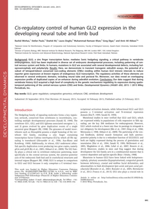
  • 2. carcinogenesis and transgenic mice studies suggest that over- expressing Gli2 in cutaneous keratinocytes develops multiple basal cell carcinoma, while the human hepatocellular carcinoma cell lines and hepatocellular tissues show high levels of GLI2 (Grachtchouk M, 2000; Cheng et al., 2009). Other human tumors associated with GLI2 are: medulloblastomas, prostate and breast cancer (Fulda et al., 2002; Okano et al., 2003; Sicklick et al., 2006). A tight temporal and spatial control of GLI2 expression is thus indispensable for multiple patterning steps in different tis- sues or organs. Gene expression plays a significant role in the evolution of vertebrate complexity and diversity in development (Carroll, 2008). Precise spatial and temporal expression of a gene is con- trolled by cis-acting elements (enhancers, repressors and silencers) that often reside hundreds of kilobases away or within an intron of the gene they control (Lettice et al., 2003; Levine and Tjian, 2003; Parveen et al., 2013). Each of these cis-acting regulatory modules achieve an explicit function, such as the acti- vation of its associated trans-dev gene in a specific cell and tissue type or at a precise phase in development (Paparidis et al., 2007; Lee et al., 2011; Pauls et al., 2012). A gene might thus contain multiple cis-regulatory modules, each of which contributes, in a somewhat combinatorial manner, to its overall spatial and tem- poral regulation (Parveen et al., 2013). Conserved non-coding DNA sequences from human to fish (evolutionary distance 450 Myr) clustered around developmental genes, are strong candi- dates for putative enhancers harbouring binding sites for multiple transcription factors (Woolfe et al., 2005; Pennacchio et al., 2006; Abbasi et al., 2007). Furthermore, some mutations in cis- acting regulatory regions can cause aberrant gene expression leading to a number of developmental defects (Lettice et al., 2003; Jeong et al., 2008; Ragvin et al., 2010). GLI paralogs are critical for early embryonic development in co-operation with Shh, triggering a sequence of reactions in downstream target genes (Lee et al., 1997; Ding et al., 1998; Dai P, 1999). The GLI family has a divergent, as well as an overlap- ping, role in growth and patterning of vertebrate development, owing to a mutual GLI code required for faithful transduction of Hh signaling (Ruiz i Altaba, 1998). Studies have already deci- phered GLI3-specific cis-acting regulatory sequences and how they control spatio-temporal expression in vertebrate develop- ment (Abbasi et al., 2007; Paparidis et al., 2007; Abbasi et al., 2010; Abbasi et al., 2013). However, the cis-acting control of the GLI2 gene remains unexplored. As an initial attempt to elucidate the cis-regulatory underpin- nings of human GLI2 expression, we have chosen tetrapod-teleost evolutionarily conserved intronic regions of GLI2, for functional assay in zebrafish. A stringent criteria of at least 50% conservation over a 50bp window across all species, was employed to highlight the tetrapod-teleost conserved regions, as shown by previous stud- ies that a criteria of 50-70% is ideal to identify putative regulatory elements (Woolfe et al., 2005; McEwen et al., 2006; Abbasi et al., 2007; Woolfe and Elgar, 2008). Our results show that intronic CNEs from the GLI2 gene act as tissue-specific enhancers and that reporter gene expression induced by these elements correlates with previously reported endogenous gli2 expression in zebrafish. Highly specific reporter expression is mainly observed in the neu- ral tube and pectoral fin. Our data reveal that tetrapod-teleost CNEs, located in the introns of GLI2, mark critical components of the cis-regulatory inventory for spatial and temporal deployment of this key developmental gene. Results Identification of Tetrapod-teleost conserved non- coding elements at the GLI2 locus by comparative sequence analysis A multi-species alignment of human GLI2 with orthologous genomic sequences from mouse, chicken, fugu, and zebrafish, revealed five anciently conserved non-coding elements embedded exclusively in the intronic intervals of GLI2 gene (Fig. 1), maintain- ing at least 50% identity over a 50bp window across all species. Comparative analysis of 100kb upstream or downstream region of the GLI2 gene did not detect any significant sequence similarity from human to fish. CNE1 and CNE5 are present within intron 1, while highly conserved CNE2 is present within intron 2, CNE3 is located in intron 4, and CNE4 is positioned within intron 7. None of the selected conserved non-coding elements overlap with exons or non-protein coding RNAs. The details of conserved tetrapod/tele- ost amplicons selected for functional analysis are described in Table 1. Moreover, highly conserved element CNE2 has two co-orthologs in the zebrafish genome. These duplicated CNEs (dCNEs) are posi- tioned within intron 2 of the zebrafish gli2a gene (Dr-gli2-CNE2a) and downstream of the zebrafish gli2b gene (Dr-gli2-CNE2b). Identification of transcription factor binding sites (TFBSs) within GLI2-associated CNEs Spatio-temporal expression of enhancers is due to the interaction between CNEs and multiple co-operative transcription factors (Parveen et al., 2013). To generate a list of candidate sites that might be responsible for the cis-regulatory activity of GLI2-associ- ated CNEs, tetrapod/teleost orthologous sequences were anlayzed. These sequences were carefully screened with the MEME motif dis- covery tool to identify conserved sequence motifs (for details see Materials and Methods). These evolutionarily conserved sequence motifs were then compared with position weight matrices (PWMs) for known vertebrate transcription factors (TFs) (Mahony and Benos, 2007). This identified putative TFBSs within each of the functionally categorized GLI2-intronic enhancers. Many of these transcription factors (Table 1) are key developmental regulators and are known to be co-expressed with Gli2 during early embry- onic development of limbs and CNS, as verified from the Mouse Genome Informatics database (http://www.informatics.jax.org/). In vivo characterization of CNEs using a co-injection assay in transiently transfected zebrafish embryos The CNEs identified through comparative genomics were next tested in vivo using zebrafish as a model organism. CNEs were co-injected with a GFP reporter into zebrafish embryos and then monitored for enhancer activity at set time points, as described previously (Woolfe et al., 2005). The reporter gene activity of all five CNEs showed reproducible tissue-specific expression after 24 (day-2) and 48 hours (day-3) post fertilization (hpf) in multiple independent transient transgenic assays. Percentage and catego- ries of cell types that are positive of each CNE are represented as a schematic (Fig. 2), and in tabular form (Table 2). CNE1 activates GFP expression predominantly in muscle cells (17%), forebrain (8%), midbrain (3%), hindbrain (19%) and spinal cord (6%) at day two; whereas at day three of development reporter expression is most frequently observed in hindbrain DEVELOPMENTALDYNAMICS 682 MINHAS ET AL.
  • 3. (45% of expressing embryos) (Fig. 3A and 3B). CNE2 induces GFP expression exclusively in notochord (79%) at $26-33 hpf, as well as at $48-56 hpf (Fig. 3C and 3D). CNE3 upregulates reporter gene expression in otic vesicle (48%) (Fig. 3E), muscle cells (52%), and hindbrain (11%) at $26-33 hpf, whereas at $48-56 hpf the main expression domains for CNE3 are hindbrain (66%) (Fig. 3F), otic vesicle (25%), and muscle cells (25%). CNE4 directs GFP expression specifically in the muscle cells at day two (93%) and at day three (96%) (Fig. 3G and 3H). CNE5 drives GFP expres- sion mainly in two major domains of the CNS, i.e. hindbrain (23%) and spinal cord (28%) at $26-33 hpf, whereas at day three CNE5 strongly enhances reporter expression in spinal cord (53%), hindbrain (40%) and muscle cells (30%) (Fig. 3I and 3J). Reproducible GFP expression of GLI2-associated CNEs by Tol2 transgenesis To validate the results from our co-injection assays, and to inves- tigate the potential cis-regulatory function of GLI2-associated CNEs in greater detail, we used a different strategy based on a Tol2 vector with a c-fos minimal promoter. Tol2 based transgene- sis has the advantage of stronger reporter expression with reduced mosaicism due to efficient and stable integration in the genome (Fisher et al., 2006; Kawakami, 2007). The injected embryos were screened ($26-33 hpf and $48-56 hpf) for compa- rable GFP expression with the co-injection assays. Moreover, zebrafish embryos showing significant and consistent GFP expression in those tissues which were not observed in co- injection assay were also noted (Table 2). With the exception of CNE1 and CNE4 (which were able to induce reporter expression with co-injection assay), the remain- der of the CNEs also upregulate GFP expression, driven by the c- fos promoter, in the Tol2 based transgenic assay. Based on our Tol2 results we categorized the human GLI2-associated CNEs into two major categories, those inducing GFP expression pre- dominantly in various domains of CNS (hindbrain and spinal cord) and those which upregulate GFP expression in the develop- ing zebrafish fin. CNE2, CNE3, and CNE5 are central nervous system specific enhancers In our zebrafish co-injection assay, the major GFP expression domain for CNE2 was notochord, whereas in the Tol2 based transgenic assay significant and reproducible reporter gene expression is detected not only in notochord (data not shown), DEVELOPMENTALDYNAMICS Fig. 1. Tetrapod-teleost conservation of the non-coding sequence across the GLI2 locusPair-wise sequence alignment of the genomic interval containing the human GLI2 locus (ENSG00000074047) and 100kb flanking upstream and downstream sequence with orthologous counterparts of tetrapod (mouse and chicken) and teleost fishes (fugu; gli2b and zebrafish; gli2a). These are shown as a VISTA (Visualization Tool for Alignment) graphical output using MLAGAN algorithm, using human sequence as a baseline. The red arrow shows the length of the GLI2 gene (257kb) and the direction of transcription. The 5 conserved non-coding elements highlighted in light blue color and indicated on top as red numbers, were selected for functional assay. Criteria of alignment were 50bp length and 50% conservation cut-off. Conserved protein-coding exons (1-14), and non-coding intronic sequences are depicted by blue and pink peaks respectively. The selected CNEs are conserved in mouse, chicken and fugu while CNE2 (marked with an asterisk) is highly conserved and has two co-orthologs in zebrafish (details are given in main text). Y-axis indicates percentage sequence identity and x-axis indicates the length of the sequences. kb, kilobase; Ex, exon; CNE, conserved non-coding elements. CIS-REGULATORY CONTROL OF HUMAN GLI2 683
  • 4. but also in hindbrain (60%) and spinal cord (70%) (Fig. 4A and 4D). CNE2 is unable to upregulate GFP expression in the develop- ing hindbrain and spinal cord using co-injection assay, probably due to high levels of mosaicism associated with this strategy (Woolfe et al., 2005; Abbasi et al., 2007; Kawakami, 2007). CNE3 and CNE5 drive reporter gene expression frequently in hindbrain and spinal cord using the co-injection strategy. In the Tol2 assay, CNE3 (63%) and CNE5 (58%) induced GFP expression is detected in the hindbrain neurons (Fig. 4B and 4C) and spinal cord (Fig. 4E and 4F). Reproducible GFP expression in the CNS with both assays using independent promoters suggests that GLI2- associated CNEs might play a role in neuronal development. CNE2 and CNE5 govern GFP expression in the developing pectoral fin In co-injection experiments none of the GLI2-associated CNEs are able to upregulate reporter gene expression in the developing pectoral fin, probably due to the high levels of mosaicism com- pared with the more penetrant Tol2 based strategy (Woolfe et al., 2005; Abbasi et al., 2007). However, when tested in the Tol2 assay, CNE2 and CNE5 are able to drive robust and reproducible GFP expression in the pectoral fin (Fig. 5A and 5B) at 48 hpf (36% and 31% for CNE2 and CNE5 respectively). Zebrafish co-orthologs of human GLI2 CNE2 appear to drive overlapping expression in hindbrain and pectoral fin with both the co-injection and Tol2 assays Zebrafish co-orthologs (Dr-gli2_CNE2a and Dr-gli2_CNE2b) of human GLI2-CNE2 share high sequence similarity within and between both lineages (human and zebrafish) (Fig. 6A). To inves- tigate the functional aspects of zebrafish duplicated enhancers we amplified Dr-gli2_CNE2a and Dr-gli2_CNE2b of almost equal length, i.e. 207 and 208bp respectively, from zebrafish genomic DNA and injected them into zebrafish embryos using the co- injection strategy. Dr-gli2_CNE2a and Dr-gli2_CNE2b drive over- lapping GFP expression at 48 hpf in the primary neurons of the hindbrain, 2.3% and 3% respectively (Fig. 6B, 6C, 7A and 7B). To verify the co-injection results, Dr-gli2_CNE2a and Dr- gli2_CNE2b were then injected in zebrafish embryos using the Tol2 based transgenic assay. These dCNEs appear to drive over- lapping GFP expression predominantly in the hindbrain region (Fig. 7C and 7D, CNE2a, 33%; CNE2b, 25%). The results are in concert with the co-injection assays for these duplicated zebra- fish CNEs, as far as reporter expression in hindbrain is concerned. In addition, both the dCNEs demonstrate robust and reproducible reporter expression in the developing pectoral fin (CNE2a, 41%; CNE2b, 45%) of zebrafish embryos (Fig. 7E and 7F). Discussion Evolutionary sequence comparison reveals candidate GLI2 enhancers Genomic comparisons of distantly related vertebrate species have revealed many genomic intervals that have remained conserved during vertebrate evolution (Abbasi et al., 2007). Some of these sequences correspond to coding genes and non-coding RNAs, however two thirds of them are unlikely to produce a functional transcript (Dermitzakis et al., 2005). These sequences fall in the DEVELOPMENTALDYNAMICS TABLE1.Tetrapod-teleostconservednon-codingelements(CNEs)fromintronsofhumanGLI2selectedforfunctionalanalysisintransgeniczebrafish assays ElementRegion Amplicon CoordinatesChr2 Amplicon Size Conservation Human-Fugu 50%;>50bpCo-injectionTol2ConservedPutativeTFBSs CNE1Intron1121667550–121668542993bp72%(131bp)þ-Ttk,frem1,SMAD3,SOX10,Hmx3,Nkx2-5,En,Ftz,Lhx3, Ptx1,GATA-3,GATA-2 CNE2Intron2121689507–121690037531bp67%(128bp)þþAML1,Pbx-1,HOXA4,CHX10,TCF11,OCT_4,NRSF,E2F, TCF11,SREBP-1 CNE3Intron4121719881–121720730850bp74%(73bp)þþEBF,RORalpha1,STAT1,Foxa2,Ptx1,POU3F2,Apex1, GATA-1,NF-kappaB CNE4Intron7121730105–1217314021298bp67%(163bp)þ-STAT5B,POU5F1,Brn-2,Pax-5,CEBP,GATA-1,DMRT2, Pbx,Alx-4,SOX17,CREB CNE5Intron1121563302–121563959658bp75%(221bp)þþAIRE,En,RAP1,Pbx,TCF11,Nrf-2,HOXA7,SOX17,FOXO3, Foxc1,TFII-I,MAZ Location,coordinates(Ensembl70:Jan2013),ampliconsize,human-fuguconservationscoreofselectedsubsetofhuman-GLI2CNEs(functionallytestedinthisstudy)are indicated.TheelementswhichinducedGFPexpressionusingco-injection(b-globinminimalpromoter)andTol2assay(c-fosminimalpromoter)inzebrafishembryosare shownaspositive.Inaddition,thetablealsoprovidesinformationaboutthetranscriptionalfactorbindingsites(resultsfromTRANSFACdatabase). 684 MINHAS ET AL.
  • 5. new category of elements, which are termed as conserved non- coding elements (CNEs) (Woolfe et al., 2005). These elements are experimentally characterized to harbour transcription factor bind- ing sites, and are implicated in the control of gene expression (Pennacchio et al., 2006). Therefore, comparative genomics based strategies have emerged as a reliable methodology to predict genomic intervals harboring transcriptional regulatory elements even in the absence of knowledge about the specific characteristics of individual cis-regulatory element (Wasserman et al., 2000). Members of the GLI family (GLI1, GLI2, and GLI3) have vital roles in many Shh-targeted developmental processes of the limb, neural tube and many internal organs. Previously, comparative analyses of human and fugu GLI3 locus has identified 11 GLI3- associated CNEs distributed throughout the introns of GLI3 gene. The regulatory potential of a subset of GLI3-associated CNEs was in agreement with the reported Gli3 endogenous expression in vertebrates (Abbasi et al., 2007; Abbasi et al., 2010; Abbasi et al., 2013). Similarly, several studies indicate that GLI2 has essential functions controlling multiple patterning steps in different tis- sues/organs, and therefore a tight temporal and spatial control of gene expression is indispensable. However, cis-regulatory under- pinnings of the human GLI2 gene remain unknown. The identifi- cation of cis-acting regulatory elements interacting with the GLI2 promoter could facilitate the detection of factors controlling the tissue-specific availability of GLI2 in trans in hedgehog target cells. In turn, identification of transcription factors for spatial and temporal control of GLI2 expression would greatly enhance our understanding of the regulatory network that coordinates the multitude of patterning events associated with the hedgehog sig- naling pathway. By employing multi-species sequence alignment we identified an ancient (tetrapod-teleost conserved) non-coding architecture within the introns of GLI2. To test possible enhancers of expres- sion we selected human CNEs encompassing >50bp tracks with more than 50% sequence similarity between tetrapod and teleost. The selected subset of intra-GLI2 CNEs were BLAST against human genome using UCSC and Ensembl genome browsers, to verify whether these genomic intervals are unique to GLI2 gene, or have a significant sequence similarity with the GLI2 paralogs (GLI1 and GLI3). This analysis revealed that all the five CNEs have significant hits against human GLI2 gene only, and no evi- dence of overlap was found nearby or within the GLI1 and GLI3 genes. Similar to the previously identified GLI3-associated enhancers (Abbasi et al., 2007), GLI2 CNEs are distributed across almost the entire gene interval (Fig. 1), with two elements in intron 1 and one in each of introns 2, 4, and 7. GLI2-associated CNEs show tissue-specific regulatory activity in vivo In order to address the in vivo role of the GLI2-associated CNEs, we used transient reporter gene expression in zebrafish embryos with two different approaches: firstly, exploiting a co-injection strategy using a minimal b-globin promoter, and secondly, through direct cloning into a Tol2 vector with a c-fos promoter (Woolfe et al., 2005; Abbasi et al., 2007; Pauls et al., 2012). These DEVELOPMENTALDYNAMICS Fig. 2. Schematic representation of GFP expression induced by GLI2-associated CNEs in zebrafish embryos at day-2 ($24 hpf) and day-3 ($48 hpf)The reporter gene was induced by individual GLI2-associated CNEs (indicated by name) and the GFP signal was noted at each stage. Embryos on the left side represent day-2 data while the embryos on the right side representing the day-3 data. GFP-positive cells are marked onto camera Lucida drawings of a zebrafish embryo on day-2 ($26-30 hpf) and day-3 ($48-54 hpf) of development. The outcomes from all embryos with expression were overlaid to generate a compound representation of the GFP expression pattern. Categories of cell type that were positive for a given CNE are color coded, with each dot representing a single GFP-expressing cell. Bar graphs show the percentage (y-axis) of GFP positive embryos with expression in each domain 1-14. The percentage of GFP expressing embryos per CNE is indicated beneath each schematic (EE¼ %). EE, expressing embryos; hpf, hours post fertilization. CIS-REGULATORY CONTROL OF HUMAN GLI2 685
  • 6. approaches, exploiting the transparency and rapid development of zebrafish embryos, have shown their potential for functionally testing enhancer elements among conserved non-coding regions (Woolfe et al., 2005; Fisher et al., 2006). GFP up-regulation is observed consistently in co-injection assay, however, it is a labo- rious technique to inject and screen hundreds of embryos to gen- erate a comprehensive view of the reporter expression pattern (Woolfe et al., 2005). Moreover, failure of detecting reporter expression with co-injection assay for some GLI2-associated CNEs in fin, illuminates the fact that this strategy is not suitable to detect the activity of limb-specific regulators. In comparison, Tol2 transposon constructs injected with transposase mRNA inte- grates in the genome of somatic cells with high efficiency and tissue-specific GFP expression can be observed easily due to non- mosaic reporter pattern. Thus, it is a more suitable strategy for smaller domains like fin (Fisher et al., 2006; Birnbaum et al., 2012). Our results with two different approaches indicate that multiple evolutionarily conserved GLI2-associated human cis- regulators control highly coordinated GFP reporter gene expres- sion in transient transgenic zebrafish, mimicking a subset of the known repertoire of endogenous gli2 expression (Table 2 and 3) (Du and Dienhart, 2001; Ke et al., 2005; Thisse and Thisse, 2005; Ke et al., 2008; Wang et al., 2013). CNE2, CNE3, and CNE5 induce reporter expression that coincides with known sites of GLI2 activity in neural tube Gli2 plays an activator role in the hindbrain and spinal cord pat- terning; studies in Gli2À /À mice have shown diverse ventral pat- terning defects in hindbrain and spinal cord with a severely affected floor plate (FP) and interneurons (Ding et al., 1998; Lebel et al., 2007). Our data shows that reporter expression driven by a subset of identified GLI2 intronic enhancers (CNE2, CNE3, and CNE5) is largely confined to the zebrafish hindbrain (Fig. 4A-4C) and dorsal spinal cord neurons (Fig. 4D-4F), reflecting the com- plex role of gli2 in neural tube development (Table 3). These in vivo data revealed the overlapping contribution of CNE2, CNE3, and CNE5 in the precise patterning of the neural tube. Further- more, in silico analysis of CNE2, CNE3, and CNE5 predicts human-fugu conserved binding sites for a number of develop- mentally important transcription factors that are known to be co- expressed with Gli2 during neural tube formation (Table 1). Cis-regulatory control of GLI2 expression in developing pectoral fin The Gli2 expression patterns within the nascent limb bud are highly dynamic and context dependent. Establishment of anterior- posterior positional identities in the limb requires integration of the spatial distribution, timing, and dosage of GLI2 expression (Bowers et al., 2012). RNA in situ studies in mice embryos have shown that at E10.5, Gli2 and Gli3 are broadly expressed in undif- ferentiated mesenchyme of the emerging limb bud, except poste- rior mesenchyme, where the genetic antagonism between Gli2 and Shh leads to reduced expression of Gli2 (Mo et al., 1997; Bai and Joyner, 2001). Gli2- /- mice show reduced ossification in various bones in limbs including; stylopod (humerus, femur), zeugopod (radius, ulna, tibia and fibula) as well as shortening of the autopod (limb) (Mo et al., 1997). In Gli3 mutant mice, Gli2 is vital for the patterning of anterior and posterior regions of the autopod (Bowers et al., 2012). Moreover, studies in zebrafish showed that gli2a expression in the fin bud appears at 30 hpf (Table 3) (Thisse and Thisse, 2005), and by 36 hpf is expressed uniformly throughout the mesenchyme of the developing pectoral fin (Karlstrom et al., 2003). In addition, limb deformities like preaxial and postaxial pol- ydactyly are well documented with GLI2 mutations (Roessler et al., 2003; Roessler et al., 2005; Bertolacini et al., 2012). Consistent with a role for gli2a in fin, we find that CNE2 and CNE5 drive reproducible reporter expression in the developing pectoral fin (Fig. 5A and 5B). The primary expansion of teleost paired fins is strikingly parallel to that of tetrapod limb buds and is controlled by a similar mechanism (Zhang et al., 2010). Evolu- tion of cis-acting elements is proposed to be the key regulator in the development and structural framework of the vertebrate fin/ limb skeleton. Shh and Gli family are the key signalling transduc- tion pathways implicated in fin and limb morphologies (Mo et al., 1997; Abbasi, 2011). Overlapping activity of two independent enhancers in fin indi- cates that GLI2 harbors multiple cis-regulatory modules required for the normal development of limb. Furthermore, the roles of CNE2 and CNE5 in the pectoral fin are reflected by the presence of binding sites for numerous established transcription factors which are known to be co-expressed with Gli2 in the developing limb (Table 1). Spatial and temporal activity of these enhancers needs to be investigated further in tetrapod model animals like mice, to completely decipher the mechanism of Gli2 in anterior- DEVELOPMENTALDYNAMICS TABLE 2. Comparison of reporter gene expression induced by intra-GLI2 CNEs Element Forebrain Midbrain Hindbrain Spinal cord Notochord Muscle cells Pectoral fin Otic vesicle CNE1 þ þ þ þ - þ - - CNE2 - - þ þ þ þ þ - CNE3 - - þ þ - þ - þ CNE4 - - - - - þ - - CNE5 - - þ þ - þ þ - Dr-gli2a_CNE2a - - þ þ - þ þ - Dr-gli2b_CNE2b - - þ - - þ þ - Comparison of CNEs mediated reporter gene expression in zebrafish embryonic domains. The “þ” sign indicates the domain where GFP signal is detected, while “-” sign indicates those domains where GFP signal is not observed. CNE, Conserved non- coding element; Dr, Danio rerio; GFP, Green fluorescent protein. 686 MINHAS ET AL.
  • 7. DEVELOPMENTALDYNAMICS Fig. 3. GLI2-associated CNEs upregulate GFP expression in live zebrafish embryos, using co-injection assaysIntronic GLI2-associated conserved non-coding elements (CNE1-CNE5) mediate GFP expression pattern in live embryos at day-2 and day-3, fluorescent views (A-D), merged bright field and fluorescence views (E-J). Embryos A, C, E, G and J are $26–33 hpf, while embryos B, D, F, H and I are $48–54 hpf. Orientation of the embryos is anterior to left, dorsal to top, with lateral views. White arrowheads indicate GFP expressing cells. CNE1 drives GFP expression in mus- cle fibers in the trunk region (A), and in the neurons of the midbrain, hindbrain (B). CNE2 expresses GFP in the developing notochord at day-2 ($26–33 hpf) (C), as well as at day-3 ($48 hpf) (D). CNE3 drives GFP expression in the otic vesicle (E), and in the primary neurons of hindbrain (F). CNE4 showed GFP signal in muscle fibers at day-2 and day-3 (G & H). GFP expression was observed in the neurons of hindbrain (I), spinal cord and muscle fibers (J) by CNE5. hpf, hours post fertilization; mb, midbrain; hb, hindbrain; nc, notochord; sc, spinal cord; m, muscle; ov, otic vesicle. CIS-REGULATORY CONTROL OF HUMAN GLI2 687
  • 8. DEVELOPMENTALDYNAMICS Fig. 4. CNE2, CNE3, and CNE5 induced GFP expression was detected in various domains of CNS, using Tol2 based transgenic assaysPatterns of GFP expression was observed by GLI2-associated CNEs in central nervous system. Images of live zebrafish embryos at $48-54 hpf, lateral views, anterior to left, dorsal to top. Arrowheads and marked area point to GFP expressing cells. Using the Tol2 system, injected zebrafish embryos demonstrate that CNE2, CNE3 and CNE5 are central nervous system enhancers; induce GFP expression in primary neurons of hindbrain (A, B and C respectively) and spinal cord (D, E and F respectively). hb, hindbrain; sc, spinal cord. Fig. 5. CNE2 and CNE5 are fin-specific enhancersImages of live zebrafish embryos at $48-54 hpf, lateral views, anterior to left, dorsal to top. Arrowheads indicate GFP expressing cells. Using the Tol2 system, CNE2 (A) and CNE5 (B) induced GFP expression in the developing fin. f, fin. 688 MINHAS ET AL.
  • 9. DEVELOPMENTALDYNAMICS Fig. 6. Sequence alignment and schematic representation of GFP expression induced by dCNEs in co-injection assayClustalW alignment of human GLI2-CNE2 and its co-orthologous sequences in zebrafish (A). The reporter gene was induced by Dr-gli2_CNE2a (B) and Dr-gli2_CNE2b (C). GFP signal was noted at $24 and $48 hpf stage. The outcomes from all embryos with expression were overlaid to give a compound represen- tation of the GFP expression pattern. The percentages of positive embryos per CNE are indicated. Dr, Danio rerio; EE, expressing embryos. Fig. 7. Duplicated copies of zebrafish CNE2 (CNE2a and CNE2b) induce overlapping GFP expression in hindbrain and pectoral finGFP expres- sion is presented in live embryos, by transiently transfected co-injection (A and B) and Tol2 transgenic assays (C, D, E, and F). All embryos were at $48-54 hpf. Lateral views, anterior to the left, dorsal to the top. GFP expression is indicated by arrowheads and marked area in the following tissue or cell types: hindbrain (A and C) and pectoral fin (E) by Dr-gli2_CNE2a; hindbrain (B and D) and pectoral fin (F) by Dr-gli2_CNE2b. hb, hind- brain; f, fin. CIS-REGULATORY CONTROL OF HUMAN GLI2 689
  • 10. posterior polarity of the limb and to define the expression pattern boundaries driven by GLI2-associated CNEs. dCNEs have overlapping expression pattern in hindbrain and pectoral fin Comparative sequence analysis revealed two copies (co-ortho- logs) of human CNE2 in zebrafish. One of the copies (CNE2a) was positioned within intron 2 of the gli2a gene whilst the other copy (CNE2b) resides downstream of the gli2b gene. CNE2a (119bp) and CNE2b (117bp) share significant sequence similarity with each other (75%), and also with human GLI2-CNE2 (117bp), i.e. 68% and 65% respectively (Fig. 6A). We analyzed each of these elements independently in transgenic zebrafish and compared reporter gene expression induced by these elements with each other. Careful examination of resultant data shows apparently overlapping reporter expression of CNE2a and CNE2b in zebrafish hindbrain (Fig. 7A-7D) and developing fin (Fig. 7E and 7F), wherein endogenous expression of gli2a/gli2b genes in a compli- mentary manner has already been reported (Table 3) (Thisse and Thisse, 2005; Ke et al., 2008). To decipher the reason for this overlapping expression of the duplicated CNEs, and any indica- tion of subfunctionalization involved in the process, more precise assays need to be performed. These findings reflect that the com- bined activity of gli2a/gli2b cis-regions is essential for the nor- mal development of zebrafish hindbrain and fin. Conclusions In this study, we have used deep evolutionary constraint as an indicator to pinpoint the cis-regulatory catalogue of the develop- mentally important human GLI2 gene. Our data demonstrate that like its evolutionary counterpart GLI3, the cis-regulatory reper- toire of GLI2 resides within its intronic intervals and has remained conserved since the divergence of the tetrapod-teleost lineages. We have shown that this ancient catalogue of intronic enhancers regulates reporter expression in a diverse set of embry- onic domains where GLI2 is known to be expressed endoge- nously. Furthermore, GLI2 enhancers depict considerable overlap in their expression territories during zebrafish development. Elu- cidation of the GLI2-associated cis-regulatory network offers a novel perspective for understanding the genetic mechanisms by which the downstream effectors of Hh signaling cascade might be regulated during embryogenesis. In addition, these cis-regula- tory sequences are strong candidates for mutational studies of GLI2-associated human birth defects, which cannot be attributed to any exonic mutation. Experimental Procedures Strategy to hunt the putative cis-acting regulatory elements of GLI2 for functional analysis The human GLI2 genomic sequence was obtained from the Ensembl genome browser (http://www.ensembl.org) release-65 (GRCh37) with 100kb flanking region along with orthologous sequences from mouse (NCBIM37), chicken (Galgal4), fugu (Fugu4) and zebrafish (Zv9). Multi-species sequence comparisons were performed using the MLAGAN alignment tool kit (Brudno et al., 2003). Human sequence was used as a baseline and anno- tated by exon/intron information available at Ensembl genome browser. The MLAGAN alignment was visualized using VISTA (Mayor et al., 2000). The conservation was measured using a 50 bp window and a cutoff score of 50% identity. Identification of putative TFBSs To identify putative conserved TFBSs for each CNE the ortholo- gous sequences of terrestrial and non-terrestrial vertebrates were retrieved from Ensembl genome database by BLASTN based simi- larity search. Each of the GLI2-associated CNE with orthologous sequences were analyzed using MEME motif discovery algorithm (Bailey et al., 2009). MEME, is a PWM (position weight matrices) based algorithm, which identifies over-represented motifs in the DEVELOPMENTALDYNAMICS TABLE 3. Reported endogenous expression pattern of Gli2 and gli2a/gli2b in vertebrates Gene Endogenous expression Source GLI2/gli2a Forebrain (Hui et al., 1994) Midbrain (Magdaleno et al., 2006), (Ke et al., 2005) Hindbrain (Ke et al., 2008), (Thisse and Thisse, 2005), (Hui et al., 1994) Spinal cord (Sasaki H, 1999), (Lee et al., 1997) Notochord (Ding et al., 1998) Limb/Pectoral fin (Mo et al., 1997), (Karlstrom et al., 2003) Muscle cells (Du and Dienhart, 2001) Otic vesicle (Hui et al., 1994) gli2b Forebrain (Thisse and Thisse, 2005) Midbrain (Ke et al., 2005) Hindbrain (Ke et al., 2008) Spinal cord (Thisse and Thisse, 2005) Notochord (Thisse and Thisse, 2005) Pectoral fin Needs to be investigated Muscle cells (Wang et al. 2013) Otic vesicle (Ke et al., 2005) This table provides information of already reported endogenous expression of Gli2 in mice and its co-orthologs (gli2a/gli2b) in zebra- fish. The last column provides the source of literature that addresses the function and endogenous expression pattern of Gli2/gli2. 690 MINHAS ET AL.
  • 11. query set. Considering the expected length of transcription fac- tors, the criteria for minimum length was set from 6 to 12bp. The identified motifs of each CNE were characterized further by using the STAMP tool (Mahony and Benos, 2007) to determine known transcription factors against TRANSFAC (v11.3) database (Matys et al., 2003). Each of the specified transcription factors were then chosen for endogenous gene expression (RNA in-situ hybridiza- tion) studies using Mouse Genome Informatics database (http:// www.informatics.jax.org/). Zebrafish transgenic assays Genomic DNA of human and zebrafish was used to amplify the conserved non-coding regions for zebrafish transgenic assays. The reporter expression cassette consisting of EGFP under the control of mouse b-globin minimal promoter, was also amplified from plasmid vector by PCR (Thermo DNA Taq, Thermo Scientific) and purified using PureLink PCR purification kit (Life Technologies). PCR puri- fied product of CNEs (30ng/ml) and b-globin-GFP promoter-reporter cassette (15ng/ml) were combined with 0.5% phenol red (Sigma), used as a tracer dye as described previously (Woolfe et al., 2005). The injection mix was injected into 1-2 cell stage of the zebrafish embryos. The injected embryos were raised at 28.5 C in 1X embryo medium containing 0.003% PTU to prevent pigmentation. The zebrafish embryos were dechorionated manually by fine forceps at day-2 and anaesthetized by Tricaine. The transgenic embryos were screened for GFP signals under an inverted fluorescent microscope (IX81, Olympus, Japan) using an FVII CCD monochrome digital software. The schematics for location and tissue-specific expression of each CNE was generated using Adobe Photoshop software. To verify the expression of CNEs using Tol2 GFP system (Fisher et al., 2006), the CNEs were first amplified with a final 10–30 min extension step. The freshly amplified PCR ($500ng/ml) products were cloned into pCR8/GW/TOPO vector (Life Technologies) to make entry clones (Pauls et al., 2012). Orientation screening to determine the sense strand was followed by a LR recombination reaction between topo entry clone ($100 ng/ml) and destination vector pGW_cfosEGFP ($100 ng/ml); Gateway LR Clonase II enzyme (Life Technologies) was used. The destination clones con- sisting of CNEs and minimal c-fos promoter were sequenced for confirmation of positive orientation into the transposon con- struct. The purified transposon construct (25ng/ml), 0.5ml trans- posase RNA enzyme (175ng/ml), and 0.5ml phenol red stock, were injected into one-cell stage zebrafish embryos. The transient transgenic embryos were screened at day-2 and day-3 using a Leica MZ 16F fluorescence stereomicroscope, and photographs taken with a Leica DFC310FX camera. Acknowledgments We are grateful to MRC NIMR Biological Services (UK) for excel- lent fish care. We also thank Joseph Grice and Dr. Boris Noyvert for useful discussions during this project. Competing interests The authors declare that they have no competing interests. Author contributions A.A.A. and G.E. conceived the project and designed the experi- ments. Computational analysis were performed by R.M. The wet lab expermients were performed by R.M., S.P., L.D., S.A., and M.R.K. The manuscript was written by A.A.A., R.M., and G.E. References Abbasi AA. 2011. Evolution of vertebrate appendicular structures: Insight from genetic and palaeontological data. Dev Dyn 240: 1005–1016. Abbasi AA, Goode DK, Amir S, Grzeschik KH. 2009. Evolution and functional diversification of the GLI family of transcription factors in vertebrates. Evol Bioinform Online 5:5–13. Abbasi AA, Minhas R, Schmidt A, Koch S, Grzeschik KH. 2013. Cis-regulatory underpinnings of human GLI3 expression in embryonic craniofacial structures and internal organs. Dev Growth Differ 55:699–709. Abbasi AA, Paparidis Z, Malik S, Bangs F, Schmidt A, Koch S, Lopez-Rios J, Grzeschik KH. 2010. Human intronic enhancers control distinct sub-domains of Gli3 expression during mouse CNS and limb development. BMC Dev Biol 10:44. Abbasi AA, Paparidis Z, Malik S, Goode DK, Callaway H, Elgar G, Grzeschik KH. 2007. Human GLI3 intragenic conserved non-coding sequences are tissue-specific enhancers. PLoS One 2:e366. Alexandre C, Jacinto A, Ingham PW. 1996. Transcriptional activa- tion of hedgehog target genes in Drosophila is mediated directly by the cubitus interruptus protein, a member of the GLI family of zinc finger DNA-binding proteins. Genes Dev 10:2003–2013. Aza-Blanc P, Kornberg T. 1999. Ci: a complex transducer of the hedgehog signal. Trends in genetics: TIG 15:458–462. Bai CB, Joyner AL. 2001. Gli1 can rescue the in vivo function of Gli2. Development 128:5161–5172. Bailey T, Boden M, Buske F, Frith M, Grant C, Clementi L, Ren J, Li W, Noble W. 2009. MEME SUITE: tools for motif discovery and searching. Nucleic acids research 37:8. Bertolacini CD, Ribeiro-Bicudo LA, Petrin A, Richieri-Costa A, Murray JC. 2012. Clinical findings in patients with GLI2 muta- tions–phenotypic variability. Clin Genet 81:70–75. Birnbaum R, Clowney E, Agamy O, Kim M, Zhao J, Yamanaka T, Pappalardo Z, Clarke S, Wenger A, Nguyen L, Gurrieri F, Everman D, Schwartz C, Birk O, Bejerano G, Lomvardas S, Ahituv N. 2012. Coding exons function as tissue-specific enhancers of nearby genes. Genome research 22:1059–1068. Bowers M, Eng L, Lao Z, Turnbull RK, Bao X, Riedel E, Mackem S, Joyner AL. 2012. Limb anterior-posterior polarity integrates activator and repressor functions of GLI2 as well as GLI3. Dev Biol 370:110–124. Brudno M, Do CB, Cooper GM, Kim MF, Davydov E, Green ED, Sidow A, Batzoglou S. 2003. LAGAN and Multi-LAGAN: efficient tools for large-scale multiple alignment of genomic DNA. Genome Res 13:721–731. Carroll SB. 2008. Evo-devo and an expanding evolutionary synthe- sis: a genetic theory of morphological evolution. Cell 134:25–36. Cheng WT, Xu K, Tian DY, Zhang ZG, Liu LJ, Chen Y. 2009. Role of Hedgehog signaling pathway in proliferation and invasiveness of hepatocellular carcinoma cells. Int J Oncol 34:829–836. Dai P AH, Tanaka Y, Maekawa T, Nakafuku M, Ishii S. 1999. Sonic Hedgehog-induced activation of the Gli1 promoter is mediated by GLI3. J Biol Chem. 274. Dermitzakis ET, Reymond A, Antonarakis SE. 2005. Conserved non-genic sequences - an unexpected feature of mammalian genomes. Nat Rev Genet 6:151–157. Ding Q, Motoyama J, Gasca S, Mo R, Sasaki H, Rossant J, Hui CC. 1998. Diminished Sonic hedgehog signaling and lack of floor plate differentiation in Gli2 mutant mice. Development 125:2533–2543. Du SJ, Dienhart M. 2001. Gli2 mediation of hedgehog signals in slow muscle induction in zebrafish. Differentiation 67:84–91. Fisher S, Grice EA, Vinton RM, Bessling SL, Urasaki A, Kawakami K, McCallion AS. 2006. Evaluating the biological relevance of putative enhancers using Tol2 transposon-mediated transgenesis in zebrafish. Nat Protoc 1:1297–1305. Franca MM, Jorge AA, Carvalho LR, Costalonga EF, Otto AP, Correa FA, Mendonca BB, Arnhold IJ. 2013. Relatively high fre- quency of non-synonymous GLI2 variants in patients with con- genital hypopituitarism without holoprosencephaly. Clin Endocrinol (Oxf) 78:551–557. DEVELOPMENTALDYNAMICS CIS-REGULATORY CONTROL OF HUMAN GLI2 691
  • 12. Fulda S, Meyer E, Debatin KM. 2002. Inhibition of TRAIL-induced apoptosis by Bcl-2 overexpression. Oncogene 21:2283–2294. Grachtchouk M MR, Yu S, Zhang X, Sasaki H, Hui CC, Dlugosz AA. 2000. Basal cell carcinomas in mice overexpressing Gli2 in skin. Nat Genet. 24:216–217. Hui CC, Slusarski D, Platt KA, Holmgren R, Joyner AL. 1994. Expression of three mouse homologs of the Drosophila segment polarity gene cubitus interruptus, Gli, Gli-2, and Gli-3, in ecto- derm- and mesoderm-derived tissues suggests multiple roles during postimplantation development. Dev Biol 162:402–413. Jeong Y, Leskow FC, El-Jaick K, Roessler E, Muenke M, Yocum A, Dubourg C, Li X, Geng X, Oliver G, Epstein DJ. 2008. Regulation of a remote Shh forebrain enhancer by the Six3 homeoprotein. Nat Genet 40:1348–1353. Karlstrom RO, Tyurina OV, Kawakami A, Nishioka N, Talbot WS, Sasaki H, Schier AF. 2003. Genetic analysis of zebrafish gli1 and gli2 reveals divergent requirements for gli genes in vertebrate development. Development 130:1549–1564. Kawakami K. 2007. Tol2: a versatile gene transfer vector in verte- brates. Genome Biol 8 Suppl 1:S7. Ke Z, Emelyanov A, Lim SE, Korzh V, Gong Z. 2005. Expression of a novel zebrafish zinc finger gene, gli2b, is affected in Hedgehog and Notch signaling related mutants during embryonic develop- ment. Dev Dyn 232:479–486. Ke Z, Kondrichin I, Gong Z, Korzh V. 2008. Combined activity of the two Gli2 genes of zebrafish play a major role in Hedgehog signaling during zebrafish neurodevelopment. Mol Cell Neurosci 37:388–401. Lebel M, Mo R, Shimamura K, Hui CC. 2007. Gli2 and Gli3 play distinct roles in the dorsoventral patterning of the mouse hind- brain. Dev Biol 302:345–355. Lee AP, Brenner S, Venkatesh B. 2011. Mouse transgenesis identifies conserved functional enhancers and cis-regulatory motif in the ver- tebrate LIM homeobox gene Lhx2 locus. PLoS One 6:e20088. Lee J, Platt KA, Censullo P, Ruiz i Altaba A. 1997. Gli1 is a target of Sonic hedgehog that induces ventral neural tube develop- ment. Development 124:2537–2552. Lettice LA, Heaney SJ, Purdie LA, Li L, de Beer P, Oostra BA, Goode D, Elgar G, Hill RE, de Graaff E. 2003. A long-range Shh enhancer regulates expression in the developing limb and fin and is associ- ated with preaxial polydactyly. Hum Mol Genet 12:1725–1735. Levine M, Tjian R. 2003. Transcription regulation and animal diver- sity. Nature 424:147–151. Magdaleno S, Jensen P, Brumwell CL, Seal A, Lehman K, Asbury A, Cheung T, Cornelius T, Batten DM, Eden C, Norland SM, Rice DS, Dosooye N, Shakya S, Mehta P, Curran T. 2006. BGEM: an in situ hybridization database of gene expression in the embry- onic and adult mouse nervous system. PLoS Biol 4:e86. Mahony S, Benos PV. 2007. STAMP: a web tool for exploring DNA- binding motif similarities. Nucleic Acids Res 35:W253-258. Matys V, Fricke E, Geffers R, Gossling E, Haubrock M, Hehl R, Hornischer K, Karas D, Kel AE, Kel-Margoulis OV, Kloos DU, Land S, Lewicki-Potapov B, Michael H, Munch R, Reuter I, Rotert S, Saxel H, Scheer M, Thiele S, Wingender E. 2003. TRANSFAC: transcriptional regulation, from patterns to profiles. Nucleic Acids Res 31:374–378. Mayor C, Brudno M, Schwartz JR, Poliakov A, Rubin EM, Frazer KA, Pachter LS, Dubchak I. 2000. VISTA: visualizing global DNA sequence alignments of arbitrary length. Bioinformatics 16:1046–1047. McDermott A, Gustafsson M, Elsam T, Hui CC, Emerson CP, Jr., Borycki AG. 2005. Gli2 and Gli3 have redundant and context- dependent function in skeletal muscle formation. Development 132:345–357. McEwen GK, Woolfe A, Goode D, Vavouri T, Callaway H, Elgar G. 2006. Ancient duplicated conserved noncoding elements in vertebrates: a genomic and functional analysis. Genome Res 16:451–465. Mo R, Freer AM, Zinyk DL, Crackower MA, Michaud J, Heng HH, Chik KW, Shi XM, Tsui LC, Cheng SH, Joyner AL, Hui C. 1997. Specific and redundant functions of Gli2 and Gli3 zinc finger genes in skeletal patterning and development. Development 124:113–123. Motoyama J LJ, Mo R, Ding Q, Post M, Hui CC. 1998. Essential function of Gli2 and Gli3 in the formation of lung, trachea and oesophagus. Nat Genet. 20:54–57. Okano H, Shiraki K, Inoue H, Kawakita T, Yamanaka T, Deguchi M, Sugimoto K, Sakai T, Ohmori S, Fujikawa K, Murata K, Nakano T. 2003. Cellular FLICE/caspase-8-inhibitory protein as a princi- pal regulator of cell death and survival in human hepatocellular carcinoma. Lab Invest 83:1033–1043. Paparidis Z, Abbasi AA, Malik S, Goode DK, Callaway H, Elgar G, deGraaff E, Lopez-Rios J, Zeller R, Grzeschik KH. 2007. Ultra- conserved non-coding sequence element controls a subset of spatiotemporal GLI3 expression. Dev Growth Differ 49:543–553. Parveen N, Masood A, Iftikhar N, Minhas BF, Minhas R, Nawaz U, Abbasi AA. 2013. Comparative genomics using teleost fish helps to systematically identify target gene bodies of functionally defined human enhancers. BMC Genomics 14:122. Pauls S, Smith SF, Elgar G. 2012. Lens development depends on a pair of highly conserved Sox21 regulatory elements. Dev Biol 365:310–318. Pennacchio LA, Ahituv N, Moses AM, Prabhakar S, Nobrega MA, Shoukry M, Minovitsky S, Dubchak I, Holt A, Lewis KD, Plajzer-Frick I, Akiyama J, De Val S, Afzal V, Black BL, Couronne O, Eisen MB, Visel A, Rubin EM. 2006. In vivo enhancer analysis of human conserved non-coding sequences. Nature 444:499– 502. Ragvin A, Moro E, Fredman D, Navratilova P, Drivenes O, Engstrom PG, Alonso ME, de la Calle Mustienes E, Gomez Skarmeta JL, Tavares MJ, Casares F, Manzanares M, van Heyningen V, Molven A, Njolstad PR, Argenton F, Lenhard B, Becker TS. 2010. Long-range gene regulation links genomic type 2 diabetes and obesity risk regions to HHEX, SOX4, and IRX3. Proc Natl Acad Sci U S A 107:775–780. Roessler E, Du YZ, Mullor JL, Casas E, Allen WP, Gillessen- Kaesbach G, Roeder ER, Ming JE, Ruiz i Altaba A, Muenke M. 2003. Loss-of-function mutations in the human GLI2 gene are associated with pituitary anomalies and holoprosencephaly-like features. Proc Natl Acad Sci U S A 100:13424–13429. Roessler E, Ermilov AN, Grange DK, Wang A, Grachtchouk M, Dlugosz AA, Muenke M. 2005. A previously unidentified amino- terminal domain regulates transcriptional activity of wild-type and disease-associated human GLI2. Hum Mol Genet 14:2181–2188. Ruiz i Altaba A. 1998. Combinatorial Gli gene function in floor plate and neuronal inductions by Sonic hedgehog. Development. Ruppert JM KK, Wong AJ, Bigner SH, Kao FT, Law ML, Seuanez HN, O’Brien SJ, Vogelstein B. 1998. The GLI-Kruppel family of human genes. Mol Cell Biol.:3104–3113. Sasaki H NY, Hui C, Nakafuku M, Kondoh H. 1999. Regulation of Gli2 and Gli3 activities by an amino-terminal repression domain: implication of Gli2 and Gli3 as primary mediators of Shh signal- ing. Development. 126:3915–3924. Sicklick JK, Li YX, Jayaraman A, Kannangai R, Qi Y, Vivekanandan P, Ludlow JW, Owzar K, Chen W, Torbenson MS, Diehl AM. 2006. Dysregulation of the Hedgehog pathway in human hepato- carcinogenesis. Carcinogenesis 27:748–757. Thisse C, Thisse B. 2005. High Throughput Expression Analysis of ZF-Models Consortium Clones. ZFIN Direct Data Submission (http://zfin.org). Wang X, Zhao Z, Muller J, Iyu A, Khng AJ, Guccione E, Ruan Y, Ingham PW. 2013. Targeted inactivation and identification of tar- gets of the Gli2a transcription factor in the zebrafish. Biol Open 2:1203–1213. Wasserman WW, Palumbo M, Thompson W, Fickett JW, Lawrence CE. 2000. Human-mouse genome comparisons to locate regula- tory sites. Nat Genet 26:225–228. Woolfe A, Elgar G. 2008. Organization of conserved elements near key developmental regulators in vertebrate genomes. Adv Genet 61:307–338. Woolfe A, Goodson M, Goode DK, Snell P, McEwen GK, Vavouri T, Smith SF, North P, Callaway H, Kelly K, Walter K, Abnizova I, Gilks W, Edwards YJ, Cooke JE, Elgar G. 2005. Highly con- served non-coding sequences are associated with vertebrate development. PLoS Biol 3:e7. Zhang J, Wagh P, Guay D, Sanchez-Pulido L, Padhi BK, Korzh V, Andrade-Navarro MA, Akimenko MA. 2010. Loss of fish actinotrichia proteins and the fin-to-limb transition. Nature 466: 234–237. DEVELOPMENTALDYNAMICS 692 MINHAS ET AL.