
Recommended
More Related Content
Similar to aPATTERNS & PHENOTYPESA Novel Planar Polarity Gene Pep.docx
Similar to aPATTERNS & PHENOTYPESA Novel Planar Polarity Gene Pep.docx (20)
More from rossskuddershamus
More from rossskuddershamus (20)
Recently uploaded
Recently uploaded (20)
aPATTERNS & PHENOTYPESA Novel Planar Polarity Gene Pep.docx
- 1. a PATTERNS & PHENOTYPES A Novel Planar Polarity Gene Pepsinogen-Like Regulates Wingless Expression in a Posttranscriptional Manner Kousuke Mouri,1 Yutaro Nishino,1 Masaki Arata,1 Dongbo Shi,1 Shin-Ya Horiuchi,1 and Tadashi Uemura1,2* 1Graduate School of Biostudies, Kyoto University, Kyoto, Japan 2 CREST, Japan Science and Technology Agency, Saitama, Japan Background: Planar cell polarity (PCP) originally referred to the coordination of global organ axes and individual cell polarity within the plane of the epithelium. More recently, it has been accepted that pertinent PCP regulators play essential roles not only in epithelial sheets, but also in various rearranging cells. Results: We identified pepsinogen-like (pcl) as a new planar polarity gene, using Drosophila wing epidermis as a model. Pcl protein is predicted to belong to a family of aspartic proteases. When pcl mutant clones were observed in pupal wings, PCP was disturbed in both mutant and wild-type cells that were juxtaposed to the clone border. We examined levels of known PCP proteins in wing imaginal discs. The amount of the seven-pass transmembrane cadherin Flamingo (Fmi), one of the PCP “core group” members, was significantly decreased in mutant clones, whereas
- 2. neither the amount of nor the polarized localization of Dachsous (Ds) at cell boundaries was affected. In addition to the PCP phenotype, the pcl mutation caused loss of wing margins. Intriguingly, this was most likely due to a dramatic decrease in the level of Wing- less (Wg) protein, but not due to a decrease in the level of wg transcripts. Conclusions: Our results raise the possibility that Pcl regulates Wg expression post-transcriptionally, and PCP, by proteolytic cleavages. Developmental Dynamics 243:791–799, 2014. VC 2014 Wiley Periodicals, Inc. Key words: aspartic protease; Wnt signaling pathway; planar cell polarity; Drosophila melanogaster Submitted 27 September 2013; First Decision 28 December 2013; Accepted 28 December 2013; Published online 8 January 2014 Introduction In epithelia, cells are polarized along a fixed axis within the plane, which is critical for many organ functions. Underlying mecha- nisms of this planar cell polarity (PCP) have been best studied in the Drosophila wing, where epidermal cells somehow sense an organ axis, localize an assembly of actin filaments at the distal cell vertexes, and produce single wing hairs in pupae (Adler, 2002). It has been shown that evolutionary conserved regulators of PCP orchestrate a variety of collective cell behaviors, such as polarized protrusive cell activity, directional cell movement, and oriented cell division, so they are crucial for the normal development of both epithelial and non-epithelial tissues (Seifert and Mlodzik,
- 3. 2007; Gray et al., 2011; Vichas and Zallen, 2011). In spite of a number of molecular players identified, a long- standing question is how exactly individual cell polarity is coordi- nated with global organ axes. At the molecular level, this coordina- tion is visible in the localization of the “core group” of the PCP regulators at selective plasma membrane domains, such as proxi- modistal cell boundaries in the Drosophila wing epidermis; and this core pathway plays an instructive role in the polarity establishment (Goodrich and Strutt, 2011). The core group includes the seven- pass transmembrane cadherin Flamingo/Starry night (Fmi/Stan) and Frizzled (Fz) (Usui et al., 1999; Chae et al., 1999; Strutt, 2001). The outstanding question above can now be rephrased as how the polarized core protein localization becomes aligned with organ axes, and what is the molecular identity of the polarizing cue. Without the function of a distinct group of PCP regulators, the localization of the core proteins is misaligned with the proximo- distal axis of the wing. This group includes atypical cadherins Fat (Ft) and Dachsous (Ds), and the Golgi kinase Four-jointed (Fj), which we refer to as the "Ft/Ds group" (Adler et al., 1998; Strutt and Strutt, 2002; Matakatsu and Blair, 2004; Ishikawa et al., 2008; Sharma and McNeill 2013). The Ft/Ds group can influence core protein localization, for example, by affecting the cell division axis and cell rearrangement (Ma et al., 2003; Aigouy
- 4. et al., 2010), or by controlling the polarity of planar microtubules that are proposed to contribute to directionally biased transport of Fz (Shimada et al., 2006; Harumoto et al., 2010). The relation- ship between the Ft/Ds group and the core group has been a tar- get of intense investigations (Casal et al., 2006; Thomas and Strutt, 2012; Brittle et al., 2012; Sagner et al., 2012; Blair, 2012). Aside from the Ft/Ds group, there has been a persistent candi- date for the polarizing cue, the Wnt family, which acts in regulat- ing development and also impacts diseases such as cancer D E V E L O P M E N T A L D
- 5. Y N A M IC S Grant sponsor: CREST; Grant sponsor: Mitsubishi Foundation; Grant sponsor: Japan Society for the Promotion of Science. *Correspondence to: Tadashi Uemura, Graduate School of Biostudies, Kyoto University, South Campus Research Building (Building G), Kyoto University, Yoshida Konoe-cho, Sakyo-ku, Kyoto 606–8507, Japan. E-mail: [email protected] Article is online at: http://onlinelibrary.wiley.com/doi/10.1002/dvdy. 24112/abstract VC 2014 Wiley Periodicals, Inc. DEVELOPMENTAL DYNAMICS 243:791–799, 2014 DOI: 10.1002/DVDY.24112 791 (Sugimura and Li, 2010; Rao and Kuhl, 2010; Clevers and Nusse, 2012; Nusse and Varmus, 2012). Vertebrate Wnts could serve an instructive role, linking both cellular and organ polarity (Gao et al., 2011; Gray et al., 2011). In insects, Wingless (Wg), the
- 6. Dro- sophila orthologue of Wnt1, was shown to be a morphogen that governs the dorsal-ventral patterning of the wing (Herranz and Milan, 2008), but it has been controversial whether Wg and other Drosophila Wnts provide the cue across the entire wing or not (Chen et al., 2008; Sagner et al., 2012; Wu et al., 2013). To identify additional components that mediate PCP establish- ment, we conducted a mosaic screen of the X chromosome and iso- lated mutations that provoked drastic misorientation of wing hairs (Mouri et al., 2012). In this study, we focused on one intriguing mutation, which mislocalized Fmi and in addition down- regulated Wg protein. The causative gene is pepsinogen-like (pcl), whose product is highly homologous to members of the aspartic protease family including cathepsin D and E, pepsin, and beta-site APP- cleaving enzyme (BACE) (Dunn, 2002). These proteases show broad substrate specificities, and their activities are kept tightly in check to prevent uncontrolled proteolysis (Conus and Simon, 2010). Com- pared to the established roles of aspartic proteases in digestion and immunity, less is known about their contributions to developmental events. We discuss how the PCP phenotype and the down- regulation of Wg in the pcl mutant clones are related to each other. Results and Discussion
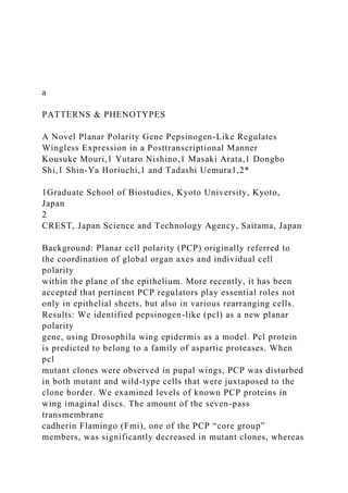
- 7. A Mutation in pepsinogen-like (pcl) Results in a Planar Polarity Phenotype and Loss of the Wing Margin To identify novel planar polarity genes, we performed a mosaic screen for X-chromosome mutations. We generated mosaic clones of about 3,000 lethal chromosomes and searched for the polarity phenotype in adult wings (see details in Mouri et al., 2012). We iso- lated 30 chromosomes that caused severe misorientation of wing hairs, and focused on one of them, no. 11166 (Fig. 1). In addition to the polarity defect, no. 11166 clones showed loss of wing margins (arrowheads in Fig. 1B). As described below, no. 11166 was mutated in pepsinogen-like (pcl)/CG13374 (McQuilton et al., 2012); thus, we designated this allele as pcl1 and hereafter refer to it as such. Next, we observed pcl1 homozygous clones in pupal wings 32 hr after pupalium formation (APF). A subpopulation of mutant cells along the distal clone border showed misorientation of prehairs (Fig. 2A–C; see left arrowhead in Fig. 2A); in contrast, mutant cells fur- ther inside the clone (e.g., near the left edge of Fig. 2A) did not. Intri- guingly, neighboring wild-type cells that were located distal to the mutant clone also showed the misorientation (right arrowhead in Fig. 2A). These local cell-autonomous and non-cell-autonomous phenotypes were also revealed by mislocalization of Fmi at
- 8. anterior- posterior cell boundaries in the clone (Fig. 2B and arrowheads in 2E) and in the adjacent wild-type cells (arrowheads in Fig. 2F), in con- trast to the normal localization at distal cell boundaries (arrowheads in Fig. 2D). This mislocalization of Fmi was reminiscent of that in Ft/Ds group mutant clones (Ma et al., 2003; Strutt and Strutt, 2002). Clones with mutations in Ft/Ds group genes mislocalize Fmi proteins at the wrong cell boundaries (anterior-posterior boundaries) both cell autonomously and non-cell autonomously. This phenotype con- trasts with mutant clones of core group genes, where Fmi no longer localizes tightly to particular cell boundaries (Usui et al., 1999). pcl Is Required for Normal Planar Polarity and Wing-Margin Formation Through genetic mapping and sequencing, we found mutations that resulted in two adjoining amino-acid substitutions in the pcl coding region, raising the possibility that pcl was the gene D E V E L
- 9. O P M E N T A L D Y N A M IC S Fig. 1. A mutation in pcl results in a planar cell polarity phenotype and loss of the margin in the wing. A, B: Wild-type (A) and pcl1 mosaic (B) adult wings. Clones of pcl1 in the adult wing caused loss of the wing margin (arrowheads). In this and all subsequent figures, distal is to the right and anterior is at the top. C, D: Higher-power images of wild- type (C) and pcl1 mosaic wings (D) that are marked by red boxes in A and B, respec- tively. D: Wing hairs were misdirected posteriorly. 792 MOURI ET AL.
- 10. responsible for the planar polarity defect (Fig. 3A, see also Exper- imental Procedures section). The Pcl protein is predicted to belong to a family of aspartic proteases, and the substituted amino acids were located just N-terminal to a sequence, SSTY, which is well conserved among aspartic proteases. To verify that pcl was the responsible gene for the no. 11166 phenotypes, we performed a rescue experiment using two dupli- cations (Venken et al., 2010) that partially overlapped each other. Dp(1; 3)DC007, which includes pcl, rescued the lethality and restored the wing phenotypes to normal (Fig. 3B and C), whereas Dp(1; 3)DC098 did not (data not shown). Because pcl is the only annotated gene that is inside DC007 and not contained on DC098, we concluded that pcl is most likely the responsible gene. We also performed a rescue experiment by expressing the pcl cDNA under the control of the armadillo-GAL4 or daughterless- GAL4. Both of the GAL4 lines rescued both lethality and the mar- gin phenotype. We note, however, the PCP phenotype was only partially rescued, and a moderate polarity defect was still observed in the rescued animals (Fig. 3D and E). pcl Regulates wg Expression in a Posttranscriptional Manner It is known that the Wingless (Wg) and Notch pathways are required for formation of the wing margin (Neumann and
- 11. Cohen, 1996; Herranz and Milan, 2008). The loss of wing mar- gins elicited by the pcl mutation implied that pcl was necessary for either the Wg or the Notch pathway. To address which path- way and which step in either pathway was primarily affected by pcl1, we examined the expression levels of various proteins or markers in wing imaginal discs in late 3rd instar larvae that were populated by pcl1 mutant clones. As previously estab- lished, in the wild-type disc, Wg is expressed in the future wing margin (Couso et al., 1994; Micchelli et al., 1997). By contrast, the Wg signal was significantly decreased in the pcl mutant clones (Fig. 4A and A’). In some smaller mutant clones, the reduction of Wg signal was less obvious than in larger clones (Fig. 4B and B’), possibly due to a perdurance effect. The level of Cut protein was also lower in the mutant clones (Fig. 4C and C’). Because the cut gene is one of the direct targets in Wg- responsive cells and is essential for the margin formation, these results imply that the pcl1 mutation reduced the amount of Wg protein, which resulted in less cut expression in the signal- receiving cells, and ultimately the loss of the wing margin (Couso et al., 1994; Micchelli et al., 1997). How, then, does the pcl mutation abrogate expression of wg? In the wing disc, transcription of the wg gene is induced by Notch signaling (Rulifson and Blair, 1995). Thus, we examined the effect of the pcl mutation on Notch signaling by using vg-lacZ (Neu- mann and Cohen, 1996) and wg-lacZ (Kassis et al., 1992) reporter genes as readouts. Intriguingly, we could not detect any change in either the vg-lacZ or the wg-lacZ signal in pcl mutant clones (Fig. 4D–E’). These results suggested that Notch signaling and its
- 12. downstream events, including transcription of wg, were not affected by the pcl mutation. Therefore, the reduction of Wg pro- tein was possibly due to a defect(s) in post-transcriptional regula- tion, such as at the level of translation, intracellular or extracellular degradation, and/or secretion. D E V E L O P M E N T A L D Y N A M IC
- 13. S Fig. 2. The pcl1 mutation produces a non-cell autonomous effect. A–C: The 32-hr APF (after puparium formation) wing was stained with phalloi- din (A), for Fmi (B), and for a clone marker GFP (blue in C). A homozygous mutant clone was recognized by the absence of the GFP marker, and wild-type cells that border the clone are indicated with yellow dots (A and B). Magenta arrowheads in A indicate misoriented prehairs of the wild- type cells (right) and in the mutant clone (left). Scale bar¼10 mm. D–F: Higher-power images of wild-type cells deep inside the clone (D), mutant cells close to the clone border (E), and wild-type cells close to the border (F) in (B). Both the mutant and wild-type cells close to the border mislo- calized Fmi at anterior-posterior cell boundaries (arrowheads in D and F), in contrast to normal localization at proximal-distal boundaries of the wild-type cells (arrowheads in D). AN ASPARTIC PROTEASE CONTROLS WINGLESS 793 In contrast to the large number of studies on the signaling pathway in Wnt signal-responsive cells, our knowledge is still limited about how Wnt proteins such as Wg are processed and secreted. Enzymes and secretory proteins that are dedicated to Wnt signals have been identified (Herr et al., 2012). Nonetheless, only a few proteins are known to regulate the secretion of Wnt protein: Porcupine (Porc) palmitoylates Wnt at the ER, whereas Wntless/Evenness Interrupted (Wls/Evi) facilitates the secretion of Wnt protein from the Golgi to the extracellular space (Kado-
- 14. waki et al., 1996; Banziger et al., 2006; Bartscherer et al., 2006). Wg proteins accumulate in Wg-producing cells that are mutant for porc or wls (van den Heuvel et al., 1993; Banziger et al., 2006; Bartscherer et al., 2006), which contrasts with the disappearance of Wg protein in pcl clones. We suspected that pcl might regulate Wg proteins by modulating the level of Porc or Wls. However, when we immunostained Wls protein, we saw no difference between pcl mutant and wild-type cells (Fig. 4F and F’). The pcl Mutation Does Not Affect the Expression of four-jointed and dachsous Because the mislocalization of Fmi in pcl1 mutant clones resembled that in fj or ds mutant clones as described above, it could be that pcl was functionally associated with these genes. To test this possibility, we observed the signal of a fj-lacZ reporter gene in pcl1 mutant clones. We did not detect significant changes in the signal between pcl mutant and wild-type cells (Fig. 5A– A’’). We also immunostained pupal wings with anti-Dachsous (Ds) antibody, and did not detect any alteration of the Ds level between mutant clones and adjacent WT clones in the wing pouch (Fig. 5B–B’). We further examined quantitatively whether the amount of Ds and its polarized localization at cell boundaries were affected or not. For this purpose, we focused on mutant clones that reached the dorsal hinge region where Ds is localized in a D
- 15. E V E L O P M E N T A L D Y N A M IC S Fig. 3. pcl is the responsible gene. A: Representation of the domain structure of Pcl protein. The thick red bar indicates a well-conserved domain among aspartic proteases. Partial amino acid sequences of Pcl and three other aspartic proteases, human cathepsin E (hCathE), human pepsinogen 5 (hPGA5), and human cathepsin D (hCathD), are aligned below. Asterisks
- 16. indicate conserved amino acids among these proteases. pcl1 had two mutations in the coding sequence of this domain (ACTCAA instead of AATAAA), which substitute amino acids NK with TQ. B, C: The misorientation phenotype of wing hairs in the pcl1 mutant was rescued by duplication Dp(1; 3)DC007. C: Higher-power image of a region that is marked by red box in B. D, E: The misorientation pheno- type was partially rescued by UAS-pcl that was expressed by armadillo-GAL4. E: Higher-power image of a region that is marked by red box in D. 794 MOURI ET AL. D E V E L O P M E N T A L D
- 17. Y N A M IC S Fig. 4. pcl regulates Wingless (Wg) expression. A–B’: Wing imaginal discs of 3rd instar larvae were stained for Wg (magenta in the merged images in A and B), Cut (magenta in C), and a clone marker GFP (green in A–F). Mutant clones of pcl were marked by the loss of GFP. Expression of Wg or Cut was reduced in the mutant clones (arrowheads in A’ and C’), but it was less obvious in clones in B’. D–F’: Expression of vestigial- lacZ (vg-lacZ) (magenta in D), wg-lacZ (magenta in E), and Wntless (Wls; magenta in F) along the dorsal-ventral boundary was not altered in the mutant clones. Scale bar¼20 mm. AN ASPARTIC PROTEASE CONTROLS WINGLESS 795 polarized fashion along the proximal-distal axis (Brittle et al., 2012), and compared “PCP nematic” (Aigouy et al., 2010) in the wild-type cells with that of the mutant cells (Fig. 5C–F, see also Experimental Procedures section). We could not detect signifi- cant differences in the amount (Fig. 5D) or the magnitude (Fig. 5E) of the axis (Fig. 5F) of the PCP nematic between the wild- type and mutant cells.
- 18. These results suggest that pcl did not regulate the expression level of fj or the amount and the polarized localization of Ds; however, it does not necessarily exclude possible functional interactions between Pcl and these proteins. For example, the Ds ectodomain is subject to endoproteolytic processing and this is modulated by Ft (Ambegaonkar et al., 2012), implying a possible involvement of the hypothetical molecular activity of Pcl. Fmi Protein Is Less Abundant in pcl Mutant Clones in Larval Imaginal Discs Although the loss of wing margins was most likely a direct con- sequence of the decrease in Wg as described above, how did the pcl mutation give rise to the defect in PCP? It has been recently reported that Wg, together with DWnt4, plays a role in PCP by providing a long-range directional cue to cells (Wu et al., 2013). However, mutant clones that remove wg, dWnt4, and two other dWnts genes cause misorientation of wing hairs along the margin (Wu et al., 2013), and adult wings comprised predominantly of wg mutant cells show a mild hair misorientation only at the distal margin (Sagner et al., 2012). In contrast, the aberrant PCP pheno- type along pcl mutant clone borders was seen when the clones were generated either along the margin or inside the wing blade D E V E L
- 19. O P M E N T A L D Y N A M IC S Fig. 5. The pcl1 mutation does not significantly affect fj and ds expression. A–A’’: Mutant clones of pcl in the wing disc were marked by the loss of GFP (green in A, and A”). Expression of fj-lacZ (magenta in A, and A’) was not significantly altered in the clones (arrowhead). B–C’: pcl mosaic clones were stained for Dachsous (Ds) (magenta in B and C, and B’ and C’) and for the clone marker GFP (green in B and C). A pcl mosaic clone in a 24-hr APF wing (B,B’) and those in the dorsal hinge region in the larval wing disc (C and C’). The nematic orders in the individual cells are overlaid on the image of Ds signals (yellow bars in C’). Distal is to the bottom
- 20. of the panel. Scale bar¼20 mm in A–B’, 5 mm in C and C’. D– F: Quantifications of the amount of Ds (D) and its polarized localization (E and F) in the wild-type cells (WT; N¼140) and pcl mutant cells (pcl; N¼96). D, E: Box-and-whisker plots depicting the signal intensity of Ds at cell boundaries (D) and the magnitudes of nematic order for individual cells (E). The box plots show median (line), top, bottom (whiskers), after removing outliers, and 25th and 75th percentile (boxes). The signal intensity of Ds is indicated by artificial unit per pixel (D) and the magnitude of nematic order (length of each yellow bar in C’) is indicated by artificial unit (E). P > 0.05, Wilcoxon rank sum test. NS: not significant. F: The axis distributions of the PCP nematic are shown by rose diagrams in a point symmetry manner. Each diagram is composed of 24 bins of 15 � each, with an approximate direction of the dorsal/ventral compartment boundary (the presumptive wing margin) set to 0 � , and concentric circles are drawn with 5% increments between them. The distributions in the two genotyped cells are not significantly different (P > 0.05, Mardia-Watson-Wheeler test). We performed the quantifications of total four discs that had mutant clones in the dorsal hinge regions, and the data of one of the four are shown in C–F, which is similar to that of the remaining three discs (data not shown). 796 MOURI ET AL.
- 21. (Figs. 1B,D, 2A–C). So a decrease in the amount of Wg in the pcl clones may not be a sole cause of the PCP phenotype. To explore the basis of the pcl PCP phenotype, we examined levels of PCP core proteins in pcl mutant clones in both pupal wings and larval discs. We found that Fmi at cell boundaries was significantly decreased, but not totally eliminated, in pcl mutant clones in 3rd instar larval discs (Fig. 6A–C) and this decrease in the apical Fmi was not associated with relocalization of Fmi at the basal level in the same cells (data not shown). In some small clones, down-regulation of Fmi was less obvious. In contrast, expression levels of a basolateral marker Discs large (Dlg), the Drosophila b-catenin Armadillo (Arm), one of the PCP core- group members Dishevelled (Dsh), or DE-cadherin was not altered in mutant clones (Fig. 6D–E’; data of Dsh and DE-cadherin were not shown). This specific reduction of Fmi in pcl mutant clones in larval imaginal discs is puzzling, because the amount of Fmi was not apparently decreased (although it was mislocalized) in pupal wings as described above (Fig. 2B and E). It has been shown that cell-boundary localization of Fmi and other core-group proteins is already polarized at a late larval stage (Classen et al., 2005; Sagner et al., 2012). It remains to be studied whether the decrease in the Fmi amount in discs is at least one cause of the clone- border selective PCP phenotype in pupal wings.
- 22. Conclusion: Pcl Acts in Both PCP and Wg Signaling In this study, we reported that both of the two well-known developmental mechanisms, PCP formation and Wnt signaling, require pcl encoding a putative aspartic protease. Aspartic pro- tease family proteins play extracellular and intracellular roles; for example, pepsinogen digests foods in the stomach lumen, whereas cathepsins function in adaptive immunity in lysosomes (Conus and Simon, 2010). Further studies will clarify where in the Drosophila wing epidermis and where in the cell Pcl is required, whether Pcl indeed possesses an aspartic protease activity or not, and if so, what are its endogenous substrates. Considering that aspartic proteases have broad substrate speci- ficity (Conus and Simon, 2010), Pcl may indirectly control the activity and/or stability of Wg and Fmi through proteolytic cleavages of distinct substrates. Experimental Procedures Mutant Screening Mutation screening was done as described previously (Mouri et al., 2012). Briefly, mutations were induced in w FRT19A/Y males, and the mutagenized chromosomes were balanced. Mutant D E V E L O P
- 23. M E N T A L D Y N A M IC S Fig. 6. Fmi is less abundant in pcl mutant clones. pcl mutant clones in wing discs were stained for Fmi (magenta in A–B’), Discs large (Dlg; magenta in D, D’), or Armadillo (Arm; magenta in E, E’). The mutant clone in E and E’ was located in the future notum where the reduction of Wg signaling had less of an effect on the expression of Arm, compared to the wing margin. pcl mutant clones were recognized by the absence of the GFP marker (A–E). B, B’: Higher-power image of A and A’, respectively. C: a and b indicate each end of the line. A plot of fluorescent intensities of Fmi (magenta) and GFP (green) along the line in A that spans a pcl mutant clone and its adjacent wild-type clone. Scale bar¼10 mm.
- 24. AN ASPARTIC PROTEASE CONTROLS WINGLESS 797 clones were induced and phenotypes were analyzed in the adult wing. pcl1/pcl1 was early larval lethal. Fly Strains and Genetics Control strains used were y w. Transgene UAS-pcl was expressed by using the GAL4/UAS system (Brand and Perrimon, 1993). Driver strains employed were armadillo (arm)-GAL4 and daughterless (ds)-GAL4, both of which were obtained from the Bloomington Drosophila Stock Center. Duplication and deficiency strains used in mapping (described below) were provided by the Drosophila Genetic Resource Center (DGRC). Other stocks were vg-lacZ (Neumann and Cohen, 1996), wg-lacZ (Kassis et al., 1992), fj-lacZ (Brodsky and Steller, 1996), Dp (1; 3)DC007 and Dp(1; 3)DC098 (Bloomington Drosophila Stock Center). All fly embryos, larvae, pupae, and adults were reared at 25 � C unless described otherwise. Exact genotypes of individual animals used in figures are as follows: Fig. 1. (A and C) y w (B and D) w pcl1 FRT19A/w FRT19A; vg-GAL4 UAS-FLP/þ
- 25. Figs. 2 and 6. w pcl1 FRT19A/y w ubi-GFP FRT19A; vg-GAL4 UAS-FLP/1 Fig. 3. (B and C) w pcl1 FRT19A/Y; Dp(1; 3)DC007/1 (D and E) w pcl1 FRT19A/Y; UAS-pcl/arm-GAL4 Fig. 4. (A–C’, F, and F’) w pcl1 FRT19A/y w ubi-GFP FRT19A; vg-GAL4 UAS-FLP/1 (D and D’) w pcl1 FRT19A/y w ubi-GFP FRT19A; vg-GAL4 UAS-FLP/vg-lacZ (E and E’) w pcl1 FRT19A/y w ubi-GFP FRT19A; vg-GAL4 UAS-FLP/wg-lacZ Fig. 5. (A) w pcl1 FRT19A/y w ubi-GFP FRT19A; vg-GAL4 UAS-FLP/ fj-lacZ (B-C’) w pcl1 FRT19A/y w ubi-GFP FRT19A; vg-GAL4 UAS-FLP/1</UL> Mapping To identify the affected genes in isolated mutants, no. 11166/ FM7 flies were mated with four deficiency and duplication stocks (DGRC stock number 108921, 108145, 108138, and 106068). Tak- ing together the information of lethality and deficiency/duplica- tion points in individual lines, we narrowed the genomic region of the responsible gene of stock 11166 to 200 kb, which included 3 genes (CG32816, l(1)sc, and pcl). We sequenced coding regions of these 3 genes in the female genome of no. 11166, and found two substitution mutations in the pcl gene. Molecular Biology
- 26. A cDNA fragment encoding the pcl gene was cloned into a pUAST-based plasmid containing a UAS promoter. To generate transgenic flies, this construct, pUAST-pcl, was microinjected into fly embryos carrying the attP2-site, and integrated into the site by phiC31-mediated site-specific recombination (Bateman et al., 2006; Bischof et al., 2007). Immunohistochemistry Wing imaginal discs in wandering 3rd instar larvae or pupal wings were fixed and used for immunohistochemistry. Primary antibodies used were rabbit anti-GFP (Molecular Probes, Eugene, OR), mouse anti-Fmi no. 74 and rat anti-Fmi (Usui et al., 1999), rabbit anti-Dlg (Woods and Bryant, 1991), rabbit anti-Ds (Strutt and Strutt, 2002), and rabbit anti-Wls (Port et al., 2008). Mouse anti-Arm N2 7A1, mouse anti-Wg 4D4, and mouse anti-Cut 2B10 were obtained from the Developmental Studies Hybridoma Bank (DSHB) at the University of Iowa. Secondary antibodies were purchased from Molecular Probes and Jackson ImmunoRe- search (West Grove, PA). Immunostaining and acquisition of con- focal images were done as described previously (Mouri et al., 2012), and fluorescent intensity was measured by using Image J software (NIH). Quantification of Ds Distribution We followed the quantification method (nematic order), which is essentially described by Aigouy et al. (2010), to determine the magnitude and axis of nematic order for a single cell as the “PCP nematic” for that cell. We calculated the average value of the intensity for I(r, u) for each u section with a 5
- 27. � range (e.g., 2.5– 7.5 � , 7.5–12.5 � , etc.), where r is the distance between the pixel and the center of the cell and u is the angle indicating the posi- tion of the pixel. Acknowledgments DGRC at Kyoto Institute of Technology, the Bloomington Stock Center, and the Developmental Studies Hybridoma Bank at the University of Iowa provided the fly stocks and the antibodies. We thank Paul Adler for his detailed advice for our mutant screening and Yukiko Miyake, Kumiko Shimizu, Jyunko Mizukoshi, Kanae Oki, and Mayumi Futamata for their technical assistance. We are also grateful to Konrad Basler, David Strutt, Dan Woods, Stephen DiNardo, Shinichi Yanagawa, Shigeo Hayashi, Ritsuko Takada, Shinji Takada, and Uemura lab members for providing other reagents, and protocols, and discussing the phenotypes. We thank James Hejna for his efforts to polish the manuscript. This work was supported by a CREST grant to T.U. from JST and a Research Grant in the Natural Sciences to T.U. from Mitsubishi Foundation. M.
- 28. K. and S. H. are recipients of fellowships from the Japan Society for the Promotion of Science. References Adler PN. 2002. Planar signaling and morphogenesis in Drosophila. Dev Cell 2:525–535. Adler PN, Charlton J, Liu J. 1998. Mutations in the cadherin super- family member gene dachsous cause a tissue polarity phenotype by altering frizzled signaling. Development 125:959–968. Aigouy B, Farhadifar R, Staple DB, Sagner A, R€oper JC, J€ulicher F, Eaton S. 2010. Cell flow reorients the axis of planar polarity in the wing epithelium of Drosophila. Cell 142:773–786. Ambegaonkar AA, Pan G, Mani M, Feng Y, Irvine KD. Propagation of Dachsous-Fat planar cell polarity. Curr Biol 2012 22:1302– 1308. Banziger C, Soldini D, Schutt C, Zipperlen P, Hausmann G, Basler K. 2006. Wntless, a conserved membrane protein dedicated to the secretion of Wnt proteins from signaling cells. Cell 125:509– 522. Bartscherer K, Pelte N, Ingelfinger D, Boutros M. 2006. Secretion of Wnt ligands requires Evi, a conserved transmembrane protein.
- 29. Cell 125:523–533. Bateman JR, Lee AM, Wu CT. 2006. Site-specific transformation of Drosophila via phiC31 integrase-mediated cassette exchange. Genetics 173:769–777. D E V E L O P M E N T A L D Y N A M IC S
- 30. 798 MOURI ET AL. Bischof J, Maeda RK, Hediger M, Karch F, Basler K. 2007. An opti- mized transgenesis system for Drosophila using germ-line- specific phiC31 integrases. Proc Natl Acad Sci USA 104:3312–3317. Blair SS. 2012. Cell polarity: overdosing on PCPs. Curr Biol. 22: R567–9. Brand A, Perrimon N. 1993. Targeted gene expression as a means of altering cell fates and generating dominant phenotypes. Development 118:401–415. Brittle A, Thomas C, Strutt D. 2012. Planar polarity specification through asymmetric subcellular localization of Fat and Dachsous. Curr Biol 22:907–14. Brodsky MH, Steller H. 1996. Positional information along the dorsal-ventral axis of the Drosophila eye: graded expression of the four-jointed gene. Dev Biol 173:428–446. Casal J, Lawrence PA, Struhl G. 2006. Two separate molecular systems, Dachsous/Fat and Starry night/Frizzled, act independ- ently to confer planar cell polarity. Development 133:4561– 4572. Chae J, Kim MJ, Goo JH, Collier S, Gubb D, Charlton J, Adler
- 31. PN, Park WJ. 1999. The Drosophila tissue polarity gene starry night encodes a member of the protocadherin family. Development 126:5421–5429. Chen WS, Antic D, Matis M, Logan CY, Povelones M, Anderson GA, Nusse R, Axelrod JD. 2008. Asymmetric homotypic interac- tions of the atypical cadherin flamingo mediate intercellular polarity signaling. Cell 133:1093–1105. Classen AK, Anderson KI, Marois E, Eaton S. 2005. Hexagonal packing of Drosophila wing epithelial cells by the planar cell polarity pathway. Dev Cell 9:805–817. Clevers H, Nusse R. 2012. Wnt/b-catenin signaling and disease. Cell 149:1192–1205. Conus S, Simon HU. 2010. Cathepsins and their involvement in immune responses. Swiss Med Wkly 140:4–11. Couso JP, Bishop SA, Martinez Arias A. 1994. The wingless signal- ling pathway and the patterning of the wing margin in Drosoph- ila. Development 120:621–636. Dunn BM. 2002. Structure and mechanism of the pepsin-like family of aspartic peptidases. Chem Rev 102:4431–4458. Gao B, Song H, Bishop K, Elliot G, Garrett L, English MA, Andre P, Robinson J, Sood R, Minami Y, et al. 2011. Wnt signaling gra- dients establish planar cell polarity by inducing Vangl2 phospho-
- 32. rylation through Ror2. Dev Cell 20:163–176. Goodrich LV, Strutt D. 2011. Principles of planar polarity in animal development. Development 138:1877–1892. Gray RS, Roszko I, Solnica-Krezel L. 2011. Planar cell polarity: coordinating morphogenetic cell behaviors with embryonic polar- ity. Dev Cell 21:120–133. Harumoto T, Ito M, Shimada Y, Kobayashi TJ, Ueda HR, Lu B, Uemura T. 2010. Atypical cadherins Dachsous and Fat control dynamics of noncentrosomal microtubules in planar cell polarity. Dev Cell 19:389–401. Herr P, Hausmann G, Basler K. 2012. WNT secretion and signalling in human disease. Trends Mol Med 18:483–493. Herranz H, Milan M. 2008. Signalling molecules, growth regulators and cell cycle control in Drosophila. Cell Cycle 7:3335–3337. Ishikawa HO, Takeuchi H, Haltiwanger RS, Irvine KD. 2008. Four- jointed is a Golgi kinase that phosphorylates a subset of cad- herin domains. Science 321:401–404. Kadowaki T, Wilder E, Klingensmith J, Zachary K, Perrimon N. 1996. The segment polarity gene porcupine encodes a putative multitransmembrane protein involved in Wingless processing. Genes Dev 10:3116–3128. Kassis JA, Noll E, VanSickle EP, Odenwald WF, Perrimon N.
- 33. 1992. Altering the insertional specificity of a Drosophila transposable element. Proc Natl Acad Sci USA 89:191–1923. Ma D, Yang C, McNeill H, Simon M, Axelrod J. 2003. Fidelity in planar cell polarity signalling. Nature 421:543–547. Matakatsu H, Blair S. 2004. Interactions between Fat and Dachs- ous and the regulation of planar cell polarity in the Drosophila wing. Development 131:3785–3794. McQuilton P, St Pierre SE, Tharmond J. FlyBase consortium. 2012. FlyBase 101: the basics of navigating FlyBase. Nucleic Acids Res 40:D706–D714. Micchelli CA, Rulifson EJ, Blair SS. 1997. The function and regula- tion of cut expression on the wing margin of Drosophila: Notch, Wingless and a dominant negative role for Delta and Serrate. Development 124:1485–1495. Mouri K, Horiuchi SY, Uemura T. 2012. Cohesin controls planar cell polarity by regulating the level of the seven-pass transmembrane cadherin Flamingo. Genes Cells 17:509–524. Neumann CJ, Cohen SM. 1996. A hierarchy of cross-regulation involving Notch, wingless, vestigial and cut organizes the dorsal/ ventral axis of the Drosophila wing. Development 122:3477– 3485.
- 34. Nusse R, Varmus H. 2012. Three decades of Wnts: a personal per- spective on how a scientific field developed. EMBO J 31:2670– 2684. Port F, Kuster M, Herr P, Furger E, Banziger C, Hausmann G, Basler K. 2008. Wingless secretion promotes and requires retromer- dependent cycling of Wntless. Nat Cell Biol 10:178–185. Rao TP, Kuhl M. 2010. An updated overview on Wnt signaling path- ways: a prelude for more. Circ Res 106:1798–1806. Rulifson EJ, Blair SS. 1995. Notch regulates wingless expression and is not required for reception of the paracrine wingless signal during wing margin neurogenesis in Drosophila. Development 121:2813–2824. Sagner A, Merkel M, Aigouy B, Gaebel J, Brankatschk M, Julicher F, Eaton S. 2012. Establishment of global patterns of planar polarity during growth of the Drosophila wing epithelium. Curr Biol 22:1296–1301. Seifert JR, Mlodzik M. 2007. Frizzled/PCP signalling: a conserved mechanism regulating cell polarity and directed motility. Nat Rev Genet 8:126–138. Sharma P, McNeill H. 2013. Fat and Dachsous cadherins. Prog Mol Biol Transl Sci 116:215–235.
- 35. Shimada Y, Yonemura S, Ohkura H, Strutt D, Uemura T. 2006. Polarized transport of Frizzled along the planar microtubule arrays in Drosophila wing epithelium. Dev Cell 10:209–222. Strutt D. 2001. Asymmetric localization of frizzled and the estab- lishment of cell polarity in the Drosophila wing. Mol Cell 7:367– 375. Strutt H, Strutt D. 2002. Nonautonomous planar polarity patterning in Drosophila: dishevelled-independent functions of frizzled. Dev Cell 3:851–863. Sugimura R, Li L. 2010. Noncanonical Wnt signaling in vertebrate development, stem cells, and diseases. Birth Defects Res C Embryo Today 90:243–256. Thomas C, Strutt D. 2012. The roles of the cadherins Fat and Dachsous in planar polarity specification in Drosophila. Dev Dyn 241:27–39. Usui T, Shima Y, Shimada Y, Hirano S, Burgess R, Schwarz T, Takeichi M, Uemura T. 1999. Flamingo, a seven-pass transmem- brane cadherin, regulates planar cell polarity under the control of Frizzled. Cell 98:585–595. van den Heuvel M, Harryman-Samos C, Klingensmith J, Perrimon
- 36. N, Nusse R. 1993. Mutations in the segment polarity genes wingless and porcupine impair secretion of the wingless protein. EMBO J 12:5293–5302. Venken KJ, Popodi E, Holtzman SL, Schulze KL, Park S, Carlson JW, Hoskins RA, Bellen HJ, Kaufman TC. 2010. A molecularly defined duplication set for the X chromosome of Drosophila mel- anogaster. Genetics 186:1111–1125. Vichas A, Zallen JA. 2011. Translating cell polarity into tissue elon- gation. Semin Cell Dev Biol 22:858–864. Woods DF, Bryant PJ. 1991. The discs-large tumor suppressor gene of Drosophila encodes a guanylate kinase homolog local- ized at septate junctions. Cell 66:451–464. Wu J, Roman AC, Carvajal-Gonzalez JM, Mlodzik M. 2013. Wg and Wnt4 provide long-range directional input to planar cell polarity orientation in Drosophila. Nat Cell Biol 15:1045–1055. D E V E L O P M E
- 37. N T A L D Y N A M IC S AN ASPARTIC PROTEASE CONTROLS WINGLESS 799 Copyright of Developmental Dynamics is the property of John Wiley & Sons, Inc. and its content may not be copied or emailed to multiple sites or posted to a listserv without the copyright holder's express written permission. However, users may print, download, or email articles for individual use. Citation: Kermeliotis, T. (2013, April 12) Somali pirates cost global economy ’18 billion a year’. Retrieved April 14, 2013, from CNN International Edition, Business, http://edition.cnn.com/2013/04/12/business/piracy-economy- world-bank/index.html. Article Title: SOMALI PIRATES COST GLOBAL ECONOMY
- 38. ’18 BILLION A YEAR Summary CNN’s April 12th article concisely summarizes the fiscal cost of piracy to the world economy. The article is full of facts and figures regarding the recent activity in world piracy with some interesting effects on the impact on related and supporting industries such as fishing and tourism. Over 1000 attacks have occurred from Somali pirates alone since 2005. The study compares to earlier reports compiled by other international organizations which tended to minimize the impact of piracy in the world. Further, the article expanded on the piracy affected countries identified by World Bank, eleven total discounting the countries of the Persian Gulf. Overall, the article provided another perspective on the piracy issue of today. Relevance Based on our readings, this article confirms the importance of policy as it relates to corruption. In this instance, lack of enforcement of both national and international policies and accountability standards are affecting the world via trade. The article also confirms some of the costs and risks of doing business internationally such as increased fuel costs to companies due to looking for different shipping routes, higher foreign insurance premiums and the cost of additional labor for security on board. This article further confirms the challenges and interconnectedness concerns most countries have with Africa. As it was rated high on the corruption index, it is now affecting the pocket books of many companies in a real way. This article shows how risk, investment, strategy, fiscal and economic controls come together to effect international trade. I learned that pirates really do still exist in the world. (CNN) -- The Somali pirates roaming the waters off the Horn of Africa push global trade costs up by billions of dollars per year and severely affect the economic activities of neighboring countries, a new World Bank report has found. Although hijackings in the region have dropped significantly
- 39. since last year, piracy could still cost the global economy an estimated $18 billion annually, according to the "Pirates of Somalia: Ending the Threat, Rebuilding a Nation" report, launched Thursday in the Somali capital of Mogadishu. Read this: Peace concert rocks Mogadishu The increased costs come as shippers are forced to change trading routes, sending fuel bills soaring, as well as pay higher insurance premiums and security bills for guards on board. Tackling piracy in Somalia Ransom payments creating problems The cost of piracy But apart from making international trade more expensive, the threat of piracy in one of the world's most important trade gateways is also an economic blow for neighboring East African countries, particularly in the pillar sectors of tourism and fishing, said the report. Since 2006, tourism spending in East African coastal countries has risen 25% more slowly than other sub-Saharan African nations, mainly because of fewer arrivals from high-income citizens of OECD countries. Piracy in the region is thought to have tarnished its image as a stable holiday destination, with visits to impacted East African coastal nations down by nearly 6.5% relative to visits to other countries. According to the World Bank, exports of fish products from piracy-hit countries have also suffered, declining by 23.8% since 2006, the year the report takes as the starting point of piracy.
- 40. The World Bank defines piracy-affected countries as Comoros, Djibouti, Kenya, Mozambique, Madagascar, Mauritius, the Seychelles, Somalia, Tanzania, as well as Yemen, Pakistan and the countries of the Persian Gulf. Read this: 'New hope for Somalia' The economy of war-torn Somalia, which last September moved closer to stability after electing its first president on home soil in decades, is also severely hit; piracy-related trade costs increase by up to $6 million annually, without considering that potential sea-based economic activities are constrained by piracy. The authors of the report said the solution to ending piracy will only come with the recreation of a functional Somali state and urged the international community to focus on helping the East African county build a viable political system. "Piracy is a symptom of the breakdown of Somalia's political system," said economist and lead author of the report Quy-Toan Do. "Go after the system, not just the pirates." According to the report, 1,068 attacks have been carried out by Somali pirates since January 2005. Of these, 218 resulted in successful hijackings, with an estimated $53 million average annual ransom paid. Between 82 and 97 casualties are believed to have occurred as a result of these attacks. Attacks peaked in 2011, but have declined sharply since, largely because of increased mobilization of international naval forces and tighter security adopted by the shipping industry. A report earlier this week by advocacy group Oceans Beyond Piracy said that maritime piracy cost the global economy between $5.7 and $6.1 billion in 2012, much lower than the World Bank figures. BA 420 Global Project Journal
- 41. Global Project Journal The assignment is intended to increase your awareness of the international business transactions happening around you as we progress through our course. You will take notice of events happening on a quarterly, daily and even hourly basis around the world which affect our lives here in the United States. You will be able to tie these events with the content we learn in class to realize the value of your increased knowledge. Being the semester has been allotted for completion of this journal, the expectation is for solid synthesis and relevancy Reminders- · 5 different articles from credible sources are required (try to pick articles throughout the semester). · Each article must be fully cited (APA is fine) and must be included in totality in your submission · Each article is to be followed by a one page synthesis and relevancy. One paragraph minimum for synthesis (what is the article about); one paragraph for relevancy (how does this relate to international business and to any of the content we have learned in class). · The Global Project Journal should have a cover page with your full name, date, course name and section. Global Project Journal Rubric · Credible article (5 pts) 25 points _____ · Credible synthesis (5 pts) each article 25 points _____ · Appropriate relevancy (10 pts) each article 50 points _____ · 100 points TOTAL ______ Please submit your Global Project Journal in eCollege via Dropbox by _______________.