Download as PDF, PPTX
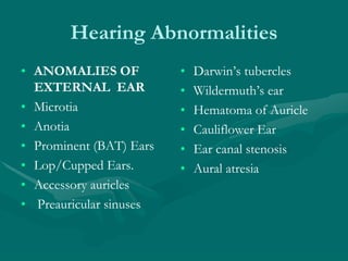
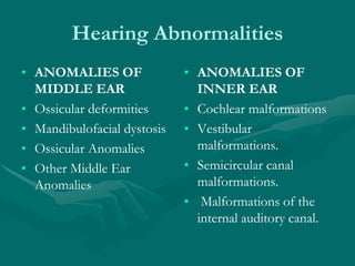

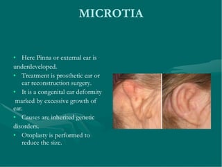
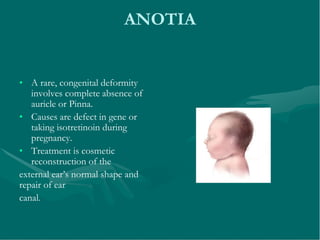
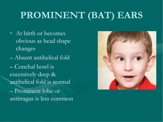


The document discusses various abnormalities that can affect the ear. It describes abnormalities in the external ear like microtia (underdeveloped pinna), anotia (absence of pinna), prominent ears, and ear canal stenosis. Abnormalities of the middle ear discussed include ossicular deformities and mandibulofacial dystosis. Inner ear abnormalities mentioned are cochlear malformations like Michel's deformity, incomplete partition types 1 and 2, vestibular malformations, semicircular canal malformations, and internal auditory canal malformations.


