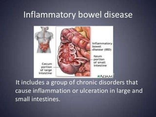
P R E S E N T E D B Y :
S H I V A L I K A L O D E 0 4 - 2 0 1 3
A I S H W A R Y A J A M W A L 3 3 - 2 0 1 3
V E N K A T E S H P A S H I N E 6 1 - 2 0 1 3
A S H L E S H A P A T W A R D H A N 6 4 - 2 0 1 3
N I S H A N T M A H E S H 9 5 - 2 0 1 3
Inflammatory
Bowel Disease
Complications of Inflammatory Bowel
Disease
 Life-threatening colonic inflammation : This
can occur in both ulcerative colitis and
Crohn’s colitis. In most extreme cases, colon
dilates (toxic megacolon) and bacterial toxins
pass freely across the diseased mucosa into
portal and then systemic circulation
 Haemorrhage : Haemorrhage due to erosion
of a major artery is rare but can occur in both
conditions
 Fistulae : They are specific o Crohn’s disease.
Enteroenteric fistulae can cause diarrhoea and
malabsorbtion. Enterovesical fistulation causes
recurrent urinary infections. Fistula from the
bowel may also cause perianal or ischiorectal
abscess,fissures and fistulae
 Cancer : The risk of dysplasia and cancer
increases with the duration and extent of
uncontrolled colonic inflammation. Thus
patients having long standing extensive colitis
are at highest risk. Tumours develop in areas of
dysplasia and maybe multiple
Patients with long standing colitis are therefore
entered into surveillance programmes beginning 10
years after diagnosis.
 Extra-intestinal complications : These are
common in IBD and may dominate the
clinical picture
Investigations
 Investigations are necessary to confirm the
diagnosis, disease distribution and activity, and
identify complications.
 Investigations - Bacteriology
 - Endoscopy
 - Radiology
 Bacteriology – The initial presentation,stool
microscopy,culture and examination for
Clostridium difficile toxin or for ova and
cysts,blood culture and serological tests should
be performed
 Endoscopy - Patients who present with diarrhoea
plus raised inflammatory markers or alarm
features, such as weight loss rectal bleeding and
anemia should undergo ileocolonoscopy
 Flexible sigmoidoscopy is ocassionally
perforemed to make diagnosis especially during
acute severe presentations
 Radiology :
 Small bowel imaging is essential to complete staging of
Crohn’s disease. Traditional contrast imaging by barium
follow-through demonstrates affected areas of the bowel as
narrowed and ulcerated .
 This has now been largely replaced by MRI enterography ,
which does not involve exposure to radiation and is a sensitive
way of detecting extraintestinal manifestations
 A plain abdominal X-ray is essential in manegment of patients
who present with severe ctive disease
 Ultrasound is also a powerful tool to detect small bladder
inflammation
 CT is limited to screening for complications
Inflammatory bowel disease

Inflammatory bowel disease

  • 1.
    P R ES E N T E D B Y : S H I V A L I K A L O D E 0 4 - 2 0 1 3 A I S H W A R Y A J A M W A L 3 3 - 2 0 1 3 V E N K A T E S H P A S H I N E 6 1 - 2 0 1 3 A S H L E S H A P A T W A R D H A N 6 4 - 2 0 1 3 N I S H A N T M A H E S H 9 5 - 2 0 1 3 Inflammatory Bowel Disease
  • 24.
    Complications of InflammatoryBowel Disease  Life-threatening colonic inflammation : This can occur in both ulcerative colitis and Crohn’s colitis. In most extreme cases, colon dilates (toxic megacolon) and bacterial toxins pass freely across the diseased mucosa into portal and then systemic circulation  Haemorrhage : Haemorrhage due to erosion of a major artery is rare but can occur in both conditions
  • 25.
     Fistulae :They are specific o Crohn’s disease. Enteroenteric fistulae can cause diarrhoea and malabsorbtion. Enterovesical fistulation causes recurrent urinary infections. Fistula from the bowel may also cause perianal or ischiorectal abscess,fissures and fistulae  Cancer : The risk of dysplasia and cancer increases with the duration and extent of uncontrolled colonic inflammation. Thus patients having long standing extensive colitis are at highest risk. Tumours develop in areas of dysplasia and maybe multiple
  • 26.
    Patients with longstanding colitis are therefore entered into surveillance programmes beginning 10 years after diagnosis.  Extra-intestinal complications : These are common in IBD and may dominate the clinical picture
  • 28.
    Investigations  Investigations arenecessary to confirm the diagnosis, disease distribution and activity, and identify complications.  Investigations - Bacteriology  - Endoscopy  - Radiology
  • 29.
     Bacteriology –The initial presentation,stool microscopy,culture and examination for Clostridium difficile toxin or for ova and cysts,blood culture and serological tests should be performed  Endoscopy - Patients who present with diarrhoea plus raised inflammatory markers or alarm features, such as weight loss rectal bleeding and anemia should undergo ileocolonoscopy  Flexible sigmoidoscopy is ocassionally perforemed to make diagnosis especially during acute severe presentations
  • 30.
     Radiology : Small bowel imaging is essential to complete staging of Crohn’s disease. Traditional contrast imaging by barium follow-through demonstrates affected areas of the bowel as narrowed and ulcerated .  This has now been largely replaced by MRI enterography , which does not involve exposure to radiation and is a sensitive way of detecting extraintestinal manifestations  A plain abdominal X-ray is essential in manegment of patients who present with severe ctive disease  Ultrasound is also a powerful tool to detect small bladder inflammation  CT is limited to screening for complications