Download to read offline
![International Journal of Computer Applications (0975 – 8887)
Volume 132 – No.8, December2015
1
Implementation of Improved Gaussian Filter Algorithm
for Retinal Fundus Images
S.A. Jameel
Assistant Professor
P.G and Research Department of
ComputerScience, Jamal Mohamed College
(Autonomous)
Tiruchirappalli 620 020, Tamil Nadu, India
A.R. Mohamed Shanavas
Associate Professor
P. G and Research Department of Computer
Science, Jamal Mohamed College (Autonomous)
Tiruchirappalli 620 020, Tamil Nadu, India
ABSTRACT
Gaussian filter has an important role in edge detection. In this
paper, a new methodology for edge detection using Gaussian
second order derivative filters is proposed. Channel separation
for Retinal Colour Fundus image is used followed by
Gaussian filter. The performance of our proposed method has
been assessed on 23 images representing the publicly
available dataset; High-Resolution Fundus (HRF) Image
Database.
Keywords
Gaussian filter, Gaussian second order derivative filter,
Fundus image
1. INTRODUCTION
The Gaussian filter is broadly used in image processing and
computer vision for long years. Generally it works with low
pass filtering. [1] Any way for directive filtering a plane
cosine wave can be multiplied to the Gaussian function. [2] Its
first and second derivatives are also widely used. Among
various smoothing filters, the most widely used smoothing
filters are Gaussian filters. These filters have been shown to
play an important role in edge detection in the human visual
system, and to be largely useful as detectors for edge and line
detection. Marr and Hildreth displayed that the Gaussian
filter (as well as with the Laplacian operator) is same to the
difference of Gaussians (DOG) filter. This is a well-known
estimation to the shape of spatial amenable fields in the visual
system of cats that has also been proposed for humans.
Babaud et al verified that when one-dimensional (1-D) signals
are softened with a Gaussian filter, the scale space
characterization of their second derivatives exhibits that
current zero-crossings disappear when moving from a fine-to-
coarse measurement, but new ones are not designed. They
also proved that for a broad category of signals, the Gaussian
function is the unique filter that has this characteristic. This
special characteristic makes it possible to track zero-crossings
over a range of scales, and also delivers the capability to
reclaim the whole signal at sufficiently small scales. Yuille
and Poggio enhanced this work to two-dimensional (2-D)
signals and demonstrated that with the Laplacian, the
Gaussian function is the only filter in extensive category that
does not produce zero-crossings as the scale increases. They
also showed that for non-sequential directional derivatives
along the gradual slope, there is no filter that does not produce
zero-crossings as the scale rises.[3] Another important
characteristic of the Gaussian filter is the only operator which
satisfies the inconstancy relation
2
1
x
(1)
Where Δx and Δw are its variance in spatial and frequency
domains, correspondingly. This property allows the Gaussian
operator to produce the finest tradeoff between the conflicting
objectives of the restriction in spatial and frequency domains
at the same time. The 2-D Gaussian filter is also the only
rotationally uniform filter that is distinguishable in Cartesian
coordinates. Separability is significant for computational
efficiency when executing the smoothing operation by
convolutions in the spatial domain. [1] Marr and Hildreth also
declared that the best smoothing filter for images should be
confined in both spatial and frequency domains, thereby
satisfying the uncertainty relation shown in equation (1).
Examine the Gaussian operator in two dimensions shown
below
)2/(
2
222
2
1
),(
yx
eyxg
(2)
Where μ = mean, with μ = 0, σ2
=variance, σ is the standard
deviation of the Gaussian function and (x,y) are the Cartesian
coordinates of the image. Marr and Hildreth suggested that by
applying Gaussian filters of different scales (σ) to an image; a
set of images with different measures of smoothness can be
acquired. To find the edges in these images it is necessary to
detect the zero-crossings of their second derivatives. The 2-D
Gaussian filter is mostly implemented as an image
preprocessing step for image softening and noise reduction.
The second derivative of Gaussian filter shown in equation (3)
is the simple supplement of the Gaussian first derivative filter
(equation 2). This can be applied independently to each
dimension. Marr and Hildreth accomplished this by applying
the Laplacian of a Gaussian (LOG) function as a filter. [1]
),(),(),(
2
2
2
2
2
yxg
dy
d
yxg
dx
d
yxg
)2/(
6
222 222
2
2
yx
e
yx
(3)
A technique for saliency estimation established on an image
concept into structurally representative elements and contrast-
based saliency measures, which can be consistently
formulated as high dimensional Gaussian filters was
presented. [4] A novel approach for achieving high-quality
edge-preserving filtering of images and videos in real time
was presented. [5] A flexible scheme for accelerating spatially
varying high-dimensional Gaussian filters. [6] An adaptive
Gaussian filter algorithm was proposed. [7] A parameterized](https://image.slidesharecdn.com/ijca-170205095838/75/IMPLEMENTATION-OF-IMPROVED-GAUSSIAN-FILTER-ALGORITHM-FOR-RETINAL-FUNDUS-IMAGES-1-2048.jpg)
![International Journal of Computer Applications (0975 – 8887)
Volume 132 – No.8, December2015
2
logarithmic image processing (PLIP) method based on
Laplacian of Gaussian filtering to improve lung swelling in
chest radiographs was proposed. [8]
2. PROPOSED GAUSSIAN SECOND
ORDER DERIVATIVE FILTERS
METHOD
2.1 Preprocessing
In the preprocessing stage, the blood vessels in the given
fundus image are enhanced as they have non-uniform
illumination. The green channel of the fundus image has best
vascular structure. Hence, this channel is only considered for
enhancement before blood vessel extraction. The first one is
channel separation in the processing step. The retinal colour
(RGB) fundus image in figure 1 composes of three channels:
red, green, and blue channel. [9]
2.2 Gaussian 2nd
order derivative filter
The blood vessels in the fundus images are enhanced by
applying Gaussian 2nd
order derivative filter as shown in
figure 2 successively by varying sigma. In order to obtain the
enhanced blood vessels, the maximum frequency response is
chosen from the responses of applied Gaussian filters. The
flowchart of the proposed algorithm is shown in figure 3. The
proposed algorithm for Gaussian Second Order Derivative
Filters is twofold.
Step 1: Design of Gaussian filter array in xx, xy and yy
direction
Step 2: Apply Gaussian filter blood vessel detection
2.3 Algorithm
Design of Gaussian filter array in xx, xy and yy direction
Gaussians(x,x)= )1/( 22
X /
4
2 *
)22/22( YX
e
Gaussians(x, y) =
)(XY /
6
2 *
)22/22( YX
e
Gaussians(y, y) = Gaussians(x, x)’
Apply Gaussian filter blood vessel detection
D(x, y) = Conv (G, Gaussians(x, x))
D(x, y) = Conv (G, Gaussians(x, y))
D(y, y) = Conv (G, Gaussians(y, y))
D(x,x)=multiply(D(x,x),
2
)
D(x,y)=multiply(D(x,y),
2
)
D(y, y) = multiply (D(y, y),
2
)
3. EXPERIMENTAL RESULTS
For performance assessment, the proposed technique is tested
using the publicly available dataset High-Resolution Fundus
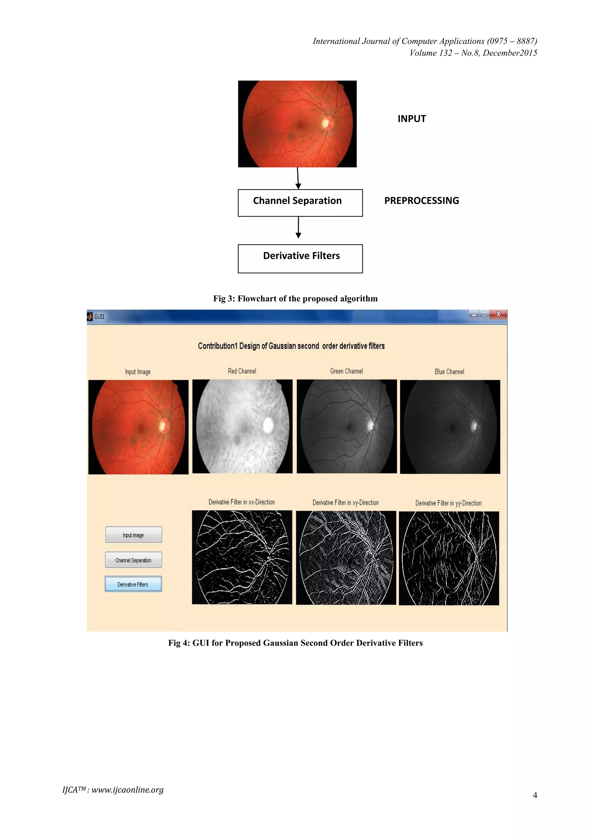
(HRF) Image Database.[10] The GUI for Proposed Gaussian
Second Order Derivative Filters is shown in figure 4.
3.1 Dataset
The public database contains at the present fifteen images of
healthy patients, fifteen images of patients with diabetic
retinopathy. The proposed method uses fifteen images of
healthy patients, eight images of patients with diabetic
retinopathy out of 15 images.[10]
4. DISCUSSION
The proposed method is used to enhance the blood vessels; in
our experiments, a set of 23 colour retinal images from the
publicly available datasets were used. This gives good
opportunity to test the algorithm on images with different
features; normal, abnormal, different sizes. The algorithm is
implemented using Matlab. For performance assessment, the
proposed technique is tested using the publicly available
dataset High-Resolution Fundus (HRF) Image Database.
5. CONCLUSION
Many works have been proposed for Gaussian Filter in image
processing. Specifically in this work a Second order
derivative filter is used for retinal fundus images. In this
paper, a new algorithm for designing of Gaussian second
order derivative filters is proposed. This is achieved by means
of preprocessing followed by derivative filters. The maximum
frequency response is chosen from the responses of applied
Gaussian filters. The proposed algorithm is tested using the
publicly available database. In future this paper is extended to
implement a novel method for segmenting the blood vessels
by thresholding approach.
6. REFERENCES
[1] Basu, Mitra. "Gaussian-based edge-detection methods-a
survey." IEEE Transactions on Systems, Man, and
Cybernetics, Part C 32.3 (2002): 252-260.
[2] Choomchuay, Somsak, and Keokanlaya Sihalath. "An
application of second derivative of gaussian filters in
fingerprint image enhancement." Bioinformatics and
Biomedical Engineering (iCBBE), 2010 4th International
Conference on. IEEE, 2010.
[3] Raman Maini (2012) Analysis and development of image
edge detection techniques. PhD thesis. Punjabi
University. http://hdl.handle.net/10603/3660.
[4] Perazzi, Federico, et al. "Saliency filters: Contrast based
filtering for salient region detection." Computer Vision
and Pattern Recognition (CVPR), 2012 IEEE Conference
on. IEEE, 2012.
[5] P Gastal, Eduardo SL, and Manuel M. Oliveira.
"Domaintransform for edge-aware image and video
processing." ACM Transactions on Graphics (TOG).
Vol. 30. No. 4. ACM, 2011.
[6] Baek, Jongmin, and David E. Jacobs. "Accelerating
spatially varying Gaussian filters." ACM Transactions on
Graphics (TOG). Vol. 29. No. 6. ACM, 2010.
[7] Deng, G., and L. W. Cahill. "An adaptive Gaussian filter
for noise reduction and edge detection." Nuclear Science
Symposium and Medical Imaging Conference, 1993.,
1993 IEEE Conference Record.. IEEE, 1993.
[8] Bao, Chen, and Chen Sheng. "A parameterized
logarithmic image processing method based on Laplacian
of Gaussian filtering for lung nodules enhancement in
chest radiographs." Instrumentation and Measurement,
Sensor Network and Automation (IMSNA), 2013 2nd
International Symposium on. IEEE, 2013.](https://image.slidesharecdn.com/ijca-170205095838/75/IMPLEMENTATION-OF-IMPROVED-GAUSSIAN-FILTER-ALGORITHM-FOR-RETINAL-FUNDUS-IMAGES-2-2048.jpg)
![International Journal of Computer Applications (0975 – 8887)
Volume 132 – No.8, December2015
3
[9] R. Hashim, Fatma. A., Nancy. M. Salem, and Ahmed
Farag Seddik. "Optic disc boundary detection from
digital fundus images." Journal of Medical Imaging and
Health Informatics 5.1 (2015): 50-56.
[10] Jan Odstrcilik, R. Kolar, A. Budai, J. Hornegger, J. Jan,
J. Gazarek, T. Kubena, P. Cernosek, O. Svoboda, and E.
Angelopoulou, “Retinal vessel segmentation by
improved matched filtering: Evaluation on a new high-
resolution fundus image database,” IET Image Process.,
vol. 7, no. 4, pp. 373–383, 2013.
7. APPENDIX
Healthy
Diabetic Retinopathy
(a) RGB image (b) Red Channel (c) Blue Channel (d) Green Channel
Fig 1: Colour Retinal Images
Healthy
Diabetic Retinopathy
(a) xx-Direction (b) xy-Direction (c) yy-Direction
Fig 2: Derivative Filters](https://image.slidesharecdn.com/ijca-170205095838/75/IMPLEMENTATION-OF-IMPROVED-GAUSSIAN-FILTER-ALGORITHM-FOR-RETINAL-FUNDUS-IMAGES-3-2048.jpg)

The document presents a methodology for edge detection in retinal fundus images using an improved Gaussian second order derivative filter algorithm. The proposed technique enhances blood vessels in these images by employing channel separation and varying the filter's sigma value, with results tested on a publicly available dataset. The findings suggest a successful performance in enhancing image features, paving the way for future methods for blood vessel segmentation.