SlideShare a Scribd company logo
1 of 21
Download to read offline
Figure:
Immunohistochemical
analysis of paraffin
embedded Human
uterus tissue. 1: Smad2
Polyclonal Antibody
was diluted at 1:200 (4
degree
Celsius,overnight). 2:
Sodium citrate pH 6.0
was used for antibody
retrieval (>98 degree
Celsius,20min). 3:
Secondary antibody was
diluted at 1:200 (room
temperature, 30min).
Negative control was
used by secondary
antibody only.
Report Number 95694-a Host Rabbit
Application IHC-P Clonality Polyclonal
Model Number STJ95694 Clone ID NA
Antibody Name Anti-Smad2 antibody
Testing Species HUMAN Testing Tissue UTERUS
ANTIBODY VALIDATION REPORT
a. (A small amount of distilled water was added into the incubation
box to prevent evaporation of antibody).
216. Secondary antibody incubation
a. Slides were washed 3 times, with PBS on a shaker for 5min.
Shortly after the slides were dried the corresponding secondary
antibody solution was added (HRP labelled), covering the
tissues, and incubated at room temperature for 30min.
b.
217. DAB staining
a. Slides were washed 3 times, with PBS on a shaker for 5min.
b. Shortly after, the slides were dried and fresh DAB staining buffer
was added inside the circles. The staining time was adjusted
under a microscope. Yellow-brown colour represented a positive
result. Slides were washed with water to stop the staining.
c.
218. Haematoxylin staining
a. Haematoxylin was used to counter-staining for 1min, and then
the slides were washed with water. 1% Hydrochloric acid and
alcohol was added for several seconds and then washed with
water. Ammonia was used to reveal blue colour, and then
flushed with water.
b.
219. Desolation and Clearing
i. Slides were incubated sequentially into: 75% alcohol 5min, 85%
alcohol 5min, Anhydrous ethanol - 5min, Anhydrous ethanol -
5min & Xylene - 5min. Shortly after slides were dried and neutral
gum was used to seal the slides.
ii.
220. Visualization
a. Results were validated with microscope, and the slides were
scanned.
Paraffin-Embedded
Immunohistochemistry Protocol
210.
211. Tissue processing
a. Slides were incubated sequentially into Xylene; 15min –
Xylene, 15min - Anhydrous ethanol, 5min - Anhydrous
ethanol, 5min - 85% alcohol, 5min - 75% alcohol & 5min –
wash in distilled water.
b.
212. Antigen retrieval
a. Tissue slides were incubated with citric acid (PH6.0) antigen
retrieval buffer and microwaved for antigen retrieval (heated
until boiled and then stopped heating) for 8min. Slides were
then heated with medium power for 7min. During this
process slides were kept from drying out. After cooling down
at room temperature, slides were washed with PBS on
shaker for 5min, repeated for 3 times.
b.
213. Inhibition of endogenous peroxidase
a. Slides were placed in 3% Hydrogen peroxide solution, and
incubated for 10 min at room temperature without light
exposure. Slides were then washed 3 times with PBS on a
shaker for 5mins.
b.
214. BSA Blocking
a. Shortly after slides were dried, a PAP pen was used to draw
circles around the tissue sections (and to prevent draining of
the antibody solution). Inside the circles, BSA was used to
cover the tissue evenly, blocking for 30min.
b.
215. Primary antibody incubation
After blocking solution was removed a 1:200 solution of
primary antibody/PBS was added on the slide, and incubated
overnight at 4°C.
St John's Laboratory Ltd.
www.stjohnslabs.com
Figure:
Immunohistochemical
analysis of paraffin
embedded Human
uterus cancer tissue. 1:
Smad2 Polyclonal
Antibody was diluted at
1:200 (4 degree
Celsius,overnight). 2:
Sodium citrate pH 6.0
was used for antibody
retrieval (>98 degree
Celsius,20min). 3:
Secondary antibody was
diluted at 1:200 (room
temperature, 30min).
Negative control was
used by secondary
antibody only.
Report Number 95694-b Host Rabbit
Application IHC-P Clonality Polyclonal
Model Number STJ95694 Clone ID NA
Antibody Name Anti-Smad2 antibody
Testing Species HUMAN Testing Tissue UTERUS CANCER
ANTIBODY VALIDATION REPORT
a. (A small amount of distilled water was added into the incubation
box to prevent evaporation of antibody).
205. Secondary antibody incubation
a. Slides were washed 3 times, with PBS on a shaker for 5min.
Shortly after the slides were dried the corresponding secondary
antibody solution was added (HRP labelled), covering the
tissues, and incubated at room temperature for 30min.
b.
206. DAB staining
a. Slides were washed 3 times, with PBS on a shaker for 5min.
b. Shortly after, the slides were dried and fresh DAB staining buffer
was added inside the circles. The staining time was adjusted
under a microscope. Yellow-brown colour represented a positive
result. Slides were washed with water to stop the staining.
c.
207. Haematoxylin staining
a. Haematoxylin was used to counter-staining for 1min, and then
the slides were washed with water. 1% Hydrochloric acid and
alcohol was added for several seconds and then washed with
water. Ammonia was used to reveal blue colour, and then
flushed with water.
b.
208. Desolation and Clearing
i. Slides were incubated sequentially into: 75% alcohol 5min, 85%
alcohol 5min, Anhydrous ethanol - 5min, Anhydrous ethanol -
5min & Xylene - 5min. Shortly after slides were dried and neutral
gum was used to seal the slides.
ii.
209. Visualization
a. Results were validated with microscope, and the slides were
scanned.
Paraffin-Embedded
Immunohistochemistry Protocol
199.
200. Tissue processing
a. Slides were incubated sequentially into Xylene; 15min –
Xylene, 15min - Anhydrous ethanol, 5min - Anhydrous
ethanol, 5min - 85% alcohol, 5min - 75% alcohol & 5min –
wash in distilled water.
b.
201. Antigen retrieval
a. Tissue slides were incubated with citric acid (PH6.0) antigen
retrieval buffer and microwaved for antigen retrieval (heated
until boiled and then stopped heating) for 8min. Slides were
then heated with medium power for 7min. During this
process slides were kept from drying out. After cooling down
at room temperature, slides were washed with PBS on
shaker for 5min, repeated for 3 times.
b.
202. Inhibition of endogenous peroxidase
a. Slides were placed in 3% Hydrogen peroxide solution, and
incubated for 10 min at room temperature without light
exposure. Slides were then washed 3 times with PBS on a
shaker for 5mins.
b.
203. BSA Blocking
a. Shortly after slides were dried, a PAP pen was used to draw
circles around the tissue sections (and to prevent draining of
the antibody solution). Inside the circles, BSA was used to
cover the tissue evenly, blocking for 30min.
b.
204. Primary antibody incubation
After blocking solution was removed a 1:200 solution of
primary antibody/PBS was added on the slide, and incubated
overnight at 4°C.
St John's Laboratory Ltd.
www.stjohnslabs.com
Figure:
Immunohistochemical
analysis of paraffin
embedded Human lung
cancer tissue. 1: Smad2
Polyclonal Antibody
was diluted at 1:200 (4
degree
Celsius,overnight). 2:
Sodium citrate pH 6.0
was used for antibody
retrieval (>98 degree
Celsius,20min). 3:
Secondary antibody was
diluted at 1:200 (room
temperature, 30min).
Negative control was
used by secondary
antibody only.
Report Number 95694-c Host Rabbit
Application IHC-P Clonality Polyclonal
Model Number STJ95694 Clone ID NA
Antibody Name Anti-Smad2 antibody
Testing Species HUMAN Testing Tissue LUNG CANCER
ANTIBODY VALIDATION REPORT
a. (A small amount of distilled water was added into the incubation
box to prevent evaporation of antibody).
194. Secondary antibody incubation
a. Slides were washed 3 times, with PBS on a shaker for 5min.
Shortly after the slides were dried the corresponding secondary
antibody solution was added (HRP labelled), covering the
tissues, and incubated at room temperature for 30min.
b.
195. DAB staining
a. Slides were washed 3 times, with PBS on a shaker for 5min.
b. Shortly after, the slides were dried and fresh DAB staining buffer
was added inside the circles. The staining time was adjusted
under a microscope. Yellow-brown colour represented a positive
result. Slides were washed with water to stop the staining.
c.
196. Haematoxylin staining
a. Haematoxylin was used to counter-staining for 1min, and then
the slides were washed with water. 1% Hydrochloric acid and
alcohol was added for several seconds and then washed with
water. Ammonia was used to reveal blue colour, and then
flushed with water.
b.
197. Desolation and Clearing
i. Slides were incubated sequentially into: 75% alcohol 5min, 85%
alcohol 5min, Anhydrous ethanol - 5min, Anhydrous ethanol -
5min & Xylene - 5min. Shortly after slides were dried and neutral
gum was used to seal the slides.
ii.
198. Visualization
a. Results were validated with microscope, and the slides were
scanned.
Paraffin-Embedded
Immunohistochemistry Protocol
188.
189. Tissue processing
a. Slides were incubated sequentially into Xylene; 15min –
Xylene, 15min - Anhydrous ethanol, 5min - Anhydrous
ethanol, 5min - 85% alcohol, 5min - 75% alcohol & 5min –
wash in distilled water.
b.
190. Antigen retrieval
a. Tissue slides were incubated with citric acid (PH6.0) antigen
retrieval buffer and microwaved for antigen retrieval (heated
until boiled and then stopped heating) for 8min. Slides were
then heated with medium power for 7min. During this
process slides were kept from drying out. After cooling down
at room temperature, slides were washed with PBS on
shaker for 5min, repeated for 3 times.
b.
191. Inhibition of endogenous peroxidase
a. Slides were placed in 3% Hydrogen peroxide solution, and
incubated for 10 min at room temperature without light
exposure. Slides were then washed 3 times with PBS on a
shaker for 5mins.
b.
192. BSA Blocking
a. Shortly after slides were dried, a PAP pen was used to draw
circles around the tissue sections (and to prevent draining of
the antibody solution). Inside the circles, BSA was used to
cover the tissue evenly, blocking for 30min.
b.
193. Primary antibody incubation
After blocking solution was removed a 1:200 solution of
primary antibody/PBS was added on the slide, and incubated
overnight at 4°C.
St John's Laboratory Ltd.
www.stjohnslabs.com
Figure:
Immunohistochemical
analysis of paraffin
embedded Human
stomach tissue. 1:
Smad2 Polyclonal
Antibody was diluted at
1:200 (4 degree
Celsius,overnight). 2:
Sodium citrate pH 6.0
was used for antibody
retrieval (>98 degree
Celsius,20min). 3:
Secondary antibody was
diluted at 1:200 (room
temperature, 30min).
Negative control was
used by secondary
antibody only.
Report Number 95694-d Host Rabbit
Application IHC-P Clonality Polyclonal
Model Number STJ95694 Clone ID NA
Antibody Name Anti-Smad2 antibody
Testing Species HUMAN Testing Tissue STOMACH
ANTIBODY VALIDATION REPORT
a. (A small amount of distilled water was added into the incubation
box to prevent evaporation of antibody).
183. Secondary antibody incubation
a. Slides were washed 3 times, with PBS on a shaker for 5min.
Shortly after the slides were dried the corresponding secondary
antibody solution was added (HRP labelled), covering the
tissues, and incubated at room temperature for 30min.
b.
184. DAB staining
a. Slides were washed 3 times, with PBS on a shaker for 5min.
b. Shortly after, the slides were dried and fresh DAB staining buffer
was added inside the circles. The staining time was adjusted
under a microscope. Yellow-brown colour represented a positive
result. Slides were washed with water to stop the staining.
c.
185. Haematoxylin staining
a. Haematoxylin was used to counter-staining for 1min, and then
the slides were washed with water. 1% Hydrochloric acid and
alcohol was added for several seconds and then washed with
water. Ammonia was used to reveal blue colour, and then
flushed with water.
b.
186. Desolation and Clearing
i. Slides were incubated sequentially into: 75% alcohol 5min, 85%
alcohol 5min, Anhydrous ethanol - 5min, Anhydrous ethanol -
5min & Xylene - 5min. Shortly after slides were dried and neutral
gum was used to seal the slides.
ii.
187. Visualization
a. Results were validated with microscope, and the slides were
scanned.
Paraffin-Embedded
Immunohistochemistry Protocol
177.
178. Tissue processing
a. Slides were incubated sequentially into Xylene; 15min –
Xylene, 15min - Anhydrous ethanol, 5min - Anhydrous
ethanol, 5min - 85% alcohol, 5min - 75% alcohol & 5min –
wash in distilled water.
b.
179. Antigen retrieval
a. Tissue slides were incubated with citric acid (PH6.0) antigen
retrieval buffer and microwaved for antigen retrieval (heated
until boiled and then stopped heating) for 8min. Slides were
then heated with medium power for 7min. During this
process slides were kept from drying out. After cooling down
at room temperature, slides were washed with PBS on
shaker for 5min, repeated for 3 times.
b.
180. Inhibition of endogenous peroxidase
a. Slides were placed in 3% Hydrogen peroxide solution, and
incubated for 10 min at room temperature without light
exposure. Slides were then washed 3 times with PBS on a
shaker for 5mins.
b.
181. BSA Blocking
a. Shortly after slides were dried, a PAP pen was used to draw
circles around the tissue sections (and to prevent draining of
the antibody solution). Inside the circles, BSA was used to
cover the tissue evenly, blocking for 30min.
b.
182. Primary antibody incubation
After blocking solution was removed a 1:200 solution of
primary antibody/PBS was added on the slide, and incubated
overnight at 4°C.
St John's Laboratory Ltd.
www.stjohnslabs.com
Figure:
Immunohistochemical
analysis of paraffin
embedded Human
stomach cancer tissue.
1: Smad2 Polyclonal
Antibody was diluted at
1:200 (4 degree
Celsius,overnight). 2:
Sodium citrate pH 6.0
was used for antibody
retrieval (>98 degree
Celsius,20min). 3:
Secondary antibody was
diluted at 1:200 (room
temperature, 30min).
Negative control was
used by secondary
antibody only.
Report Number 95694-e Host Rabbit
Application IHC-P Clonality Polyclonal
Model Number STJ95694 Clone ID NA
Antibody Name Anti-Smad2 antibody
Testing Species HUMAN Testing Tissue STOMACH CANCER
ANTIBODY VALIDATION REPORT
a. (A small amount of distilled water was added into the incubation
box to prevent evaporation of antibody).
172. Secondary antibody incubation
a. Slides were washed 3 times, with PBS on a shaker for 5min.
Shortly after the slides were dried the corresponding secondary
antibody solution was added (HRP labelled), covering the
tissues, and incubated at room temperature for 30min.
b.
173. DAB staining
a. Slides were washed 3 times, with PBS on a shaker for 5min.
b. Shortly after, the slides were dried and fresh DAB staining buffer
was added inside the circles. The staining time was adjusted
under a microscope. Yellow-brown colour represented a positive
result. Slides were washed with water to stop the staining.
c.
174. Haematoxylin staining
a. Haematoxylin was used to counter-staining for 1min, and then
the slides were washed with water. 1% Hydrochloric acid and
alcohol was added for several seconds and then washed with
water. Ammonia was used to reveal blue colour, and then
flushed with water.
b.
175. Desolation and Clearing
i. Slides were incubated sequentially into: 75% alcohol 5min, 85%
alcohol 5min, Anhydrous ethanol - 5min, Anhydrous ethanol -
5min & Xylene - 5min. Shortly after slides were dried and neutral
gum was used to seal the slides.
ii.
176. Visualization
a. Results were validated with microscope, and the slides were
scanned.
Paraffin-Embedded
Immunohistochemistry Protocol
166.
167. Tissue processing
a. Slides were incubated sequentially into Xylene; 15min –
Xylene, 15min - Anhydrous ethanol, 5min - Anhydrous
ethanol, 5min - 85% alcohol, 5min - 75% alcohol & 5min –
wash in distilled water.
b.
168. Antigen retrieval
a. Tissue slides were incubated with citric acid (PH6.0) antigen
retrieval buffer and microwaved for antigen retrieval (heated
until boiled and then stopped heating) for 8min. Slides were
then heated with medium power for 7min. During this
process slides were kept from drying out. After cooling down
at room temperature, slides were washed with PBS on
shaker for 5min, repeated for 3 times.
b.
169. Inhibition of endogenous peroxidase
a. Slides were placed in 3% Hydrogen peroxide solution, and
incubated for 10 min at room temperature without light
exposure. Slides were then washed 3 times with PBS on a
shaker for 5mins.
b.
170. BSA Blocking
a. Shortly after slides were dried, a PAP pen was used to draw
circles around the tissue sections (and to prevent draining of
the antibody solution). Inside the circles, BSA was used to
cover the tissue evenly, blocking for 30min.
b.
171. Primary antibody incubation
After blocking solution was removed a 1:200 solution of
primary antibody/PBS was added on the slide, and incubated
overnight at 4°C.
St John's Laboratory Ltd.
www.stjohnslabs.com
Figure:
Immunohistochemical
analysis of paraffin
embedded Human
appendix tissue. 1:
Smad2 Polyclonal
Antibody was diluted at
1:200 (4 degree
Celsius,overnight). 2:
Sodium citrate pH 6.0
was used for antibody
retrieval (>98 degree
Celsius,20min). 3:
Secondary antibody was
diluted at 1:200 (room
temperature, 30min).
Negative control was
used by secondary
antibody only.
Report Number 95694-f Host Rabbit
Application IHC-P Clonality Polyclonal
Model Number STJ95694 Clone ID NA
Antibody Name Anti-Smad2 antibody
Testing Species HUMAN Testing Tissue APPENDIX
ANTIBODY VALIDATION REPORT
a. (A small amount of distilled water was added into the incubation
box to prevent evaporation of antibody).
161. Secondary antibody incubation
a. Slides were washed 3 times, with PBS on a shaker for 5min.
Shortly after the slides were dried the corresponding secondary
antibody solution was added (HRP labelled), covering the
tissues, and incubated at room temperature for 30min.
b.
162. DAB staining
a. Slides were washed 3 times, with PBS on a shaker for 5min.
b. Shortly after, the slides were dried and fresh DAB staining buffer
was added inside the circles. The staining time was adjusted
under a microscope. Yellow-brown colour represented a positive
result. Slides were washed with water to stop the staining.
c.
163. Haematoxylin staining
a. Haematoxylin was used to counter-staining for 1min, and then
the slides were washed with water. 1% Hydrochloric acid and
alcohol was added for several seconds and then washed with
water. Ammonia was used to reveal blue colour, and then
flushed with water.
b.
164. Desolation and Clearing
i. Slides were incubated sequentially into: 75% alcohol 5min, 85%
alcohol 5min, Anhydrous ethanol - 5min, Anhydrous ethanol -
5min & Xylene - 5min. Shortly after slides were dried and neutral
gum was used to seal the slides.
ii.
165. Visualization
a. Results were validated with microscope, and the slides were
scanned.
Paraffin-Embedded
Immunohistochemistry Protocol
155.
156. Tissue processing
a. Slides were incubated sequentially into Xylene; 15min –
Xylene, 15min - Anhydrous ethanol, 5min - Anhydrous
ethanol, 5min - 85% alcohol, 5min - 75% alcohol & 5min –
wash in distilled water.
b.
157. Antigen retrieval
a. Tissue slides were incubated with citric acid (PH6.0) antigen
retrieval buffer and microwaved for antigen retrieval (heated
until boiled and then stopped heating) for 8min. Slides were
then heated with medium power for 7min. During this
process slides were kept from drying out. After cooling down
at room temperature, slides were washed with PBS on
shaker for 5min, repeated for 3 times.
b.
158. Inhibition of endogenous peroxidase
a. Slides were placed in 3% Hydrogen peroxide solution, and
incubated for 10 min at room temperature without light
exposure. Slides were then washed 3 times with PBS on a
shaker for 5mins.
b.
159. BSA Blocking
a. Shortly after slides were dried, a PAP pen was used to draw
circles around the tissue sections (and to prevent draining of
the antibody solution). Inside the circles, BSA was used to
cover the tissue evenly, blocking for 30min.
b.
160. Primary antibody incubation
After blocking solution was removed a 1:200 solution of
primary antibody/PBS was added on the slide, and incubated
overnight at 4°C.
St John's Laboratory Ltd.
www.stjohnslabs.com
Figure:
Immunohistochemical
analysis of paraffin
embedded Rat heart
tissue. 1: Smad2
Polyclonal Antibody
was diluted at 1:200 (4
degree
Celsius,overnight). 2:
Sodium citrate pH 6.0
was used for antibody
retrieval (>98 degree
Celsius,20min). 3:
Secondary antibody was
diluted at 1:200 (room
temperature, 30min).
Negative control was
used by secondary
antibody only.
Report Number 95694-g Host Rabbit
Application IHC-P Clonality Polyclonal
Model Number STJ95694 Clone ID NA
Antibody Name Anti-Smad2 antibody
Testing Species RAT Testing Tissue HEART
ANTIBODY VALIDATION REPORT
a. (A small amount of distilled water was added into the incubation
box to prevent evaporation of antibody).
150. Secondary antibody incubation
a. Slides were washed 3 times, with PBS on a shaker for 5min.
Shortly after the slides were dried the corresponding secondary
antibody solution was added (HRP labelled), covering the
tissues, and incubated at room temperature for 30min.
b.
151. DAB staining
a. Slides were washed 3 times, with PBS on a shaker for 5min.
b. Shortly after, the slides were dried and fresh DAB staining buffer
was added inside the circles. The staining time was adjusted
under a microscope. Yellow-brown colour represented a positive
result. Slides were washed with water to stop the staining.
c.
152. Haematoxylin staining
a. Haematoxylin was used to counter-staining for 1min, and then
the slides were washed with water. 1% Hydrochloric acid and
alcohol was added for several seconds and then washed with
water. Ammonia was used to reveal blue colour, and then
flushed with water.
b.
153. Desolation and Clearing
i. Slides were incubated sequentially into: 75% alcohol 5min, 85%
alcohol 5min, Anhydrous ethanol - 5min, Anhydrous ethanol -
5min & Xylene - 5min. Shortly after slides were dried and neutral
gum was used to seal the slides.
ii.
154. Visualization
a. Results were validated with microscope, and the slides were
scanned.
Paraffin-Embedded
Immunohistochemistry Protocol
144.
145. Tissue processing
a. Slides were incubated sequentially into Xylene; 15min –
Xylene, 15min - Anhydrous ethanol, 5min - Anhydrous
ethanol, 5min - 85% alcohol, 5min - 75% alcohol & 5min –
wash in distilled water.
b.
146. Antigen retrieval
a. Tissue slides were incubated with citric acid (PH6.0) antigen
retrieval buffer and microwaved for antigen retrieval (heated
until boiled and then stopped heating) for 8min. Slides were
then heated with medium power for 7min. During this
process slides were kept from drying out. After cooling down
at room temperature, slides were washed with PBS on
shaker for 5min, repeated for 3 times.
b.
147. Inhibition of endogenous peroxidase
a. Slides were placed in 3% Hydrogen peroxide solution, and
incubated for 10 min at room temperature without light
exposure. Slides were then washed 3 times with PBS on a
shaker for 5mins.
b.
148. BSA Blocking
a. Shortly after slides were dried, a PAP pen was used to draw
circles around the tissue sections (and to prevent draining of
the antibody solution). Inside the circles, BSA was used to
cover the tissue evenly, blocking for 30min.
b.
149. Primary antibody incubation
After blocking solution was removed a 1:200 solution of
primary antibody/PBS was added on the slide, and incubated
overnight at 4°C.
St John's Laboratory Ltd.
www.stjohnslabs.com
Figure:
Immunohistochemical
analysis of paraffin
embedded Rat testis
tissue. 1: Smad2
Polyclonal Antibody
was diluted at 1:200 (4
degree
Celsius,overnight). 2:
Sodium citrate pH 6.0
was used for antibody
retrieval (>98 degree
Celsius,20min). 3:
Secondary antibody was
diluted at 1:200 (room
temperature, 30min).
Negative control was
used by secondary
antibody only.
Report Number 95694-h Host Rabbit
Application IHC-P Clonality Polyclonal
Model Number STJ95694 Clone ID NA
Antibody Name Anti-Smad2 antibody
Testing Species RAT Testing Tissue TESTIS
ANTIBODY VALIDATION REPORT
a. (A small amount of distilled water was added into the incubation
box to prevent evaporation of antibody).
139. Secondary antibody incubation
a. Slides were washed 3 times, with PBS on a shaker for 5min.
Shortly after the slides were dried the corresponding secondary
antibody solution was added (HRP labelled), covering the
tissues, and incubated at room temperature for 30min.
b.
140. DAB staining
a. Slides were washed 3 times, with PBS on a shaker for 5min.
b. Shortly after, the slides were dried and fresh DAB staining buffer
was added inside the circles. The staining time was adjusted
under a microscope. Yellow-brown colour represented a positive
result. Slides were washed with water to stop the staining.
c.
141. Haematoxylin staining
a. Haematoxylin was used to counter-staining for 1min, and then
the slides were washed with water. 1% Hydrochloric acid and
alcohol was added for several seconds and then washed with
water. Ammonia was used to reveal blue colour, and then
flushed with water.
b.
142. Desolation and Clearing
i. Slides were incubated sequentially into: 75% alcohol 5min, 85%
alcohol 5min, Anhydrous ethanol - 5min, Anhydrous ethanol -
5min & Xylene - 5min. Shortly after slides were dried and neutral
gum was used to seal the slides.
ii.
143. Visualization
a. Results were validated with microscope, and the slides were
scanned.
Paraffin-Embedded
Immunohistochemistry Protocol
133.
134. Tissue processing
a. Slides were incubated sequentially into Xylene; 15min –
Xylene, 15min - Anhydrous ethanol, 5min - Anhydrous
ethanol, 5min - 85% alcohol, 5min - 75% alcohol & 5min –
wash in distilled water.
b.
135. Antigen retrieval
a. Tissue slides were incubated with citric acid (PH6.0) antigen
retrieval buffer and microwaved for antigen retrieval (heated
until boiled and then stopped heating) for 8min. Slides were
then heated with medium power for 7min. During this
process slides were kept from drying out. After cooling down
at room temperature, slides were washed with PBS on
shaker for 5min, repeated for 3 times.
b.
136. Inhibition of endogenous peroxidase
a. Slides were placed in 3% Hydrogen peroxide solution, and
incubated for 10 min at room temperature without light
exposure. Slides were then washed 3 times with PBS on a
shaker for 5mins.
b.
137. BSA Blocking
a. Shortly after slides were dried, a PAP pen was used to draw
circles around the tissue sections (and to prevent draining of
the antibody solution). Inside the circles, BSA was used to
cover the tissue evenly, blocking for 30min.
b.
138. Primary antibody incubation
After blocking solution was removed a 1:200 solution of
primary antibody/PBS was added on the slide, and incubated
overnight at 4°C.
St John's Laboratory Ltd.
www.stjohnslabs.com
Figure:
Immunohistochemical
analysis of paraffin
embedded Rat liver
tissue. 1: Smad2
Polyclonal Antibody
was diluted at 1:200 (4
degree
Celsius,overnight). 2:
Sodium citrate pH 6.0
was used for antibody
retrieval (>98 degree
Celsius,20min). 3:
Secondary antibody was
diluted at 1:200 (room
temperature, 30min).
Negative control was
used by secondary
antibody only.
Report Number 95694-i Host Rabbit
Application IHC-P Clonality Polyclonal
Model Number STJ95694 Clone ID NA
Antibody Name Anti-Smad2 antibody
Testing Species RAT Testing Tissue LIVER
ANTIBODY VALIDATION REPORT
a. (A small amount of distilled water was added into the incubation
box to prevent evaporation of antibody).
128. Secondary antibody incubation
a. Slides were washed 3 times, with PBS on a shaker for 5min.
Shortly after the slides were dried the corresponding secondary
antibody solution was added (HRP labelled), covering the
tissues, and incubated at room temperature for 30min.
b.
129. DAB staining
a. Slides were washed 3 times, with PBS on a shaker for 5min.
b. Shortly after, the slides were dried and fresh DAB staining buffer
was added inside the circles. The staining time was adjusted
under a microscope. Yellow-brown colour represented a positive
result. Slides were washed with water to stop the staining.
c.
130. Haematoxylin staining
a. Haematoxylin was used to counter-staining for 1min, and then
the slides were washed with water. 1% Hydrochloric acid and
alcohol was added for several seconds and then washed with
water. Ammonia was used to reveal blue colour, and then
flushed with water.
b.
131. Desolation and Clearing
i. Slides were incubated sequentially into: 75% alcohol 5min, 85%
alcohol 5min, Anhydrous ethanol - 5min, Anhydrous ethanol -
5min & Xylene - 5min. Shortly after slides were dried and neutral
gum was used to seal the slides.
ii.
132. Visualization
a. Results were validated with microscope, and the slides were
scanned.
Paraffin-Embedded
Immunohistochemistry Protocol
122.
123. Tissue processing
a. Slides were incubated sequentially into Xylene; 15min –
Xylene, 15min - Anhydrous ethanol, 5min - Anhydrous
ethanol, 5min - 85% alcohol, 5min - 75% alcohol & 5min –
wash in distilled water.
b.
124. Antigen retrieval
a. Tissue slides were incubated with citric acid (PH6.0) antigen
retrieval buffer and microwaved for antigen retrieval (heated
until boiled and then stopped heating) for 8min. Slides were
then heated with medium power for 7min. During this
process slides were kept from drying out. After cooling down
at room temperature, slides were washed with PBS on
shaker for 5min, repeated for 3 times.
b.
125. Inhibition of endogenous peroxidase
a. Slides were placed in 3% Hydrogen peroxide solution, and
incubated for 10 min at room temperature without light
exposure. Slides were then washed 3 times with PBS on a
shaker for 5mins.
b.
126. BSA Blocking
a. Shortly after slides were dried, a PAP pen was used to draw
circles around the tissue sections (and to prevent draining of
the antibody solution). Inside the circles, BSA was used to
cover the tissue evenly, blocking for 30min.
b.
127. Primary antibody incubation
After blocking solution was removed a 1:200 solution of
primary antibody/PBS was added on the slide, and incubated
overnight at 4°C.
St John's Laboratory Ltd.
www.stjohnslabs.com
Figure:
Immunohistochemical
analysis of paraffin
embedded Rat lung
tissue. 1: Smad2
Polyclonal Antibody
was diluted at 1:200 (4
degree
Celsius,overnight). 2:
Sodium citrate pH 6.0
was used for antibody
retrieval (>98 degree
Celsius,20min). 3:
Secondary antibody was
diluted at 1:200 (room
temperature, 30min).
Negative control was
used by secondary
antibody only.
Report Number 95694-j Host Rabbit
Application IHC-P Clonality Polyclonal
Model Number STJ95694 Clone ID NA
Antibody Name Anti-Smad2 antibody
Testing Species RAT Testing Tissue LUNG
ANTIBODY VALIDATION REPORT
a. (A small amount of distilled water was added into the incubation
box to prevent evaporation of antibody).
117. Secondary antibody incubation
a. Slides were washed 3 times, with PBS on a shaker for 5min.
Shortly after the slides were dried the corresponding secondary
antibody solution was added (HRP labelled), covering the
tissues, and incubated at room temperature for 30min.
b.
118. DAB staining
a. Slides were washed 3 times, with PBS on a shaker for 5min.
b. Shortly after, the slides were dried and fresh DAB staining buffer
was added inside the circles. The staining time was adjusted
under a microscope. Yellow-brown colour represented a positive
result. Slides were washed with water to stop the staining.
c.
119. Haematoxylin staining
a. Haematoxylin was used to counter-staining for 1min, and then
the slides were washed with water. 1% Hydrochloric acid and
alcohol was added for several seconds and then washed with
water. Ammonia was used to reveal blue colour, and then
flushed with water.
b.
120. Desolation and Clearing
i. Slides were incubated sequentially into: 75% alcohol 5min, 85%
alcohol 5min, Anhydrous ethanol - 5min, Anhydrous ethanol -
5min & Xylene - 5min. Shortly after slides were dried and neutral
gum was used to seal the slides.
ii.
121. Visualization
a. Results were validated with microscope, and the slides were
scanned.
Paraffin-Embedded
Immunohistochemistry Protocol
111.
112. Tissue processing
a. Slides were incubated sequentially into Xylene; 15min –
Xylene, 15min - Anhydrous ethanol, 5min - Anhydrous
ethanol, 5min - 85% alcohol, 5min - 75% alcohol & 5min –
wash in distilled water.
b.
113. Antigen retrieval
a. Tissue slides were incubated with citric acid (PH6.0) antigen
retrieval buffer and microwaved for antigen retrieval (heated
until boiled and then stopped heating) for 8min. Slides were
then heated with medium power for 7min. During this
process slides were kept from drying out. After cooling down
at room temperature, slides were washed with PBS on
shaker for 5min, repeated for 3 times.
b.
114. Inhibition of endogenous peroxidase
a. Slides were placed in 3% Hydrogen peroxide solution, and
incubated for 10 min at room temperature without light
exposure. Slides were then washed 3 times with PBS on a
shaker for 5mins.
b.
115. BSA Blocking
a. Shortly after slides were dried, a PAP pen was used to draw
circles around the tissue sections (and to prevent draining of
the antibody solution). Inside the circles, BSA was used to
cover the tissue evenly, blocking for 30min.
b.
116. Primary antibody incubation
After blocking solution was removed a 1:200 solution of
primary antibody/PBS was added on the slide, and incubated
overnight at 4°C.
St John's Laboratory Ltd.
www.stjohnslabs.com
Figure:
Immunohistochemical
analysis of paraffin
embedded Rat kidney
tissue. 1: Smad2
Polyclonal Antibody
was diluted at 1:200 (4
degree
Celsius,overnight). 2:
Sodium citrate pH 6.0
was used for antibody
retrieval (>98 degree
Celsius,20min). 3:
Secondary antibody was
diluted at 1:200 (room
temperature, 30min).
Negative control was
used by secondary
antibody only.
Report Number 95694-k Host Rabbit
Application IHC-P Clonality Polyclonal
Model Number STJ95694 Clone ID NA
Antibody Name Anti-Smad2 antibody
Testing Species RAT Testing Tissue KIDNEY
ANTIBODY VALIDATION REPORT
a. (A small amount of distilled water was added into the incubation
box to prevent evaporation of antibody).
106. Secondary antibody incubation
a. Slides were washed 3 times, with PBS on a shaker for 5min.
Shortly after the slides were dried the corresponding secondary
antibody solution was added (HRP labelled), covering the
tissues, and incubated at room temperature for 30min.
b.
107. DAB staining
a. Slides were washed 3 times, with PBS on a shaker for 5min.
b. Shortly after, the slides were dried and fresh DAB staining buffer
was added inside the circles. The staining time was adjusted
under a microscope. Yellow-brown colour represented a positive
result. Slides were washed with water to stop the staining.
c.
108. Haematoxylin staining
a. Haematoxylin was used to counter-staining for 1min, and then
the slides were washed with water. 1% Hydrochloric acid and
alcohol was added for several seconds and then washed with
water. Ammonia was used to reveal blue colour, and then
flushed with water.
b.
109. Desolation and Clearing
i. Slides were incubated sequentially into: 75% alcohol 5min, 85%
alcohol 5min, Anhydrous ethanol - 5min, Anhydrous ethanol -
5min & Xylene - 5min. Shortly after slides were dried and neutral
gum was used to seal the slides.
ii.
110. Visualization
a. Results were validated with microscope, and the slides were
scanned.
Paraffin-Embedded
Immunohistochemistry Protocol
100.
101. Tissue processing
a. Slides were incubated sequentially into Xylene; 15min –
Xylene, 15min - Anhydrous ethanol, 5min - Anhydrous
ethanol, 5min - 85% alcohol, 5min - 75% alcohol & 5min –
wash in distilled water.
b.
102. Antigen retrieval
a. Tissue slides were incubated with citric acid (PH6.0) antigen
retrieval buffer and microwaved for antigen retrieval (heated
until boiled and then stopped heating) for 8min. Slides were
then heated with medium power for 7min. During this
process slides were kept from drying out. After cooling down
at room temperature, slides were washed with PBS on
shaker for 5min, repeated for 3 times.
b.
103. Inhibition of endogenous peroxidase
a. Slides were placed in 3% Hydrogen peroxide solution, and
incubated for 10 min at room temperature without light
exposure. Slides were then washed 3 times with PBS on a
shaker for 5mins.
b.
104. BSA Blocking
a. Shortly after slides were dried, a PAP pen was used to draw
circles around the tissue sections (and to prevent draining of
the antibody solution). Inside the circles, BSA was used to
cover the tissue evenly, blocking for 30min.
b.
105. Primary antibody incubation
After blocking solution was removed a 1:200 solution of
primary antibody/PBS was added on the slide, and incubated
overnight at 4°C.
St John's Laboratory Ltd.
www.stjohnslabs.com
Figure:
Immunohistochemical
analysis of paraffin
embedded Rat spinal
cord tissue. 1: Smad2
Polyclonal Antibody
was diluted at 1:200 (4
degree
Celsius,overnight). 2:
Sodium citrate pH 6.0
was used for antibody
retrieval (>98 degree
Celsius,20min). 3:
Secondary antibody was
diluted at 1:200 (room
temperature, 30min).
Negative control was
used by secondary
antibody only.
Report Number 95694-l Host Rabbit
Application IHC-P Clonality Polyclonal
Model Number STJ95694 Clone ID NA
Antibody Name Anti-Smad2 antibody
Testing Species RAT Testing Tissue SPINAL
ANTIBODY VALIDATION REPORT
a. (A small amount of distilled water was added into the incubation
box to prevent evaporation of antibody).
95. Secondary antibody incubation
a. Slides were washed 3 times, with PBS on a shaker for 5min.
Shortly after the slides were dried the corresponding secondary
antibody solution was added (HRP labelled), covering the
tissues, and incubated at room temperature for 30min.
b.
96. DAB staining
a. Slides were washed 3 times, with PBS on a shaker for 5min.
b. Shortly after, the slides were dried and fresh DAB staining buffer
was added inside the circles. The staining time was adjusted
under a microscope. Yellow-brown colour represented a positive
result. Slides were washed with water to stop the staining.
c.
97. Haematoxylin staining
a. Haematoxylin was used to counter-staining for 1min, and then
the slides were washed with water. 1% Hydrochloric acid and
alcohol was added for several seconds and then washed with
water. Ammonia was used to reveal blue colour, and then
flushed with water.
b.
98. Desolation and Clearing
i. Slides were incubated sequentially into: 75% alcohol 5min, 85%
alcohol 5min, Anhydrous ethanol - 5min, Anhydrous ethanol -
5min & Xylene - 5min. Shortly after slides were dried and neutral
gum was used to seal the slides.
ii.
99. Visualization
a. Results were validated with microscope, and the slides were
scanned.
Paraffin-Embedded
Immunohistochemistry Protocol
89.
90. Tissue processing
a. Slides were incubated sequentially into Xylene; 15min –
Xylene, 15min - Anhydrous ethanol, 5min - Anhydrous
ethanol, 5min - 85% alcohol, 5min - 75% alcohol & 5min –
wash in distilled water.
b.
91. Antigen retrieval
a. Tissue slides were incubated with citric acid (PH6.0) antigen
retrieval buffer and microwaved for antigen retrieval (heated
until boiled and then stopped heating) for 8min. Slides were
then heated with medium power for 7min. During this
process slides were kept from drying out. After cooling down
at room temperature, slides were washed with PBS on
shaker for 5min, repeated for 3 times.
b.
92. Inhibition of endogenous peroxidase
a. Slides were placed in 3% Hydrogen peroxide solution, and
incubated for 10 min at room temperature without light
exposure. Slides were then washed 3 times with PBS on a
shaker for 5mins.
b.
93. BSA Blocking
a. Shortly after slides were dried, a PAP pen was used to draw
circles around the tissue sections (and to prevent draining of
the antibody solution). Inside the circles, BSA was used to
cover the tissue evenly, blocking for 30min.
b.
94. Primary antibody incubation
After blocking solution was removed a 1:200 solution of
primary antibody/PBS was added on the slide, and incubated
overnight at 4°C.
St John's Laboratory Ltd.
www.stjohnslabs.com
Figure:
Immunohistochemical
analysis of paraffin
embedded Rat brain
tissue. 1: Smad2
Polyclonal Antibody
was diluted at 1:200 (4
degree
Celsius,overnight). 2:
Sodium citrate pH 6.0
was used for antibody
retrieval (>98 degree
Celsius,20min). 3:
Secondary antibody was
diluted at 1:200 (room
temperature, 30min).
Negative control was
used by secondary
antibody only.
Report Number 95694-m Host Rabbit
Application IHC-P Clonality Polyclonal
Model Number STJ95694 Clone ID NA
Antibody Name Anti-Smad2 antibody
Testing Species RAT Testing Tissue BRAIN
ANTIBODY VALIDATION REPORT
a. (A small amount of distilled water was added into the incubation
box to prevent evaporation of antibody).
84. Secondary antibody incubation
a. Slides were washed 3 times, with PBS on a shaker for 5min.
Shortly after the slides were dried the corresponding secondary
antibody solution was added (HRP labelled), covering the
tissues, and incubated at room temperature for 30min.
b.
85. DAB staining
a. Slides were washed 3 times, with PBS on a shaker for 5min.
b. Shortly after, the slides were dried and fresh DAB staining buffer
was added inside the circles. The staining time was adjusted
under a microscope. Yellow-brown colour represented a positive
result. Slides were washed with water to stop the staining.
c.
86. Haematoxylin staining
a. Haematoxylin was used to counter-staining for 1min, and then
the slides were washed with water. 1% Hydrochloric acid and
alcohol was added for several seconds and then washed with
water. Ammonia was used to reveal blue colour, and then
flushed with water.
b.
87. Desolation and Clearing
i. Slides were incubated sequentially into: 75% alcohol 5min, 85%
alcohol 5min, Anhydrous ethanol - 5min, Anhydrous ethanol -
5min & Xylene - 5min. Shortly after slides were dried and neutral
gum was used to seal the slides.
ii.
88. Visualization
a. Results were validated with microscope, and the slides were
scanned.
Paraffin-Embedded
Immunohistochemistry Protocol
78.
79. Tissue processing
a. Slides were incubated sequentially into Xylene; 15min –
Xylene, 15min - Anhydrous ethanol, 5min - Anhydrous
ethanol, 5min - 85% alcohol, 5min - 75% alcohol & 5min –
wash in distilled water.
b.
80. Antigen retrieval
a. Tissue slides were incubated with citric acid (PH6.0) antigen
retrieval buffer and microwaved for antigen retrieval (heated
until boiled and then stopped heating) for 8min. Slides were
then heated with medium power for 7min. During this
process slides were kept from drying out. After cooling down
at room temperature, slides were washed with PBS on
shaker for 5min, repeated for 3 times.
b.
81. Inhibition of endogenous peroxidase
a. Slides were placed in 3% Hydrogen peroxide solution, and
incubated for 10 min at room temperature without light
exposure. Slides were then washed 3 times with PBS on a
shaker for 5mins.
b.
82. BSA Blocking
a. Shortly after slides were dried, a PAP pen was used to draw
circles around the tissue sections (and to prevent draining of
the antibody solution). Inside the circles, BSA was used to
cover the tissue evenly, blocking for 30min.
b.
83. Primary antibody incubation
After blocking solution was removed a 1:200 solution of
primary antibody/PBS was added on the slide, and incubated
overnight at 4°C.
St John's Laboratory Ltd.
www.stjohnslabs.com
Figure:
Immunohistochemical
analysis of paraffin
embedded Rat spleen
tissue. 1: Smad2
Polyclonal Antibody
was diluted at 1:200 (4
degree
Celsius,overnight). 2:
Sodium citrate pH 6.0
was used for antibody
retrieval (>98 degree
Celsius,20min). 3:
Secondary antibody was
diluted at 1:200 (room
temperature, 30min).
Negative control was
used by secondary
antibody only.
Report Number 95694-n Host Rabbit
Application IHC-P Clonality Polyclonal
Model Number STJ95694 Clone ID NA
Antibody Name Anti-Smad2 antibody
Testing Species RAT Testing Tissue SPLEEN
ANTIBODY VALIDATION REPORT
a. (A small amount of distilled water was added into the incubation
box to prevent evaporation of antibody).
73. Secondary antibody incubation
a. Slides were washed 3 times, with PBS on a shaker for 5min.
Shortly after the slides were dried the corresponding secondary
antibody solution was added (HRP labelled), covering the
tissues, and incubated at room temperature for 30min.
b.
74. DAB staining
a. Slides were washed 3 times, with PBS on a shaker for 5min.
b. Shortly after, the slides were dried and fresh DAB staining buffer
was added inside the circles. The staining time was adjusted
under a microscope. Yellow-brown colour represented a positive
result. Slides were washed with water to stop the staining.
c.
75. Haematoxylin staining
a. Haematoxylin was used to counter-staining for 1min, and then
the slides were washed with water. 1% Hydrochloric acid and
alcohol was added for several seconds and then washed with
water. Ammonia was used to reveal blue colour, and then
flushed with water.
b.
76. Desolation and Clearing
i. Slides were incubated sequentially into: 75% alcohol 5min, 85%
alcohol 5min, Anhydrous ethanol - 5min, Anhydrous ethanol -
5min & Xylene - 5min. Shortly after slides were dried and neutral
gum was used to seal the slides.
ii.
77. Visualization
a. Results were validated with microscope, and the slides were
scanned.
Paraffin-Embedded
Immunohistochemistry Protocol
67.
68. Tissue processing
a. Slides were incubated sequentially into Xylene; 15min –
Xylene, 15min - Anhydrous ethanol, 5min - Anhydrous
ethanol, 5min - 85% alcohol, 5min - 75% alcohol & 5min –
wash in distilled water.
b.
69. Antigen retrieval
a. Tissue slides were incubated with citric acid (PH6.0) antigen
retrieval buffer and microwaved for antigen retrieval (heated
until boiled and then stopped heating) for 8min. Slides were
then heated with medium power for 7min. During this
process slides were kept from drying out. After cooling down
at room temperature, slides were washed with PBS on
shaker for 5min, repeated for 3 times.
b.
70. Inhibition of endogenous peroxidase
a. Slides were placed in 3% Hydrogen peroxide solution, and
incubated for 10 min at room temperature without light
exposure. Slides were then washed 3 times with PBS on a
shaker for 5mins.
b.
71. BSA Blocking
a. Shortly after slides were dried, a PAP pen was used to draw
circles around the tissue sections (and to prevent draining of
the antibody solution). Inside the circles, BSA was used to
cover the tissue evenly, blocking for 30min.
b.
72. Primary antibody incubation
After blocking solution was removed a 1:200 solution of
primary antibody/PBS was added on the slide, and incubated
overnight at 4°C.
St John's Laboratory Ltd.
www.stjohnslabs.com
Figure:
Immunohistochemical
analysis of paraffin
embedded Mouse testis
tissue. 1: Smad2
Polyclonal Antibody
was diluted at 1:200 (4
degree
Celsius,overnight). 2:
Sodium citrate pH 6.0
was used for antibody
retrieval (>98 degree
Celsius,20min). 3:
Secondary antibody was
diluted at 1:200 (room
temperature, 30min).
Negative control was
used by secondary
antibody only.
Report Number 95694-o Host Rabbit
Application IHC-P Clonality Polyclonal
Model Number STJ95694 Clone ID NA
Antibody Name Anti-Smad2 antibody
Testing Species MOUSE Testing Tissue TESTIS
ANTIBODY VALIDATION REPORT
a. (A small amount of distilled water was added into the incubation
box to prevent evaporation of antibody).
62. Secondary antibody incubation
a. Slides were washed 3 times, with PBS on a shaker for 5min.
Shortly after the slides were dried the corresponding secondary
antibody solution was added (HRP labelled), covering the
tissues, and incubated at room temperature for 30min.
b.
63. DAB staining
a. Slides were washed 3 times, with PBS on a shaker for 5min.
b. Shortly after, the slides were dried and fresh DAB staining buffer
was added inside the circles. The staining time was adjusted
under a microscope. Yellow-brown colour represented a positive
result. Slides were washed with water to stop the staining.
c.
64. Haematoxylin staining
a. Haematoxylin was used to counter-staining for 1min, and then
the slides were washed with water. 1% Hydrochloric acid and
alcohol was added for several seconds and then washed with
water. Ammonia was used to reveal blue colour, and then
flushed with water.
b.
65. Desolation and Clearing
i. Slides were incubated sequentially into: 75% alcohol 5min, 85%
alcohol 5min, Anhydrous ethanol - 5min, Anhydrous ethanol -
5min & Xylene - 5min. Shortly after slides were dried and neutral
gum was used to seal the slides.
ii.
66. Visualization
a. Results were validated with microscope, and the slides were
scanned.
Paraffin-Embedded
Immunohistochemistry Protocol
56.
57. Tissue processing
a. Slides were incubated sequentially into Xylene; 15min –
Xylene, 15min - Anhydrous ethanol, 5min - Anhydrous
ethanol, 5min - 85% alcohol, 5min - 75% alcohol & 5min –
wash in distilled water.
b.
58. Antigen retrieval
a. Tissue slides were incubated with citric acid (PH6.0) antigen
retrieval buffer and microwaved for antigen retrieval (heated
until boiled and then stopped heating) for 8min. Slides were
then heated with medium power for 7min. During this
process slides were kept from drying out. After cooling down
at room temperature, slides were washed with PBS on
shaker for 5min, repeated for 3 times.
b.
59. Inhibition of endogenous peroxidase
a. Slides were placed in 3% Hydrogen peroxide solution, and
incubated for 10 min at room temperature without light
exposure. Slides were then washed 3 times with PBS on a
shaker for 5mins.
b.
60. BSA Blocking
a. Shortly after slides were dried, a PAP pen was used to draw
circles around the tissue sections (and to prevent draining of
the antibody solution). Inside the circles, BSA was used to
cover the tissue evenly, blocking for 30min.
b.
61. Primary antibody incubation
After blocking solution was removed a 1:200 solution of
primary antibody/PBS was added on the slide, and incubated
overnight at 4°C.
St John's Laboratory Ltd.
www.stjohnslabs.com
Figure:
Immunohistochemical
analysis of paraffin
embedded Mouse colon
tissue. 1: Smad2
Polyclonal Antibody
was diluted at 1:200 (4
degree
Celsius,overnight). 2:
Sodium citrate pH 6.0
was used for antibody
retrieval (>98 degree
Celsius,20min). 3:
Secondary antibody was
diluted at 1:200 (room
temperature, 30min).
Negative control was
used by secondary
antibody only.
Report Number 95694-p Host Rabbit
Application IHC-P Clonality Polyclonal
Model Number STJ95694 Clone ID NA
Antibody Name Anti-Smad2 antibody
Testing Species MOUSE Testing Tissue COLON
ANTIBODY VALIDATION REPORT
a. (A small amount of distilled water was added into the incubation
box to prevent evaporation of antibody).
51. Secondary antibody incubation
a. Slides were washed 3 times, with PBS on a shaker for 5min.
Shortly after the slides were dried the corresponding secondary
antibody solution was added (HRP labelled), covering the
tissues, and incubated at room temperature for 30min.
b.
52. DAB staining
a. Slides were washed 3 times, with PBS on a shaker for 5min.
b. Shortly after, the slides were dried and fresh DAB staining buffer
was added inside the circles. The staining time was adjusted
under a microscope. Yellow-brown colour represented a positive
result. Slides were washed with water to stop the staining.
c.
53. Haematoxylin staining
a. Haematoxylin was used to counter-staining for 1min, and then
the slides were washed with water. 1% Hydrochloric acid and
alcohol was added for several seconds and then washed with
water. Ammonia was used to reveal blue colour, and then
flushed with water.
b.
54. Desolation and Clearing
i. Slides were incubated sequentially into: 75% alcohol 5min, 85%
alcohol 5min, Anhydrous ethanol - 5min, Anhydrous ethanol -
5min & Xylene - 5min. Shortly after slides were dried and neutral
gum was used to seal the slides.
ii.
55. Visualization
a. Results were validated with microscope, and the slides were
scanned.
Paraffin-Embedded
Immunohistochemistry Protocol
45.
46. Tissue processing
a. Slides were incubated sequentially into Xylene; 15min –
Xylene, 15min - Anhydrous ethanol, 5min - Anhydrous
ethanol, 5min - 85% alcohol, 5min - 75% alcohol & 5min –
wash in distilled water.
b.
47. Antigen retrieval
a. Tissue slides were incubated with citric acid (PH6.0) antigen
retrieval buffer and microwaved for antigen retrieval (heated
until boiled and then stopped heating) for 8min. Slides were
then heated with medium power for 7min. During this
process slides were kept from drying out. After cooling down
at room temperature, slides were washed with PBS on
shaker for 5min, repeated for 3 times.
b.
48. Inhibition of endogenous peroxidase
a. Slides were placed in 3% Hydrogen peroxide solution, and
incubated for 10 min at room temperature without light
exposure. Slides were then washed 3 times with PBS on a
shaker for 5mins.
b.
49. BSA Blocking
a. Shortly after slides were dried, a PAP pen was used to draw
circles around the tissue sections (and to prevent draining of
the antibody solution). Inside the circles, BSA was used to
cover the tissue evenly, blocking for 30min.
b.
50. Primary antibody incubation
After blocking solution was removed a 1:200 solution of
primary antibody/PBS was added on the slide, and incubated
overnight at 4°C.
St John's Laboratory Ltd.
www.stjohnslabs.com
Figure:
Immunohistochemical
analysis of paraffin
embedded Mouse liver
tissue. 1: Smad2
Polyclonal Antibody
was diluted at 1:200 (4
degree
Celsius,overnight). 2:
Sodium citrate pH 6.0
was used for antibody
retrieval (>98 degree
Celsius,20min). 3:
Secondary antibody was
diluted at 1:200 (room
temperature, 30min).
Negative control was
used by secondary
antibody only.
Report Number 95694-q Host Rabbit
Application IHC-P Clonality Polyclonal
Model Number STJ95694 Clone ID NA
Antibody Name Anti-Smad2 antibody
Testing Species MOUSE Testing Tissue LIVER
ANTIBODY VALIDATION REPORT
a. (A small amount of distilled water was added into the incubation
box to prevent evaporation of antibody).
40. Secondary antibody incubation
a. Slides were washed 3 times, with PBS on a shaker for 5min.
Shortly after the slides were dried the corresponding secondary
antibody solution was added (HRP labelled), covering the
tissues, and incubated at room temperature for 30min.
b.
41. DAB staining
a. Slides were washed 3 times, with PBS on a shaker for 5min.
b. Shortly after, the slides were dried and fresh DAB staining buffer
was added inside the circles. The staining time was adjusted
under a microscope. Yellow-brown colour represented a positive
result. Slides were washed with water to stop the staining.
c.
42. Haematoxylin staining
a. Haematoxylin was used to counter-staining for 1min, and then
the slides were washed with water. 1% Hydrochloric acid and
alcohol was added for several seconds and then washed with
water. Ammonia was used to reveal blue colour, and then
flushed with water.
b.
43. Desolation and Clearing
i. Slides were incubated sequentially into: 75% alcohol 5min, 85%
alcohol 5min, Anhydrous ethanol - 5min, Anhydrous ethanol -
5min & Xylene - 5min. Shortly after slides were dried and neutral
gum was used to seal the slides.
ii.
44. Visualization
a. Results were validated with microscope, and the slides were
scanned.
Paraffin-Embedded
Immunohistochemistry Protocol
34.
35. Tissue processing
a. Slides were incubated sequentially into Xylene; 15min –
Xylene, 15min - Anhydrous ethanol, 5min - Anhydrous
ethanol, 5min - 85% alcohol, 5min - 75% alcohol & 5min –
wash in distilled water.
b.
36. Antigen retrieval
a. Tissue slides were incubated with citric acid (PH6.0) antigen
retrieval buffer and microwaved for antigen retrieval (heated
until boiled and then stopped heating) for 8min. Slides were
then heated with medium power for 7min. During this
process slides were kept from drying out. After cooling down
at room temperature, slides were washed with PBS on
shaker for 5min, repeated for 3 times.
b.
37. Inhibition of endogenous peroxidase
a. Slides were placed in 3% Hydrogen peroxide solution, and
incubated for 10 min at room temperature without light
exposure. Slides were then washed 3 times with PBS on a
shaker for 5mins.
b.
38. BSA Blocking
a. Shortly after slides were dried, a PAP pen was used to draw
circles around the tissue sections (and to prevent draining of
the antibody solution). Inside the circles, BSA was used to
cover the tissue evenly, blocking for 30min.
b.
39. Primary antibody incubation
After blocking solution was removed a 1:200 solution of
primary antibody/PBS was added on the slide, and incubated
overnight at 4°C.
St John's Laboratory Ltd.
www.stjohnslabs.com
Figure:
Immunohistochemical
analysis of paraffin
embedded Mouse lung
tissue. 1: Smad2
Polyclonal Antibody
was diluted at 1:200 (4
degree
Celsius,overnight). 2:
Sodium citrate pH 6.0
was used for antibody
retrieval (>98 degree
Celsius,20min). 3:
Secondary antibody was
diluted at 1:200 (room
temperature, 30min).
Negative control was
used by secondary
antibody only.
Report Number 95694-r Host Rabbit
Application IHC-P Clonality Polyclonal
Model Number STJ95694 Clone ID NA
Antibody Name Anti-Smad2 antibody
Testing Species MOUSE Testing Tissue LUNG
ANTIBODY VALIDATION REPORT
a. (A small amount of distilled water was added into the incubation
box to prevent evaporation of antibody).
29. Secondary antibody incubation
a. Slides were washed 3 times, with PBS on a shaker for 5min.
Shortly after the slides were dried the corresponding secondary
antibody solution was added (HRP labelled), covering the
tissues, and incubated at room temperature for 30min.
b.
30. DAB staining
a. Slides were washed 3 times, with PBS on a shaker for 5min.
b. Shortly after, the slides were dried and fresh DAB staining buffer
was added inside the circles. The staining time was adjusted
under a microscope. Yellow-brown colour represented a positive
result. Slides were washed with water to stop the staining.
c.
31. Haematoxylin staining
a. Haematoxylin was used to counter-staining for 1min, and then
the slides were washed with water. 1% Hydrochloric acid and
alcohol was added for several seconds and then washed with
water. Ammonia was used to reveal blue colour, and then
flushed with water.
b.
32. Desolation and Clearing
i. Slides were incubated sequentially into: 75% alcohol 5min, 85%
alcohol 5min, Anhydrous ethanol - 5min, Anhydrous ethanol -
5min & Xylene - 5min. Shortly after slides were dried and neutral
gum was used to seal the slides.
ii.
33. Visualization
a. Results were validated with microscope, and the slides were
scanned.
Paraffin-Embedded
Immunohistochemistry Protocol
23.
24. Tissue processing
a. Slides were incubated sequentially into Xylene; 15min –
Xylene, 15min - Anhydrous ethanol, 5min - Anhydrous
ethanol, 5min - 85% alcohol, 5min - 75% alcohol & 5min –
wash in distilled water.
b.
25. Antigen retrieval
a. Tissue slides were incubated with citric acid (PH6.0) antigen
retrieval buffer and microwaved for antigen retrieval (heated
until boiled and then stopped heating) for 8min. Slides were
then heated with medium power for 7min. During this
process slides were kept from drying out. After cooling down
at room temperature, slides were washed with PBS on
shaker for 5min, repeated for 3 times.
b.
26. Inhibition of endogenous peroxidase
a. Slides were placed in 3% Hydrogen peroxide solution, and
incubated for 10 min at room temperature without light
exposure. Slides were then washed 3 times with PBS on a
shaker for 5mins.
b.
27. BSA Blocking
a. Shortly after slides were dried, a PAP pen was used to draw
circles around the tissue sections (and to prevent draining of
the antibody solution). Inside the circles, BSA was used to
cover the tissue evenly, blocking for 30min.
b.
28. Primary antibody incubation
After blocking solution was removed a 1:200 solution of
primary antibody/PBS was added on the slide, and incubated
overnight at 4°C.
St John's Laboratory Ltd.
www.stjohnslabs.com
Figure:
Immunohistochemical
analysis of paraffin
embedded Mouse
kidney tissue. 1: Smad2
Polyclonal Antibody
was diluted at 1:200 (4
degree
Celsius,overnight). 2:
Sodium citrate pH 6.0
was used for antibody
retrieval (>98 degree
Celsius,20min). 3:
Secondary antibody was
diluted at 1:200 (room
temperature, 30min).
Negative control was
used by secondary
antibody only.
Report Number 95694-s Host Rabbit
Application IHC-P Clonality Polyclonal
Model Number STJ95694 Clone ID NA
Antibody Name Anti-Smad2 antibody
Testing Species MOUSE Testing Tissue KIDNEY
ANTIBODY VALIDATION REPORT
a. (A small amount of distilled water was added into the incubation
box to prevent evaporation of antibody).
18. Secondary antibody incubation
a. Slides were washed 3 times, with PBS on a shaker for 5min.
Shortly after the slides were dried the corresponding secondary
antibody solution was added (HRP labelled), covering the
tissues, and incubated at room temperature for 30min.
b.
19. DAB staining
a. Slides were washed 3 times, with PBS on a shaker for 5min.
b. Shortly after, the slides were dried and fresh DAB staining buffer
was added inside the circles. The staining time was adjusted
under a microscope. Yellow-brown colour represented a positive
result. Slides were washed with water to stop the staining.
c.
20. Haematoxylin staining
a. Haematoxylin was used to counter-staining for 1min, and then
the slides were washed with water. 1% Hydrochloric acid and
alcohol was added for several seconds and then washed with
water. Ammonia was used to reveal blue colour, and then
flushed with water.
b.
21. Desolation and Clearing
i. Slides were incubated sequentially into: 75% alcohol 5min, 85%
alcohol 5min, Anhydrous ethanol - 5min, Anhydrous ethanol -
5min & Xylene - 5min. Shortly after slides were dried and neutral
gum was used to seal the slides.
ii.
22. Visualization
a. Results were validated with microscope, and the slides were
scanned.
Paraffin-Embedded
Immunohistochemistry Protocol
12.
13. Tissue processing
a. Slides were incubated sequentially into Xylene; 15min –
Xylene, 15min - Anhydrous ethanol, 5min - Anhydrous
ethanol, 5min - 85% alcohol, 5min - 75% alcohol & 5min –
wash in distilled water.
b.
14. Antigen retrieval
a. Tissue slides were incubated with citric acid (PH6.0) antigen
retrieval buffer and microwaved for antigen retrieval (heated
until boiled and then stopped heating) for 8min. Slides were
then heated with medium power for 7min. During this
process slides were kept from drying out. After cooling down
at room temperature, slides were washed with PBS on
shaker for 5min, repeated for 3 times.
b.
15. Inhibition of endogenous peroxidase
a. Slides were placed in 3% Hydrogen peroxide solution, and
incubated for 10 min at room temperature without light
exposure. Slides were then washed 3 times with PBS on a
shaker for 5mins.
b.
16. BSA Blocking
a. Shortly after slides were dried, a PAP pen was used to draw
circles around the tissue sections (and to prevent draining of
the antibody solution). Inside the circles, BSA was used to
cover the tissue evenly, blocking for 30min.
b.
17. Primary antibody incubation
After blocking solution was removed a 1:200 solution of
primary antibody/PBS was added on the slide, and incubated
overnight at 4°C.
St John's Laboratory Ltd.
www.stjohnslabs.com
Figure:
Immunohistochemical
analysis of paraffin
embedded Mouse brain
tissue. 1: Smad2
Polyclonal Antibody
was diluted at 1:200 (4
degree
Celsius,overnight). 2:
Sodium citrate pH 6.0
was used for antibody
retrieval (>98 degree
Celsius,20min). 3:
Secondary antibody was
diluted at 1:200 (room
temperature, 30min).
Negative control was
used by secondary
antibody only.
Report Number 95694-t Host Rabbit
Application IHC-P Clonality Polyclonal
Model Number STJ95694 Clone ID NA
Antibody Name Anti-Smad2 antibody
Testing Species MOUSE Testing Tissue BRAIN
ANTIBODY VALIDATION REPORT
a. (A small amount of distilled water was added into the incubation
box to prevent evaporation of antibody).
7. Secondary antibody incubation
a. Slides were washed 3 times, with PBS on a shaker for 5min.
Shortly after the slides were dried the corresponding secondary
antibody solution was added (HRP labelled), covering the
tissues, and incubated at room temperature for 30min.
b.
8. DAB staining
a. Slides were washed 3 times, with PBS on a shaker for 5min.
b. Shortly after, the slides were dried and fresh DAB staining buffer
was added inside the circles. The staining time was adjusted
under a microscope. Yellow-brown colour represented a positive
result. Slides were washed with water to stop the staining.
c.
9. Haematoxylin staining
a. Haematoxylin was used to counter-staining for 1min, and then
the slides were washed with water. 1% Hydrochloric acid and
alcohol was added for several seconds and then washed with
water. Ammonia was used to reveal blue colour, and then
flushed with water.
b.
10. Desolation and Clearing
i. Slides were incubated sequentially into: 75% alcohol 5min, 85%
alcohol 5min, Anhydrous ethanol - 5min, Anhydrous ethanol -
5min & Xylene - 5min. Shortly after slides were dried and neutral
gum was used to seal the slides.
ii.
11. Visualization
a. Results were validated with microscope, and the slides were
scanned.
Paraffin-Embedded
Immunohistochemistry Protocol
1.
2. Tissue processing
a. Slides were incubated sequentially into Xylene; 15min –
Xylene, 15min - Anhydrous ethanol, 5min - Anhydrous
ethanol, 5min - 85% alcohol, 5min - 75% alcohol & 5min –
wash in distilled water.
b.
3. Antigen retrieval
a. Tissue slides were incubated with citric acid (PH6.0) antigen
retrieval buffer and microwaved for antigen retrieval (heated
until boiled and then stopped heating) for 8min. Slides were
then heated with medium power for 7min. During this
process slides were kept from drying out. After cooling down
at room temperature, slides were washed with PBS on
shaker for 5min, repeated for 3 times.
b.
4. Inhibition of endogenous peroxidase
a. Slides were placed in 3% Hydrogen peroxide solution, and
incubated for 10 min at room temperature without light
exposure. Slides were then washed 3 times with PBS on a
shaker for 5mins.
b.
5. BSA Blocking
a. Shortly after slides were dried, a PAP pen was used to draw
circles around the tissue sections (and to prevent draining of
the antibody solution). Inside the circles, BSA was used to
cover the tissue evenly, blocking for 30min.
b.
6. Primary antibody incubation
After blocking solution was removed a 1:200 solution of
primary antibody/PBS was added on the slide, and incubated
overnight at 4°C.
St John's Laboratory Ltd.
www.stjohnslabs.com
Figure:
Immunohistochemical
analysis of paraffin
embedded Mouse
spleen tissue. 1: Smad2
Polyclonal Antibody
was diluted at 1:200 (4
degree
Celsius,overnight). 2:
Sodium citrate pH 6.0
was used for antibody
retrieval (>98 degree
Celsius,20min). 3:
Secondary antibody was
diluted at 1:200 (room
temperature, 30min).
Negative control was
used by secondary
antibody only.
Report Number 95694-u Host Rabbit
Application IHC-P Clonality Polyclonal
Model Number STJ95694 Clone ID NA
Antibody Name Anti-Smad2 antibody
Testing Species MOUSE Testing Tissue SPLEEN
ANTIBODY VALIDATION REPORT
c. (A small amount of distilled water was added into the incubation
box to prevent evaporation of antibody).
221. Secondary antibody incubation
a. Slides were washed 3 times, with PBS on a shaker for 5min.
Shortly after the slides were dried the corresponding secondary
antibody solution was added (HRP labelled), covering the
tissues, and incubated at room temperature for 30min.
b.
222. DAB staining
a. Slides were washed 3 times, with PBS on a shaker for 5min.
b. Shortly after, the slides were dried and fresh DAB staining buffer
was added inside the circles. The staining time was adjusted
under a microscope. Yellow-brown colour represented a positive
result. Slides were washed with water to stop the staining.
c.
223. Haematoxylin staining
a. Haematoxylin was used to counter-staining for 1min, and then
the slides were washed with water. 1% Hydrochloric acid and
alcohol was added for several seconds and then washed with
water. Ammonia was used to reveal blue colour, and then
flushed with water.
b.
224. Desolation and Clearing
i. Slides were incubated sequentially into: 75% alcohol 5min, 85%
alcohol 5min, Anhydrous ethanol - 5min, Anhydrous ethanol -
5min & Xylene - 5min. Shortly after slides were dried and neutral
gum was used to seal the slides.
ii.
225. Visualization
a. Results were validated with microscope, and the slides were
scanned.
Paraffin-Embedded
Immunohistochemistry Protocol
226.
227. Tissue processing
a. Slides were incubated sequentially into Xylene; 15min –
Xylene, 15min - Anhydrous ethanol, 5min - Anhydrous
ethanol, 5min - 85% alcohol, 5min - 75% alcohol & 5min –
wash in distilled water.
b.
228. Antigen retrieval
a. Tissue slides were incubated with citric acid (PH6.0) antigen
retrieval buffer and microwaved for antigen retrieval (heated
until boiled and then stopped heating) for 8min. Slides were
then heated with medium power for 7min. During this
process slides were kept from drying out. After cooling down
at room temperature, slides were washed with PBS on
shaker for 5min, repeated for 3 times.
b.
229. Inhibition of endogenous peroxidase
a. Slides were placed in 3% Hydrogen peroxide solution, and
incubated for 10 min at room temperature without light
exposure. Slides were then washed 3 times with PBS on a
shaker for 5mins.
b.
230. BSA Blocking
a. Shortly after slides were dried, a PAP pen was used to draw
circles around the tissue sections (and to prevent draining of
the antibody solution). Inside the circles, BSA was used to
cover the tissue evenly, blocking for 30min.
b.
231. Primary antibody incubation
After blocking solution was removed a 1:200 solution of
primary antibody/PBS was added on the slide, and incubated
overnight at 4°C.
St John's Laboratory Ltd.
www.stjohnslabs.com

More Related Content

What's hot

Immunohistochemistry Antibody Validation Report for Anti-EGFR Antibody (STJ92...
Immunohistochemistry Antibody Validation Report for Anti-EGFR Antibody (STJ92...Immunohistochemistry Antibody Validation Report for Anti-EGFR Antibody (STJ92...
Immunohistochemistry Antibody Validation Report for Anti-EGFR Antibody (STJ92...St John's Laboratory Ltd
 
Immunohistochemistry Antibody Validation Report for Anti-Catalase Antibody (S...
Immunohistochemistry Antibody Validation Report for Anti-Catalase Antibody (S...Immunohistochemistry Antibody Validation Report for Anti-Catalase Antibody (S...
Immunohistochemistry Antibody Validation Report for Anti-Catalase Antibody (S...St John's Laboratory Ltd
 
Immunohistochemistry Antibody Validation Report for Anti-MEK-1/2 Antibody (ST...
Immunohistochemistry Antibody Validation Report for Anti-MEK-1/2 Antibody (ST...Immunohistochemistry Antibody Validation Report for Anti-MEK-1/2 Antibody (ST...
Immunohistochemistry Antibody Validation Report for Anti-MEK-1/2 Antibody (ST...St John's Laboratory Ltd
 
Immunohistochemistry Antibody Validation Report for Anti-p300 Antibody (STJ97...
Immunohistochemistry Antibody Validation Report for Anti-p300 Antibody (STJ97...Immunohistochemistry Antibody Validation Report for Anti-p300 Antibody (STJ97...
Immunohistochemistry Antibody Validation Report for Anti-p300 Antibody (STJ97...St John's Laboratory Ltd
 
Immunohistochemistry Antibody Validation Report for Anti-Cyclin B1 Antibody (...
Immunohistochemistry Antibody Validation Report for Anti-Cyclin B1 Antibody (...Immunohistochemistry Antibody Validation Report for Anti-Cyclin B1 Antibody (...
Immunohistochemistry Antibody Validation Report for Anti-Cyclin B1 Antibody (...St John's Laboratory Ltd
 
Immunohistochemistry Antibody Validation Report for Anti-MDM2 Antibody (STJ96...
Immunohistochemistry Antibody Validation Report for Anti-MDM2 Antibody (STJ96...Immunohistochemistry Antibody Validation Report for Anti-MDM2 Antibody (STJ96...
Immunohistochemistry Antibody Validation Report for Anti-MDM2 Antibody (STJ96...St John's Laboratory Ltd
 
Immunohistochemistry Antibody Validation Report for Anti-Claudin-5 Antibody (...
Immunohistochemistry Antibody Validation Report for Anti-Claudin-5 Antibody (...Immunohistochemistry Antibody Validation Report for Anti-Claudin-5 Antibody (...
Immunohistochemistry Antibody Validation Report for Anti-Claudin-5 Antibody (...St John's Laboratory Ltd
 
Immunohistochemistry Antibody Validation Report for Anti-Akt Antibody (STJ91550)
Immunohistochemistry Antibody Validation Report for Anti-Akt Antibody (STJ91550)Immunohistochemistry Antibody Validation Report for Anti-Akt Antibody (STJ91550)
Immunohistochemistry Antibody Validation Report for Anti-Akt Antibody (STJ91550)St John's Laboratory Ltd
 
Immunohistochemistry Antibody Validation Report for Anti-PKM2 Antibody (STJ95...
Immunohistochemistry Antibody Validation Report for Anti-PKM2 Antibody (STJ95...Immunohistochemistry Antibody Validation Report for Anti-PKM2 Antibody (STJ95...
Immunohistochemistry Antibody Validation Report for Anti-PKM2 Antibody (STJ95...St John's Laboratory Ltd
 
Immunohistochemistry Antibody Validation Report for Anti-JAK1 Antibody (STJ93...
Immunohistochemistry Antibody Validation Report for Anti-JAK1 Antibody (STJ93...Immunohistochemistry Antibody Validation Report for Anti-JAK1 Antibody (STJ93...
Immunohistochemistry Antibody Validation Report for Anti-JAK1 Antibody (STJ93...St John's Laboratory Ltd
 
Immunohistochemistry Antibody Validation Report for Anti-Connexin 43 Antibody...
Immunohistochemistry Antibody Validation Report for Anti-Connexin 43 Antibody...Immunohistochemistry Antibody Validation Report for Anti-Connexin 43 Antibody...
Immunohistochemistry Antibody Validation Report for Anti-Connexin 43 Antibody...St John's Laboratory Ltd
 
Immunohistochemistry Antibody Validation Report for Anti-Cyclophilin B Antibo...
Immunohistochemistry Antibody Validation Report for Anti-Cyclophilin B Antibo...Immunohistochemistry Antibody Validation Report for Anti-Cyclophilin B Antibo...
Immunohistochemistry Antibody Validation Report for Anti-Cyclophilin B Antibo...St John's Laboratory Ltd
 
Immunohistochemistry Antibody Validation Report for Anti-Tau Antibody (STJ98827)
Immunohistochemistry Antibody Validation Report for Anti-Tau Antibody (STJ98827)Immunohistochemistry Antibody Validation Report for Anti-Tau Antibody (STJ98827)
Immunohistochemistry Antibody Validation Report for Anti-Tau Antibody (STJ98827)St John's Laboratory Ltd
 
Immunohistochemistry Antibody Validation Report for Anti-Akt1 Antibody (STJ91...
Immunohistochemistry Antibody Validation Report for Anti-Akt1 Antibody (STJ91...Immunohistochemistry Antibody Validation Report for Anti-Akt1 Antibody (STJ91...
Immunohistochemistry Antibody Validation Report for Anti-Akt1 Antibody (STJ91...St John's Laboratory Ltd
 
Immunohistochemistry Antibody Validation Report for Anti-Cytokeratin 18 Antib...
Immunohistochemistry Antibody Validation Report for Anti-Cytokeratin 18 Antib...Immunohistochemistry Antibody Validation Report for Anti-Cytokeratin 18 Antib...
Immunohistochemistry Antibody Validation Report for Anti-Cytokeratin 18 Antib...St John's Laboratory Ltd
 
Immunohistochemistry Antibody Validation Report for Anti-PDGF-B Antibody (STJ...
Immunohistochemistry Antibody Validation Report for Anti-PDGF-B Antibody (STJ...Immunohistochemistry Antibody Validation Report for Anti-PDGF-B Antibody (STJ...
Immunohistochemistry Antibody Validation Report for Anti-PDGF-B Antibody (STJ...St John's Laboratory Ltd
 
Immunohistochemistry Antibody Validation Report for Anti-Bad Antibody (STJ91800)
Immunohistochemistry Antibody Validation Report for Anti-Bad Antibody (STJ91800)Immunohistochemistry Antibody Validation Report for Anti-Bad Antibody (STJ91800)
Immunohistochemistry Antibody Validation Report for Anti-Bad Antibody (STJ91800)St John's Laboratory Ltd
 
Immunohistochemistry Antibody Validation Report for Anti-MIF Antibody (STJ94126)
Immunohistochemistry Antibody Validation Report for Anti-MIF Antibody (STJ94126)Immunohistochemistry Antibody Validation Report for Anti-MIF Antibody (STJ94126)
Immunohistochemistry Antibody Validation Report for Anti-MIF Antibody (STJ94126)St John's Laboratory Ltd
 
Immunohistochemistry Antibody Validation Report for Anti-Smad3 Antibody (STJ9...
Immunohistochemistry Antibody Validation Report for Anti-Smad3 Antibody (STJ9...Immunohistochemistry Antibody Validation Report for Anti-Smad3 Antibody (STJ9...
Immunohistochemistry Antibody Validation Report for Anti-Smad3 Antibody (STJ9...St John's Laboratory Ltd
 
Immunohistochemistry Antibody Validation Report for Anti-Smad4 Antibody (STJ9...
Immunohistochemistry Antibody Validation Report for Anti-Smad4 Antibody (STJ9...Immunohistochemistry Antibody Validation Report for Anti-Smad4 Antibody (STJ9...
Immunohistochemistry Antibody Validation Report for Anti-Smad4 Antibody (STJ9...St John's Laboratory Ltd
 

What's hot (20)

Immunohistochemistry Antibody Validation Report for Anti-EGFR Antibody (STJ92...
Immunohistochemistry Antibody Validation Report for Anti-EGFR Antibody (STJ92...Immunohistochemistry Antibody Validation Report for Anti-EGFR Antibody (STJ92...
Immunohistochemistry Antibody Validation Report for Anti-EGFR Antibody (STJ92...
 
Immunohistochemistry Antibody Validation Report for Anti-Catalase Antibody (S...
Immunohistochemistry Antibody Validation Report for Anti-Catalase Antibody (S...Immunohistochemistry Antibody Validation Report for Anti-Catalase Antibody (S...
Immunohistochemistry Antibody Validation Report for Anti-Catalase Antibody (S...
 
Immunohistochemistry Antibody Validation Report for Anti-MEK-1/2 Antibody (ST...
Immunohistochemistry Antibody Validation Report for Anti-MEK-1/2 Antibody (ST...Immunohistochemistry Antibody Validation Report for Anti-MEK-1/2 Antibody (ST...
Immunohistochemistry Antibody Validation Report for Anti-MEK-1/2 Antibody (ST...
 
Immunohistochemistry Antibody Validation Report for Anti-p300 Antibody (STJ97...
Immunohistochemistry Antibody Validation Report for Anti-p300 Antibody (STJ97...Immunohistochemistry Antibody Validation Report for Anti-p300 Antibody (STJ97...
Immunohistochemistry Antibody Validation Report for Anti-p300 Antibody (STJ97...
 
Immunohistochemistry Antibody Validation Report for Anti-Cyclin B1 Antibody (...
Immunohistochemistry Antibody Validation Report for Anti-Cyclin B1 Antibody (...Immunohistochemistry Antibody Validation Report for Anti-Cyclin B1 Antibody (...
Immunohistochemistry Antibody Validation Report for Anti-Cyclin B1 Antibody (...
 
Immunohistochemistry Antibody Validation Report for Anti-MDM2 Antibody (STJ96...
Immunohistochemistry Antibody Validation Report for Anti-MDM2 Antibody (STJ96...Immunohistochemistry Antibody Validation Report for Anti-MDM2 Antibody (STJ96...
Immunohistochemistry Antibody Validation Report for Anti-MDM2 Antibody (STJ96...
 
Immunohistochemistry Antibody Validation Report for Anti-Claudin-5 Antibody (...
Immunohistochemistry Antibody Validation Report for Anti-Claudin-5 Antibody (...Immunohistochemistry Antibody Validation Report for Anti-Claudin-5 Antibody (...
Immunohistochemistry Antibody Validation Report for Anti-Claudin-5 Antibody (...
 
Immunohistochemistry Antibody Validation Report for Anti-Akt Antibody (STJ91550)
Immunohistochemistry Antibody Validation Report for Anti-Akt Antibody (STJ91550)Immunohistochemistry Antibody Validation Report for Anti-Akt Antibody (STJ91550)
Immunohistochemistry Antibody Validation Report for Anti-Akt Antibody (STJ91550)
 
Immunohistochemistry Antibody Validation Report for Anti-PKM2 Antibody (STJ95...
Immunohistochemistry Antibody Validation Report for Anti-PKM2 Antibody (STJ95...Immunohistochemistry Antibody Validation Report for Anti-PKM2 Antibody (STJ95...
Immunohistochemistry Antibody Validation Report for Anti-PKM2 Antibody (STJ95...
 
Immunohistochemistry Antibody Validation Report for Anti-JAK1 Antibody (STJ93...
Immunohistochemistry Antibody Validation Report for Anti-JAK1 Antibody (STJ93...Immunohistochemistry Antibody Validation Report for Anti-JAK1 Antibody (STJ93...
Immunohistochemistry Antibody Validation Report for Anti-JAK1 Antibody (STJ93...
 
Immunohistochemistry Antibody Validation Report for Anti-Connexin 43 Antibody...
Immunohistochemistry Antibody Validation Report for Anti-Connexin 43 Antibody...Immunohistochemistry Antibody Validation Report for Anti-Connexin 43 Antibody...
Immunohistochemistry Antibody Validation Report for Anti-Connexin 43 Antibody...
 
Immunohistochemistry Antibody Validation Report for Anti-Cyclophilin B Antibo...
Immunohistochemistry Antibody Validation Report for Anti-Cyclophilin B Antibo...Immunohistochemistry Antibody Validation Report for Anti-Cyclophilin B Antibo...
Immunohistochemistry Antibody Validation Report for Anti-Cyclophilin B Antibo...
 
Immunohistochemistry Antibody Validation Report for Anti-Tau Antibody (STJ98827)
Immunohistochemistry Antibody Validation Report for Anti-Tau Antibody (STJ98827)Immunohistochemistry Antibody Validation Report for Anti-Tau Antibody (STJ98827)
Immunohistochemistry Antibody Validation Report for Anti-Tau Antibody (STJ98827)
 
Immunohistochemistry Antibody Validation Report for Anti-Akt1 Antibody (STJ91...
Immunohistochemistry Antibody Validation Report for Anti-Akt1 Antibody (STJ91...Immunohistochemistry Antibody Validation Report for Anti-Akt1 Antibody (STJ91...
Immunohistochemistry Antibody Validation Report for Anti-Akt1 Antibody (STJ91...
 
Immunohistochemistry Antibody Validation Report for Anti-Cytokeratin 18 Antib...
Immunohistochemistry Antibody Validation Report for Anti-Cytokeratin 18 Antib...Immunohistochemistry Antibody Validation Report for Anti-Cytokeratin 18 Antib...
Immunohistochemistry Antibody Validation Report for Anti-Cytokeratin 18 Antib...
 
Immunohistochemistry Antibody Validation Report for Anti-PDGF-B Antibody (STJ...
Immunohistochemistry Antibody Validation Report for Anti-PDGF-B Antibody (STJ...Immunohistochemistry Antibody Validation Report for Anti-PDGF-B Antibody (STJ...
Immunohistochemistry Antibody Validation Report for Anti-PDGF-B Antibody (STJ...
 
Immunohistochemistry Antibody Validation Report for Anti-Bad Antibody (STJ91800)
Immunohistochemistry Antibody Validation Report for Anti-Bad Antibody (STJ91800)Immunohistochemistry Antibody Validation Report for Anti-Bad Antibody (STJ91800)
Immunohistochemistry Antibody Validation Report for Anti-Bad Antibody (STJ91800)
 
Immunohistochemistry Antibody Validation Report for Anti-MIF Antibody (STJ94126)
Immunohistochemistry Antibody Validation Report for Anti-MIF Antibody (STJ94126)Immunohistochemistry Antibody Validation Report for Anti-MIF Antibody (STJ94126)
Immunohistochemistry Antibody Validation Report for Anti-MIF Antibody (STJ94126)
 
Immunohistochemistry Antibody Validation Report for Anti-Smad3 Antibody (STJ9...
Immunohistochemistry Antibody Validation Report for Anti-Smad3 Antibody (STJ9...Immunohistochemistry Antibody Validation Report for Anti-Smad3 Antibody (STJ9...
Immunohistochemistry Antibody Validation Report for Anti-Smad3 Antibody (STJ9...
 
Immunohistochemistry Antibody Validation Report for Anti-Smad4 Antibody (STJ9...
Immunohistochemistry Antibody Validation Report for Anti-Smad4 Antibody (STJ9...Immunohistochemistry Antibody Validation Report for Anti-Smad4 Antibody (STJ9...
Immunohistochemistry Antibody Validation Report for Anti-Smad4 Antibody (STJ9...
 

Viewers also liked

Immunohistochemistry Antibody Validation Report for Anti-Synapsin I Antibody ...
Immunohistochemistry Antibody Validation Report for Anti-Synapsin I Antibody ...Immunohistochemistry Antibody Validation Report for Anti-Synapsin I Antibody ...
Immunohistochemistry Antibody Validation Report for Anti-Synapsin I Antibody ...St John's Laboratory Ltd
 
Immunohistochemistry Antibody Validation Report for Anti-Survivin Antibody (S...
Immunohistochemistry Antibody Validation Report for Anti-Survivin Antibody (S...Immunohistochemistry Antibody Validation Report for Anti-Survivin Antibody (S...
Immunohistochemistry Antibody Validation Report for Anti-Survivin Antibody (S...St John's Laboratory Ltd
 
Immunohistochemistry Antibody Validation Report for Anti-Stat3 Antibody (STJ9...
Immunohistochemistry Antibody Validation Report for Anti-Stat3 Antibody (STJ9...Immunohistochemistry Antibody Validation Report for Anti-Stat3 Antibody (STJ9...
Immunohistochemistry Antibody Validation Report for Anti-Stat3 Antibody (STJ9...St John's Laboratory Ltd
 
Immunohistochemistry Antibody Validation Report for Anti-Rock-1 Antibody (STJ...
Immunohistochemistry Antibody Validation Report for Anti-Rock-1 Antibody (STJ...Immunohistochemistry Antibody Validation Report for Anti-Rock-1 Antibody (STJ...
Immunohistochemistry Antibody Validation Report for Anti-Rock-1 Antibody (STJ...St John's Laboratory Ltd
 
Immunohistochemistry Antibody Validation Report for Anti-IL8 Antibody (STJ96518)
Immunohistochemistry Antibody Validation Report for Anti-IL8 Antibody (STJ96518)Immunohistochemistry Antibody Validation Report for Anti-IL8 Antibody (STJ96518)
Immunohistochemistry Antibody Validation Report for Anti-IL8 Antibody (STJ96518)St John's Laboratory Ltd
 
Immunohistochemistry Antibody Validation Report for Anti-Rho A Antibody (STJ9...
Immunohistochemistry Antibody Validation Report for Anti-Rho A Antibody (STJ9...Immunohistochemistry Antibody Validation Report for Anti-Rho A Antibody (STJ9...
Immunohistochemistry Antibody Validation Report for Anti-Rho A Antibody (STJ9...St John's Laboratory Ltd
 
Immunohistochemistry Antibody Validation Report for Anti-Tak1 Antibody (STJ95...
Immunohistochemistry Antibody Validation Report for Anti-Tak1 Antibody (STJ95...Immunohistochemistry Antibody Validation Report for Anti-Tak1 Antibody (STJ95...
Immunohistochemistry Antibody Validation Report for Anti-Tak1 Antibody (STJ95...St John's Laboratory Ltd
 
Immunohistochemistry Antibody Validation Report for Anti-Rb Antibody (STJ95388)
Immunohistochemistry Antibody Validation Report for Anti-Rb Antibody (STJ95388)Immunohistochemistry Antibody Validation Report for Anti-Rb Antibody (STJ95388)
Immunohistochemistry Antibody Validation Report for Anti-Rb Antibody (STJ95388)St John's Laboratory Ltd
 
Immunohistochemistry Antibody Validation Report for Anti-Raf-1 Antibody (STJ9...
Immunohistochemistry Antibody Validation Report for Anti-Raf-1 Antibody (STJ9...Immunohistochemistry Antibody Validation Report for Anti-Raf-1 Antibody (STJ9...
Immunohistochemistry Antibody Validation Report for Anti-Raf-1 Antibody (STJ9...St John's Laboratory Ltd
 
Immunofluorescence Antibody Validation Report for Anti-NOS2 Antibody (STJ94534)
Immunofluorescence Antibody Validation Report for Anti-NOS2 Antibody (STJ94534)Immunofluorescence Antibody Validation Report for Anti-NOS2 Antibody (STJ94534)
Immunofluorescence Antibody Validation Report for Anti-NOS2 Antibody (STJ94534)St John's Laboratory Ltd
 
Immunohistochemistry Antibody Validation Report for Anti-Stat1 Antibody (STJ9...
Immunohistochemistry Antibody Validation Report for Anti-Stat1 Antibody (STJ9...Immunohistochemistry Antibody Validation Report for Anti-Stat1 Antibody (STJ9...
Immunohistochemistry Antibody Validation Report for Anti-Stat1 Antibody (STJ9...St John's Laboratory Ltd
 
Immunohistochemistry Antibody Validation Report for Anti-Smad2/3 Antibody (ST...
Immunohistochemistry Antibody Validation Report for Anti-Smad2/3 Antibody (ST...Immunohistochemistry Antibody Validation Report for Anti-Smad2/3 Antibody (ST...
Immunohistochemistry Antibody Validation Report for Anti-Smad2/3 Antibody (ST...St John's Laboratory Ltd
 
Immunohistochemistry Antibody Validation Report for Anti-SOCS-1 Antibody (STJ...
Immunohistochemistry Antibody Validation Report for Anti-SOCS-1 Antibody (STJ...Immunohistochemistry Antibody Validation Report for Anti-SOCS-1 Antibody (STJ...
Immunohistochemistry Antibody Validation Report for Anti-SOCS-1 Antibody (STJ...St John's Laboratory Ltd
 
Immunohistochemistry Antibody Validation Report for Anti-SDF-1 Antibody (STJ9...
Immunohistochemistry Antibody Validation Report for Anti-SDF-1 Antibody (STJ9...Immunohistochemistry Antibody Validation Report for Anti-SDF-1 Antibody (STJ9...
Immunohistochemistry Antibody Validation Report for Anti-SDF-1 Antibody (STJ9...St John's Laboratory Ltd
 
Immunohistochemistry Antibody Validation Report for Anti-SIRT1 Antibody (STJ9...
Immunohistochemistry Antibody Validation Report for Anti-SIRT1 Antibody (STJ9...Immunohistochemistry Antibody Validation Report for Anti-SIRT1 Antibody (STJ9...
Immunohistochemistry Antibody Validation Report for Anti-SIRT1 Antibody (STJ9...St John's Laboratory Ltd
 
Immunohistochemistry Antibody Validation Report for Anti-Tubulin α Antibody (...
Immunohistochemistry Antibody Validation Report for Anti-Tubulin α Antibody (...Immunohistochemistry Antibody Validation Report for Anti-Tubulin α Antibody (...
Immunohistochemistry Antibody Validation Report for Anti-Tubulin α Antibody (...St John's Laboratory Ltd
 
Immunohistochemistry Antibody Validation Report for Anti-Caspase-3 Antibody (...
Immunohistochemistry Antibody Validation Report for Anti-Caspase-3 Antibody (...Immunohistochemistry Antibody Validation Report for Anti-Caspase-3 Antibody (...
Immunohistochemistry Antibody Validation Report for Anti-Caspase-3 Antibody (...St John's Laboratory Ltd
 
Immunohistochemistry Antibody Validation Report for Anti-Bcl-6 Antibody (STJ9...
Immunohistochemistry Antibody Validation Report for Anti-Bcl-6 Antibody (STJ9...Immunohistochemistry Antibody Validation Report for Anti-Bcl-6 Antibody (STJ9...
Immunohistochemistry Antibody Validation Report for Anti-Bcl-6 Antibody (STJ9...St John's Laboratory Ltd
 
Immunohistochemistry Antibody Validation Report for Anti-TGFβ1 Antibody (STJ9...
Immunohistochemistry Antibody Validation Report for Anti-TGFβ1 Antibody (STJ9...Immunohistochemistry Antibody Validation Report for Anti-TGFβ1 Antibody (STJ9...
Immunohistochemistry Antibody Validation Report for Anti-TGFβ1 Antibody (STJ9...St John's Laboratory Ltd
 
Immunohistochemistry Antibody Validation Report for Anti-TIMP-1 Antibody (STJ...
Immunohistochemistry Antibody Validation Report for Anti-TIMP-1 Antibody (STJ...Immunohistochemistry Antibody Validation Report for Anti-TIMP-1 Antibody (STJ...
Immunohistochemistry Antibody Validation Report for Anti-TIMP-1 Antibody (STJ...St John's Laboratory Ltd
 

Viewers also liked (20)

Immunohistochemistry Antibody Validation Report for Anti-Synapsin I Antibody ...
Immunohistochemistry Antibody Validation Report for Anti-Synapsin I Antibody ...Immunohistochemistry Antibody Validation Report for Anti-Synapsin I Antibody ...
Immunohistochemistry Antibody Validation Report for Anti-Synapsin I Antibody ...
 
Immunohistochemistry Antibody Validation Report for Anti-Survivin Antibody (S...
Immunohistochemistry Antibody Validation Report for Anti-Survivin Antibody (S...Immunohistochemistry Antibody Validation Report for Anti-Survivin Antibody (S...
Immunohistochemistry Antibody Validation Report for Anti-Survivin Antibody (S...
 
Immunohistochemistry Antibody Validation Report for Anti-Stat3 Antibody (STJ9...
Immunohistochemistry Antibody Validation Report for Anti-Stat3 Antibody (STJ9...Immunohistochemistry Antibody Validation Report for Anti-Stat3 Antibody (STJ9...
Immunohistochemistry Antibody Validation Report for Anti-Stat3 Antibody (STJ9...
 
Immunohistochemistry Antibody Validation Report for Anti-Rock-1 Antibody (STJ...
Immunohistochemistry Antibody Validation Report for Anti-Rock-1 Antibody (STJ...Immunohistochemistry Antibody Validation Report for Anti-Rock-1 Antibody (STJ...
Immunohistochemistry Antibody Validation Report for Anti-Rock-1 Antibody (STJ...
 
Immunohistochemistry Antibody Validation Report for Anti-IL8 Antibody (STJ96518)
Immunohistochemistry Antibody Validation Report for Anti-IL8 Antibody (STJ96518)Immunohistochemistry Antibody Validation Report for Anti-IL8 Antibody (STJ96518)
Immunohistochemistry Antibody Validation Report for Anti-IL8 Antibody (STJ96518)
 
Immunohistochemistry Antibody Validation Report for Anti-Rho A Antibody (STJ9...
Immunohistochemistry Antibody Validation Report for Anti-Rho A Antibody (STJ9...Immunohistochemistry Antibody Validation Report for Anti-Rho A Antibody (STJ9...
Immunohistochemistry Antibody Validation Report for Anti-Rho A Antibody (STJ9...
 
Immunohistochemistry Antibody Validation Report for Anti-Tak1 Antibody (STJ95...
Immunohistochemistry Antibody Validation Report for Anti-Tak1 Antibody (STJ95...Immunohistochemistry Antibody Validation Report for Anti-Tak1 Antibody (STJ95...
Immunohistochemistry Antibody Validation Report for Anti-Tak1 Antibody (STJ95...
 
Immunohistochemistry Antibody Validation Report for Anti-Rb Antibody (STJ95388)
Immunohistochemistry Antibody Validation Report for Anti-Rb Antibody (STJ95388)Immunohistochemistry Antibody Validation Report for Anti-Rb Antibody (STJ95388)
Immunohistochemistry Antibody Validation Report for Anti-Rb Antibody (STJ95388)
 
Immunohistochemistry Antibody Validation Report for Anti-Raf-1 Antibody (STJ9...
Immunohistochemistry Antibody Validation Report for Anti-Raf-1 Antibody (STJ9...Immunohistochemistry Antibody Validation Report for Anti-Raf-1 Antibody (STJ9...
Immunohistochemistry Antibody Validation Report for Anti-Raf-1 Antibody (STJ9...
 
Immunofluorescence Antibody Validation Report for Anti-NOS2 Antibody (STJ94534)
Immunofluorescence Antibody Validation Report for Anti-NOS2 Antibody (STJ94534)Immunofluorescence Antibody Validation Report for Anti-NOS2 Antibody (STJ94534)
Immunofluorescence Antibody Validation Report for Anti-NOS2 Antibody (STJ94534)
 
Immunohistochemistry Antibody Validation Report for Anti-Stat1 Antibody (STJ9...
Immunohistochemistry Antibody Validation Report for Anti-Stat1 Antibody (STJ9...Immunohistochemistry Antibody Validation Report for Anti-Stat1 Antibody (STJ9...
Immunohistochemistry Antibody Validation Report for Anti-Stat1 Antibody (STJ9...
 
Immunohistochemistry Antibody Validation Report for Anti-Smad2/3 Antibody (ST...
Immunohistochemistry Antibody Validation Report for Anti-Smad2/3 Antibody (ST...Immunohistochemistry Antibody Validation Report for Anti-Smad2/3 Antibody (ST...
Immunohistochemistry Antibody Validation Report for Anti-Smad2/3 Antibody (ST...
 
Immunohistochemistry Antibody Validation Report for Anti-SOCS-1 Antibody (STJ...
Immunohistochemistry Antibody Validation Report for Anti-SOCS-1 Antibody (STJ...Immunohistochemistry Antibody Validation Report for Anti-SOCS-1 Antibody (STJ...
Immunohistochemistry Antibody Validation Report for Anti-SOCS-1 Antibody (STJ...
 
Immunohistochemistry Antibody Validation Report for Anti-SDF-1 Antibody (STJ9...
Immunohistochemistry Antibody Validation Report for Anti-SDF-1 Antibody (STJ9...Immunohistochemistry Antibody Validation Report for Anti-SDF-1 Antibody (STJ9...
Immunohistochemistry Antibody Validation Report for Anti-SDF-1 Antibody (STJ9...
 
Immunohistochemistry Antibody Validation Report for Anti-SIRT1 Antibody (STJ9...
Immunohistochemistry Antibody Validation Report for Anti-SIRT1 Antibody (STJ9...Immunohistochemistry Antibody Validation Report for Anti-SIRT1 Antibody (STJ9...
Immunohistochemistry Antibody Validation Report for Anti-SIRT1 Antibody (STJ9...
 
Immunohistochemistry Antibody Validation Report for Anti-Tubulin α Antibody (...
Immunohistochemistry Antibody Validation Report for Anti-Tubulin α Antibody (...Immunohistochemistry Antibody Validation Report for Anti-Tubulin α Antibody (...
Immunohistochemistry Antibody Validation Report for Anti-Tubulin α Antibody (...
 
Immunohistochemistry Antibody Validation Report for Anti-Caspase-3 Antibody (...
Immunohistochemistry Antibody Validation Report for Anti-Caspase-3 Antibody (...Immunohistochemistry Antibody Validation Report for Anti-Caspase-3 Antibody (...
Immunohistochemistry Antibody Validation Report for Anti-Caspase-3 Antibody (...
 
Immunohistochemistry Antibody Validation Report for Anti-Bcl-6 Antibody (STJ9...
Immunohistochemistry Antibody Validation Report for Anti-Bcl-6 Antibody (STJ9...Immunohistochemistry Antibody Validation Report for Anti-Bcl-6 Antibody (STJ9...
Immunohistochemistry Antibody Validation Report for Anti-Bcl-6 Antibody (STJ9...
 
Immunohistochemistry Antibody Validation Report for Anti-TGFβ1 Antibody (STJ9...
Immunohistochemistry Antibody Validation Report for Anti-TGFβ1 Antibody (STJ9...Immunohistochemistry Antibody Validation Report for Anti-TGFβ1 Antibody (STJ9...
Immunohistochemistry Antibody Validation Report for Anti-TGFβ1 Antibody (STJ9...
 
Immunohistochemistry Antibody Validation Report for Anti-TIMP-1 Antibody (STJ...
Immunohistochemistry Antibody Validation Report for Anti-TIMP-1 Antibody (STJ...Immunohistochemistry Antibody Validation Report for Anti-TIMP-1 Antibody (STJ...
Immunohistochemistry Antibody Validation Report for Anti-TIMP-1 Antibody (STJ...
 

Similar to Immunohistochemistry Antibody Validation Report for Anti-Smad2 Antibody (STJ95694)

Immunohistochemistry Antibody Validation Report for Anti-Ki67 Antibody (STJ93...
Immunohistochemistry Antibody Validation Report for Anti-Ki67 Antibody (STJ93...Immunohistochemistry Antibody Validation Report for Anti-Ki67 Antibody (STJ93...
Immunohistochemistry Antibody Validation Report for Anti-Ki67 Antibody (STJ93...St John's Laboratory Ltd
 
Immunohistochemistry Antibody Validation Report for Anti-α-SMA Antibody (STJ9...
Immunohistochemistry Antibody Validation Report for Anti-α-SMA Antibody (STJ9...Immunohistochemistry Antibody Validation Report for Anti-α-SMA Antibody (STJ9...
Immunohistochemistry Antibody Validation Report for Anti-α-SMA Antibody (STJ9...St John's Laboratory Ltd
 
Immunohistochemistry Antibody Validation Report for Anti-FAS-L Antibody (STJ9...
Immunohistochemistry Antibody Validation Report for Anti-FAS-L Antibody (STJ9...Immunohistochemistry Antibody Validation Report for Anti-FAS-L Antibody (STJ9...
Immunohistochemistry Antibody Validation Report for Anti-FAS-L Antibody (STJ9...St John's Laboratory Ltd
 
Immunohistochemistry Antibody Validation Report for Anti-N-cadherin Antibody ...
Immunohistochemistry Antibody Validation Report for Anti-N-cadherin Antibody ...Immunohistochemistry Antibody Validation Report for Anti-N-cadherin Antibody ...
Immunohistochemistry Antibody Validation Report for Anti-N-cadherin Antibody ...St John's Laboratory Ltd
 
Immunohistochemistry Antibody Validation Report for Anti-MMP-9 Antibody (STJ9...
Immunohistochemistry Antibody Validation Report for Anti-MMP-9 Antibody (STJ9...Immunohistochemistry Antibody Validation Report for Anti-MMP-9 Antibody (STJ9...
Immunohistochemistry Antibody Validation Report for Anti-MMP-9 Antibody (STJ9...St John's Laboratory Ltd
 
Immunohistochemistry Antibody Validation Report for Anti-Caspase-8 Antibody (...
Immunohistochemistry Antibody Validation Report for Anti-Caspase-8 Antibody (...Immunohistochemistry Antibody Validation Report for Anti-Caspase-8 Antibody (...
Immunohistochemistry Antibody Validation Report for Anti-Caspase-8 Antibody (...St John's Laboratory Ltd
 
Immunohistochemistry Antibody Validation Report for Anti-Cdk2 Antibody (STJ92...
Immunohistochemistry Antibody Validation Report for Anti-Cdk2 Antibody (STJ92...Immunohistochemistry Antibody Validation Report for Anti-Cdk2 Antibody (STJ92...
Immunohistochemistry Antibody Validation Report for Anti-Cdk2 Antibody (STJ92...St John's Laboratory Ltd
 
Immunohistochemistry Antibody Validation Report for Anti-YAP Antibody (STJ96289)
Immunohistochemistry Antibody Validation Report for Anti-YAP Antibody (STJ96289)Immunohistochemistry Antibody Validation Report for Anti-YAP Antibody (STJ96289)
Immunohistochemistry Antibody Validation Report for Anti-YAP Antibody (STJ96289)St John's Laboratory Ltd
 
Immunohistochemistry Antibody Validation Report for Anti-PPAR-α Antibody (STJ...
Immunohistochemistry Antibody Validation Report for Anti-PPAR-α Antibody (STJ...Immunohistochemistry Antibody Validation Report for Anti-PPAR-α Antibody (STJ...
Immunohistochemistry Antibody Validation Report for Anti-PPAR-α Antibody (STJ...St John's Laboratory Ltd
 
Immunohistochemistry Antibody Validation Report for Anti-HIF-1α Antibody (STJ...
Immunohistochemistry Antibody Validation Report for Anti-HIF-1α Antibody (STJ...Immunohistochemistry Antibody Validation Report for Anti-HIF-1α Antibody (STJ...
Immunohistochemistry Antibody Validation Report for Anti-HIF-1α Antibody (STJ...St John's Laboratory Ltd
 
Immunohistochemistry Antibody Validation Report for Anti-Amyloid-β Antibody (...
Immunohistochemistry Antibody Validation Report for Anti-Amyloid-β Antibody (...Immunohistochemistry Antibody Validation Report for Anti-Amyloid-β Antibody (...
Immunohistochemistry Antibody Validation Report for Anti-Amyloid-β Antibody (...St John's Laboratory Ltd
 
Immunohistochemistry Antibody Validation Report for Anti-Lamin A/C Antibody (...
Immunohistochemistry Antibody Validation Report for Anti-Lamin A/C Antibody (...Immunohistochemistry Antibody Validation Report for Anti-Lamin A/C Antibody (...
Immunohistochemistry Antibody Validation Report for Anti-Lamin A/C Antibody (...St John's Laboratory Ltd
 
Immunohistochemistry Antibody Validation Report for Anti-Cox-2 Antibody (STJ9...
Immunohistochemistry Antibody Validation Report for Anti-Cox-2 Antibody (STJ9...Immunohistochemistry Antibody Validation Report for Anti-Cox-2 Antibody (STJ9...
Immunohistochemistry Antibody Validation Report for Anti-Cox-2 Antibody (STJ9...St John's Laboratory Ltd
 
Immunohistochemistry Antibody Validation Report for Anti-RANKL Antibody (STJ9...
Immunohistochemistry Antibody Validation Report for Anti-RANKL Antibody (STJ9...Immunohistochemistry Antibody Validation Report for Anti-RANKL Antibody (STJ9...
Immunohistochemistry Antibody Validation Report for Anti-RANKL Antibody (STJ9...St John's Laboratory Ltd
 
Immunohistochemistry Antibody Validation Report for Anti-CD21 Antibody (STJ96...
Immunohistochemistry Antibody Validation Report for Anti-CD21 Antibody (STJ96...Immunohistochemistry Antibody Validation Report for Anti-CD21 Antibody (STJ96...
Immunohistochemistry Antibody Validation Report for Anti-CD21 Antibody (STJ96...St John's Laboratory Ltd
 
Immunohistochemistry Antibody Validation Report for Anti-AMACR Antibody (STJ9...
Immunohistochemistry Antibody Validation Report for Anti-AMACR Antibody (STJ9...Immunohistochemistry Antibody Validation Report for Anti-AMACR Antibody (STJ9...
Immunohistochemistry Antibody Validation Report for Anti-AMACR Antibody (STJ9...St John's Laboratory Ltd
 
Immunohistochemistry Antibody Validation Report for Anti-MAP2 Antibody (STJ96...
Immunohistochemistry Antibody Validation Report for Anti-MAP2 Antibody (STJ96...Immunohistochemistry Antibody Validation Report for Anti-MAP2 Antibody (STJ96...
Immunohistochemistry Antibody Validation Report for Anti-MAP2 Antibody (STJ96...St John's Laboratory Ltd
 
Immunohistochemistry Antibody Validation Report for Anti-ERα Antibody (STJ92999)
Immunohistochemistry Antibody Validation Report for Anti-ERα Antibody (STJ92999)Immunohistochemistry Antibody Validation Report for Anti-ERα Antibody (STJ92999)
Immunohistochemistry Antibody Validation Report for Anti-ERα Antibody (STJ92999)St John's Laboratory Ltd
 

Similar to Immunohistochemistry Antibody Validation Report for Anti-Smad2 Antibody (STJ95694) (18)

Immunohistochemistry Antibody Validation Report for Anti-Ki67 Antibody (STJ93...
Immunohistochemistry Antibody Validation Report for Anti-Ki67 Antibody (STJ93...Immunohistochemistry Antibody Validation Report for Anti-Ki67 Antibody (STJ93...
Immunohistochemistry Antibody Validation Report for Anti-Ki67 Antibody (STJ93...
 
Immunohistochemistry Antibody Validation Report for Anti-α-SMA Antibody (STJ9...
Immunohistochemistry Antibody Validation Report for Anti-α-SMA Antibody (STJ9...Immunohistochemistry Antibody Validation Report for Anti-α-SMA Antibody (STJ9...
Immunohistochemistry Antibody Validation Report for Anti-α-SMA Antibody (STJ9...
 
Immunohistochemistry Antibody Validation Report for Anti-FAS-L Antibody (STJ9...
Immunohistochemistry Antibody Validation Report for Anti-FAS-L Antibody (STJ9...Immunohistochemistry Antibody Validation Report for Anti-FAS-L Antibody (STJ9...
Immunohistochemistry Antibody Validation Report for Anti-FAS-L Antibody (STJ9...
 
Immunohistochemistry Antibody Validation Report for Anti-N-cadherin Antibody ...
Immunohistochemistry Antibody Validation Report for Anti-N-cadherin Antibody ...Immunohistochemistry Antibody Validation Report for Anti-N-cadherin Antibody ...
Immunohistochemistry Antibody Validation Report for Anti-N-cadherin Antibody ...
 
Immunohistochemistry Antibody Validation Report for Anti-MMP-9 Antibody (STJ9...
Immunohistochemistry Antibody Validation Report for Anti-MMP-9 Antibody (STJ9...Immunohistochemistry Antibody Validation Report for Anti-MMP-9 Antibody (STJ9...
Immunohistochemistry Antibody Validation Report for Anti-MMP-9 Antibody (STJ9...
 
Immunohistochemistry Antibody Validation Report for Anti-Caspase-8 Antibody (...
Immunohistochemistry Antibody Validation Report for Anti-Caspase-8 Antibody (...Immunohistochemistry Antibody Validation Report for Anti-Caspase-8 Antibody (...
Immunohistochemistry Antibody Validation Report for Anti-Caspase-8 Antibody (...
 
Immunohistochemistry Antibody Validation Report for Anti-Cdk2 Antibody (STJ92...
Immunohistochemistry Antibody Validation Report for Anti-Cdk2 Antibody (STJ92...Immunohistochemistry Antibody Validation Report for Anti-Cdk2 Antibody (STJ92...
Immunohistochemistry Antibody Validation Report for Anti-Cdk2 Antibody (STJ92...
 
Immunohistochemistry Antibody Validation Report for Anti-YAP Antibody (STJ96289)
Immunohistochemistry Antibody Validation Report for Anti-YAP Antibody (STJ96289)Immunohistochemistry Antibody Validation Report for Anti-YAP Antibody (STJ96289)
Immunohistochemistry Antibody Validation Report for Anti-YAP Antibody (STJ96289)
 
Immunohistochemistry Antibody Validation Report for Anti-PPAR-α Antibody (STJ...
Immunohistochemistry Antibody Validation Report for Anti-PPAR-α Antibody (STJ...Immunohistochemistry Antibody Validation Report for Anti-PPAR-α Antibody (STJ...
Immunohistochemistry Antibody Validation Report for Anti-PPAR-α Antibody (STJ...
 
Immunohistochemistry Antibody Validation Report for Anti-HIF-1α Antibody (STJ...
Immunohistochemistry Antibody Validation Report for Anti-HIF-1α Antibody (STJ...Immunohistochemistry Antibody Validation Report for Anti-HIF-1α Antibody (STJ...
Immunohistochemistry Antibody Validation Report for Anti-HIF-1α Antibody (STJ...
 
Immunohistochemistry Antibody Validation Report for Anti-Amyloid-β Antibody (...
Immunohistochemistry Antibody Validation Report for Anti-Amyloid-β Antibody (...Immunohistochemistry Antibody Validation Report for Anti-Amyloid-β Antibody (...
Immunohistochemistry Antibody Validation Report for Anti-Amyloid-β Antibody (...
 
Immunohistochemistry Antibody Validation Report for Anti-Lamin A/C Antibody (...
Immunohistochemistry Antibody Validation Report for Anti-Lamin A/C Antibody (...Immunohistochemistry Antibody Validation Report for Anti-Lamin A/C Antibody (...
Immunohistochemistry Antibody Validation Report for Anti-Lamin A/C Antibody (...
 
Immunohistochemistry Antibody Validation Report for Anti-Cox-2 Antibody (STJ9...
Immunohistochemistry Antibody Validation Report for Anti-Cox-2 Antibody (STJ9...Immunohistochemistry Antibody Validation Report for Anti-Cox-2 Antibody (STJ9...
Immunohistochemistry Antibody Validation Report for Anti-Cox-2 Antibody (STJ9...
 
Immunohistochemistry Antibody Validation Report for Anti-RANKL Antibody (STJ9...
Immunohistochemistry Antibody Validation Report for Anti-RANKL Antibody (STJ9...Immunohistochemistry Antibody Validation Report for Anti-RANKL Antibody (STJ9...
Immunohistochemistry Antibody Validation Report for Anti-RANKL Antibody (STJ9...
 
Immunohistochemistry Antibody Validation Report for Anti-CD21 Antibody (STJ96...
Immunohistochemistry Antibody Validation Report for Anti-CD21 Antibody (STJ96...Immunohistochemistry Antibody Validation Report for Anti-CD21 Antibody (STJ96...
Immunohistochemistry Antibody Validation Report for Anti-CD21 Antibody (STJ96...
 
Immunohistochemistry Antibody Validation Report for Anti-AMACR Antibody (STJ9...
Immunohistochemistry Antibody Validation Report for Anti-AMACR Antibody (STJ9...Immunohistochemistry Antibody Validation Report for Anti-AMACR Antibody (STJ9...
Immunohistochemistry Antibody Validation Report for Anti-AMACR Antibody (STJ9...
 
Immunohistochemistry Antibody Validation Report for Anti-MAP2 Antibody (STJ96...
Immunohistochemistry Antibody Validation Report for Anti-MAP2 Antibody (STJ96...Immunohistochemistry Antibody Validation Report for Anti-MAP2 Antibody (STJ96...
Immunohistochemistry Antibody Validation Report for Anti-MAP2 Antibody (STJ96...
 
Immunohistochemistry Antibody Validation Report for Anti-ERα Antibody (STJ92999)
Immunohistochemistry Antibody Validation Report for Anti-ERα Antibody (STJ92999)Immunohistochemistry Antibody Validation Report for Anti-ERα Antibody (STJ92999)
Immunohistochemistry Antibody Validation Report for Anti-ERα Antibody (STJ92999)
 

More from St John's Laboratory Ltd

Immunohistochemistry Customer Review Anti-Smo antibody (STJ95710)
Immunohistochemistry Customer Review Anti-Smo antibody (STJ95710)Immunohistochemistry Customer Review Anti-Smo antibody (STJ95710)
Immunohistochemistry Customer Review Anti-Smo antibody (STJ95710)St John's Laboratory Ltd
 
Western Blot Customer Review Anti Glucocorticoid Receptor antibody (STJ97101)
Western Blot Customer Review Anti Glucocorticoid Receptor antibody (STJ97101)Western Blot Customer Review Anti Glucocorticoid Receptor antibody (STJ97101)
Western Blot Customer Review Anti Glucocorticoid Receptor antibody (STJ97101)St John's Laboratory Ltd
 
Western Blot Customer Review Anti-Phospho-Cofilin (S3) Antibody (STJ90230)
Western Blot Customer Review Anti-Phospho-Cofilin (S3) Antibody (STJ90230)Western Blot Customer Review Anti-Phospho-Cofilin (S3) Antibody (STJ90230)
Western Blot Customer Review Anti-Phospho-Cofilin (S3) Antibody (STJ90230)St John's Laboratory Ltd
 
Western Blot Customer Review Anti-WAVE2 Antibody (STJ96263)
Western Blot Customer Review Anti-WAVE2 Antibody (STJ96263)Western Blot Customer Review Anti-WAVE2 Antibody (STJ96263)
Western Blot Customer Review Anti-WAVE2 Antibody (STJ96263)St John's Laboratory Ltd
 
Western Blot Customer Review Anti-Endophilin I Antibody (STJ92924)
Western Blot Customer Review Anti-Endophilin I Antibody (STJ92924)Western Blot Customer Review Anti-Endophilin I Antibody (STJ92924)
Western Blot Customer Review Anti-Endophilin I Antibody (STJ92924)St John's Laboratory Ltd
 
Western Blot Customer Review Anti-β-tubulin Antibody (STJ97037)
Western Blot Customer Review Anti-β-tubulin Antibody (STJ97037)Western Blot Customer Review Anti-β-tubulin Antibody (STJ97037)
Western Blot Customer Review Anti-β-tubulin Antibody (STJ97037)St John's Laboratory Ltd
 
Immunohistochemistry Antibody Validation Report for Anti-Gamma Tubulin Antibo...
Immunohistochemistry Antibody Validation Report for Anti-Gamma Tubulin Antibo...Immunohistochemistry Antibody Validation Report for Anti-Gamma Tubulin Antibo...
Immunohistochemistry Antibody Validation Report for Anti-Gamma Tubulin Antibo...St John's Laboratory Ltd
 
Immunohistochemistry Antibody Validation Report for Anti-Epsilon Tubulin Anti...
Immunohistochemistry Antibody Validation Report for Anti-Epsilon Tubulin Anti...Immunohistochemistry Antibody Validation Report for Anti-Epsilon Tubulin Anti...
Immunohistochemistry Antibody Validation Report for Anti-Epsilon Tubulin Anti...St John's Laboratory Ltd
 
Immunohistochemistry Antibody Validation Report for Anti-Luciferase Antibody ...
Immunohistochemistry Antibody Validation Report for Anti-Luciferase Antibody ...Immunohistochemistry Antibody Validation Report for Anti-Luciferase Antibody ...
Immunohistochemistry Antibody Validation Report for Anti-Luciferase Antibody ...St John's Laboratory Ltd
 
Immunohistochemistry Antibody Validation Report for Anti-LC3A Antibody (STJ97...
Immunohistochemistry Antibody Validation Report for Anti-LC3A Antibody (STJ97...Immunohistochemistry Antibody Validation Report for Anti-LC3A Antibody (STJ97...
Immunohistochemistry Antibody Validation Report for Anti-LC3A Antibody (STJ97...St John's Laboratory Ltd
 
Immunohistochemistry Antibody Validation Report for Anti-CHOP Antibody (STJ97...
Immunohistochemistry Antibody Validation Report for Anti-CHOP Antibody (STJ97...Immunohistochemistry Antibody Validation Report for Anti-CHOP Antibody (STJ97...
Immunohistochemistry Antibody Validation Report for Anti-CHOP Antibody (STJ97...St John's Laboratory Ltd
 
Immunohistochemistry Antibody Validation Report for Anti-phospho-MLKL (S358) ...
Immunohistochemistry Antibody Validation Report for Anti-phospho-MLKL (S358) ...Immunohistochemistry Antibody Validation Report for Anti-phospho-MLKL (S358) ...
Immunohistochemistry Antibody Validation Report for Anti-phospho-MLKL (S358) ...St John's Laboratory Ltd
 
Immunohistochemistry Antibody Validation Report for Anti-ERK1 Antibody (STJ97...
Immunohistochemistry Antibody Validation Report for Anti-ERK1 Antibody (STJ97...Immunohistochemistry Antibody Validation Report for Anti-ERK1 Antibody (STJ97...
Immunohistochemistry Antibody Validation Report for Anti-ERK1 Antibody (STJ97...St John's Laboratory Ltd
 
Immunohistochemistry Antibody Validation Report for Anti-PDGFRα Antibody (STJ...
Immunohistochemistry Antibody Validation Report for Anti-PDGFRα Antibody (STJ...Immunohistochemistry Antibody Validation Report for Anti-PDGFRα Antibody (STJ...
Immunohistochemistry Antibody Validation Report for Anti-PDGFRα Antibody (STJ...St John's Laboratory Ltd
 
Immunohistochemistry Antibody Validation Report for Anti-Caspase-1 Antibody (...
Immunohistochemistry Antibody Validation Report for Anti-Caspase-1 Antibody (...Immunohistochemistry Antibody Validation Report for Anti-Caspase-1 Antibody (...
Immunohistochemistry Antibody Validation Report for Anti-Caspase-1 Antibody (...St John's Laboratory Ltd
 
Immunohistochemistry Antibody Validation Report for Anti-Bcl-2 Antibody (STJ9...
Immunohistochemistry Antibody Validation Report for Anti-Bcl-2 Antibody (STJ9...Immunohistochemistry Antibody Validation Report for Anti-Bcl-2 Antibody (STJ9...
Immunohistochemistry Antibody Validation Report for Anti-Bcl-2 Antibody (STJ9...St John's Laboratory Ltd
 
Immunohistochemistry Antibody Validation Report for Anti-E2F-1 Antibody (STJ9...
Immunohistochemistry Antibody Validation Report for Anti-E2F-1 Antibody (STJ9...Immunohistochemistry Antibody Validation Report for Anti-E2F-1 Antibody (STJ9...
Immunohistochemistry Antibody Validation Report for Anti-E2F-1 Antibody (STJ9...St John's Laboratory Ltd
 
Immunohistochemistry Antibody Validation Report for Anti-Annexin I Antibody (...
Immunohistochemistry Antibody Validation Report for Anti-Annexin I Antibody (...Immunohistochemistry Antibody Validation Report for Anti-Annexin I Antibody (...
Immunohistochemistry Antibody Validation Report for Anti-Annexin I Antibody (...St John's Laboratory Ltd
 
Immunohistochemistry Antibody Validation Report for Anti-TTR Antibody (STJ98876)
Immunohistochemistry Antibody Validation Report for Anti-TTR Antibody (STJ98876)Immunohistochemistry Antibody Validation Report for Anti-TTR Antibody (STJ98876)
Immunohistochemistry Antibody Validation Report for Anti-TTR Antibody (STJ98876)St John's Laboratory Ltd
 

More from St John's Laboratory Ltd (20)

Immunohistochemistry Customer Review Anti-Smo antibody (STJ95710)
Immunohistochemistry Customer Review Anti-Smo antibody (STJ95710)Immunohistochemistry Customer Review Anti-Smo antibody (STJ95710)
Immunohistochemistry Customer Review Anti-Smo antibody (STJ95710)
 
Western Blot Customer Review Anti Glucocorticoid Receptor antibody (STJ97101)
Western Blot Customer Review Anti Glucocorticoid Receptor antibody (STJ97101)Western Blot Customer Review Anti Glucocorticoid Receptor antibody (STJ97101)
Western Blot Customer Review Anti Glucocorticoid Receptor antibody (STJ97101)
 
Western Blot Customer Review Anti-Phospho-Cofilin (S3) Antibody (STJ90230)
Western Blot Customer Review Anti-Phospho-Cofilin (S3) Antibody (STJ90230)Western Blot Customer Review Anti-Phospho-Cofilin (S3) Antibody (STJ90230)
Western Blot Customer Review Anti-Phospho-Cofilin (S3) Antibody (STJ90230)
 
Scientist of the Month - May Byron Baron
Scientist of the Month - May Byron BaronScientist of the Month - May Byron Baron
Scientist of the Month - May Byron Baron
 
Western Blot Customer Review Anti-WAVE2 Antibody (STJ96263)
Western Blot Customer Review Anti-WAVE2 Antibody (STJ96263)Western Blot Customer Review Anti-WAVE2 Antibody (STJ96263)
Western Blot Customer Review Anti-WAVE2 Antibody (STJ96263)
 
Western Blot Customer Review Anti-Endophilin I Antibody (STJ92924)
Western Blot Customer Review Anti-Endophilin I Antibody (STJ92924)Western Blot Customer Review Anti-Endophilin I Antibody (STJ92924)
Western Blot Customer Review Anti-Endophilin I Antibody (STJ92924)
 
Western Blot Customer Review Anti-β-tubulin Antibody (STJ97037)
Western Blot Customer Review Anti-β-tubulin Antibody (STJ97037)Western Blot Customer Review Anti-β-tubulin Antibody (STJ97037)
Western Blot Customer Review Anti-β-tubulin Antibody (STJ97037)
 
Immunohistochemistry Antibody Validation Report for Anti-Gamma Tubulin Antibo...
Immunohistochemistry Antibody Validation Report for Anti-Gamma Tubulin Antibo...Immunohistochemistry Antibody Validation Report for Anti-Gamma Tubulin Antibo...
Immunohistochemistry Antibody Validation Report for Anti-Gamma Tubulin Antibo...
 
Immunohistochemistry Antibody Validation Report for Anti-Epsilon Tubulin Anti...
Immunohistochemistry Antibody Validation Report for Anti-Epsilon Tubulin Anti...Immunohistochemistry Antibody Validation Report for Anti-Epsilon Tubulin Anti...
Immunohistochemistry Antibody Validation Report for Anti-Epsilon Tubulin Anti...
 
Immunohistochemistry Antibody Validation Report for Anti-Luciferase Antibody ...
Immunohistochemistry Antibody Validation Report for Anti-Luciferase Antibody ...Immunohistochemistry Antibody Validation Report for Anti-Luciferase Antibody ...
Immunohistochemistry Antibody Validation Report for Anti-Luciferase Antibody ...
 
Immunohistochemistry Antibody Validation Report for Anti-LC3A Antibody (STJ97...
Immunohistochemistry Antibody Validation Report for Anti-LC3A Antibody (STJ97...Immunohistochemistry Antibody Validation Report for Anti-LC3A Antibody (STJ97...
Immunohistochemistry Antibody Validation Report for Anti-LC3A Antibody (STJ97...
 
Immunohistochemistry Antibody Validation Report for Anti-CHOP Antibody (STJ97...
Immunohistochemistry Antibody Validation Report for Anti-CHOP Antibody (STJ97...Immunohistochemistry Antibody Validation Report for Anti-CHOP Antibody (STJ97...
Immunohistochemistry Antibody Validation Report for Anti-CHOP Antibody (STJ97...
 
Immunohistochemistry Antibody Validation Report for Anti-phospho-MLKL (S358) ...
Immunohistochemistry Antibody Validation Report for Anti-phospho-MLKL (S358) ...Immunohistochemistry Antibody Validation Report for Anti-phospho-MLKL (S358) ...
Immunohistochemistry Antibody Validation Report for Anti-phospho-MLKL (S358) ...
 
Immunohistochemistry Antibody Validation Report for Anti-ERK1 Antibody (STJ97...
Immunohistochemistry Antibody Validation Report for Anti-ERK1 Antibody (STJ97...Immunohistochemistry Antibody Validation Report for Anti-ERK1 Antibody (STJ97...
Immunohistochemistry Antibody Validation Report for Anti-ERK1 Antibody (STJ97...
 
Immunohistochemistry Antibody Validation Report for Anti-PDGFRα Antibody (STJ...
Immunohistochemistry Antibody Validation Report for Anti-PDGFRα Antibody (STJ...Immunohistochemistry Antibody Validation Report for Anti-PDGFRα Antibody (STJ...
Immunohistochemistry Antibody Validation Report for Anti-PDGFRα Antibody (STJ...
 
Immunohistochemistry Antibody Validation Report for Anti-Caspase-1 Antibody (...
Immunohistochemistry Antibody Validation Report for Anti-Caspase-1 Antibody (...Immunohistochemistry Antibody Validation Report for Anti-Caspase-1 Antibody (...
Immunohistochemistry Antibody Validation Report for Anti-Caspase-1 Antibody (...
 
Immunohistochemistry Antibody Validation Report for Anti-Bcl-2 Antibody (STJ9...
Immunohistochemistry Antibody Validation Report for Anti-Bcl-2 Antibody (STJ9...Immunohistochemistry Antibody Validation Report for Anti-Bcl-2 Antibody (STJ9...
Immunohistochemistry Antibody Validation Report for Anti-Bcl-2 Antibody (STJ9...
 
Immunohistochemistry Antibody Validation Report for Anti-E2F-1 Antibody (STJ9...
Immunohistochemistry Antibody Validation Report for Anti-E2F-1 Antibody (STJ9...Immunohistochemistry Antibody Validation Report for Anti-E2F-1 Antibody (STJ9...
Immunohistochemistry Antibody Validation Report for Anti-E2F-1 Antibody (STJ9...
 
Immunohistochemistry Antibody Validation Report for Anti-Annexin I Antibody (...
Immunohistochemistry Antibody Validation Report for Anti-Annexin I Antibody (...Immunohistochemistry Antibody Validation Report for Anti-Annexin I Antibody (...
Immunohistochemistry Antibody Validation Report for Anti-Annexin I Antibody (...
 
Immunohistochemistry Antibody Validation Report for Anti-TTR Antibody (STJ98876)
Immunohistochemistry Antibody Validation Report for Anti-TTR Antibody (STJ98876)Immunohistochemistry Antibody Validation Report for Anti-TTR Antibody (STJ98876)
Immunohistochemistry Antibody Validation Report for Anti-TTR Antibody (STJ98876)
 

Recently uploaded

CYTOGENETIC MAP................ ppt.pptx
CYTOGENETIC MAP................ ppt.pptxCYTOGENETIC MAP................ ppt.pptx
CYTOGENETIC MAP................ ppt.pptxSilpa
 
Digital Dentistry.Digital Dentistryvv.pptx
Digital Dentistry.Digital Dentistryvv.pptxDigital Dentistry.Digital Dentistryvv.pptx
Digital Dentistry.Digital Dentistryvv.pptxMohamedFarag457087
 
THE ROLE OF BIOTECHNOLOGY IN THE ECONOMIC UPLIFT.pptx
THE ROLE OF BIOTECHNOLOGY IN THE ECONOMIC UPLIFT.pptxTHE ROLE OF BIOTECHNOLOGY IN THE ECONOMIC UPLIFT.pptx
THE ROLE OF BIOTECHNOLOGY IN THE ECONOMIC UPLIFT.pptxANSARKHAN96
 
Role of AI in seed science Predictive modelling and Beyond.pptx
Role of AI in seed science  Predictive modelling and  Beyond.pptxRole of AI in seed science  Predictive modelling and  Beyond.pptx
Role of AI in seed science Predictive modelling and Beyond.pptxArvind Kumar
 
Human & Veterinary Respiratory Physilogy_DR.E.Muralinath_Associate Professor....
Human & Veterinary Respiratory Physilogy_DR.E.Muralinath_Associate Professor....Human & Veterinary Respiratory Physilogy_DR.E.Muralinath_Associate Professor....
Human & Veterinary Respiratory Physilogy_DR.E.Muralinath_Associate Professor....muralinath2
 
CURRENT SCENARIO OF POULTRY PRODUCTION IN INDIA
CURRENT SCENARIO OF POULTRY PRODUCTION IN INDIACURRENT SCENARIO OF POULTRY PRODUCTION IN INDIA
CURRENT SCENARIO OF POULTRY PRODUCTION IN INDIADr. TATHAGAT KHOBRAGADE
 
Selaginella: features, morphology ,anatomy and reproduction.
Selaginella: features, morphology ,anatomy and reproduction.Selaginella: features, morphology ,anatomy and reproduction.
Selaginella: features, morphology ,anatomy and reproduction.Silpa
 
Dr. E. Muralinath_ Blood indices_clinical aspects
Dr. E. Muralinath_ Blood indices_clinical  aspectsDr. E. Muralinath_ Blood indices_clinical  aspects
Dr. E. Muralinath_ Blood indices_clinical aspectsmuralinath2
 
Cyanide resistant respiration pathway.pptx
Cyanide resistant respiration pathway.pptxCyanide resistant respiration pathway.pptx
Cyanide resistant respiration pathway.pptxSilpa
 
Reboulia: features, anatomy, morphology etc.
Reboulia: features, anatomy, morphology etc.Reboulia: features, anatomy, morphology etc.
Reboulia: features, anatomy, morphology etc.Silpa
 
Cyathodium bryophyte: morphology, anatomy, reproduction etc.
Cyathodium bryophyte: morphology, anatomy, reproduction etc.Cyathodium bryophyte: morphology, anatomy, reproduction etc.
Cyathodium bryophyte: morphology, anatomy, reproduction etc.Silpa
 
Factory Acceptance Test( FAT).pptx .
Factory Acceptance Test( FAT).pptx       .Factory Acceptance Test( FAT).pptx       .
Factory Acceptance Test( FAT).pptx .Poonam Aher Patil
 
Thyroid Physiology_Dr.E. Muralinath_ Associate Professor
Thyroid Physiology_Dr.E. Muralinath_ Associate ProfessorThyroid Physiology_Dr.E. Muralinath_ Associate Professor
Thyroid Physiology_Dr.E. Muralinath_ Associate Professormuralinath2
 
The Mariana Trench remarkable geological features on Earth.pptx
The Mariana Trench remarkable geological features on Earth.pptxThe Mariana Trench remarkable geological features on Earth.pptx
The Mariana Trench remarkable geological features on Earth.pptxseri bangash
 
GBSN - Biochemistry (Unit 2) Basic concept of organic chemistry
GBSN - Biochemistry (Unit 2) Basic concept of organic chemistry GBSN - Biochemistry (Unit 2) Basic concept of organic chemistry
GBSN - Biochemistry (Unit 2) Basic concept of organic chemistry Areesha Ahmad
 
development of diagnostic enzyme assay to detect leuser virus
development of diagnostic enzyme assay to detect leuser virusdevelopment of diagnostic enzyme assay to detect leuser virus
development of diagnostic enzyme assay to detect leuser virusNazaninKarimi6
 
TransientOffsetin14CAftertheCarringtonEventRecordedbyPolarTreeRings
TransientOffsetin14CAftertheCarringtonEventRecordedbyPolarTreeRingsTransientOffsetin14CAftertheCarringtonEventRecordedbyPolarTreeRings
TransientOffsetin14CAftertheCarringtonEventRecordedbyPolarTreeRingsSérgio Sacani
 

Recently uploaded (20)

PATNA CALL GIRLS 8617370543 LOW PRICE ESCORT SERVICE
PATNA CALL GIRLS 8617370543 LOW PRICE ESCORT SERVICEPATNA CALL GIRLS 8617370543 LOW PRICE ESCORT SERVICE
PATNA CALL GIRLS 8617370543 LOW PRICE ESCORT SERVICE
 
CYTOGENETIC MAP................ ppt.pptx
CYTOGENETIC MAP................ ppt.pptxCYTOGENETIC MAP................ ppt.pptx
CYTOGENETIC MAP................ ppt.pptx
 
Digital Dentistry.Digital Dentistryvv.pptx
Digital Dentistry.Digital Dentistryvv.pptxDigital Dentistry.Digital Dentistryvv.pptx
Digital Dentistry.Digital Dentistryvv.pptx
 
THE ROLE OF BIOTECHNOLOGY IN THE ECONOMIC UPLIFT.pptx
THE ROLE OF BIOTECHNOLOGY IN THE ECONOMIC UPLIFT.pptxTHE ROLE OF BIOTECHNOLOGY IN THE ECONOMIC UPLIFT.pptx
THE ROLE OF BIOTECHNOLOGY IN THE ECONOMIC UPLIFT.pptx
 
Role of AI in seed science Predictive modelling and Beyond.pptx
Role of AI in seed science  Predictive modelling and  Beyond.pptxRole of AI in seed science  Predictive modelling and  Beyond.pptx
Role of AI in seed science Predictive modelling and Beyond.pptx
 
Human & Veterinary Respiratory Physilogy_DR.E.Muralinath_Associate Professor....
Human & Veterinary Respiratory Physilogy_DR.E.Muralinath_Associate Professor....Human & Veterinary Respiratory Physilogy_DR.E.Muralinath_Associate Professor....
Human & Veterinary Respiratory Physilogy_DR.E.Muralinath_Associate Professor....
 
CURRENT SCENARIO OF POULTRY PRODUCTION IN INDIA
CURRENT SCENARIO OF POULTRY PRODUCTION IN INDIACURRENT SCENARIO OF POULTRY PRODUCTION IN INDIA
CURRENT SCENARIO OF POULTRY PRODUCTION IN INDIA
 
Site Acceptance Test .
Site Acceptance Test                    .Site Acceptance Test                    .
Site Acceptance Test .
 
Selaginella: features, morphology ,anatomy and reproduction.
Selaginella: features, morphology ,anatomy and reproduction.Selaginella: features, morphology ,anatomy and reproduction.
Selaginella: features, morphology ,anatomy and reproduction.
 
Dr. E. Muralinath_ Blood indices_clinical aspects
Dr. E. Muralinath_ Blood indices_clinical  aspectsDr. E. Muralinath_ Blood indices_clinical  aspects
Dr. E. Muralinath_ Blood indices_clinical aspects
 
+971581248768>> SAFE AND ORIGINAL ABORTION PILLS FOR SALE IN DUBAI AND ABUDHA...
+971581248768>> SAFE AND ORIGINAL ABORTION PILLS FOR SALE IN DUBAI AND ABUDHA...+971581248768>> SAFE AND ORIGINAL ABORTION PILLS FOR SALE IN DUBAI AND ABUDHA...
+971581248768>> SAFE AND ORIGINAL ABORTION PILLS FOR SALE IN DUBAI AND ABUDHA...
 
Cyanide resistant respiration pathway.pptx
Cyanide resistant respiration pathway.pptxCyanide resistant respiration pathway.pptx
Cyanide resistant respiration pathway.pptx
 
Reboulia: features, anatomy, morphology etc.
Reboulia: features, anatomy, morphology etc.Reboulia: features, anatomy, morphology etc.
Reboulia: features, anatomy, morphology etc.
 
Cyathodium bryophyte: morphology, anatomy, reproduction etc.
Cyathodium bryophyte: morphology, anatomy, reproduction etc.Cyathodium bryophyte: morphology, anatomy, reproduction etc.
Cyathodium bryophyte: morphology, anatomy, reproduction etc.
 
Factory Acceptance Test( FAT).pptx .
Factory Acceptance Test( FAT).pptx       .Factory Acceptance Test( FAT).pptx       .
Factory Acceptance Test( FAT).pptx .
 
Thyroid Physiology_Dr.E. Muralinath_ Associate Professor
Thyroid Physiology_Dr.E. Muralinath_ Associate ProfessorThyroid Physiology_Dr.E. Muralinath_ Associate Professor
Thyroid Physiology_Dr.E. Muralinath_ Associate Professor
 
The Mariana Trench remarkable geological features on Earth.pptx
The Mariana Trench remarkable geological features on Earth.pptxThe Mariana Trench remarkable geological features on Earth.pptx
The Mariana Trench remarkable geological features on Earth.pptx
 
GBSN - Biochemistry (Unit 2) Basic concept of organic chemistry
GBSN - Biochemistry (Unit 2) Basic concept of organic chemistry GBSN - Biochemistry (Unit 2) Basic concept of organic chemistry
GBSN - Biochemistry (Unit 2) Basic concept of organic chemistry
 
development of diagnostic enzyme assay to detect leuser virus
development of diagnostic enzyme assay to detect leuser virusdevelopment of diagnostic enzyme assay to detect leuser virus
development of diagnostic enzyme assay to detect leuser virus
 
TransientOffsetin14CAftertheCarringtonEventRecordedbyPolarTreeRings
TransientOffsetin14CAftertheCarringtonEventRecordedbyPolarTreeRingsTransientOffsetin14CAftertheCarringtonEventRecordedbyPolarTreeRings
TransientOffsetin14CAftertheCarringtonEventRecordedbyPolarTreeRings
 

Immunohistochemistry Antibody Validation Report for Anti-Smad2 Antibody (STJ95694)

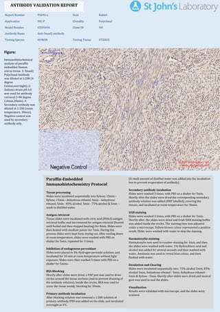
  • 1. Figure: Immunohistochemical analysis of paraffin embedded Human uterus tissue. 1: Smad2 Polyclonal Antibody was diluted at 1:200 (4 degree Celsius,overnight). 2: Sodium citrate pH 6.0 was used for antibody retrieval (>98 degree Celsius,20min). 3: Secondary antibody was diluted at 1:200 (room temperature, 30min). Negative control was used by secondary antibody only. Report Number 95694-a Host Rabbit Application IHC-P Clonality Polyclonal Model Number STJ95694 Clone ID NA Antibody Name Anti-Smad2 antibody Testing Species HUMAN Testing Tissue UTERUS ANTIBODY VALIDATION REPORT a. (A small amount of distilled water was added into the incubation box to prevent evaporation of antibody). 216. Secondary antibody incubation a. Slides were washed 3 times, with PBS on a shaker for 5min. Shortly after the slides were dried the corresponding secondary antibody solution was added (HRP labelled), covering the tissues, and incubated at room temperature for 30min. b. 217. DAB staining a. Slides were washed 3 times, with PBS on a shaker for 5min. b. Shortly after, the slides were dried and fresh DAB staining buffer was added inside the circles. The staining time was adjusted under a microscope. Yellow-brown colour represented a positive result. Slides were washed with water to stop the staining. c. 218. Haematoxylin staining a. Haematoxylin was used to counter-staining for 1min, and then the slides were washed with water. 1% Hydrochloric acid and alcohol was added for several seconds and then washed with water. Ammonia was used to reveal blue colour, and then flushed with water. b. 219. Desolation and Clearing i. Slides were incubated sequentially into: 75% alcohol 5min, 85% alcohol 5min, Anhydrous ethanol - 5min, Anhydrous ethanol - 5min & Xylene - 5min. Shortly after slides were dried and neutral gum was used to seal the slides. ii. 220. Visualization a. Results were validated with microscope, and the slides were scanned. Paraffin-Embedded Immunohistochemistry Protocol 210. 211. Tissue processing a. Slides were incubated sequentially into Xylene; 15min – Xylene, 15min - Anhydrous ethanol, 5min - Anhydrous ethanol, 5min - 85% alcohol, 5min - 75% alcohol & 5min – wash in distilled water. b. 212. Antigen retrieval a. Tissue slides were incubated with citric acid (PH6.0) antigen retrieval buffer and microwaved for antigen retrieval (heated until boiled and then stopped heating) for 8min. Slides were then heated with medium power for 7min. During this process slides were kept from drying out. After cooling down at room temperature, slides were washed with PBS on shaker for 5min, repeated for 3 times. b. 213. Inhibition of endogenous peroxidase a. Slides were placed in 3% Hydrogen peroxide solution, and incubated for 10 min at room temperature without light exposure. Slides were then washed 3 times with PBS on a shaker for 5mins. b. 214. BSA Blocking a. Shortly after slides were dried, a PAP pen was used to draw circles around the tissue sections (and to prevent draining of the antibody solution). Inside the circles, BSA was used to cover the tissue evenly, blocking for 30min. b. 215. Primary antibody incubation After blocking solution was removed a 1:200 solution of primary antibody/PBS was added on the slide, and incubated overnight at 4°C. St John's Laboratory Ltd. www.stjohnslabs.com
  • 2. Figure: Immunohistochemical analysis of paraffin embedded Human uterus cancer tissue. 1: Smad2 Polyclonal Antibody was diluted at 1:200 (4 degree Celsius,overnight). 2: Sodium citrate pH 6.0 was used for antibody retrieval (>98 degree Celsius,20min). 3: Secondary antibody was diluted at 1:200 (room temperature, 30min). Negative control was used by secondary antibody only. Report Number 95694-b Host Rabbit Application IHC-P Clonality Polyclonal Model Number STJ95694 Clone ID NA Antibody Name Anti-Smad2 antibody Testing Species HUMAN Testing Tissue UTERUS CANCER ANTIBODY VALIDATION REPORT a. (A small amount of distilled water was added into the incubation box to prevent evaporation of antibody). 205. Secondary antibody incubation a. Slides were washed 3 times, with PBS on a shaker for 5min. Shortly after the slides were dried the corresponding secondary antibody solution was added (HRP labelled), covering the tissues, and incubated at room temperature for 30min. b. 206. DAB staining a. Slides were washed 3 times, with PBS on a shaker for 5min. b. Shortly after, the slides were dried and fresh DAB staining buffer was added inside the circles. The staining time was adjusted under a microscope. Yellow-brown colour represented a positive result. Slides were washed with water to stop the staining. c. 207. Haematoxylin staining a. Haematoxylin was used to counter-staining for 1min, and then the slides were washed with water. 1% Hydrochloric acid and alcohol was added for several seconds and then washed with water. Ammonia was used to reveal blue colour, and then flushed with water. b. 208. Desolation and Clearing i. Slides were incubated sequentially into: 75% alcohol 5min, 85% alcohol 5min, Anhydrous ethanol - 5min, Anhydrous ethanol - 5min & Xylene - 5min. Shortly after slides were dried and neutral gum was used to seal the slides. ii. 209. Visualization a. Results were validated with microscope, and the slides were scanned. Paraffin-Embedded Immunohistochemistry Protocol 199. 200. Tissue processing a. Slides were incubated sequentially into Xylene; 15min – Xylene, 15min - Anhydrous ethanol, 5min - Anhydrous ethanol, 5min - 85% alcohol, 5min - 75% alcohol & 5min – wash in distilled water. b. 201. Antigen retrieval a. Tissue slides were incubated with citric acid (PH6.0) antigen retrieval buffer and microwaved for antigen retrieval (heated until boiled and then stopped heating) for 8min. Slides were then heated with medium power for 7min. During this process slides were kept from drying out. After cooling down at room temperature, slides were washed with PBS on shaker for 5min, repeated for 3 times. b. 202. Inhibition of endogenous peroxidase a. Slides were placed in 3% Hydrogen peroxide solution, and incubated for 10 min at room temperature without light exposure. Slides were then washed 3 times with PBS on a shaker for 5mins. b. 203. BSA Blocking a. Shortly after slides were dried, a PAP pen was used to draw circles around the tissue sections (and to prevent draining of the antibody solution). Inside the circles, BSA was used to cover the tissue evenly, blocking for 30min. b. 204. Primary antibody incubation After blocking solution was removed a 1:200 solution of primary antibody/PBS was added on the slide, and incubated overnight at 4°C. St John's Laboratory Ltd. www.stjohnslabs.com
  • 3. Figure: Immunohistochemical analysis of paraffin embedded Human lung cancer tissue. 1: Smad2 Polyclonal Antibody was diluted at 1:200 (4 degree Celsius,overnight). 2: Sodium citrate pH 6.0 was used for antibody retrieval (>98 degree Celsius,20min). 3: Secondary antibody was diluted at 1:200 (room temperature, 30min). Negative control was used by secondary antibody only. Report Number 95694-c Host Rabbit Application IHC-P Clonality Polyclonal Model Number STJ95694 Clone ID NA Antibody Name Anti-Smad2 antibody Testing Species HUMAN Testing Tissue LUNG CANCER ANTIBODY VALIDATION REPORT a. (A small amount of distilled water was added into the incubation box to prevent evaporation of antibody). 194. Secondary antibody incubation a. Slides were washed 3 times, with PBS on a shaker for 5min. Shortly after the slides were dried the corresponding secondary antibody solution was added (HRP labelled), covering the tissues, and incubated at room temperature for 30min. b. 195. DAB staining a. Slides were washed 3 times, with PBS on a shaker for 5min. b. Shortly after, the slides were dried and fresh DAB staining buffer was added inside the circles. The staining time was adjusted under a microscope. Yellow-brown colour represented a positive result. Slides were washed with water to stop the staining. c. 196. Haematoxylin staining a. Haematoxylin was used to counter-staining for 1min, and then the slides were washed with water. 1% Hydrochloric acid and alcohol was added for several seconds and then washed with water. Ammonia was used to reveal blue colour, and then flushed with water. b. 197. Desolation and Clearing i. Slides were incubated sequentially into: 75% alcohol 5min, 85% alcohol 5min, Anhydrous ethanol - 5min, Anhydrous ethanol - 5min & Xylene - 5min. Shortly after slides were dried and neutral gum was used to seal the slides. ii. 198. Visualization a. Results were validated with microscope, and the slides were scanned. Paraffin-Embedded Immunohistochemistry Protocol 188. 189. Tissue processing a. Slides were incubated sequentially into Xylene; 15min – Xylene, 15min - Anhydrous ethanol, 5min - Anhydrous ethanol, 5min - 85% alcohol, 5min - 75% alcohol & 5min – wash in distilled water. b. 190. Antigen retrieval a. Tissue slides were incubated with citric acid (PH6.0) antigen retrieval buffer and microwaved for antigen retrieval (heated until boiled and then stopped heating) for 8min. Slides were then heated with medium power for 7min. During this process slides were kept from drying out. After cooling down at room temperature, slides were washed with PBS on shaker for 5min, repeated for 3 times. b. 191. Inhibition of endogenous peroxidase a. Slides were placed in 3% Hydrogen peroxide solution, and incubated for 10 min at room temperature without light exposure. Slides were then washed 3 times with PBS on a shaker for 5mins. b. 192. BSA Blocking a. Shortly after slides were dried, a PAP pen was used to draw circles around the tissue sections (and to prevent draining of the antibody solution). Inside the circles, BSA was used to cover the tissue evenly, blocking for 30min. b. 193. Primary antibody incubation After blocking solution was removed a 1:200 solution of primary antibody/PBS was added on the slide, and incubated overnight at 4°C. St John's Laboratory Ltd. www.stjohnslabs.com
  • 4. Figure: Immunohistochemical analysis of paraffin embedded Human stomach tissue. 1: Smad2 Polyclonal Antibody was diluted at 1:200 (4 degree Celsius,overnight). 2: Sodium citrate pH 6.0 was used for antibody retrieval (>98 degree Celsius,20min). 3: Secondary antibody was diluted at 1:200 (room temperature, 30min). Negative control was used by secondary antibody only. Report Number 95694-d Host Rabbit Application IHC-P Clonality Polyclonal Model Number STJ95694 Clone ID NA Antibody Name Anti-Smad2 antibody Testing Species HUMAN Testing Tissue STOMACH ANTIBODY VALIDATION REPORT a. (A small amount of distilled water was added into the incubation box to prevent evaporation of antibody). 183. Secondary antibody incubation a. Slides were washed 3 times, with PBS on a shaker for 5min. Shortly after the slides were dried the corresponding secondary antibody solution was added (HRP labelled), covering the tissues, and incubated at room temperature for 30min. b. 184. DAB staining a. Slides were washed 3 times, with PBS on a shaker for 5min. b. Shortly after, the slides were dried and fresh DAB staining buffer was added inside the circles. The staining time was adjusted under a microscope. Yellow-brown colour represented a positive result. Slides were washed with water to stop the staining. c. 185. Haematoxylin staining a. Haematoxylin was used to counter-staining for 1min, and then the slides were washed with water. 1% Hydrochloric acid and alcohol was added for several seconds and then washed with water. Ammonia was used to reveal blue colour, and then flushed with water. b. 186. Desolation and Clearing i. Slides were incubated sequentially into: 75% alcohol 5min, 85% alcohol 5min, Anhydrous ethanol - 5min, Anhydrous ethanol - 5min & Xylene - 5min. Shortly after slides were dried and neutral gum was used to seal the slides. ii. 187. Visualization a. Results were validated with microscope, and the slides were scanned. Paraffin-Embedded Immunohistochemistry Protocol 177. 178. Tissue processing a. Slides were incubated sequentially into Xylene; 15min – Xylene, 15min - Anhydrous ethanol, 5min - Anhydrous ethanol, 5min - 85% alcohol, 5min - 75% alcohol & 5min – wash in distilled water. b. 179. Antigen retrieval a. Tissue slides were incubated with citric acid (PH6.0) antigen retrieval buffer and microwaved for antigen retrieval (heated until boiled and then stopped heating) for 8min. Slides were then heated with medium power for 7min. During this process slides were kept from drying out. After cooling down at room temperature, slides were washed with PBS on shaker for 5min, repeated for 3 times. b. 180. Inhibition of endogenous peroxidase a. Slides were placed in 3% Hydrogen peroxide solution, and incubated for 10 min at room temperature without light exposure. Slides were then washed 3 times with PBS on a shaker for 5mins. b. 181. BSA Blocking a. Shortly after slides were dried, a PAP pen was used to draw circles around the tissue sections (and to prevent draining of the antibody solution). Inside the circles, BSA was used to cover the tissue evenly, blocking for 30min. b. 182. Primary antibody incubation After blocking solution was removed a 1:200 solution of primary antibody/PBS was added on the slide, and incubated overnight at 4°C. St John's Laboratory Ltd. www.stjohnslabs.com
  • 5. Figure: Immunohistochemical analysis of paraffin embedded Human stomach cancer tissue. 1: Smad2 Polyclonal Antibody was diluted at 1:200 (4 degree Celsius,overnight). 2: Sodium citrate pH 6.0 was used for antibody retrieval (>98 degree Celsius,20min). 3: Secondary antibody was diluted at 1:200 (room temperature, 30min). Negative control was used by secondary antibody only. Report Number 95694-e Host Rabbit Application IHC-P Clonality Polyclonal Model Number STJ95694 Clone ID NA Antibody Name Anti-Smad2 antibody Testing Species HUMAN Testing Tissue STOMACH CANCER ANTIBODY VALIDATION REPORT a. (A small amount of distilled water was added into the incubation box to prevent evaporation of antibody). 172. Secondary antibody incubation a. Slides were washed 3 times, with PBS on a shaker for 5min. Shortly after the slides were dried the corresponding secondary antibody solution was added (HRP labelled), covering the tissues, and incubated at room temperature for 30min. b. 173. DAB staining a. Slides were washed 3 times, with PBS on a shaker for 5min. b. Shortly after, the slides were dried and fresh DAB staining buffer was added inside the circles. The staining time was adjusted under a microscope. Yellow-brown colour represented a positive result. Slides were washed with water to stop the staining. c. 174. Haematoxylin staining a. Haematoxylin was used to counter-staining for 1min, and then the slides were washed with water. 1% Hydrochloric acid and alcohol was added for several seconds and then washed with water. Ammonia was used to reveal blue colour, and then flushed with water. b. 175. Desolation and Clearing i. Slides were incubated sequentially into: 75% alcohol 5min, 85% alcohol 5min, Anhydrous ethanol - 5min, Anhydrous ethanol - 5min & Xylene - 5min. Shortly after slides were dried and neutral gum was used to seal the slides. ii. 176. Visualization a. Results were validated with microscope, and the slides were scanned. Paraffin-Embedded Immunohistochemistry Protocol 166. 167. Tissue processing a. Slides were incubated sequentially into Xylene; 15min – Xylene, 15min - Anhydrous ethanol, 5min - Anhydrous ethanol, 5min - 85% alcohol, 5min - 75% alcohol & 5min – wash in distilled water. b. 168. Antigen retrieval a. Tissue slides were incubated with citric acid (PH6.0) antigen retrieval buffer and microwaved for antigen retrieval (heated until boiled and then stopped heating) for 8min. Slides were then heated with medium power for 7min. During this process slides were kept from drying out. After cooling down at room temperature, slides were washed with PBS on shaker for 5min, repeated for 3 times. b. 169. Inhibition of endogenous peroxidase a. Slides were placed in 3% Hydrogen peroxide solution, and incubated for 10 min at room temperature without light exposure. Slides were then washed 3 times with PBS on a shaker for 5mins. b. 170. BSA Blocking a. Shortly after slides were dried, a PAP pen was used to draw circles around the tissue sections (and to prevent draining of the antibody solution). Inside the circles, BSA was used to cover the tissue evenly, blocking for 30min. b. 171. Primary antibody incubation After blocking solution was removed a 1:200 solution of primary antibody/PBS was added on the slide, and incubated overnight at 4°C. St John's Laboratory Ltd. www.stjohnslabs.com
  • 6. Figure: Immunohistochemical analysis of paraffin embedded Human appendix tissue. 1: Smad2 Polyclonal Antibody was diluted at 1:200 (4 degree Celsius,overnight). 2: Sodium citrate pH 6.0 was used for antibody retrieval (>98 degree Celsius,20min). 3: Secondary antibody was diluted at 1:200 (room temperature, 30min). Negative control was used by secondary antibody only. Report Number 95694-f Host Rabbit Application IHC-P Clonality Polyclonal Model Number STJ95694 Clone ID NA Antibody Name Anti-Smad2 antibody Testing Species HUMAN Testing Tissue APPENDIX ANTIBODY VALIDATION REPORT a. (A small amount of distilled water was added into the incubation box to prevent evaporation of antibody). 161. Secondary antibody incubation a. Slides were washed 3 times, with PBS on a shaker for 5min. Shortly after the slides were dried the corresponding secondary antibody solution was added (HRP labelled), covering the tissues, and incubated at room temperature for 30min. b. 162. DAB staining a. Slides were washed 3 times, with PBS on a shaker for 5min. b. Shortly after, the slides were dried and fresh DAB staining buffer was added inside the circles. The staining time was adjusted under a microscope. Yellow-brown colour represented a positive result. Slides were washed with water to stop the staining. c. 163. Haematoxylin staining a. Haematoxylin was used to counter-staining for 1min, and then the slides were washed with water. 1% Hydrochloric acid and alcohol was added for several seconds and then washed with water. Ammonia was used to reveal blue colour, and then flushed with water. b. 164. Desolation and Clearing i. Slides were incubated sequentially into: 75% alcohol 5min, 85% alcohol 5min, Anhydrous ethanol - 5min, Anhydrous ethanol - 5min & Xylene - 5min. Shortly after slides were dried and neutral gum was used to seal the slides. ii. 165. Visualization a. Results were validated with microscope, and the slides were scanned. Paraffin-Embedded Immunohistochemistry Protocol 155. 156. Tissue processing a. Slides were incubated sequentially into Xylene; 15min – Xylene, 15min - Anhydrous ethanol, 5min - Anhydrous ethanol, 5min - 85% alcohol, 5min - 75% alcohol & 5min – wash in distilled water. b. 157. Antigen retrieval a. Tissue slides were incubated with citric acid (PH6.0) antigen retrieval buffer and microwaved for antigen retrieval (heated until boiled and then stopped heating) for 8min. Slides were then heated with medium power for 7min. During this process slides were kept from drying out. After cooling down at room temperature, slides were washed with PBS on shaker for 5min, repeated for 3 times. b. 158. Inhibition of endogenous peroxidase a. Slides were placed in 3% Hydrogen peroxide solution, and incubated for 10 min at room temperature without light exposure. Slides were then washed 3 times with PBS on a shaker for 5mins. b. 159. BSA Blocking a. Shortly after slides were dried, a PAP pen was used to draw circles around the tissue sections (and to prevent draining of the antibody solution). Inside the circles, BSA was used to cover the tissue evenly, blocking for 30min. b. 160. Primary antibody incubation After blocking solution was removed a 1:200 solution of primary antibody/PBS was added on the slide, and incubated overnight at 4°C. St John's Laboratory Ltd. www.stjohnslabs.com
  • 7. Figure: Immunohistochemical analysis of paraffin embedded Rat heart tissue. 1: Smad2 Polyclonal Antibody was diluted at 1:200 (4 degree Celsius,overnight). 2: Sodium citrate pH 6.0 was used for antibody retrieval (>98 degree Celsius,20min). 3: Secondary antibody was diluted at 1:200 (room temperature, 30min). Negative control was used by secondary antibody only. Report Number 95694-g Host Rabbit Application IHC-P Clonality Polyclonal Model Number STJ95694 Clone ID NA Antibody Name Anti-Smad2 antibody Testing Species RAT Testing Tissue HEART ANTIBODY VALIDATION REPORT a. (A small amount of distilled water was added into the incubation box to prevent evaporation of antibody). 150. Secondary antibody incubation a. Slides were washed 3 times, with PBS on a shaker for 5min. Shortly after the slides were dried the corresponding secondary antibody solution was added (HRP labelled), covering the tissues, and incubated at room temperature for 30min. b. 151. DAB staining a. Slides were washed 3 times, with PBS on a shaker for 5min. b. Shortly after, the slides were dried and fresh DAB staining buffer was added inside the circles. The staining time was adjusted under a microscope. Yellow-brown colour represented a positive result. Slides were washed with water to stop the staining. c. 152. Haematoxylin staining a. Haematoxylin was used to counter-staining for 1min, and then the slides were washed with water. 1% Hydrochloric acid and alcohol was added for several seconds and then washed with water. Ammonia was used to reveal blue colour, and then flushed with water. b. 153. Desolation and Clearing i. Slides were incubated sequentially into: 75% alcohol 5min, 85% alcohol 5min, Anhydrous ethanol - 5min, Anhydrous ethanol - 5min & Xylene - 5min. Shortly after slides were dried and neutral gum was used to seal the slides. ii. 154. Visualization a. Results were validated with microscope, and the slides were scanned. Paraffin-Embedded Immunohistochemistry Protocol 144. 145. Tissue processing a. Slides were incubated sequentially into Xylene; 15min – Xylene, 15min - Anhydrous ethanol, 5min - Anhydrous ethanol, 5min - 85% alcohol, 5min - 75% alcohol & 5min – wash in distilled water. b. 146. Antigen retrieval a. Tissue slides were incubated with citric acid (PH6.0) antigen retrieval buffer and microwaved for antigen retrieval (heated until boiled and then stopped heating) for 8min. Slides were then heated with medium power for 7min. During this process slides were kept from drying out. After cooling down at room temperature, slides were washed with PBS on shaker for 5min, repeated for 3 times. b. 147. Inhibition of endogenous peroxidase a. Slides were placed in 3% Hydrogen peroxide solution, and incubated for 10 min at room temperature without light exposure. Slides were then washed 3 times with PBS on a shaker for 5mins. b. 148. BSA Blocking a. Shortly after slides were dried, a PAP pen was used to draw circles around the tissue sections (and to prevent draining of the antibody solution). Inside the circles, BSA was used to cover the tissue evenly, blocking for 30min. b. 149. Primary antibody incubation After blocking solution was removed a 1:200 solution of primary antibody/PBS was added on the slide, and incubated overnight at 4°C. St John's Laboratory Ltd. www.stjohnslabs.com
  • 8. Figure: Immunohistochemical analysis of paraffin embedded Rat testis tissue. 1: Smad2 Polyclonal Antibody was diluted at 1:200 (4 degree Celsius,overnight). 2: Sodium citrate pH 6.0 was used for antibody retrieval (>98 degree Celsius,20min). 3: Secondary antibody was diluted at 1:200 (room temperature, 30min). Negative control was used by secondary antibody only. Report Number 95694-h Host Rabbit Application IHC-P Clonality Polyclonal Model Number STJ95694 Clone ID NA Antibody Name Anti-Smad2 antibody Testing Species RAT Testing Tissue TESTIS ANTIBODY VALIDATION REPORT a. (A small amount of distilled water was added into the incubation box to prevent evaporation of antibody). 139. Secondary antibody incubation a. Slides were washed 3 times, with PBS on a shaker for 5min. Shortly after the slides were dried the corresponding secondary antibody solution was added (HRP labelled), covering the tissues, and incubated at room temperature for 30min. b. 140. DAB staining a. Slides were washed 3 times, with PBS on a shaker for 5min. b. Shortly after, the slides were dried and fresh DAB staining buffer was added inside the circles. The staining time was adjusted under a microscope. Yellow-brown colour represented a positive result. Slides were washed with water to stop the staining. c. 141. Haematoxylin staining a. Haematoxylin was used to counter-staining for 1min, and then the slides were washed with water. 1% Hydrochloric acid and alcohol was added for several seconds and then washed with water. Ammonia was used to reveal blue colour, and then flushed with water. b. 142. Desolation and Clearing i. Slides were incubated sequentially into: 75% alcohol 5min, 85% alcohol 5min, Anhydrous ethanol - 5min, Anhydrous ethanol - 5min & Xylene - 5min. Shortly after slides were dried and neutral gum was used to seal the slides. ii. 143. Visualization a. Results were validated with microscope, and the slides were scanned. Paraffin-Embedded Immunohistochemistry Protocol 133. 134. Tissue processing a. Slides were incubated sequentially into Xylene; 15min – Xylene, 15min - Anhydrous ethanol, 5min - Anhydrous ethanol, 5min - 85% alcohol, 5min - 75% alcohol & 5min – wash in distilled water. b. 135. Antigen retrieval a. Tissue slides were incubated with citric acid (PH6.0) antigen retrieval buffer and microwaved for antigen retrieval (heated until boiled and then stopped heating) for 8min. Slides were then heated with medium power for 7min. During this process slides were kept from drying out. After cooling down at room temperature, slides were washed with PBS on shaker for 5min, repeated for 3 times. b. 136. Inhibition of endogenous peroxidase a. Slides were placed in 3% Hydrogen peroxide solution, and incubated for 10 min at room temperature without light exposure. Slides were then washed 3 times with PBS on a shaker for 5mins. b. 137. BSA Blocking a. Shortly after slides were dried, a PAP pen was used to draw circles around the tissue sections (and to prevent draining of the antibody solution). Inside the circles, BSA was used to cover the tissue evenly, blocking for 30min. b. 138. Primary antibody incubation After blocking solution was removed a 1:200 solution of primary antibody/PBS was added on the slide, and incubated overnight at 4°C. St John's Laboratory Ltd. www.stjohnslabs.com
  • 9. Figure: Immunohistochemical analysis of paraffin embedded Rat liver tissue. 1: Smad2 Polyclonal Antibody was diluted at 1:200 (4 degree Celsius,overnight). 2: Sodium citrate pH 6.0 was used for antibody retrieval (>98 degree Celsius,20min). 3: Secondary antibody was diluted at 1:200 (room temperature, 30min). Negative control was used by secondary antibody only. Report Number 95694-i Host Rabbit Application IHC-P Clonality Polyclonal Model Number STJ95694 Clone ID NA Antibody Name Anti-Smad2 antibody Testing Species RAT Testing Tissue LIVER ANTIBODY VALIDATION REPORT a. (A small amount of distilled water was added into the incubation box to prevent evaporation of antibody). 128. Secondary antibody incubation a. Slides were washed 3 times, with PBS on a shaker for 5min. Shortly after the slides were dried the corresponding secondary antibody solution was added (HRP labelled), covering the tissues, and incubated at room temperature for 30min. b. 129. DAB staining a. Slides were washed 3 times, with PBS on a shaker for 5min. b. Shortly after, the slides were dried and fresh DAB staining buffer was added inside the circles. The staining time was adjusted under a microscope. Yellow-brown colour represented a positive result. Slides were washed with water to stop the staining. c. 130. Haematoxylin staining a. Haematoxylin was used to counter-staining for 1min, and then the slides were washed with water. 1% Hydrochloric acid and alcohol was added for several seconds and then washed with water. Ammonia was used to reveal blue colour, and then flushed with water. b. 131. Desolation and Clearing i. Slides were incubated sequentially into: 75% alcohol 5min, 85% alcohol 5min, Anhydrous ethanol - 5min, Anhydrous ethanol - 5min & Xylene - 5min. Shortly after slides were dried and neutral gum was used to seal the slides. ii. 132. Visualization a. Results were validated with microscope, and the slides were scanned. Paraffin-Embedded Immunohistochemistry Protocol 122. 123. Tissue processing a. Slides were incubated sequentially into Xylene; 15min – Xylene, 15min - Anhydrous ethanol, 5min - Anhydrous ethanol, 5min - 85% alcohol, 5min - 75% alcohol & 5min – wash in distilled water. b. 124. Antigen retrieval a. Tissue slides were incubated with citric acid (PH6.0) antigen retrieval buffer and microwaved for antigen retrieval (heated until boiled and then stopped heating) for 8min. Slides were then heated with medium power for 7min. During this process slides were kept from drying out. After cooling down at room temperature, slides were washed with PBS on shaker for 5min, repeated for 3 times. b. 125. Inhibition of endogenous peroxidase a. Slides were placed in 3% Hydrogen peroxide solution, and incubated for 10 min at room temperature without light exposure. Slides were then washed 3 times with PBS on a shaker for 5mins. b. 126. BSA Blocking a. Shortly after slides were dried, a PAP pen was used to draw circles around the tissue sections (and to prevent draining of the antibody solution). Inside the circles, BSA was used to cover the tissue evenly, blocking for 30min. b. 127. Primary antibody incubation After blocking solution was removed a 1:200 solution of primary antibody/PBS was added on the slide, and incubated overnight at 4°C. St John's Laboratory Ltd. www.stjohnslabs.com
  • 10. Figure: Immunohistochemical analysis of paraffin embedded Rat lung tissue. 1: Smad2 Polyclonal Antibody was diluted at 1:200 (4 degree Celsius,overnight). 2: Sodium citrate pH 6.0 was used for antibody retrieval (>98 degree Celsius,20min). 3: Secondary antibody was diluted at 1:200 (room temperature, 30min). Negative control was used by secondary antibody only. Report Number 95694-j Host Rabbit Application IHC-P Clonality Polyclonal Model Number STJ95694 Clone ID NA Antibody Name Anti-Smad2 antibody Testing Species RAT Testing Tissue LUNG ANTIBODY VALIDATION REPORT a. (A small amount of distilled water was added into the incubation box to prevent evaporation of antibody). 117. Secondary antibody incubation a. Slides were washed 3 times, with PBS on a shaker for 5min. Shortly after the slides were dried the corresponding secondary antibody solution was added (HRP labelled), covering the tissues, and incubated at room temperature for 30min. b. 118. DAB staining a. Slides were washed 3 times, with PBS on a shaker for 5min. b. Shortly after, the slides were dried and fresh DAB staining buffer was added inside the circles. The staining time was adjusted under a microscope. Yellow-brown colour represented a positive result. Slides were washed with water to stop the staining. c. 119. Haematoxylin staining a. Haematoxylin was used to counter-staining for 1min, and then the slides were washed with water. 1% Hydrochloric acid and alcohol was added for several seconds and then washed with water. Ammonia was used to reveal blue colour, and then flushed with water. b. 120. Desolation and Clearing i. Slides were incubated sequentially into: 75% alcohol 5min, 85% alcohol 5min, Anhydrous ethanol - 5min, Anhydrous ethanol - 5min & Xylene - 5min. Shortly after slides were dried and neutral gum was used to seal the slides. ii. 121. Visualization a. Results were validated with microscope, and the slides were scanned. Paraffin-Embedded Immunohistochemistry Protocol 111. 112. Tissue processing a. Slides were incubated sequentially into Xylene; 15min – Xylene, 15min - Anhydrous ethanol, 5min - Anhydrous ethanol, 5min - 85% alcohol, 5min - 75% alcohol & 5min – wash in distilled water. b. 113. Antigen retrieval a. Tissue slides were incubated with citric acid (PH6.0) antigen retrieval buffer and microwaved for antigen retrieval (heated until boiled and then stopped heating) for 8min. Slides were then heated with medium power for 7min. During this process slides were kept from drying out. After cooling down at room temperature, slides were washed with PBS on shaker for 5min, repeated for 3 times. b. 114. Inhibition of endogenous peroxidase a. Slides were placed in 3% Hydrogen peroxide solution, and incubated for 10 min at room temperature without light exposure. Slides were then washed 3 times with PBS on a shaker for 5mins. b. 115. BSA Blocking a. Shortly after slides were dried, a PAP pen was used to draw circles around the tissue sections (and to prevent draining of the antibody solution). Inside the circles, BSA was used to cover the tissue evenly, blocking for 30min. b. 116. Primary antibody incubation After blocking solution was removed a 1:200 solution of primary antibody/PBS was added on the slide, and incubated overnight at 4°C. St John's Laboratory Ltd. www.stjohnslabs.com
  • 11. Figure: Immunohistochemical analysis of paraffin embedded Rat kidney tissue. 1: Smad2 Polyclonal Antibody was diluted at 1:200 (4 degree Celsius,overnight). 2: Sodium citrate pH 6.0 was used for antibody retrieval (>98 degree Celsius,20min). 3: Secondary antibody was diluted at 1:200 (room temperature, 30min). Negative control was used by secondary antibody only. Report Number 95694-k Host Rabbit Application IHC-P Clonality Polyclonal Model Number STJ95694 Clone ID NA Antibody Name Anti-Smad2 antibody Testing Species RAT Testing Tissue KIDNEY ANTIBODY VALIDATION REPORT a. (A small amount of distilled water was added into the incubation box to prevent evaporation of antibody). 106. Secondary antibody incubation a. Slides were washed 3 times, with PBS on a shaker for 5min. Shortly after the slides were dried the corresponding secondary antibody solution was added (HRP labelled), covering the tissues, and incubated at room temperature for 30min. b. 107. DAB staining a. Slides were washed 3 times, with PBS on a shaker for 5min. b. Shortly after, the slides were dried and fresh DAB staining buffer was added inside the circles. The staining time was adjusted under a microscope. Yellow-brown colour represented a positive result. Slides were washed with water to stop the staining. c. 108. Haematoxylin staining a. Haematoxylin was used to counter-staining for 1min, and then the slides were washed with water. 1% Hydrochloric acid and alcohol was added for several seconds and then washed with water. Ammonia was used to reveal blue colour, and then flushed with water. b. 109. Desolation and Clearing i. Slides were incubated sequentially into: 75% alcohol 5min, 85% alcohol 5min, Anhydrous ethanol - 5min, Anhydrous ethanol - 5min & Xylene - 5min. Shortly after slides were dried and neutral gum was used to seal the slides. ii. 110. Visualization a. Results were validated with microscope, and the slides were scanned. Paraffin-Embedded Immunohistochemistry Protocol 100. 101. Tissue processing a. Slides were incubated sequentially into Xylene; 15min – Xylene, 15min - Anhydrous ethanol, 5min - Anhydrous ethanol, 5min - 85% alcohol, 5min - 75% alcohol & 5min – wash in distilled water. b. 102. Antigen retrieval a. Tissue slides were incubated with citric acid (PH6.0) antigen retrieval buffer and microwaved for antigen retrieval (heated until boiled and then stopped heating) for 8min. Slides were then heated with medium power for 7min. During this process slides were kept from drying out. After cooling down at room temperature, slides were washed with PBS on shaker for 5min, repeated for 3 times. b. 103. Inhibition of endogenous peroxidase a. Slides were placed in 3% Hydrogen peroxide solution, and incubated for 10 min at room temperature without light exposure. Slides were then washed 3 times with PBS on a shaker for 5mins. b. 104. BSA Blocking a. Shortly after slides were dried, a PAP pen was used to draw circles around the tissue sections (and to prevent draining of the antibody solution). Inside the circles, BSA was used to cover the tissue evenly, blocking for 30min. b. 105. Primary antibody incubation After blocking solution was removed a 1:200 solution of primary antibody/PBS was added on the slide, and incubated overnight at 4°C. St John's Laboratory Ltd. www.stjohnslabs.com
  • 12. Figure: Immunohistochemical analysis of paraffin embedded Rat spinal cord tissue. 1: Smad2 Polyclonal Antibody was diluted at 1:200 (4 degree Celsius,overnight). 2: Sodium citrate pH 6.0 was used for antibody retrieval (>98 degree Celsius,20min). 3: Secondary antibody was diluted at 1:200 (room temperature, 30min). Negative control was used by secondary antibody only. Report Number 95694-l Host Rabbit Application IHC-P Clonality Polyclonal Model Number STJ95694 Clone ID NA Antibody Name Anti-Smad2 antibody Testing Species RAT Testing Tissue SPINAL ANTIBODY VALIDATION REPORT a. (A small amount of distilled water was added into the incubation box to prevent evaporation of antibody). 95. Secondary antibody incubation a. Slides were washed 3 times, with PBS on a shaker for 5min. Shortly after the slides were dried the corresponding secondary antibody solution was added (HRP labelled), covering the tissues, and incubated at room temperature for 30min. b. 96. DAB staining a. Slides were washed 3 times, with PBS on a shaker for 5min. b. Shortly after, the slides were dried and fresh DAB staining buffer was added inside the circles. The staining time was adjusted under a microscope. Yellow-brown colour represented a positive result. Slides were washed with water to stop the staining. c. 97. Haematoxylin staining a. Haematoxylin was used to counter-staining for 1min, and then the slides were washed with water. 1% Hydrochloric acid and alcohol was added for several seconds and then washed with water. Ammonia was used to reveal blue colour, and then flushed with water. b. 98. Desolation and Clearing i. Slides were incubated sequentially into: 75% alcohol 5min, 85% alcohol 5min, Anhydrous ethanol - 5min, Anhydrous ethanol - 5min & Xylene - 5min. Shortly after slides were dried and neutral gum was used to seal the slides. ii. 99. Visualization a. Results were validated with microscope, and the slides were scanned. Paraffin-Embedded Immunohistochemistry Protocol 89. 90. Tissue processing a. Slides were incubated sequentially into Xylene; 15min – Xylene, 15min - Anhydrous ethanol, 5min - Anhydrous ethanol, 5min - 85% alcohol, 5min - 75% alcohol & 5min – wash in distilled water. b. 91. Antigen retrieval a. Tissue slides were incubated with citric acid (PH6.0) antigen retrieval buffer and microwaved for antigen retrieval (heated until boiled and then stopped heating) for 8min. Slides were then heated with medium power for 7min. During this process slides were kept from drying out. After cooling down at room temperature, slides were washed with PBS on shaker for 5min, repeated for 3 times. b. 92. Inhibition of endogenous peroxidase a. Slides were placed in 3% Hydrogen peroxide solution, and incubated for 10 min at room temperature without light exposure. Slides were then washed 3 times with PBS on a shaker for 5mins. b. 93. BSA Blocking a. Shortly after slides were dried, a PAP pen was used to draw circles around the tissue sections (and to prevent draining of the antibody solution). Inside the circles, BSA was used to cover the tissue evenly, blocking for 30min. b. 94. Primary antibody incubation After blocking solution was removed a 1:200 solution of primary antibody/PBS was added on the slide, and incubated overnight at 4°C. St John's Laboratory Ltd. www.stjohnslabs.com
  • 13. Figure: Immunohistochemical analysis of paraffin embedded Rat brain tissue. 1: Smad2 Polyclonal Antibody was diluted at 1:200 (4 degree Celsius,overnight). 2: Sodium citrate pH 6.0 was used for antibody retrieval (>98 degree Celsius,20min). 3: Secondary antibody was diluted at 1:200 (room temperature, 30min). Negative control was used by secondary antibody only. Report Number 95694-m Host Rabbit Application IHC-P Clonality Polyclonal Model Number STJ95694 Clone ID NA Antibody Name Anti-Smad2 antibody Testing Species RAT Testing Tissue BRAIN ANTIBODY VALIDATION REPORT a. (A small amount of distilled water was added into the incubation box to prevent evaporation of antibody). 84. Secondary antibody incubation a. Slides were washed 3 times, with PBS on a shaker for 5min. Shortly after the slides were dried the corresponding secondary antibody solution was added (HRP labelled), covering the tissues, and incubated at room temperature for 30min. b. 85. DAB staining a. Slides were washed 3 times, with PBS on a shaker for 5min. b. Shortly after, the slides were dried and fresh DAB staining buffer was added inside the circles. The staining time was adjusted under a microscope. Yellow-brown colour represented a positive result. Slides were washed with water to stop the staining. c. 86. Haematoxylin staining a. Haematoxylin was used to counter-staining for 1min, and then the slides were washed with water. 1% Hydrochloric acid and alcohol was added for several seconds and then washed with water. Ammonia was used to reveal blue colour, and then flushed with water. b. 87. Desolation and Clearing i. Slides were incubated sequentially into: 75% alcohol 5min, 85% alcohol 5min, Anhydrous ethanol - 5min, Anhydrous ethanol - 5min & Xylene - 5min. Shortly after slides were dried and neutral gum was used to seal the slides. ii. 88. Visualization a. Results were validated with microscope, and the slides were scanned. Paraffin-Embedded Immunohistochemistry Protocol 78. 79. Tissue processing a. Slides were incubated sequentially into Xylene; 15min – Xylene, 15min - Anhydrous ethanol, 5min - Anhydrous ethanol, 5min - 85% alcohol, 5min - 75% alcohol & 5min – wash in distilled water. b. 80. Antigen retrieval a. Tissue slides were incubated with citric acid (PH6.0) antigen retrieval buffer and microwaved for antigen retrieval (heated until boiled and then stopped heating) for 8min. Slides were then heated with medium power for 7min. During this process slides were kept from drying out. After cooling down at room temperature, slides were washed with PBS on shaker for 5min, repeated for 3 times. b. 81. Inhibition of endogenous peroxidase a. Slides were placed in 3% Hydrogen peroxide solution, and incubated for 10 min at room temperature without light exposure. Slides were then washed 3 times with PBS on a shaker for 5mins. b. 82. BSA Blocking a. Shortly after slides were dried, a PAP pen was used to draw circles around the tissue sections (and to prevent draining of the antibody solution). Inside the circles, BSA was used to cover the tissue evenly, blocking for 30min. b. 83. Primary antibody incubation After blocking solution was removed a 1:200 solution of primary antibody/PBS was added on the slide, and incubated overnight at 4°C. St John's Laboratory Ltd. www.stjohnslabs.com
  • 14. Figure: Immunohistochemical analysis of paraffin embedded Rat spleen tissue. 1: Smad2 Polyclonal Antibody was diluted at 1:200 (4 degree Celsius,overnight). 2: Sodium citrate pH 6.0 was used for antibody retrieval (>98 degree Celsius,20min). 3: Secondary antibody was diluted at 1:200 (room temperature, 30min). Negative control was used by secondary antibody only. Report Number 95694-n Host Rabbit Application IHC-P Clonality Polyclonal Model Number STJ95694 Clone ID NA Antibody Name Anti-Smad2 antibody Testing Species RAT Testing Tissue SPLEEN ANTIBODY VALIDATION REPORT a. (A small amount of distilled water was added into the incubation box to prevent evaporation of antibody). 73. Secondary antibody incubation a. Slides were washed 3 times, with PBS on a shaker for 5min. Shortly after the slides were dried the corresponding secondary antibody solution was added (HRP labelled), covering the tissues, and incubated at room temperature for 30min. b. 74. DAB staining a. Slides were washed 3 times, with PBS on a shaker for 5min. b. Shortly after, the slides were dried and fresh DAB staining buffer was added inside the circles. The staining time was adjusted under a microscope. Yellow-brown colour represented a positive result. Slides were washed with water to stop the staining. c. 75. Haematoxylin staining a. Haematoxylin was used to counter-staining for 1min, and then the slides were washed with water. 1% Hydrochloric acid and alcohol was added for several seconds and then washed with water. Ammonia was used to reveal blue colour, and then flushed with water. b. 76. Desolation and Clearing i. Slides were incubated sequentially into: 75% alcohol 5min, 85% alcohol 5min, Anhydrous ethanol - 5min, Anhydrous ethanol - 5min & Xylene - 5min. Shortly after slides were dried and neutral gum was used to seal the slides. ii. 77. Visualization a. Results were validated with microscope, and the slides were scanned. Paraffin-Embedded Immunohistochemistry Protocol 67. 68. Tissue processing a. Slides were incubated sequentially into Xylene; 15min – Xylene, 15min - Anhydrous ethanol, 5min - Anhydrous ethanol, 5min - 85% alcohol, 5min - 75% alcohol & 5min – wash in distilled water. b. 69. Antigen retrieval a. Tissue slides were incubated with citric acid (PH6.0) antigen retrieval buffer and microwaved for antigen retrieval (heated until boiled and then stopped heating) for 8min. Slides were then heated with medium power for 7min. During this process slides were kept from drying out. After cooling down at room temperature, slides were washed with PBS on shaker for 5min, repeated for 3 times. b. 70. Inhibition of endogenous peroxidase a. Slides were placed in 3% Hydrogen peroxide solution, and incubated for 10 min at room temperature without light exposure. Slides were then washed 3 times with PBS on a shaker for 5mins. b. 71. BSA Blocking a. Shortly after slides were dried, a PAP pen was used to draw circles around the tissue sections (and to prevent draining of the antibody solution). Inside the circles, BSA was used to cover the tissue evenly, blocking for 30min. b. 72. Primary antibody incubation After blocking solution was removed a 1:200 solution of primary antibody/PBS was added on the slide, and incubated overnight at 4°C. St John's Laboratory Ltd. www.stjohnslabs.com
  • 15. Figure: Immunohistochemical analysis of paraffin embedded Mouse testis tissue. 1: Smad2 Polyclonal Antibody was diluted at 1:200 (4 degree Celsius,overnight). 2: Sodium citrate pH 6.0 was used for antibody retrieval (>98 degree Celsius,20min). 3: Secondary antibody was diluted at 1:200 (room temperature, 30min). Negative control was used by secondary antibody only. Report Number 95694-o Host Rabbit Application IHC-P Clonality Polyclonal Model Number STJ95694 Clone ID NA Antibody Name Anti-Smad2 antibody Testing Species MOUSE Testing Tissue TESTIS ANTIBODY VALIDATION REPORT a. (A small amount of distilled water was added into the incubation box to prevent evaporation of antibody). 62. Secondary antibody incubation a. Slides were washed 3 times, with PBS on a shaker for 5min. Shortly after the slides were dried the corresponding secondary antibody solution was added (HRP labelled), covering the tissues, and incubated at room temperature for 30min. b. 63. DAB staining a. Slides were washed 3 times, with PBS on a shaker for 5min. b. Shortly after, the slides were dried and fresh DAB staining buffer was added inside the circles. The staining time was adjusted under a microscope. Yellow-brown colour represented a positive result. Slides were washed with water to stop the staining. c. 64. Haematoxylin staining a. Haematoxylin was used to counter-staining for 1min, and then the slides were washed with water. 1% Hydrochloric acid and alcohol was added for several seconds and then washed with water. Ammonia was used to reveal blue colour, and then flushed with water. b. 65. Desolation and Clearing i. Slides were incubated sequentially into: 75% alcohol 5min, 85% alcohol 5min, Anhydrous ethanol - 5min, Anhydrous ethanol - 5min & Xylene - 5min. Shortly after slides were dried and neutral gum was used to seal the slides. ii. 66. Visualization a. Results were validated with microscope, and the slides were scanned. Paraffin-Embedded Immunohistochemistry Protocol 56. 57. Tissue processing a. Slides were incubated sequentially into Xylene; 15min – Xylene, 15min - Anhydrous ethanol, 5min - Anhydrous ethanol, 5min - 85% alcohol, 5min - 75% alcohol & 5min – wash in distilled water. b. 58. Antigen retrieval a. Tissue slides were incubated with citric acid (PH6.0) antigen retrieval buffer and microwaved for antigen retrieval (heated until boiled and then stopped heating) for 8min. Slides were then heated with medium power for 7min. During this process slides were kept from drying out. After cooling down at room temperature, slides were washed with PBS on shaker for 5min, repeated for 3 times. b. 59. Inhibition of endogenous peroxidase a. Slides were placed in 3% Hydrogen peroxide solution, and incubated for 10 min at room temperature without light exposure. Slides were then washed 3 times with PBS on a shaker for 5mins. b. 60. BSA Blocking a. Shortly after slides were dried, a PAP pen was used to draw circles around the tissue sections (and to prevent draining of the antibody solution). Inside the circles, BSA was used to cover the tissue evenly, blocking for 30min. b. 61. Primary antibody incubation After blocking solution was removed a 1:200 solution of primary antibody/PBS was added on the slide, and incubated overnight at 4°C. St John's Laboratory Ltd. www.stjohnslabs.com
  • 16. Figure: Immunohistochemical analysis of paraffin embedded Mouse colon tissue. 1: Smad2 Polyclonal Antibody was diluted at 1:200 (4 degree Celsius,overnight). 2: Sodium citrate pH 6.0 was used for antibody retrieval (>98 degree Celsius,20min). 3: Secondary antibody was diluted at 1:200 (room temperature, 30min). Negative control was used by secondary antibody only. Report Number 95694-p Host Rabbit Application IHC-P Clonality Polyclonal Model Number STJ95694 Clone ID NA Antibody Name Anti-Smad2 antibody Testing Species MOUSE Testing Tissue COLON ANTIBODY VALIDATION REPORT a. (A small amount of distilled water was added into the incubation box to prevent evaporation of antibody). 51. Secondary antibody incubation a. Slides were washed 3 times, with PBS on a shaker for 5min. Shortly after the slides were dried the corresponding secondary antibody solution was added (HRP labelled), covering the tissues, and incubated at room temperature for 30min. b. 52. DAB staining a. Slides were washed 3 times, with PBS on a shaker for 5min. b. Shortly after, the slides were dried and fresh DAB staining buffer was added inside the circles. The staining time was adjusted under a microscope. Yellow-brown colour represented a positive result. Slides were washed with water to stop the staining. c. 53. Haematoxylin staining a. Haematoxylin was used to counter-staining for 1min, and then the slides were washed with water. 1% Hydrochloric acid and alcohol was added for several seconds and then washed with water. Ammonia was used to reveal blue colour, and then flushed with water. b. 54. Desolation and Clearing i. Slides were incubated sequentially into: 75% alcohol 5min, 85% alcohol 5min, Anhydrous ethanol - 5min, Anhydrous ethanol - 5min & Xylene - 5min. Shortly after slides were dried and neutral gum was used to seal the slides. ii. 55. Visualization a. Results were validated with microscope, and the slides were scanned. Paraffin-Embedded Immunohistochemistry Protocol 45. 46. Tissue processing a. Slides were incubated sequentially into Xylene; 15min – Xylene, 15min - Anhydrous ethanol, 5min - Anhydrous ethanol, 5min - 85% alcohol, 5min - 75% alcohol & 5min – wash in distilled water. b. 47. Antigen retrieval a. Tissue slides were incubated with citric acid (PH6.0) antigen retrieval buffer and microwaved for antigen retrieval (heated until boiled and then stopped heating) for 8min. Slides were then heated with medium power for 7min. During this process slides were kept from drying out. After cooling down at room temperature, slides were washed with PBS on shaker for 5min, repeated for 3 times. b. 48. Inhibition of endogenous peroxidase a. Slides were placed in 3% Hydrogen peroxide solution, and incubated for 10 min at room temperature without light exposure. Slides were then washed 3 times with PBS on a shaker for 5mins. b. 49. BSA Blocking a. Shortly after slides were dried, a PAP pen was used to draw circles around the tissue sections (and to prevent draining of the antibody solution). Inside the circles, BSA was used to cover the tissue evenly, blocking for 30min. b. 50. Primary antibody incubation After blocking solution was removed a 1:200 solution of primary antibody/PBS was added on the slide, and incubated overnight at 4°C. St John's Laboratory Ltd. www.stjohnslabs.com
  • 17. Figure: Immunohistochemical analysis of paraffin embedded Mouse liver tissue. 1: Smad2 Polyclonal Antibody was diluted at 1:200 (4 degree Celsius,overnight). 2: Sodium citrate pH 6.0 was used for antibody retrieval (>98 degree Celsius,20min). 3: Secondary antibody was diluted at 1:200 (room temperature, 30min). Negative control was used by secondary antibody only. Report Number 95694-q Host Rabbit Application IHC-P Clonality Polyclonal Model Number STJ95694 Clone ID NA Antibody Name Anti-Smad2 antibody Testing Species MOUSE Testing Tissue LIVER ANTIBODY VALIDATION REPORT a. (A small amount of distilled water was added into the incubation box to prevent evaporation of antibody). 40. Secondary antibody incubation a. Slides were washed 3 times, with PBS on a shaker for 5min. Shortly after the slides were dried the corresponding secondary antibody solution was added (HRP labelled), covering the tissues, and incubated at room temperature for 30min. b. 41. DAB staining a. Slides were washed 3 times, with PBS on a shaker for 5min. b. Shortly after, the slides were dried and fresh DAB staining buffer was added inside the circles. The staining time was adjusted under a microscope. Yellow-brown colour represented a positive result. Slides were washed with water to stop the staining. c. 42. Haematoxylin staining a. Haematoxylin was used to counter-staining for 1min, and then the slides were washed with water. 1% Hydrochloric acid and alcohol was added for several seconds and then washed with water. Ammonia was used to reveal blue colour, and then flushed with water. b. 43. Desolation and Clearing i. Slides were incubated sequentially into: 75% alcohol 5min, 85% alcohol 5min, Anhydrous ethanol - 5min, Anhydrous ethanol - 5min & Xylene - 5min. Shortly after slides were dried and neutral gum was used to seal the slides. ii. 44. Visualization a. Results were validated with microscope, and the slides were scanned. Paraffin-Embedded Immunohistochemistry Protocol 34. 35. Tissue processing a. Slides were incubated sequentially into Xylene; 15min – Xylene, 15min - Anhydrous ethanol, 5min - Anhydrous ethanol, 5min - 85% alcohol, 5min - 75% alcohol & 5min – wash in distilled water. b. 36. Antigen retrieval a. Tissue slides were incubated with citric acid (PH6.0) antigen retrieval buffer and microwaved for antigen retrieval (heated until boiled and then stopped heating) for 8min. Slides were then heated with medium power for 7min. During this process slides were kept from drying out. After cooling down at room temperature, slides were washed with PBS on shaker for 5min, repeated for 3 times. b. 37. Inhibition of endogenous peroxidase a. Slides were placed in 3% Hydrogen peroxide solution, and incubated for 10 min at room temperature without light exposure. Slides were then washed 3 times with PBS on a shaker for 5mins. b. 38. BSA Blocking a. Shortly after slides were dried, a PAP pen was used to draw circles around the tissue sections (and to prevent draining of the antibody solution). Inside the circles, BSA was used to cover the tissue evenly, blocking for 30min. b. 39. Primary antibody incubation After blocking solution was removed a 1:200 solution of primary antibody/PBS was added on the slide, and incubated overnight at 4°C. St John's Laboratory Ltd. www.stjohnslabs.com
  • 18. Figure: Immunohistochemical analysis of paraffin embedded Mouse lung tissue. 1: Smad2 Polyclonal Antibody was diluted at 1:200 (4 degree Celsius,overnight). 2: Sodium citrate pH 6.0 was used for antibody retrieval (>98 degree Celsius,20min). 3: Secondary antibody was diluted at 1:200 (room temperature, 30min). Negative control was used by secondary antibody only. Report Number 95694-r Host Rabbit Application IHC-P Clonality Polyclonal Model Number STJ95694 Clone ID NA Antibody Name Anti-Smad2 antibody Testing Species MOUSE Testing Tissue LUNG ANTIBODY VALIDATION REPORT a. (A small amount of distilled water was added into the incubation box to prevent evaporation of antibody). 29. Secondary antibody incubation a. Slides were washed 3 times, with PBS on a shaker for 5min. Shortly after the slides were dried the corresponding secondary antibody solution was added (HRP labelled), covering the tissues, and incubated at room temperature for 30min. b. 30. DAB staining a. Slides were washed 3 times, with PBS on a shaker for 5min. b. Shortly after, the slides were dried and fresh DAB staining buffer was added inside the circles. The staining time was adjusted under a microscope. Yellow-brown colour represented a positive result. Slides were washed with water to stop the staining. c. 31. Haematoxylin staining a. Haematoxylin was used to counter-staining for 1min, and then the slides were washed with water. 1% Hydrochloric acid and alcohol was added for several seconds and then washed with water. Ammonia was used to reveal blue colour, and then flushed with water. b. 32. Desolation and Clearing i. Slides were incubated sequentially into: 75% alcohol 5min, 85% alcohol 5min, Anhydrous ethanol - 5min, Anhydrous ethanol - 5min & Xylene - 5min. Shortly after slides were dried and neutral gum was used to seal the slides. ii. 33. Visualization a. Results were validated with microscope, and the slides were scanned. Paraffin-Embedded Immunohistochemistry Protocol 23. 24. Tissue processing a. Slides were incubated sequentially into Xylene; 15min – Xylene, 15min - Anhydrous ethanol, 5min - Anhydrous ethanol, 5min - 85% alcohol, 5min - 75% alcohol & 5min – wash in distilled water. b. 25. Antigen retrieval a. Tissue slides were incubated with citric acid (PH6.0) antigen retrieval buffer and microwaved for antigen retrieval (heated until boiled and then stopped heating) for 8min. Slides were then heated with medium power for 7min. During this process slides were kept from drying out. After cooling down at room temperature, slides were washed with PBS on shaker for 5min, repeated for 3 times. b. 26. Inhibition of endogenous peroxidase a. Slides were placed in 3% Hydrogen peroxide solution, and incubated for 10 min at room temperature without light exposure. Slides were then washed 3 times with PBS on a shaker for 5mins. b. 27. BSA Blocking a. Shortly after slides were dried, a PAP pen was used to draw circles around the tissue sections (and to prevent draining of the antibody solution). Inside the circles, BSA was used to cover the tissue evenly, blocking for 30min. b. 28. Primary antibody incubation After blocking solution was removed a 1:200 solution of primary antibody/PBS was added on the slide, and incubated overnight at 4°C. St John's Laboratory Ltd. www.stjohnslabs.com
  • 19. Figure: Immunohistochemical analysis of paraffin embedded Mouse kidney tissue. 1: Smad2 Polyclonal Antibody was diluted at 1:200 (4 degree Celsius,overnight). 2: Sodium citrate pH 6.0 was used for antibody retrieval (>98 degree Celsius,20min). 3: Secondary antibody was diluted at 1:200 (room temperature, 30min). Negative control was used by secondary antibody only. Report Number 95694-s Host Rabbit Application IHC-P Clonality Polyclonal Model Number STJ95694 Clone ID NA Antibody Name Anti-Smad2 antibody Testing Species MOUSE Testing Tissue KIDNEY ANTIBODY VALIDATION REPORT a. (A small amount of distilled water was added into the incubation box to prevent evaporation of antibody). 18. Secondary antibody incubation a. Slides were washed 3 times, with PBS on a shaker for 5min. Shortly after the slides were dried the corresponding secondary antibody solution was added (HRP labelled), covering the tissues, and incubated at room temperature for 30min. b. 19. DAB staining a. Slides were washed 3 times, with PBS on a shaker for 5min. b. Shortly after, the slides were dried and fresh DAB staining buffer was added inside the circles. The staining time was adjusted under a microscope. Yellow-brown colour represented a positive result. Slides were washed with water to stop the staining. c. 20. Haematoxylin staining a. Haematoxylin was used to counter-staining for 1min, and then the slides were washed with water. 1% Hydrochloric acid and alcohol was added for several seconds and then washed with water. Ammonia was used to reveal blue colour, and then flushed with water. b. 21. Desolation and Clearing i. Slides were incubated sequentially into: 75% alcohol 5min, 85% alcohol 5min, Anhydrous ethanol - 5min, Anhydrous ethanol - 5min & Xylene - 5min. Shortly after slides were dried and neutral gum was used to seal the slides. ii. 22. Visualization a. Results were validated with microscope, and the slides were scanned. Paraffin-Embedded Immunohistochemistry Protocol 12. 13. Tissue processing a. Slides were incubated sequentially into Xylene; 15min – Xylene, 15min - Anhydrous ethanol, 5min - Anhydrous ethanol, 5min - 85% alcohol, 5min - 75% alcohol & 5min – wash in distilled water. b. 14. Antigen retrieval a. Tissue slides were incubated with citric acid (PH6.0) antigen retrieval buffer and microwaved for antigen retrieval (heated until boiled and then stopped heating) for 8min. Slides were then heated with medium power for 7min. During this process slides were kept from drying out. After cooling down at room temperature, slides were washed with PBS on shaker for 5min, repeated for 3 times. b. 15. Inhibition of endogenous peroxidase a. Slides were placed in 3% Hydrogen peroxide solution, and incubated for 10 min at room temperature without light exposure. Slides were then washed 3 times with PBS on a shaker for 5mins. b. 16. BSA Blocking a. Shortly after slides were dried, a PAP pen was used to draw circles around the tissue sections (and to prevent draining of the antibody solution). Inside the circles, BSA was used to cover the tissue evenly, blocking for 30min. b. 17. Primary antibody incubation After blocking solution was removed a 1:200 solution of primary antibody/PBS was added on the slide, and incubated overnight at 4°C. St John's Laboratory Ltd. www.stjohnslabs.com
  • 20. Figure: Immunohistochemical analysis of paraffin embedded Mouse brain tissue. 1: Smad2 Polyclonal Antibody was diluted at 1:200 (4 degree Celsius,overnight). 2: Sodium citrate pH 6.0 was used for antibody retrieval (>98 degree Celsius,20min). 3: Secondary antibody was diluted at 1:200 (room temperature, 30min). Negative control was used by secondary antibody only. Report Number 95694-t Host Rabbit Application IHC-P Clonality Polyclonal Model Number STJ95694 Clone ID NA Antibody Name Anti-Smad2 antibody Testing Species MOUSE Testing Tissue BRAIN ANTIBODY VALIDATION REPORT a. (A small amount of distilled water was added into the incubation box to prevent evaporation of antibody). 7. Secondary antibody incubation a. Slides were washed 3 times, with PBS on a shaker for 5min. Shortly after the slides were dried the corresponding secondary antibody solution was added (HRP labelled), covering the tissues, and incubated at room temperature for 30min. b. 8. DAB staining a. Slides were washed 3 times, with PBS on a shaker for 5min. b. Shortly after, the slides were dried and fresh DAB staining buffer was added inside the circles. The staining time was adjusted under a microscope. Yellow-brown colour represented a positive result. Slides were washed with water to stop the staining. c. 9. Haematoxylin staining a. Haematoxylin was used to counter-staining for 1min, and then the slides were washed with water. 1% Hydrochloric acid and alcohol was added for several seconds and then washed with water. Ammonia was used to reveal blue colour, and then flushed with water. b. 10. Desolation and Clearing i. Slides were incubated sequentially into: 75% alcohol 5min, 85% alcohol 5min, Anhydrous ethanol - 5min, Anhydrous ethanol - 5min & Xylene - 5min. Shortly after slides were dried and neutral gum was used to seal the slides. ii. 11. Visualization a. Results were validated with microscope, and the slides were scanned. Paraffin-Embedded Immunohistochemistry Protocol 1. 2. Tissue processing a. Slides were incubated sequentially into Xylene; 15min – Xylene, 15min - Anhydrous ethanol, 5min - Anhydrous ethanol, 5min - 85% alcohol, 5min - 75% alcohol & 5min – wash in distilled water. b. 3. Antigen retrieval a. Tissue slides were incubated with citric acid (PH6.0) antigen retrieval buffer and microwaved for antigen retrieval (heated until boiled and then stopped heating) for 8min. Slides were then heated with medium power for 7min. During this process slides were kept from drying out. After cooling down at room temperature, slides were washed with PBS on shaker for 5min, repeated for 3 times. b. 4. Inhibition of endogenous peroxidase a. Slides were placed in 3% Hydrogen peroxide solution, and incubated for 10 min at room temperature without light exposure. Slides were then washed 3 times with PBS on a shaker for 5mins. b. 5. BSA Blocking a. Shortly after slides were dried, a PAP pen was used to draw circles around the tissue sections (and to prevent draining of the antibody solution). Inside the circles, BSA was used to cover the tissue evenly, blocking for 30min. b. 6. Primary antibody incubation After blocking solution was removed a 1:200 solution of primary antibody/PBS was added on the slide, and incubated overnight at 4°C. St John's Laboratory Ltd. www.stjohnslabs.com
  • 21. Figure: Immunohistochemical analysis of paraffin embedded Mouse spleen tissue. 1: Smad2 Polyclonal Antibody was diluted at 1:200 (4 degree Celsius,overnight). 2: Sodium citrate pH 6.0 was used for antibody retrieval (>98 degree Celsius,20min). 3: Secondary antibody was diluted at 1:200 (room temperature, 30min). Negative control was used by secondary antibody only. Report Number 95694-u Host Rabbit Application IHC-P Clonality Polyclonal Model Number STJ95694 Clone ID NA Antibody Name Anti-Smad2 antibody Testing Species MOUSE Testing Tissue SPLEEN ANTIBODY VALIDATION REPORT c. (A small amount of distilled water was added into the incubation box to prevent evaporation of antibody). 221. Secondary antibody incubation a. Slides were washed 3 times, with PBS on a shaker for 5min. Shortly after the slides were dried the corresponding secondary antibody solution was added (HRP labelled), covering the tissues, and incubated at room temperature for 30min. b. 222. DAB staining a. Slides were washed 3 times, with PBS on a shaker for 5min. b. Shortly after, the slides were dried and fresh DAB staining buffer was added inside the circles. The staining time was adjusted under a microscope. Yellow-brown colour represented a positive result. Slides were washed with water to stop the staining. c. 223. Haematoxylin staining a. Haematoxylin was used to counter-staining for 1min, and then the slides were washed with water. 1% Hydrochloric acid and alcohol was added for several seconds and then washed with water. Ammonia was used to reveal blue colour, and then flushed with water. b. 224. Desolation and Clearing i. Slides were incubated sequentially into: 75% alcohol 5min, 85% alcohol 5min, Anhydrous ethanol - 5min, Anhydrous ethanol - 5min & Xylene - 5min. Shortly after slides were dried and neutral gum was used to seal the slides. ii. 225. Visualization a. Results were validated with microscope, and the slides were scanned. Paraffin-Embedded Immunohistochemistry Protocol 226. 227. Tissue processing a. Slides were incubated sequentially into Xylene; 15min – Xylene, 15min - Anhydrous ethanol, 5min - Anhydrous ethanol, 5min - 85% alcohol, 5min - 75% alcohol & 5min – wash in distilled water. b. 228. Antigen retrieval a. Tissue slides were incubated with citric acid (PH6.0) antigen retrieval buffer and microwaved for antigen retrieval (heated until boiled and then stopped heating) for 8min. Slides were then heated with medium power for 7min. During this process slides were kept from drying out. After cooling down at room temperature, slides were washed with PBS on shaker for 5min, repeated for 3 times. b. 229. Inhibition of endogenous peroxidase a. Slides were placed in 3% Hydrogen peroxide solution, and incubated for 10 min at room temperature without light exposure. Slides were then washed 3 times with PBS on a shaker for 5mins. b. 230. BSA Blocking a. Shortly after slides were dried, a PAP pen was used to draw circles around the tissue sections (and to prevent draining of the antibody solution). Inside the circles, BSA was used to cover the tissue evenly, blocking for 30min. b. 231. Primary antibody incubation After blocking solution was removed a 1:200 solution of primary antibody/PBS was added on the slide, and incubated overnight at 4°C. St John's Laboratory Ltd. www.stjohnslabs.com