Download to read offline

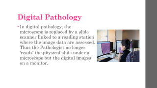
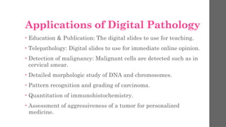


Digital pathology involves the digitization, sharing, and interpretation of pathology information, allowing pathologists to analyze high-resolution digital images instead of physical glass slides. It offers advantages such as quicker diagnoses, collaboration among experts, and reduced patient anxiety but also faces challenges like high costs and significant storage requirements. Key applications include education, telepathology, malignancy detection, and personalized medicine.


