Download to read offline
![NANOSECOND FIBER LASER MICRO-TEXTURING OF TITANIUM SURFACE FOR
BIOMEDICAL APPLICATIONS
M502
Habib Abou Saleh
#1
, Ehsan Toyserkani
*2
, Fathy Ismail
#3
#1*2#3
Department of Mechanical and Mechatronics Engineering, University of Waterloo
200 University Avenue West, Waterloo, N2L 3G1, Ontario, Canada
#1
habousal@uwaterloo.ca
*2
etoyserk@uwaterloo.ca
#3
fmismail@mecheng1.uwaterloo.ca
Abstract
This paper investigates the feasibility of generating
micro-self-assembled structures on pure titanium
using a nanosecond Ytterbium fiber laser. The effect
of process parameters, including laser frequency,
power, processing speed and spot size, on the
induction of the micro-self-assembled structures is
investigated. Scanning Electron Microscope (SEM)
and Profilometry analyses are carried out to
demonstrate the size, shape, and roughness of the
generated micro-structures. Analysis of the
experimental results suggests that the generation of
self-assembled structures with a desired roughness is
viable. It is also observed that the laser spot size can
potentially control the local surface roughness when
the other process parameters are fixed.
Keywords - Laser micro-texturing, Ytterbium fiber
laser, Micro-self-assembled structures.
Introduction
The understanding of surface modification of
materials with laser beams along with the
investigation of the underlying principles of laser
radiation interaction with the ablated surfaces are
essential to realize industrial and medical
applications of laser beams [1, 2]. Laser surface
modification of titanium and its alloys are of great
interest [1, 2, 3]. Titanium has been widely used in
biomedical applications due to its bio-stability,
biocompatibility, light weight, high mechanical
strength and long term durability [1, 2, 3]. Surface
topography has a great influence on the implant
performance during in vivo and in vitro studies [1, 3].
The micro structures generated at the surface of
titanium influence the biological processes at the
implant interface after the interaction with the
biological environment [1, 3]. To generate desired
microstructures on the surface of titanium, several
methods have been employed and studied such as
grit-blasting, chemical etching, titanium plasma
spray, electrochemical treatment as well as
combinations of these methods [1, 3]. These methods
generate microstructures with different irregular
patterns, with varying forms and diameters of
structural elements [4]. Osteoinductive qualities and
mechanical stability is determined by the degree of
contamination of the titanium surface. Considerable
amounts of contamination with numerous foreign
elements were found on the processed surfaces using
the above methods [4]. Such surfaces can lead to scar
tissue formation due to the development of random
bone cell orientations and an increase in cytotoxic
concentration elements at the implant surface which
will lead eventually to biological rejection of the
implant, and thus implant failure [1]. Recent studies
have shown that laser processing of implanted
surfaces are of a great importance since they provide
uniformly distributed surface topography with
uniform patterns and less surface contamination
when compared with other methods [3, 4].
Microstructures have been produced using long-pulse
lasers, including nanosecond Nd:YAG laser, copper
vapor laser, nanosecond excimer lasers, picosecond
Nd:YAG laser, and sub-picosecond excimer laser [2,
3, 5, 6, 7, 8, 9]. The laser patterned surfaces were
found suitable for cell adhesion, proliferation and
spreading, but none of the studies have dealt with
generating self-assembled micro-patterns using
nanosecond Ytterbium fiber laser, and its interaction
with Titanium surfaces.
In this paper, the feasibility of generating self-
assembled structures on pure titanium samples using
a nanosecond Ytterbium fiber laser is investigated.
Titanium samples were laser irradiated at different
laser process parameters which include laser
frequency, laser power, interaction time and spot
size. The process parameters that result in the local
surface roughness ranging from 0.9 to 2 µm (which is](https://image.slidesharecdn.com/c370ac04-d943-4a53-9993-9ca3c2a5545e-150303145706-conversion-gate01/85/ICALEO-2009-Habib-Abou-Saleh-ET-1-320.jpg)
![Figure 4. Profilometry Result of one specimen (# 4), at P = 17 Watts, F = 20 KHz, V = 2 mm/s, Dz = 400 µm.
Results and Discussion
The goal of this study was to investigate the
feasibility of producing microstructures with surface
roughness ranging from 1 µm to 2 µm which
enhances osteoblasts tissue integration. Laser
irradiated zones produced at a processing speed of 2
mm/s with laser power of 17 Watts, frequency of 20
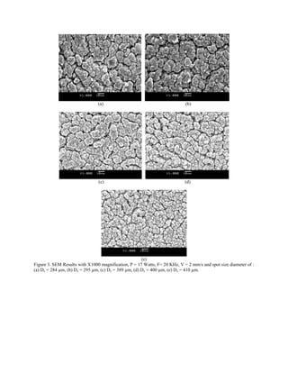
KHz, and spot size diameter of 284 µm, 295 µm, 389
µm, 400 µm, and 410 µm were selected for detailed
visual and surface metrological characterization using
SEM and profilometry tests. It should be noted that
numerous experiments were conducted; however, the
abovementioned parameters have resulted in the
generation of microstructures with the desired surface
roughness for the specific application. Table 1 lists
surface roughness parameters including the average
roughness, Rl and root mean square Rq, which are
calculated over the measured array, Furthermore, the
value of the peak-to-valley was measured and found
to be between 10 to 16 µm. The surface roughness
parameters were obtained by selecting a subregion of
dimension of 75 µm x 75 µm of the entire measured
array. Furthermore, the corresponding values for the
effective energy are listed in the table. The effective
energy (Ep) is calculated as:
Where, E is the laser pulse energy (J); F is laser
frequency (Hz); V is the laser processing speed (m/s);
and D is the spot size diameter (m).
Fig. 3 shows SEM images at different process
parameters. As seen, the size of the self-assembled
structures (grains) reduces when the spot size
increases. The profilometry result is shown in Fig. 4.
The figure also shows the extreme peak-to-valley
value in the measured array.
Table1. Specimens Surface Roughness Parameters
Specimens Spot Size Ep Rl Rq
1 284 µm 3.52x10⁷
1.68 µm 2.25µm
2 295 µm 3.38x10⁷
1.32 µm 1.72µm
3 389 µm 2.57x10⁷
1.33 µm 1.73µm
4 400 µm 2.50x10⁷
0.926µm 1.23µm
5 410 µm 2.44x10⁷
0.957µm 1.27µm
The generated surface parameters Rl and Rt can be
used to study the effect of surface roughness on the
cell integration and adhesion. It has been reported
that the osteoblasts tissue integration is favoured with
surface roughness ≤ 2 µm, and features depth
between 8 µm to 12 µm [1, 7, 10, 11, 12, 13, 14, 15,
16]. According to Ball et al. 2005 [16], osteoblasts
tissue integration is favoured with microstructures of
micron scale topography. In his study,](https://image.slidesharecdn.com/c370ac04-d943-4a53-9993-9ca3c2a5545e-150303145706-conversion-gate01/85/ICALEO-2009-Habib-Abou-Saleh-ET-4-320.jpg)
![osteointegration was noticed on titanium surfaces
with surface roughness ranging from 0.05 µm to 1.75
µm. According to our results, osteoblasts tissue
integration would occur with our generated
microstructures which have a local surface roughness
range approximately between 1 µm to 2 µm. As a
result, nanosecond Ytterbium fiber laser for titanium
micro-texturing is well suited for micron scale
topographical surface modifications to influence and
increase osteointegration of bone contacting devices.
Surface roughness was found to be inversely
proportional to the spot size diameter of the laser
beam. A decrease in the surface roughness and
surface grain size was noticed at the same time with
the increase in the spot size of the laser beam which
also coincided with decrease in the laser effective
energy. As a future work, we will analyse the role of
wide range of the process parameters on the top
viewed features size of micro self-assembled
structures.
Conclusion
Nanosecond Ytterbium fiber laser micro-texturing of
titanium specimens was investigated in this paper In
conclusion:
1- Nanosecond Ytterbium fiber laser has the
potential to be used for generating self-
assembled micro-structures.
2- The roughness ranging from 1 µm to 2 µm
was produced at a processing speed of 2
mm/s with 17 Watts laser power, frequency
of 20 KHz, and spot size diameter of
approximately 284 µm, 295 µm, 389 µm,
400 µm, and 410 µm.
3- A decrease in the surface roughness was
identified with an increase in the spot
diameter of the laser beam.
Acknowledgments
The authors would like to acknowledge technical
help of Negar Rasti and Hamidreza Alemohammad.
The authors also acknowledge the financial support
provided by NSERC.
References
[1] Mwenifumbo Steven, Li Mingwei, Chen Jianbo,
Beye Aboubaker, Soboyejo Wol´e, “Cell/surface
interactions on laser micro-textured titanium-
coatedsilicon surfaces”, J Mater Sci: Mater Med
(2007) 18:9–23.
[2] Trtica M.S., Radak B.B., Gakovic B.M.,
Milovanovic D.S., Batani D., Desai T., “Surface
modifications of Ti6Al4V by a picoseconds Nd:YAG
laser”, Laser and Particle Beams, 2009, 27, pp 85–90.
[3] Vorobyev A.Y., Guo Chunlei, “Femtosecond
laser structuring of titanium implants”, Applied
Surface Science 253, 2007, pp 7272-7280.
[4] Gaggl A., Schultes G., MuKller W.D., KaKrcher
H., “Scanning electron microscopical analysis of
laser-treated titanium implant surfaces*a comparative
study”, Biomaterials 21, 2000, pp: 1067 - 1073
[5] Tian Y.S, Chen C.Z, Li S.T. , Huo Q.H,
“Research progress on laser surface modification of
titanium alloys” . Appl. Surf. Sci. 2005, 242, pp 177–
184.
[6] Tritca M., Gakovic B. Batani D. Desai T., Panjan
P. Radak B. “ Surface modifications of a titanium
implant by a picosecond Nd:YAG laser operating at
1064 and 532 nm” , Appl. Surf. Sci. 2006, 253, pp
2551–2556.
[7] Mirhosseini N., Crouse P.L., Schmidth M.J.J., Li
L. Garrod D., “Laser surface micro-texturing of Ti–
6Al–4V substrates for improved cell integration”,
Appl. Surf. Sci. 2007, 253, pp 7738–7743
[8] Khosroshahi M.E, Magmoodi M. Tavakoli J. “
Characterization of Ti6Al4V implant surface treated
by Nd:YAG laser and emery paper for orthopaedic
applications”, Appl. Surf. Sci. 2007, 253, pp 8772–
8781.
[9] Zelinski A., Jazdzewska M., Narozniak-Luksza
A., Serbinski W., “Surface structure and properties of
Ti6A4V alloy laser melted at cryogenic conditions",
J. Achiev. Mat. Manufact. Engin. 2006, 18, pp 423–
426.
[10] Lloyd Robert, Abdolvand Amin, Schmidt Marc,
Crouse Philips, Whitehead David, Liu Zhu, and Li
Lin, “Laser Assisted Generation of Self-assembled
Microstructures on Stainless Steel” , Applied Physics
A: Materials Science & Processing, 2008, Volume
93, Number 1, pp 117-122.
[11] Bacakova L., Filova E., Rypacek F., Svorcik V.,
“Cell Adhesion on Artificial Materials for Tissue
Engineering”, Physiological Research, 2004, ISSN
0862-8408, Volume 53, pp 35-45.](https://image.slidesharecdn.com/c370ac04-d943-4a53-9993-9ca3c2a5545e-150303145706-conversion-gate01/85/ICALEO-2009-Habib-Abou-Saleh-ET-5-320.jpg)
![[12] Gerald M. Edelman, “Cell Adhesion and the
Molecular Process of Morphogenesis”, Ann. Rev.
Biochem. 1985.54:135-169.
[13] L. Marcotte a,b, M. Tabrizian, “General review:
Sensing surfaces: Challenges in studying the cell
adhesion process and the cell adhesion forces on
biomaterials”, ScienceDirect, 2008, ITBM-RBM 29,
pp 77–88.
[14] Kurella Anil, Dahotre Narendra B., “Review
paper: Surface Modification for Bioimplants: The
Role of Laser Surface Engineering”, J Biomater Appl
2005; 20; 5, pp 4 – 50.
[15] Lincks J, Boyan BD, Blanchard CR, et al.
Response of MG63 osteoblast-like cells to titanium
and titanium alloy is dependent on surface roughness
and composition. Biomaterials 1998; 19(23), pp:
2219 - 2232
[16] Ball Michael, Grant David M., Lo Wei-Jen,
Scotchford Colin A., “The effect of different surface
morphology and roughness on osteoblast-like cells”,
Wiley InterScience, 2005, pp 637 – 647.](https://image.slidesharecdn.com/c370ac04-d943-4a53-9993-9ca3c2a5545e-150303145706-conversion-gate01/85/ICALEO-2009-Habib-Abou-Saleh-ET-6-320.jpg)

This document summarizes research on using a nanosecond ytterbium fiber laser to micro-texture titanium surfaces for biomedical applications. The researchers investigated how laser process parameters like power, frequency, speed and spot size affect the generation of micro-scale self-assembled structures on titanium surfaces. Scanning electron microscopy and profilometry analysis showed that surface roughness between 1-2 microns, suitable for osteoblast tissue integration, could be achieved by adjusting the laser parameters. In particular, a spot size of 284-410 microns at 17 watts power, 20 kHz frequency and 2 mm/s speed produced the desired roughness. The study demonstrated the potential of this laser for controlled micro-texturing of titanium implants.


![Microstructurally_Modified_TiAl6V[1]](https://cdn.slidesharecdn.com/ss_thumbnails/4dabef30-550b-45b5-9916-714e6bd1987b-150205034829-conversion-gate01-thumbnail.jpg?width=640&height=640&fit=bounds)


