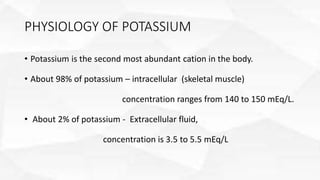
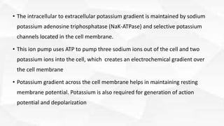
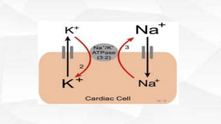
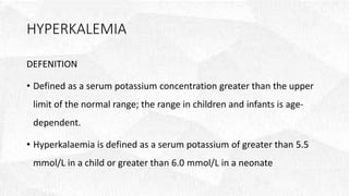
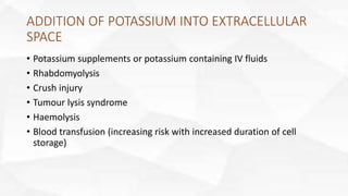
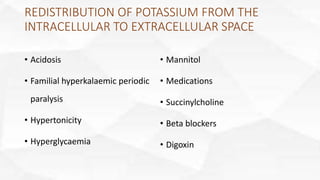
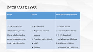
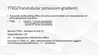
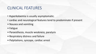
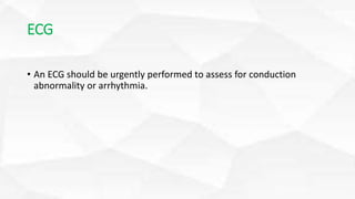
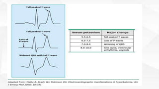
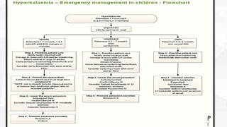
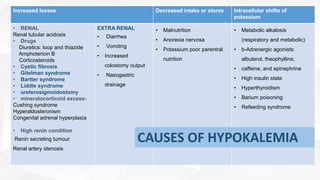
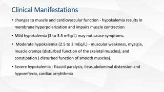
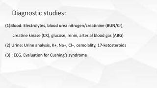
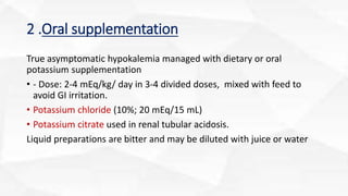
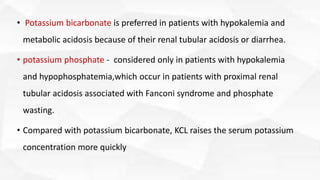
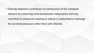
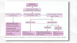
HYPOKALEMIA
AND
HYPERKALEMIA
PRESENTER : DR VIJITHA A S
PHYSIOLOGY OF POTASSIUM
• Potassium is the second most abundant cation in the body.
• About 98% of potassium – intracellular (skeletal muscle)
concentration ranges from 140 to 150 mEq/L.
• About 2% of potassium - Extracellular fluid,
concentration is 3.5 to 5.5 mEq/L
• The intracellular to extracellular potassium gradient is maintained by sodium
potassium adenosine triphosphatase (NaK-ATPase) and selective potassium
channels located in the cell membrane.
• This ion pump uses ATP to pump three sodium ions out of the cell and two
potassium ions into the cell, which creates an electrochemical gradient over
the cell membrane
• Potassium gradient across the cell membrane helps in maintaining resting
membrane potential. Potassium is also required for generation of action
potential and depolarization
Potassium homeostasis
• Daily requirement of potassium is about 1–2 mEq/kg.
• 90% is absorbed in small intestine and eliminated through kidney
5–10% is excreted in stool and less than 5% in sweat.
• Nearly 85–90% of the potassium is reabsorbed up to distal tubules and only 10–15% reaches cortical
and outer medullary collecting duct, which is the principle site of regulation of potassium excretion.
• Aldosterone and insulin play important roles in potassium homeostasis
• Aldosterone- high potassium level stimulate renal secretion via aldosterone mediated enhancement
of distal expression of secretory potassium channels(ROMK)
• Insulin and β-adrenergic receptor agonist activate Na-K-ATPase and
promote potassium uptake by the cells.
• During osmotic diuresis, the kidney reabsorbs less potassium, and
thus hypokalemia occur. eg: diabetic ketoacidosis
• Major factors influence transcellular shift are - metabolic acidosis,
alkalosis, insulin, glucagon, catecholamine, hyperosmolality, failure of
Na-K-ATPase pump, and cellular injury.
• Acidosis promotes extracellular movement of potassium
• Alkalosis leads to potassium uptake by the cells.
HYPERKALEMIA
DEFENITION
• Defined as a serum potassium concentration greater than the upper
limit of the normal range; the range in children and infants is age-
dependent.
• Hyperkalaemia is defined as a serum potassium of greater than 5.5
mmol/L in a child or greater than 6.0 mmol/L in a neonate
CLASSIFICATION BY SEVERITY:
Mild: 5.5 – 6 mmol/L
Moderate: 6.1 – 7 mmol/L
Severe: greater than 7 mmol/L or greater than 6.5 mmol/L with ECG changes
Pseudohyperkalaemia : Falsely elevated serum potassium level greater than 5.5mmol/L
CAUSES HYPERKALEMIA
1. Pseudohyperkalaemia (Factitious hyperkalaemia)
2. Impaired potassium excretion
3. Redistribution of potassium from the intracellular to extracellular space
4. Addition of potassium into extracellular space
PSEUDOHYPERKALAEMIA (FACTITIOUS
HYPERKALAEMIA)
• Collection technique (haemolysis) - most common cause of
hyperkalemia, from hemolysis of the blood sample when the sample
is obtained from a heel stick or a small bore intravenous line or fist
clenching, either of which causes local potassium release from
muscle.
• Significant thrombocytosis (platelets > 1,000 x 109/L)
• Significant leucocytosis (WCC >70 x 109/L)
ADDITION OF POTASSIUM INTO EXTRACELLULAR
SPACE
• Potassium supplements or potassium containing IV fluids
• Rhabdomyolysis
• Crush injury
• Tumour lysis syndrome
• Haemolysis
• Blood transfusion (increasing risk with increased duration of cell
storage)
REDISTRIBUTION OF POTASSIUM FROM THE
INTRACELLULAR TO EXTRACELLULAR SPACE
• Acidosis
• Familial hyperkalaemic periodic
paralysis
• Hypertonicity
• Hyperglycaemia
• Mannitol
• Medications
• Succinylcholine
• Beta blockers
• Digoxin
DECREASED LOSS
RENAL DRUGS Mineralocorticoid deficiency
Acute renal failure
Chronic kidney disease
Renal tubular disorders-
pseudo hypoaldosteronism
Urinary tract obstruction
 ACE inhibitors
 Angiotensin receptor
blockers
 Potassium sparing diuretics
 NSAID
 Heparin
 Addison disease
 21 hydroxylase deficiency
 3 β hydroxysteroid
dehydrogenase deficiency
 Calcineurin inhibitors
(tacrolimus and cyclosporin)
TTKG(Transtubular potassium gradient)
• Accounts confounding effect of urine concentration on interpretation of
urine potassium excretion
• TTKG = urine K * serum osmolality
serum K*urine osmolality
Normal TTKG – between 6 and 12
Hyperkalemia >10
<5 - in appropriate aldosterone effect
Increase in TTKG >7- after administration of fludrocortisone suggest-
mineralocorticoid deficiency ; <7 - resistance
CLINICAL FEATURES
• Hyperkalaemia is usually asymptomatic.
• cardiac and neurological features tend to predominate if present
• Nausea and vomiting
• Fatigue
• Paraesthesia, muscle weakness, paralysis
• Respiratory distress and failure
• Palpitations, syncope, cardiac arrest
ECG
• An ECG should be urgently performed to assess for conduction
abnormality or arrhythmia.
Management
• Clinical evaluation of children with hyperkalemia
1. detailed history and examination - hydration, hypertension,
hyperpyrexia, hyperglycemia, hypoxia, acidosis, and cardiac and
renal status.
2. ECG should be done for changes in cardiac rhythm
TREATMENT
• Depends on the serum potassium level, as well as the presence or
absence of symptoms and ECG changes
1. Identify and remove all sources of oral or parenteral potassium
intake (oral potassium supplements and intravenous maintenance
fluids or parenteral nutrition must be considered)
2. Evaluate drugs that can increase the serum potassium level (e.g.,
potassium-sparing diuretics, angiotensin-converting enzyme
inhibitors, and nonsteroidal antiinflammatory agents).
The goals of hyperkalemia treatment
• Antagonize the cardiac effects of potassium reverse symptoms, and return the
serum potassium level to normal while avoid in overcorrection.
Three principle methods are used to treat
hyperkalemia
1. calcium is administered to counteract the effects of excess
potassium on the heart.
2. Medications used to shift potassium from extracellular to
intracellular fluid compartments.
3. Exchange resins, diuretics, or dialysis are used to remove potassium
from the body .
CALCIUM
• Calcium increases the cellular threshold potential, thereby restoring the
normal difference between the resting membrane potential and the
firing threshold, which is elevated abnormally in persons with
hyperkalemia
• Temporary to antagonize the effects of hyperkalemia on cardiac muscle
and will not remove potassium from the body
• For membrane stabilization
• calcium gluconate is used as 10% solution, 0.5–1 mL/kg (maximum 10 mL)
• 1:1 diluted with saline over 10 min under cardiac monitoring.
• If slowing of heart rate - slow the rate of infusion.
• Onset of action is within 1 min and lasts up to 20–60 min.
• If patient is taking digitalis, give infusion over 30 min.
• cardiac glycosides are synergistic with parenteral calcium salts and thus the
combination of digitalis and calcium may increase the risk of precipitating
hypokalemia-related arrhythmias
TREATMENT - SHIFTS POTASSIUM INTO
CELLS
I. Increasing the Serum pH of the Acidotic Patient
• If there is nonanion gap metabolic acidosis, 1–2 mEq/kg of sodium
bicarbonate may be given intravenously; onset occurs within 30 min
and lasts for 60 min.
II . Glucose Plus Insulin
• Insulin stimulates cellular uptake of glucose with potassium
• If the patient is hyperglycemic, only the administration of insulin is
recommended to treat the hyperkalemia
• Infants and young children - 2 mL/kg of 25% dextrose with 0.1 unit/kg
of regular insulin to be infused over 30 min.
• For older children - 50 mL of 50% dextrose with 10 units of regular
insulin to be infused over 30 min.
• If blood glucose level is more than 300 mg/dL, insulin may be given
alone.
• All cases should be monitored for hypoglycemia.
III. b-Adrenergic Agonists
• β-Adrenergic agonists stimulate the Na+,K+-ATPase, increasing
cellular uptake of potassium.
• Salbutamol, 2.5–5 mg in 3–4 mL of saline may be nebulized over 20
min and may be repeated if required.
• It has onset within 30 min and effect lasts for about 2 hours.
TREATMENT THAT REMOVES POTASSIUM
• Exchange Resins
• Sodium polystyrene sulfonate or Kayexalate mixed in sorbitol is a cation-exchange
resin that binds potassium in the gastrointestinal tract and eliminates it from the
body
• Each gram of resin will bind approximately 1 mEq of potassium and release 2 to 3
mEq of sodium.
• It should be given at a dose of 1 g/kg orally or per rectum (maximum dose:
15g/dose) and repeated every 1 to 2 hours until the serum potassium level is
lowered
• The onset of action of sodium polystyrene sulfonate administered orally is at least 2
hours, and the maximal effect may take 6 hours
• complications - hypernatremia and necrotizing enterocolitis
• Kayexalate use in neonates should be reserved for refractory cases
Diuretics
• If renal function is maintained,the administration of furosemide, a
loop diuretic, will produce an increase in the renal excretion of
potassium.
• The onset of action of parenteral furosemide is within 5 minutes; the
peak effect is observed within 30 minutes.
• The furosemide dose for children - 1 mg/kg/dose (maximum 40
mg/dose
• The amount of potassium excreted is unreliable and does not
correlate with the diuretic dose
Renal Replacement Therapy
• when conservative methods fail or for patients with life-threatening
hyperkalemia.
• Hemodialysis (or continuous venovenous hemofiltration in
hemodynamically unstable patients) is more effective than peritoneal
dialysis and is the preferred method when hyperkalemia is the result
of cell breakdown
• lower the K+ levels by 1.2–1.5 mEq/h. Peritoneal dialysis with
potassium-free fluid is also an effective alternative, if hemodialysis is
not possible
Prevention of Recurrence
• After hyperkalemia is treated, it is essential to determine the cause
and implement measures to prevent recurrence.
• In patients with renal dysfunction, management for sustained
hyperkalemia is to reduce the overall total dietary potassium intake,
which includes restriction in the use of salt substitutes because they
contain potassium chloride (KCl).
Monitoring
• Once initial interventions have been made, the serum potassium level
should be rechecked within 1 to 2 hours to ensure the effectiveness
of the correction.
HYPOKALEMIA
• occurs when a serum potassium concentration is < 3.5 mEq/L
• it can become life threatening when the serum potassium concentration falls
below 2.5 mEq/L
• Hypokalemia can result from intracellular shifts of potassium, increased losses of
potassium, or decreased ingestion or administration of potassium
• Main cause of hypokalemia in pediatric patients - excessive gastrointestinal losses
(diarrhea or vomiting)- volume depletion and metabolic alkalosis
• volume depletion –> secondary hyper aldosteronism-->enhances sodium
reabsorption and potassium secretion in cortical CT
• Metabolic alkalosis – increased potassium secretion due to decreased
availability of hydrogen ions for secretion in response to sodium
reabsorption
• serum potassium levels do not correlate with intracellular potassium
levels, hypokalemia does not reflect total body potassium stores
Increased losses Decreased intake or stores Intracellular shifts of
potassium
• RENAL
Renal tubular acidosis
• Drugs
Diuretics: loop and thiazide
Amphotericin B
Corticosteroids
• Cystic fibrosis
• Gitelman syndrome
• Bartter syndrome
• Liddle syndrome
• ureterosigmoidostomy
• mineralocorticoid excess-
Cushing syndrome
Hyperaldosteronism
Congenital adrenal hyperplasia
• High renin condition
Renin secreting tumour
Renal artery stenosis
EXTRA RENAL
• Diarrhea
• Vomiting
• Increased
colostomy output
• Nasogastric
drainage
• Malnutrition
• Anorexia nervosa
• Potassium poor parentral
nutrition
• Metabolic alkalosis
(respiratory and metabolic)
• b-Adrenergic agonists:
albuterol, theophylline,
• caffeine, and epinephrine
• High insulin state
• Hyperthyroidism
• Barium poisoning
• Refeeding syndrome
CAUSES OF HYPOKALEMIA
Clinical Manifestations
• changes to muscle and cardiovascular function - hypokalemia results in
membrane hyperpolarization and impairs muscle contraction
• Mild hypokalemia (3 to 3.5 mEq/L) may not cause symptoms.
• Moderate hypokalemia (2.5 to 3 mEq/L) - muscular weakness, myalgia,
muscle cramps (disturbed function of the skeletal muscles), and
constipation ( disturbed function of smooth muscles).
• Severe hypokalemia - flaccid paralysis, ileus,abdominal distension and
hyporeflexia, cardiac arryhthmia
• Chronic hypokalemia- associated with interstitial renal disease
Diagnostic studies:
(1)Blood: Electrolytes, blood urea nitrogen/creatinine (BUN/Cr),
creatine kinase (CK), glucose, renin, arterial blood gas (ABG)
(2) Urine: Urine analysis, K+, Na+, Cl−, osmolality, 17-ketosteroids
(3) : ECG, Evaluation for Cushing’s syndrome
ECG
• Early ECG changes include - ST segment depression, T wave flattening,
and the presence of U waves .
TREATMENT OF HYPOKALEMIA
• goals of therapy for hypokalemia include avoidance or resolution of
symptoms and return of the serum potassium Concentration to normal
1. Decreasing ongoing loss - discontinuation od diuretics,alpha 2 agonist
2.Replenishing potassium stores (oral/ IV potassium)
3. Disease specific therapy
Treatment of hypokalemia
1. IV supplementation –
• Indication: Symptomatic patients,
severe hypokalemia ; ( <2.5 mEq/L)
ECG abnormalities
• Potassium chloride (15%; 2 mEq/mL)
• 0.5-1 m.Eq/kg/ dose IV infusion over 1-2 hr.
• Infusion rate should not exceed 1 mEq/kg/hr; concentration of
potassium should not exceed 60 mEq/L (peripheral line) 'and 80
mEq/L (central line)
• As the plasma K+ concentration improves the rate of infusion should
be reduced to maintenance.
• Once the serum potassium level is stabilized, the oral route of
administration is preferable
• Children who respond poorly to potassium replacement should also be
checked and corrected for hypomagnesemia and hypophosphatemia.
2 .Oral supplementation
True asymptomatic hypokalemia managed with dietary or oral
potassium supplementation
• - Dose: 2-4 mEq/kg/ day in 3-4 divided doses, mixed with feed to
avoid GI irritation.
• Potassium chloride (10%; 20 mEq/15 mL)
• Potassium citrate used in renal tubular acidosis.
Liquid preparations are bitter and may be diluted with juice or water
• Potassium bicarbonate is preferred in patients with hypokalemia and
metabolic acidosis because of their renal tubular acidosis or diarrhea.
• potassium phosphate - considered only in patients with hypokalemia
and hypophosphatemia,which occur in patients with proximal renal
tubular acidosis associated with Fanconi syndrome and phosphate
wasting.
• Compared with potassium bicarbonate, KCL raises the serum potassium
concentration more quickly
• Chloride depletion contributes to maintenance of the metabolic
alkalosis by enhancing renal bicarbonate reabsorption and may
contribute to potassium wasting as sodium is reabsorbed in exchange
for secreted potassium rather than with chloride.
Monitoring
• The timing of a repeat serum potassium level depends on the severity of the
initial value, the patient’s symptoms, and the form of potassium
administered to the patient.
• In a symptomatic patient who receives an intravenous dose of KCl, the dose
should be repeated without measuring a serum value if the patient’s
symptoms persist. If the symptoms resolve, the serum potassium level can
be obtained 1 hour after completion of an intravenous dose
THANK YOU
References
• OP GHAI
• NELSON TEXT BOOK OF PAEDIATRICS
• PIYUSH GUPTA

Hyperkalemia&amp;hypokalemia by dr vijitha

  • 1.
  • 2.
    PHYSIOLOGY OF POTASSIUM •Potassium is the second most abundant cation in the body. • About 98% of potassium – intracellular (skeletal muscle) concentration ranges from 140 to 150 mEq/L. • About 2% of potassium - Extracellular fluid, concentration is 3.5 to 5.5 mEq/L
  • 3.
    • The intracellularto extracellular potassium gradient is maintained by sodium potassium adenosine triphosphatase (NaK-ATPase) and selective potassium channels located in the cell membrane. • This ion pump uses ATP to pump three sodium ions out of the cell and two potassium ions into the cell, which creates an electrochemical gradient over the cell membrane • Potassium gradient across the cell membrane helps in maintaining resting membrane potential. Potassium is also required for generation of action potential and depolarization
  • 5.
    Potassium homeostasis • Dailyrequirement of potassium is about 1–2 mEq/kg. • 90% is absorbed in small intestine and eliminated through kidney 5–10% is excreted in stool and less than 5% in sweat. • Nearly 85–90% of the potassium is reabsorbed up to distal tubules and only 10–15% reaches cortical and outer medullary collecting duct, which is the principle site of regulation of potassium excretion. • Aldosterone and insulin play important roles in potassium homeostasis • Aldosterone- high potassium level stimulate renal secretion via aldosterone mediated enhancement of distal expression of secretory potassium channels(ROMK)
  • 6.
    • Insulin andβ-adrenergic receptor agonist activate Na-K-ATPase and promote potassium uptake by the cells. • During osmotic diuresis, the kidney reabsorbs less potassium, and thus hypokalemia occur. eg: diabetic ketoacidosis • Major factors influence transcellular shift are - metabolic acidosis, alkalosis, insulin, glucagon, catecholamine, hyperosmolality, failure of Na-K-ATPase pump, and cellular injury.
  • 7.
    • Acidosis promotesextracellular movement of potassium • Alkalosis leads to potassium uptake by the cells.
  • 9.
    HYPERKALEMIA DEFENITION • Defined asa serum potassium concentration greater than the upper limit of the normal range; the range in children and infants is age- dependent. • Hyperkalaemia is defined as a serum potassium of greater than 5.5 mmol/L in a child or greater than 6.0 mmol/L in a neonate
  • 10.
    CLASSIFICATION BY SEVERITY: Mild:5.5 – 6 mmol/L Moderate: 6.1 – 7 mmol/L Severe: greater than 7 mmol/L or greater than 6.5 mmol/L with ECG changes Pseudohyperkalaemia : Falsely elevated serum potassium level greater than 5.5mmol/L
  • 12.
    CAUSES HYPERKALEMIA 1. Pseudohyperkalaemia(Factitious hyperkalaemia) 2. Impaired potassium excretion 3. Redistribution of potassium from the intracellular to extracellular space 4. Addition of potassium into extracellular space
  • 13.
    PSEUDOHYPERKALAEMIA (FACTITIOUS HYPERKALAEMIA) • Collectiontechnique (haemolysis) - most common cause of hyperkalemia, from hemolysis of the blood sample when the sample is obtained from a heel stick or a small bore intravenous line or fist clenching, either of which causes local potassium release from muscle. • Significant thrombocytosis (platelets > 1,000 x 109/L) • Significant leucocytosis (WCC >70 x 109/L)
  • 14.
    ADDITION OF POTASSIUMINTO EXTRACELLULAR SPACE • Potassium supplements or potassium containing IV fluids • Rhabdomyolysis • Crush injury • Tumour lysis syndrome • Haemolysis • Blood transfusion (increasing risk with increased duration of cell storage)
  • 15.
    REDISTRIBUTION OF POTASSIUMFROM THE INTRACELLULAR TO EXTRACELLULAR SPACE • Acidosis • Familial hyperkalaemic periodic paralysis • Hypertonicity • Hyperglycaemia • Mannitol • Medications • Succinylcholine • Beta blockers • Digoxin
  • 16.
    DECREASED LOSS RENAL DRUGSMineralocorticoid deficiency Acute renal failure Chronic kidney disease Renal tubular disorders- pseudo hypoaldosteronism Urinary tract obstruction  ACE inhibitors  Angiotensin receptor blockers  Potassium sparing diuretics  NSAID  Heparin  Addison disease  21 hydroxylase deficiency  3 β hydroxysteroid dehydrogenase deficiency  Calcineurin inhibitors (tacrolimus and cyclosporin)
  • 17.
    TTKG(Transtubular potassium gradient) •Accounts confounding effect of urine concentration on interpretation of urine potassium excretion • TTKG = urine K * serum osmolality serum K*urine osmolality Normal TTKG – between 6 and 12 Hyperkalemia >10 <5 - in appropriate aldosterone effect Increase in TTKG >7- after administration of fludrocortisone suggest- mineralocorticoid deficiency ; <7 - resistance
  • 18.
    CLINICAL FEATURES • Hyperkalaemiais usually asymptomatic. • cardiac and neurological features tend to predominate if present • Nausea and vomiting • Fatigue • Paraesthesia, muscle weakness, paralysis • Respiratory distress and failure • Palpitations, syncope, cardiac arrest
  • 19.
    ECG • An ECGshould be urgently performed to assess for conduction abnormality or arrhythmia.
  • 22.
    Management • Clinical evaluationof children with hyperkalemia 1. detailed history and examination - hydration, hypertension, hyperpyrexia, hyperglycemia, hypoxia, acidosis, and cardiac and renal status. 2. ECG should be done for changes in cardiac rhythm
  • 23.
    TREATMENT • Depends onthe serum potassium level, as well as the presence or absence of symptoms and ECG changes 1. Identify and remove all sources of oral or parenteral potassium intake (oral potassium supplements and intravenous maintenance fluids or parenteral nutrition must be considered) 2. Evaluate drugs that can increase the serum potassium level (e.g., potassium-sparing diuretics, angiotensin-converting enzyme inhibitors, and nonsteroidal antiinflammatory agents).
  • 24.
    The goals ofhyperkalemia treatment • Antagonize the cardiac effects of potassium reverse symptoms, and return the serum potassium level to normal while avoid in overcorrection.
  • 25.
    Three principle methodsare used to treat hyperkalemia 1. calcium is administered to counteract the effects of excess potassium on the heart. 2. Medications used to shift potassium from extracellular to intracellular fluid compartments. 3. Exchange resins, diuretics, or dialysis are used to remove potassium from the body .
  • 26.
    CALCIUM • Calcium increasesthe cellular threshold potential, thereby restoring the normal difference between the resting membrane potential and the firing threshold, which is elevated abnormally in persons with hyperkalemia • Temporary to antagonize the effects of hyperkalemia on cardiac muscle and will not remove potassium from the body
  • 27.
    • For membranestabilization • calcium gluconate is used as 10% solution, 0.5–1 mL/kg (maximum 10 mL) • 1:1 diluted with saline over 10 min under cardiac monitoring. • If slowing of heart rate - slow the rate of infusion. • Onset of action is within 1 min and lasts up to 20–60 min. • If patient is taking digitalis, give infusion over 30 min. • cardiac glycosides are synergistic with parenteral calcium salts and thus the combination of digitalis and calcium may increase the risk of precipitating hypokalemia-related arrhythmias
  • 28.
    TREATMENT - SHIFTSPOTASSIUM INTO CELLS I. Increasing the Serum pH of the Acidotic Patient • If there is nonanion gap metabolic acidosis, 1–2 mEq/kg of sodium bicarbonate may be given intravenously; onset occurs within 30 min and lasts for 60 min.
  • 29.
    II . GlucosePlus Insulin • Insulin stimulates cellular uptake of glucose with potassium • If the patient is hyperglycemic, only the administration of insulin is recommended to treat the hyperkalemia • Infants and young children - 2 mL/kg of 25% dextrose with 0.1 unit/kg of regular insulin to be infused over 30 min. • For older children - 50 mL of 50% dextrose with 10 units of regular insulin to be infused over 30 min. • If blood glucose level is more than 300 mg/dL, insulin may be given alone. • All cases should be monitored for hypoglycemia.
  • 30.
    III. b-Adrenergic Agonists •β-Adrenergic agonists stimulate the Na+,K+-ATPase, increasing cellular uptake of potassium. • Salbutamol, 2.5–5 mg in 3–4 mL of saline may be nebulized over 20 min and may be repeated if required. • It has onset within 30 min and effect lasts for about 2 hours.
  • 31.
    TREATMENT THAT REMOVESPOTASSIUM • Exchange Resins • Sodium polystyrene sulfonate or Kayexalate mixed in sorbitol is a cation-exchange resin that binds potassium in the gastrointestinal tract and eliminates it from the body • Each gram of resin will bind approximately 1 mEq of potassium and release 2 to 3 mEq of sodium. • It should be given at a dose of 1 g/kg orally or per rectum (maximum dose: 15g/dose) and repeated every 1 to 2 hours until the serum potassium level is lowered • The onset of action of sodium polystyrene sulfonate administered orally is at least 2 hours, and the maximal effect may take 6 hours • complications - hypernatremia and necrotizing enterocolitis • Kayexalate use in neonates should be reserved for refractory cases
  • 32.
    Diuretics • If renalfunction is maintained,the administration of furosemide, a loop diuretic, will produce an increase in the renal excretion of potassium. • The onset of action of parenteral furosemide is within 5 minutes; the peak effect is observed within 30 minutes. • The furosemide dose for children - 1 mg/kg/dose (maximum 40 mg/dose • The amount of potassium excreted is unreliable and does not correlate with the diuretic dose
  • 33.
    Renal Replacement Therapy •when conservative methods fail or for patients with life-threatening hyperkalemia. • Hemodialysis (or continuous venovenous hemofiltration in hemodynamically unstable patients) is more effective than peritoneal dialysis and is the preferred method when hyperkalemia is the result of cell breakdown • lower the K+ levels by 1.2–1.5 mEq/h. Peritoneal dialysis with potassium-free fluid is also an effective alternative, if hemodialysis is not possible
  • 35.
    Prevention of Recurrence •After hyperkalemia is treated, it is essential to determine the cause and implement measures to prevent recurrence. • In patients with renal dysfunction, management for sustained hyperkalemia is to reduce the overall total dietary potassium intake, which includes restriction in the use of salt substitutes because they contain potassium chloride (KCl).
  • 36.
    Monitoring • Once initialinterventions have been made, the serum potassium level should be rechecked within 1 to 2 hours to ensure the effectiveness of the correction.
  • 37.
    HYPOKALEMIA • occurs whena serum potassium concentration is < 3.5 mEq/L • it can become life threatening when the serum potassium concentration falls below 2.5 mEq/L • Hypokalemia can result from intracellular shifts of potassium, increased losses of potassium, or decreased ingestion or administration of potassium • Main cause of hypokalemia in pediatric patients - excessive gastrointestinal losses (diarrhea or vomiting)- volume depletion and metabolic alkalosis • volume depletion –> secondary hyper aldosteronism-->enhances sodium reabsorption and potassium secretion in cortical CT
  • 38.
    • Metabolic alkalosis– increased potassium secretion due to decreased availability of hydrogen ions for secretion in response to sodium reabsorption • serum potassium levels do not correlate with intracellular potassium levels, hypokalemia does not reflect total body potassium stores
  • 39.
    Increased losses Decreasedintake or stores Intracellular shifts of potassium • RENAL Renal tubular acidosis • Drugs Diuretics: loop and thiazide Amphotericin B Corticosteroids • Cystic fibrosis • Gitelman syndrome • Bartter syndrome • Liddle syndrome • ureterosigmoidostomy • mineralocorticoid excess- Cushing syndrome Hyperaldosteronism Congenital adrenal hyperplasia • High renin condition Renin secreting tumour Renal artery stenosis EXTRA RENAL • Diarrhea • Vomiting • Increased colostomy output • Nasogastric drainage • Malnutrition • Anorexia nervosa • Potassium poor parentral nutrition • Metabolic alkalosis (respiratory and metabolic) • b-Adrenergic agonists: albuterol, theophylline, • caffeine, and epinephrine • High insulin state • Hyperthyroidism • Barium poisoning • Refeeding syndrome CAUSES OF HYPOKALEMIA
  • 40.
    Clinical Manifestations • changesto muscle and cardiovascular function - hypokalemia results in membrane hyperpolarization and impairs muscle contraction • Mild hypokalemia (3 to 3.5 mEq/L) may not cause symptoms. • Moderate hypokalemia (2.5 to 3 mEq/L) - muscular weakness, myalgia, muscle cramps (disturbed function of the skeletal muscles), and constipation ( disturbed function of smooth muscles). • Severe hypokalemia - flaccid paralysis, ileus,abdominal distension and hyporeflexia, cardiac arryhthmia
  • 41.
    • Chronic hypokalemia-associated with interstitial renal disease
  • 42.
    Diagnostic studies: (1)Blood: Electrolytes,blood urea nitrogen/creatinine (BUN/Cr), creatine kinase (CK), glucose, renin, arterial blood gas (ABG) (2) Urine: Urine analysis, K+, Na+, Cl−, osmolality, 17-ketosteroids (3) : ECG, Evaluation for Cushing’s syndrome
  • 43.
    ECG • Early ECGchanges include - ST segment depression, T wave flattening, and the presence of U waves .
  • 44.
    TREATMENT OF HYPOKALEMIA •goals of therapy for hypokalemia include avoidance or resolution of symptoms and return of the serum potassium Concentration to normal 1. Decreasing ongoing loss - discontinuation od diuretics,alpha 2 agonist 2.Replenishing potassium stores (oral/ IV potassium) 3. Disease specific therapy
  • 45.
    Treatment of hypokalemia 1.IV supplementation – • Indication: Symptomatic patients, severe hypokalemia ; ( <2.5 mEq/L) ECG abnormalities • Potassium chloride (15%; 2 mEq/mL) • 0.5-1 m.Eq/kg/ dose IV infusion over 1-2 hr. • Infusion rate should not exceed 1 mEq/kg/hr; concentration of potassium should not exceed 60 mEq/L (peripheral line) 'and 80 mEq/L (central line)
  • 46.
    • As theplasma K+ concentration improves the rate of infusion should be reduced to maintenance. • Once the serum potassium level is stabilized, the oral route of administration is preferable • Children who respond poorly to potassium replacement should also be checked and corrected for hypomagnesemia and hypophosphatemia.
  • 47.
    2 .Oral supplementation Trueasymptomatic hypokalemia managed with dietary or oral potassium supplementation • - Dose: 2-4 mEq/kg/ day in 3-4 divided doses, mixed with feed to avoid GI irritation. • Potassium chloride (10%; 20 mEq/15 mL) • Potassium citrate used in renal tubular acidosis. Liquid preparations are bitter and may be diluted with juice or water
  • 48.
    • Potassium bicarbonateis preferred in patients with hypokalemia and metabolic acidosis because of their renal tubular acidosis or diarrhea. • potassium phosphate - considered only in patients with hypokalemia and hypophosphatemia,which occur in patients with proximal renal tubular acidosis associated with Fanconi syndrome and phosphate wasting. • Compared with potassium bicarbonate, KCL raises the serum potassium concentration more quickly
  • 49.
    • Chloride depletioncontributes to maintenance of the metabolic alkalosis by enhancing renal bicarbonate reabsorption and may contribute to potassium wasting as sodium is reabsorbed in exchange for secreted potassium rather than with chloride.
  • 51.
    Monitoring • The timingof a repeat serum potassium level depends on the severity of the initial value, the patient’s symptoms, and the form of potassium administered to the patient. • In a symptomatic patient who receives an intravenous dose of KCl, the dose should be repeated without measuring a serum value if the patient’s symptoms persist. If the symptoms resolve, the serum potassium level can be obtained 1 hour after completion of an intravenous dose
  • 52.
  • 53.
    References • OP GHAI •NELSON TEXT BOOK OF PAEDIATRICS • PIYUSH GUPTA