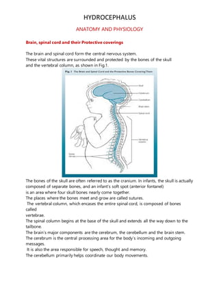
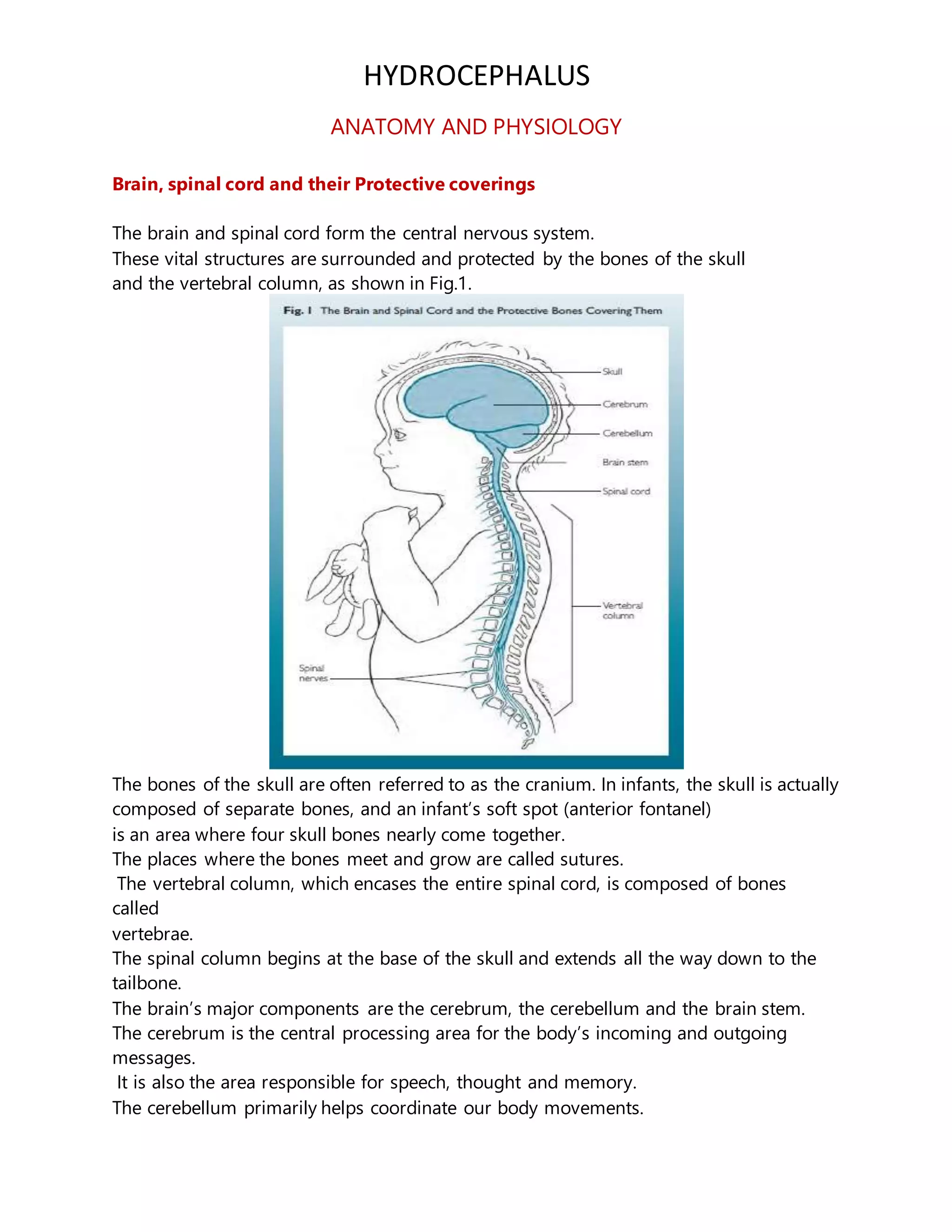
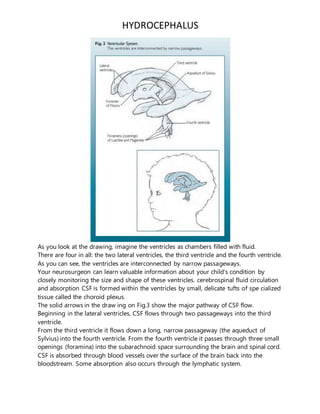
HYDROCEPHALUS
ANATOMY AND PHYSIOLOGY
Brain, spinal cord and their Protective coverings
The brain and spinal cord form the central nervous system.
These vital structures are surrounded and protected by the bones of the skull
and the vertebral column, as shown in Fig.1.
The bones of the skull are often referred to as the cranium. In infants, the skull is actually
composed of separate bones, and an infant’s soft spot (anterior fontanel)
is an area where four skull bones nearly come together.
The places where the bones meet and grow are called sutures.
The vertebral column, which encases the entire spinal cord, is composed of bones
called
vertebrae.
The spinal column begins at the base of the skull and extends all the way down to the
tailbone.
The brain’s major components are the cerebrum, the cerebellum and the brain stem.
The cerebrum is the central processing area for the body’s incoming and outgoing
messages.
It is also the area responsible for speech, thought and memory.
The cerebellum primarily helps coordinate our body movements.
HYDROCEPHALUS
The brain stem controls basic functions like heart rate, breathing and blood pressure.
The spinal cord extends from the brain stem, through a very large opening (the foramen
magnum) in the base of the skull, and down the spine.
At the level of each vertebra in the spine, nerve fibers arise from the spinal
cord and emerge through openings between the vertebrae.
These are the spinal nerves, which carry messages to and from various regions
of our bodies.
Lying between the brain and skull are three other pro tective coverings.
These are the membranes (meninges), which completely surround the brain and spinal
cord. An important fluid—the cerebrospinal fluid (CSF)—flows in a space between these
membranes that is called the subarachnoid space. CSF is essentially salt water, and it is
in constant circulation and serves several important functions. The brain floats in CSF.
Ventricles
CSF is produced within the cavities of the brain that are called ventricles. Fig. 2 is a
drawing of the ventricles.
HYDROCEPHALUS
As you look at the drawing, imagine the ventricles as chambers filled with fluid.
There are four in all: the two lateral ventricles, the third ventricle and the fourth ventricle.
As you can see, the ventricles are interconnected by narrow passageways.
Your neurosurgeon can learn valuable information about your child’s condition by
closely monitoring the size and shape of these ventricles. cerebrospinal fluid circulation
and absorption CSF is formed within the ventricles by small, delicate tufts of spe cialized
tissue called the choroid plexus.
The solid arrows in the draw ing on Fig.3 show the major pathway of CSF flow.
Beginning in the lateral ventricles, CSF flows through two passageways into the third
ventricle.
From the third ventricle it flows down a long, narrow passageway (the aqueduct of
Sylvius) into the fourth ventricle. From the fourth ventricle it passes through three small
openings (foramina) into the subarachnoid space surrounding the brain and spinal cord.
CSF is absorbed through blood vessels over the surface of the brain back into the
bloodstream. Some absorption also occurs through the lymphatic system.
HYDROCEPHALUS
Once in the bloodstream, it is carried away and filtered by our kidneys and liver in the
same way as are our other body fluids.
The ventricular system is the major pathway for the flow of CSF.
CSF also flows directly from the ventricles into the brain tissue sur rounding them.
This is shown by the broken arrows.
Here the CSF passes through the spaces between the cells to where it eventually enters
the subarachnoid space.
It is believed that the brain tissue does not absorb any CSF, but simply provides another
pathway for the fluid moving to the subarachnoid space.
Some small amounts of CSF are also absorbed into lymphatic channels along the
membranes covering the nerves (nerve sheaths) as they leave the brain stem and spinal
cord.
Our bodies produce approximately a pint (500 ml) of CSF daily, continuously replacing
CSF as it is absorbed.
Under normal conditions there is a delicate balance between the amount of CSF that is
produced and the rate at which it is absorbed.
Hydrocephalus occurs when this balance is disrupted.
Although there are many factors that can disrupt this balance, the most common is a
blockage, or obstruc tion, somewhere along the circulatory pathway of CSF.
The obstruction may develop from a variety of causes, such as brain tumors, cysts,
scarring and infection. Specific causes will be discussed more fully in a later section.
Because CSF is produced continuously, when it is blocked it will begin to accumulate
upstream from the site of the obstruction, much like a river swells behind a dam.
Eventually, as the amount of fluid accumulates, it causes the ventricles to enlarge and
pressure to increase inside the head.This condition is known as hydrocephalus.
Obstruction of the CSF pathway often occurs within the ventricles.
Although it can occur anywhere in the ventricular system, the site of blockage usually
lies either within the narrow passageways connecting the ventricles or where the CSF
exits the fourth ventricle into the sub arachnoid space. For example, because of its long,
narrow structure, the aqueduct of Sylvius is especially vulnerable to becoming narrowed
or obstructed, so that it blocks the flow of CSF.
Likewise, when the small openings of the fourth ventricle fail to develop, or develop
improperly, they also may obstruct the flow of CSF.
Hydrocephalus of this kind is called noncommunicating hydrocephalus because the
ventricles no longer provide free passage of CSF through them into the subarachnoid
space.
In some cases of hydrocephalus, CSF flows unrestricted through the ventricles, but once
it reaches the subarachnoid space its flow is impeded as it passes over the surfaces of
the brain.
HYDROCEPHALUS
In other cases, the absorptive sites (arachnoid villi) are blocked. Because the ventricles
remain open and communicate with each other, this type of hydro - cephalus is called
communicating hydrocephalus.
Normal physiologyof CSF (production and absoption)
The CSF is secreted by the choroid plexus within the ventricles by ultrafiltration and
active secretion.
It passes from the lateral ventricles to the third and fourth ventricles and exits from
foramen of Luschka and Magendie into the basal cisterns and then the cerebral and
spinal subarachnoid spaces where it is absorbed via the arachnoid villi (granulations)
into the venous channels and sinuses.
About 20 ml of CSF is secreted in an hour and its turnover is 3 or 4 times in a day.
Our bodies produce approximately a pint (500 ml) of CSF daily, continuously
replacing CSF as it is absorbed.
Under normal conditions there is a delicate balance between the amount of CSF that is
pro duced and the rate at which it is absorbed. Hydrocephalus occurs when this balance
is disrupted.
Definition
Hydrocephalus is an abnormal accumulation of fluid—cerebro spinal
fluid, or CSF—within cavities called ventricles inside the brain.
Etiology
Hydrocephalus results from an imbalance between production and absorption of
cerebrospinal fluid.
It may be communicating or noncommunicating.
Communicatinghydrocephalus.
There is no blockage in the CSF pathway but reabsorption may be affected. Excess CSF
may be produced in papilloma of choroid plexus.
Obstructive or noncommunicatinghydrocephalus.
The block is at any level in the ventricular system, commonly at the level of aqueduct or
foramina of Luschka and Magendie (Fig. 18.10).
HYDROCEPHALUS
Fig. 18.10: Arnold-Chiari II malformation: MRI T1W sagittal view
showing obstructive hydrocephalus stretched brainstem and tonsillar
herniation (patient also had a meningomyelocele) (Courtesy: Dr Atin
Kumar, Deptt. of Radiodiagnosis, AIIMS, New Delhi)
In obstructive hydrocephalus, the ventricles are dilated above the block. In cerebral
atrophy, ventricles are dilated but pressure is not raised (hydrocephalus ex vacuo).
Presence of periventricular ooze on CT or MR imaging helps to identify the former.
Hydrocephalus may be congenital or acquired (Table 18.8).
Congenital hydrocephalus
starts in fetal life and may manifest or even develop subsequently.
The large head size at birth causes difficulty in delivery of the head during labor.
There may be associated congenital malformations.
Table 18.8: Causes of hydrocephalus
Congenital hydrocephalus
In trauterine infec tions: Rubella, cytomegalovirus, toxoplasmosis,
intracranial bleeds, intraventricular hemorrhage
Congenital malformations: Aqueduct stenosis, Dandy-Walker
HYDROCEPHALUS
syndrome (posterior fossa cyst continuous with fourth
ventricle), Arnold-Chiari syndrome (portions of cerebellum
and brainstem herniating into cervical spinal canal, blocking
the flow of CSF to the posterior fossa)
Midline tumors obstructing CSF flow
Acquired hydrocephalus
Tuberculosis, chronic and pyogenic meningitis
Post-intraventricular hemorrhage
Pos terior fossa tumors: Medulloblastoma, astrocytoma,
ependymoma
Arteriovenous malformation, in tracranial hemorrhage,
ruptured aneurysm
Hydrocephalus ex vacuo
Pathology
Ventricles are dilated, at times unevenly.
Ependymal lining of ventricles is disrupted resulting in periventricular ooze and hence
periventricular white matter is compressed. Cortex is generally preserved until late but
cortical atrophy may occur. The process may be reversible if the treatment is initiated
early.
Clinicalfeatures
Hydrocephalus may manifest with enlarging head size, delayed closure of fontanel and
sutures.
Associated symptoms include headache, nausea, vomiting, personality and behavior
disturbances such as irritability, head banging, apathy and drowsiness.
Papilledema, pyramidal tract signs and cranial nerve palsies may occur.
Skull contour becomes abnormal and forehead is prominent.
Scalp veins become prominent and dilated. A sunset sign is seen in the eyes, i.e. sclera
above the cornea becomes visible. Upward gaze is impaired.Limbs become spastic
because of stretching of cortical fibers.
Distortion of the brainstem may lead to bradycardia, systemic hypertension and altered
respiration rate.
HYDROCEPHALUS
Diagnosis
Accurate serial recording of the head circumference is essential for early diagnosis of
hydrocephalus and should be supported by serial USG.
An increase in the head circumference in the first 3 months of life > 1 cm every fortnight
should arouse suspicion of hydrocephalus.
Brain grows very rapidly in the first few weeks of life and therefore sagittal and coronal
sutures may be separated up to 0.5 cm.
This physiological separation disappears after the first fortnight of life.
Persistent widening of squamoparietal sutures is not physiological and should arouse
suspicion of hydrocephalus. Cranial ultrasound and computed tomography help to
evaluate serial ventricular size while the latter gives information about cortical mantle,
periventricular ooze and etiology of hydrocephalus.
MRI may be necessary to determine the site of obstruction and in congenital
hydrocephalus to identify associated malformations.
Arnold-Chiari malformation has downward displacement of cerebellum and medulla,
obstruction of CSF pathway or migration defects.
Dandy-Walker malformation reveals a cystic malformation, atresia of outlet foramina or
any brain malformations.
Differential diagnosis
Megalencephaly refers to the increase in volume of brain parenchyma.
There are no signs of increased intracranial pressure.
The ventricles are neither large, nor under increased pressure.
Causes include Hurler syndrome, metachromatic leukodystrophy and Tay-Sachs
disease. Chronic subdural hematoma causes large head, mostly located in the parietal
region without prominent scalp veins or sunset sign.
Large head size is also observed in hydranencephaly, rickets, achondroplasia, hemolytic
anemia and familial macrocephaly.
Treatment
Management includes making a precise etiological diagnosis and identification of
associated malformations, clinical course and severity of hydrocephalus.
If hydrocephalus is arrested spontaneously, surgical intervention may not be necessary.
Medical management should be instituted if surgery is not indicated.
Acetazolamide at a dose of 25-100 mg/kg/day diminishes CSF production in mild,
slowly progressive hydrocephalus.
HYDROCEPHALUS
Oral glycerol has also been used for similar purpose.
A conservative approach is better in most cases.
If the head size enlarges rapidly, or is associated with progressive symptoms, where
vision or life is endangered it is desirable to treat surgically before irreparable damage
occurs. In congenital obstructive hydrocephalus, acquired hydrocephalus, periventricular
ooze with hydrocephalus a ventriculoatrial or preferably a ventriculoperitoneal shunt
should be done to drain the CSF directly into the circulation or into the peritoneal cavity.
Third ventriculostomy by endoscopic approach is another option particularly in children
with obstructive hydrocephalus. In cases of bacterial meningitis, an acute hydrocephalus
may set in which is self limited.
Patients with tuberculous meningitis and progressive hydrocephalus require a shunt,
specially if it is obstructive.
A variety of shunts are now available.
It is usually necessary to keep the shunt for the entire life. As the child grows in size it
may be necessary to revise the shunt, using
a longer tube.
Blockage and infection are the two most common shunt complications.
Shunt revision may also be necessary if there is bacterial colonization of the shunt.
Prognosis
Even with the best of treatment, prognosis is guarded.
Almost two-thirds patients have variable mental and developmental disabilities.
Prognosis of hydrocephalus associated with spina bifida is not satisfactory.

Hydrocephalus

  • 1.
    HYDROCEPHALUS ANATOMY AND PHYSIOLOGY Brain,spinal cord and their Protective coverings The brain and spinal cord form the central nervous system. These vital structures are surrounded and protected by the bones of the skull and the vertebral column, as shown in Fig.1. The bones of the skull are often referred to as the cranium. In infants, the skull is actually composed of separate bones, and an infant’s soft spot (anterior fontanel) is an area where four skull bones nearly come together. The places where the bones meet and grow are called sutures. The vertebral column, which encases the entire spinal cord, is composed of bones called vertebrae. The spinal column begins at the base of the skull and extends all the way down to the tailbone. The brain’s major components are the cerebrum, the cerebellum and the brain stem. The cerebrum is the central processing area for the body’s incoming and outgoing messages. It is also the area responsible for speech, thought and memory. The cerebellum primarily helps coordinate our body movements.
  • 2.
    HYDROCEPHALUS The brain stemcontrols basic functions like heart rate, breathing and blood pressure. The spinal cord extends from the brain stem, through a very large opening (the foramen magnum) in the base of the skull, and down the spine. At the level of each vertebra in the spine, nerve fibers arise from the spinal cord and emerge through openings between the vertebrae. These are the spinal nerves, which carry messages to and from various regions of our bodies. Lying between the brain and skull are three other pro tective coverings. These are the membranes (meninges), which completely surround the brain and spinal cord. An important fluid—the cerebrospinal fluid (CSF)—flows in a space between these membranes that is called the subarachnoid space. CSF is essentially salt water, and it is in constant circulation and serves several important functions. The brain floats in CSF. Ventricles CSF is produced within the cavities of the brain that are called ventricles. Fig. 2 is a drawing of the ventricles.
  • 3.
    HYDROCEPHALUS As you lookat the drawing, imagine the ventricles as chambers filled with fluid. There are four in all: the two lateral ventricles, the third ventricle and the fourth ventricle. As you can see, the ventricles are interconnected by narrow passageways. Your neurosurgeon can learn valuable information about your child’s condition by closely monitoring the size and shape of these ventricles. cerebrospinal fluid circulation and absorption CSF is formed within the ventricles by small, delicate tufts of spe cialized tissue called the choroid plexus. The solid arrows in the draw ing on Fig.3 show the major pathway of CSF flow. Beginning in the lateral ventricles, CSF flows through two passageways into the third ventricle. From the third ventricle it flows down a long, narrow passageway (the aqueduct of Sylvius) into the fourth ventricle. From the fourth ventricle it passes through three small openings (foramina) into the subarachnoid space surrounding the brain and spinal cord. CSF is absorbed through blood vessels over the surface of the brain back into the bloodstream. Some absorption also occurs through the lymphatic system.
  • 4.
    HYDROCEPHALUS Once in thebloodstream, it is carried away and filtered by our kidneys and liver in the same way as are our other body fluids. The ventricular system is the major pathway for the flow of CSF. CSF also flows directly from the ventricles into the brain tissue sur rounding them. This is shown by the broken arrows. Here the CSF passes through the spaces between the cells to where it eventually enters the subarachnoid space. It is believed that the brain tissue does not absorb any CSF, but simply provides another pathway for the fluid moving to the subarachnoid space. Some small amounts of CSF are also absorbed into lymphatic channels along the membranes covering the nerves (nerve sheaths) as they leave the brain stem and spinal cord. Our bodies produce approximately a pint (500 ml) of CSF daily, continuously replacing CSF as it is absorbed. Under normal conditions there is a delicate balance between the amount of CSF that is produced and the rate at which it is absorbed. Hydrocephalus occurs when this balance is disrupted. Although there are many factors that can disrupt this balance, the most common is a blockage, or obstruc tion, somewhere along the circulatory pathway of CSF. The obstruction may develop from a variety of causes, such as brain tumors, cysts, scarring and infection. Specific causes will be discussed more fully in a later section. Because CSF is produced continuously, when it is blocked it will begin to accumulate upstream from the site of the obstruction, much like a river swells behind a dam. Eventually, as the amount of fluid accumulates, it causes the ventricles to enlarge and pressure to increase inside the head.This condition is known as hydrocephalus. Obstruction of the CSF pathway often occurs within the ventricles. Although it can occur anywhere in the ventricular system, the site of blockage usually lies either within the narrow passageways connecting the ventricles or where the CSF exits the fourth ventricle into the sub arachnoid space. For example, because of its long, narrow structure, the aqueduct of Sylvius is especially vulnerable to becoming narrowed or obstructed, so that it blocks the flow of CSF. Likewise, when the small openings of the fourth ventricle fail to develop, or develop improperly, they also may obstruct the flow of CSF. Hydrocephalus of this kind is called noncommunicating hydrocephalus because the ventricles no longer provide free passage of CSF through them into the subarachnoid space. In some cases of hydrocephalus, CSF flows unrestricted through the ventricles, but once it reaches the subarachnoid space its flow is impeded as it passes over the surfaces of the brain.
  • 5.
    HYDROCEPHALUS In other cases,the absorptive sites (arachnoid villi) are blocked. Because the ventricles remain open and communicate with each other, this type of hydro - cephalus is called communicating hydrocephalus. Normal physiologyof CSF (production and absoption) The CSF is secreted by the choroid plexus within the ventricles by ultrafiltration and active secretion. It passes from the lateral ventricles to the third and fourth ventricles and exits from foramen of Luschka and Magendie into the basal cisterns and then the cerebral and spinal subarachnoid spaces where it is absorbed via the arachnoid villi (granulations) into the venous channels and sinuses. About 20 ml of CSF is secreted in an hour and its turnover is 3 or 4 times in a day. Our bodies produce approximately a pint (500 ml) of CSF daily, continuously replacing CSF as it is absorbed. Under normal conditions there is a delicate balance between the amount of CSF that is pro duced and the rate at which it is absorbed. Hydrocephalus occurs when this balance is disrupted. Definition Hydrocephalus is an abnormal accumulation of fluid—cerebro spinal fluid, or CSF—within cavities called ventricles inside the brain. Etiology Hydrocephalus results from an imbalance between production and absorption of cerebrospinal fluid. It may be communicating or noncommunicating. Communicatinghydrocephalus. There is no blockage in the CSF pathway but reabsorption may be affected. Excess CSF may be produced in papilloma of choroid plexus. Obstructive or noncommunicatinghydrocephalus. The block is at any level in the ventricular system, commonly at the level of aqueduct or foramina of Luschka and Magendie (Fig. 18.10).
  • 6.
    HYDROCEPHALUS Fig. 18.10: Arnold-ChiariII malformation: MRI T1W sagittal view showing obstructive hydrocephalus stretched brainstem and tonsillar herniation (patient also had a meningomyelocele) (Courtesy: Dr Atin Kumar, Deptt. of Radiodiagnosis, AIIMS, New Delhi) In obstructive hydrocephalus, the ventricles are dilated above the block. In cerebral atrophy, ventricles are dilated but pressure is not raised (hydrocephalus ex vacuo). Presence of periventricular ooze on CT or MR imaging helps to identify the former. Hydrocephalus may be congenital or acquired (Table 18.8). Congenital hydrocephalus starts in fetal life and may manifest or even develop subsequently. The large head size at birth causes difficulty in delivery of the head during labor. There may be associated congenital malformations. Table 18.8: Causes of hydrocephalus Congenital hydrocephalus In trauterine infec tions: Rubella, cytomegalovirus, toxoplasmosis, intracranial bleeds, intraventricular hemorrhage Congenital malformations: Aqueduct stenosis, Dandy-Walker
  • 7.
    HYDROCEPHALUS syndrome (posterior fossacyst continuous with fourth ventricle), Arnold-Chiari syndrome (portions of cerebellum and brainstem herniating into cervical spinal canal, blocking the flow of CSF to the posterior fossa) Midline tumors obstructing CSF flow Acquired hydrocephalus Tuberculosis, chronic and pyogenic meningitis Post-intraventricular hemorrhage Pos terior fossa tumors: Medulloblastoma, astrocytoma, ependymoma Arteriovenous malformation, in tracranial hemorrhage, ruptured aneurysm Hydrocephalus ex vacuo Pathology Ventricles are dilated, at times unevenly. Ependymal lining of ventricles is disrupted resulting in periventricular ooze and hence periventricular white matter is compressed. Cortex is generally preserved until late but cortical atrophy may occur. The process may be reversible if the treatment is initiated early. Clinicalfeatures Hydrocephalus may manifest with enlarging head size, delayed closure of fontanel and sutures. Associated symptoms include headache, nausea, vomiting, personality and behavior disturbances such as irritability, head banging, apathy and drowsiness. Papilledema, pyramidal tract signs and cranial nerve palsies may occur. Skull contour becomes abnormal and forehead is prominent. Scalp veins become prominent and dilated. A sunset sign is seen in the eyes, i.e. sclera above the cornea becomes visible. Upward gaze is impaired.Limbs become spastic because of stretching of cortical fibers. Distortion of the brainstem may lead to bradycardia, systemic hypertension and altered respiration rate.
  • 8.
    HYDROCEPHALUS Diagnosis Accurate serial recordingof the head circumference is essential for early diagnosis of hydrocephalus and should be supported by serial USG. An increase in the head circumference in the first 3 months of life > 1 cm every fortnight should arouse suspicion of hydrocephalus. Brain grows very rapidly in the first few weeks of life and therefore sagittal and coronal sutures may be separated up to 0.5 cm. This physiological separation disappears after the first fortnight of life. Persistent widening of squamoparietal sutures is not physiological and should arouse suspicion of hydrocephalus. Cranial ultrasound and computed tomography help to evaluate serial ventricular size while the latter gives information about cortical mantle, periventricular ooze and etiology of hydrocephalus. MRI may be necessary to determine the site of obstruction and in congenital hydrocephalus to identify associated malformations. Arnold-Chiari malformation has downward displacement of cerebellum and medulla, obstruction of CSF pathway or migration defects. Dandy-Walker malformation reveals a cystic malformation, atresia of outlet foramina or any brain malformations. Differential diagnosis Megalencephaly refers to the increase in volume of brain parenchyma. There are no signs of increased intracranial pressure. The ventricles are neither large, nor under increased pressure. Causes include Hurler syndrome, metachromatic leukodystrophy and Tay-Sachs disease. Chronic subdural hematoma causes large head, mostly located in the parietal region without prominent scalp veins or sunset sign. Large head size is also observed in hydranencephaly, rickets, achondroplasia, hemolytic anemia and familial macrocephaly. Treatment Management includes making a precise etiological diagnosis and identification of associated malformations, clinical course and severity of hydrocephalus. If hydrocephalus is arrested spontaneously, surgical intervention may not be necessary. Medical management should be instituted if surgery is not indicated. Acetazolamide at a dose of 25-100 mg/kg/day diminishes CSF production in mild, slowly progressive hydrocephalus.
  • 9.
    HYDROCEPHALUS Oral glycerol hasalso been used for similar purpose. A conservative approach is better in most cases. If the head size enlarges rapidly, or is associated with progressive symptoms, where vision or life is endangered it is desirable to treat surgically before irreparable damage occurs. In congenital obstructive hydrocephalus, acquired hydrocephalus, periventricular ooze with hydrocephalus a ventriculoatrial or preferably a ventriculoperitoneal shunt should be done to drain the CSF directly into the circulation or into the peritoneal cavity. Third ventriculostomy by endoscopic approach is another option particularly in children with obstructive hydrocephalus. In cases of bacterial meningitis, an acute hydrocephalus may set in which is self limited. Patients with tuberculous meningitis and progressive hydrocephalus require a shunt, specially if it is obstructive. A variety of shunts are now available. It is usually necessary to keep the shunt for the entire life. As the child grows in size it may be necessary to revise the shunt, using a longer tube. Blockage and infection are the two most common shunt complications. Shunt revision may also be necessary if there is bacterial colonization of the shunt. Prognosis Even with the best of treatment, prognosis is guarded. Almost two-thirds patients have variable mental and developmental disabilities. Prognosis of hydrocephalus associated with spina bifida is not satisfactory.