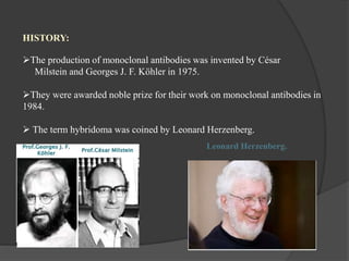
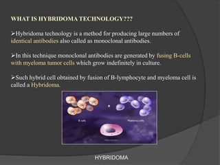
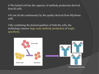
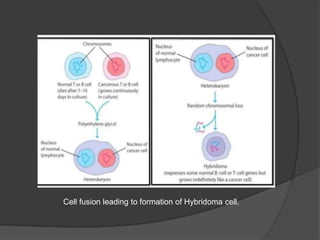
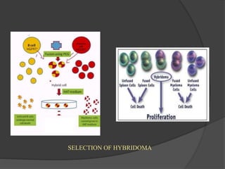
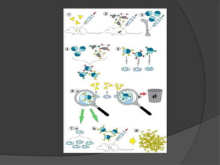
Hybridoma technology is a method for generating monoclonal antibodies by fusing B cells with myeloma cells. Georges Köhler and César Milstein developed this technique in 1975 and were awarded the Nobel Prize for it. The hybridoma cells produced from this fusion have the antibody production ability of B cells and indefinite growth ability of myeloma cells, allowing large-scale monoclonal antibody production. Hybridomas are selected using HAT medium, which eliminates unfused B and myeloma cells. The resulting hybridomas are then screened and cultured to produce monoclonal antibodies, which have wide applications in areas like cancer diagnosis, treatment, and research.
Hybridoma technology is a method for producing large number of identical antibodies called monoclonal antibodies.
It was discovered by G.kohler and C.milstein in 1975. they were awarded nobel prize for physiology and medicine in 1975.
The hybrid cells are produced by fusing B- lumphocyte with myeloma cells or tumour cells.
The B-lymphocyte have the ability to produce large number of antibodies and tumour cells have indefinite growth.
This is why two cells are used for the production of hybrid cell
Hybridoma technology is a method for producing large numbers of identical antibodies (also called monoclonal antibodies). This process starts by injecting a mouse (or other mammals) with an antigen that provokes an immune response.
Hybridoma
Hybridomas are cells that have been engineered to produce a desired antibody in large amounts, to produce monoclonal antibodies.
Monoclonal antibodies can be produced in specialized cells through a technique now popularly known as hybridoma technology.
Hybridoma technology was discovered in 1975 by two scientists, G. Kohler and C. Milstein, were awarded Noble prize for physiology and medicine in 1984.
Production and applications of monoclonal antibodiesKaayathri Devi
production and applications of monoclonal antibodies, monoclonal antibodies ,applications of monoclonal antibodies, production of monoclonal antibodies,
Protein engineering is the process of developing useful or valuable proteins. It is a young discipline, with much research taking place into the understanding of protein folding and recognition for protein design principles
INTRODUCTION: Monoclonal antibodies can be produced through a technique known as hybridoma technology.
HISTORY: The production of monoclonal antibodies was invented by Niels K.J. Georges, J.F. Kohler and Cesar Milstein in 1975.
PRINCIPLE FOR CREATION OF HYBRIDOMA CELLS: HAT (hypoxanthine aminopterin and thymidine) medium – Only hybridoma cells can proliferate in HAT medium.
PRODUCTION OF MONOCLONAL ANTIBODIES (HYBRIDOMA TECHNOLOGY): The establishment of hybridomas and production of monoclonal antibodies involves the following steps-
Immunization (ii) Cell fusion (iii) Selection of hybridomas (iv) Screening the products (v) Cloning and propagation (vi) Characterization and storage.
ADVANTAGES AND DISADVANTAGES OF MONOCLONAL ANTIBODIES:
Advantages- Monoclonal antibodies is specific to a given antigenic determinant.
Disadvantages- There is no guarantee that monoclonal antibodies produced is totally virus-free, despite the purification.
APPLICATIONS OF MONOCLONAL ANTIBODIES: Diagnostic applications, therapeutic applications, protein purification and miscellaneous applications.
REFERENCES:
• Satyanarayana, U. 2016. Biotechnology. Books and Allied (P) Ltd, Kolkata. pp. 213-226.
• Gupta, P.K. 2016. Biotechnology and Genomics. Rastogi Publications, Meerut. pp. 299-311.
• Owen, J.A., Punt J., Stranford, S.A. and Patricia, P.J. 2013. Kuby Immunology. 7th Ed. W.H. Freeman and Company, New York. pp.645-655.
• Singh, B.D. 2017. Biotechnology Expanding Horizons. Kalyani Publishers, New Delhi. pp. 172-174.
• Dubey, R.C. and Maheshwari, D.K. 2018. A Textbook of Microbiology. S Chand and Company Limited, New Delhi. pp. 662-663.
BIOTECHNOLOGY IS
CHALLENGING SUBJECT TO TEACH AND UNDERSTAND ......
ITS A VERY INTERESTING TO LEARN ABOUT HYBRIDOMA TECHNOLOGY .. THEIR PRODUCTION AND
APPLICATION ALSO ....
Various diagnostic tools now a days relied on the Hybriodoma technology and monoclonal antibodies,so this presentation will give some basic information about mAb and Hybridoma technology.
A hybridoma is a hybrid cell obtained by fusion of B lymphocyte with usually a tumor cell of antibody forming system or B lymphocyte (these are called myelomas).
HYBRIDOMA TECHNOLOGY IT IS DEFINED AS THE PROCESS WERE THERE IS A FUSION OF SPLLEN CELL AND MYELOMA CELLS IN THE PRESENCE OF POLYETHYLENE GLYCOL OR SENDAI VIRUS AND LEADS TO THE PRODUCTION OF MONOCLONL ANTIBODY.
Hybridoma Technology ( Production , Purification , and Application ) Sakshi Ghasle
Hybridoma technology revolutionized the field of immunology by enabling the production of monoclonal antibodies with high specificity and affinity. This presentation delves into the principles of DNA hybridoma technology, highlighting its significance in antibody production, therapeutic applications, and biomedical research. Learn about the key steps involved in generating hybridomas, from immunization to antibody screening, and discover the potential of recombinant DNA techniques in enhancing antibody engineering. Whether you're a student, researcher, or industry professional, this overview will provide valuable insights into the innovative world of hybridoma technology."
Uncover the wide-ranging applications of monoclonal antibodies in areas such as cancer therapy, autoimmune diseases, infectious diseases, and beyond. Learn about the latest advancements in antibody engineering and the development of novel therapeutic modalities, including bispecific antibodies, antibody-drug conjugates, and immune checkpoint inhibitors.
Whether you're a seasoned researcher or a newcomer to the field, this SlideShare presentation serves as a valuable resource for understanding the principles, techniques, and applications of hybridoma technology in modern biomedicine. Join a journey through the fascinating world of monoclonal antibodies and the groundbreaking science behind their creation.
Unlock the potential of hybridoma technology and propel your research to new heights. Dive into this SlideShare presentation now and explore the limitless possibilities of monoclonal antibody production with hybridoma technology.
Earliest Galaxies in the JADES Origins Field: Luminosity Function and Cosmic ...Sérgio Sacani
We characterize the earliest galaxy population in the JADES Origins Field (JOF), the deepest
imaging field observed with JWST. We make use of the ancillary Hubble optical images (5 filters
spanning 0.4−0.9µm) and novel JWST images with 14 filters spanning 0.8−5µm, including 7 mediumband filters, and reaching total exposure times of up to 46 hours per filter. We combine all our data
at > 2.3µm to construct an ultradeep image, reaching as deep as ≈ 31.4 AB mag in the stack and
30.3-31.0 AB mag (5σ, r = 0.1” circular aperture) in individual filters. We measure photometric
redshifts and use robust selection criteria to identify a sample of eight galaxy candidates at redshifts
z = 11.5 − 15. These objects show compact half-light radii of R1/2 ∼ 50 − 200pc, stellar masses of
M⋆ ∼ 107−108M⊙, and star-formation rates of SFR ∼ 0.1−1 M⊙ yr−1
. Our search finds no candidates
at 15 < z < 20, placing upper limits at these redshifts. We develop a forward modeling approach to
infer the properties of the evolving luminosity function without binning in redshift or luminosity that
marginalizes over the photometric redshift uncertainty of our candidate galaxies and incorporates the
impact of non-detections. We find a z = 12 luminosity function in good agreement with prior results,
and that the luminosity function normalization and UV luminosity density decline by a factor of ∼ 2.5
from z = 12 to z = 14. We discuss the possible implications of our results in the context of theoretical
models for evolution of the dark matter halo mass function.
The increased availability of biomedical data, particularly in the public domain, offers the opportunity to better understand human health and to develop effective therapeutics for a wide range of unmet medical needs. However, data scientists remain stymied by the fact that data remain hard to find and to productively reuse because data and their metadata i) are wholly inaccessible, ii) are in non-standard or incompatible representations, iii) do not conform to community standards, and iv) have unclear or highly restricted terms and conditions that preclude legitimate reuse. These limitations require a rethink on data can be made machine and AI-ready - the key motivation behind the FAIR Guiding Principles. Concurrently, while recent efforts have explored the use of deep learning to fuse disparate data into predictive models for a wide range of biomedical applications, these models often fail even when the correct answer is already known, and fail to explain individual predictions in terms that data scientists can appreciate. These limitations suggest that new methods to produce practical artificial intelligence are still needed.
In this talk, I will discuss our work in (1) building an integrative knowledge infrastructure to prepare FAIR and "AI-ready" data and services along with (2) neurosymbolic AI methods to improve the quality of predictions and to generate plausible explanations. Attention is given to standards, platforms, and methods to wrangle knowledge into simple, but effective semantic and latent representations, and to make these available into standards-compliant and discoverable interfaces that can be used in model building, validation, and explanation. Our work, and those of others in the field, creates a baseline for building trustworthy and easy to deploy AI models in biomedicine.
Bio
Dr. Michel Dumontier is the Distinguished Professor of Data Science at Maastricht University, founder and executive director of the Institute of Data Science, and co-founder of the FAIR (Findable, Accessible, Interoperable and Reusable) data principles. His research explores socio-technological approaches for responsible discovery science, which includes collaborative multi-modal knowledge graphs, privacy-preserving distributed data mining, and AI methods for drug discovery and personalized medicine. His work is supported through the Dutch National Research Agenda, the Netherlands Organisation for Scientific Research, Horizon Europe, the European Open Science Cloud, the US National Institutes of Health, and a Marie-Curie Innovative Training Network. He is the editor-in-chief for the journal Data Science and is internationally recognized for his contributions in bioinformatics, biomedical informatics, and semantic technologies including ontologies and linked data.
Professional air quality monitoring systems provide immediate, on-site data for analysis, compliance, and decision-making.
Monitor common gases, weather parameters, particulates.
Richard's entangled aventures in wonderlandRichard Gill
Since the loophole-free Bell experiments of 2020 and the Nobel prizes in physics of 2022, critics of Bell's work have retreated to the fortress of super-determinism. Now, super-determinism is a derogatory word - it just means "determinism". Palmer, Hance and Hossenfelder argue that quantum mechanics and determinism are not incompatible, using a sophisticated mathematical construction based on a subtle thinning of allowed states and measurements in quantum mechanics, such that what is left appears to make Bell's argument fail, without altering the empirical predictions of quantum mechanics. I think however that it is a smoke screen, and the slogan "lost in math" comes to my mind. I will discuss some other recent disproofs of Bell's theorem using the language of causality based on causal graphs. Causal thinking is also central to law and justice. I will mention surprising connections to my work on serial killer nurse cases, in particular the Dutch case of Lucia de Berk and the current UK case of Lucy Letby.
This pdf is about the Schizophrenia.
For more details visit on YouTube; @SELF-EXPLANATORY;
https://www.youtube.com/channel/UCAiarMZDNhe1A3Rnpr_WkzA/videos
Thanks...!
Slide 1: Title Slide
Extrachromosomal Inheritance
Slide 2: Introduction to Extrachromosomal Inheritance
Definition: Extrachromosomal inheritance refers to the transmission of genetic material that is not found within the nucleus.
Key Components: Involves genes located in mitochondria, chloroplasts, and plasmids.
Slide 3: Mitochondrial Inheritance
Mitochondria: Organelles responsible for energy production.
Mitochondrial DNA (mtDNA): Circular DNA molecule found in mitochondria.
Inheritance Pattern: Maternally inherited, meaning it is passed from mothers to all their offspring.
Diseases: Examples include Leber’s hereditary optic neuropathy (LHON) and mitochondrial myopathy.
Slide 4: Chloroplast Inheritance
Chloroplasts: Organelles responsible for photosynthesis in plants.
Chloroplast DNA (cpDNA): Circular DNA molecule found in chloroplasts.
Inheritance Pattern: Often maternally inherited in most plants, but can vary in some species.
Examples: Variegation in plants, where leaf color patterns are determined by chloroplast DNA.
Slide 5: Plasmid Inheritance
Plasmids: Small, circular DNA molecules found in bacteria and some eukaryotes.
Features: Can carry antibiotic resistance genes and can be transferred between cells through processes like conjugation.
Significance: Important in biotechnology for gene cloning and genetic engineering.
Slide 6: Mechanisms of Extrachromosomal Inheritance
Non-Mendelian Patterns: Do not follow Mendel’s laws of inheritance.
Cytoplasmic Segregation: During cell division, organelles like mitochondria and chloroplasts are randomly distributed to daughter cells.
Heteroplasmy: Presence of more than one type of organellar genome within a cell, leading to variation in expression.
Slide 7: Examples of Extrachromosomal Inheritance
Four O’clock Plant (Mirabilis jalapa): Shows variegated leaves due to different cpDNA in leaf cells.
Petite Mutants in Yeast: Result from mutations in mitochondrial DNA affecting respiration.
Slide 8: Importance of Extrachromosomal Inheritance
Evolution: Provides insight into the evolution of eukaryotic cells.
Medicine: Understanding mitochondrial inheritance helps in diagnosing and treating mitochondrial diseases.
Agriculture: Chloroplast inheritance can be used in plant breeding and genetic modification.
Slide 9: Recent Research and Advances
Gene Editing: Techniques like CRISPR-Cas9 are being used to edit mitochondrial and chloroplast DNA.
Therapies: Development of mitochondrial replacement therapy (MRT) for preventing mitochondrial diseases.
Slide 10: Conclusion
Summary: Extrachromosomal inheritance involves the transmission of genetic material outside the nucleus and plays a crucial role in genetics, medicine, and biotechnology.
Future Directions: Continued research and technological advancements hold promise for new treatments and applications.
Slide 11: Questions and Discussion
Invite Audience: Open the floor for any questions or further discussion on the topic.
3. HISTORY:
The production of monoclonal antibodies was invented by César
Milstein and Georges J. F. Köhler in 1975.
They were awarded noble prize for their work on monoclonal antibodies in
1984.
The term hybridoma was coined by Leonard Herzenberg.
Leonard Herzenberg.
4. WHAT IS HYBRIDOMA TECHNOLOGY???
Hybridoma technology is a method for producing large numbers of
identical antibodies also called as monoclonal antibodies.
In this technique monoclonal antibodies are generated by fusing B-cells
with myeloma tumor cells which grow indefinitely in culture.
Such hybrid cell obtained by fusion of B-lymphocyte and myeloma cell is
called a Hybridoma.
HYBRIDOMA
5. The hybrid cell has the capacity of antibody production derived
from B cells.
It can divide continuously by the quality derived from Myeloma
cells.
By combining the desired qualities of both the cells, the
technology ensures large scale antibody production of single
specificity.
Monoclonal antibodies
6. METHODOLOGY:
Laboratory animals (mammals, e.g. mice) are first exposed to the antigen
that an antibody is to be generated against.
Usually this is done by a series of injections of the antigen, over the course
of several weeks.
These injections are typically followed by the use of in
vivo electroporation, which significantly enhances the immune response.
The spleenocytes from spleen and myeloma cells from bone marrow of the
immunized mouse are isolated.
7. The fusion of the B cells with myeloma cells can be done using:
Physical method: Electrofusion.
Chemical method : Polyethylene glycol.
PEG mediated Cell fusion Electrofusion
9. Nucleotide synthesis:
Nucleotide synthesis is essential for cell survival.
The two pathways for nucleotide synthesis are :
Denovo pathway. (synthesis of nucleotides from simple sugars, amino
acids etc.)
Salvage pathway. (synthesis of nucleotides from intermediate of
nucleotide degradation pathways)
An important enzyme of salvage pathway is HGPRT.
10. SELECTION:
Process through which only hybridoma cells are selected by eliminating
unfused myeloma cells and unfused B cells
HAT medium is used for the selection of hybrid cells.
Fused cells are incubated in HAT medium (hypoxanthine- aminopterin -
thymidine medium) for roughly 10 to 14 days.
Elimination of unfused myeloma cells:
Removal of the unfused myeloma cells is necessary because they have the
potential to outgrow other cells, especially weakly established hybridomas
The myeloma cells first selected for fusion are hypoxanthine-guanine
phosphoribosyl transferase (HGPRT) mutant cells(raised by using 8-
azaguanine which causes mutations).
Thus the unfused myeloma cells in the medium cannot carryout nucleotide
synthesis through salvage pathway due to the deficiency of HGPRT.
11. Where as, Aminopterin blocks the denovo pathway.
Hence, unfused myeloma cells die, as they cannot synthesize nucleotides.
Elimination of unfused B cells:
Unlike the unfused myeloma cells, the B cells can carryout nucleotide
synthesis through Denovo pathway as they are not HGPRT mutants.
But due to the lack of infinite cell division capability the B cells which
survive in the HAT medium die within 7-10 days.
Thus, only the Hybrid cells survive on HAT as they have HGPRT enzyme
from B cells and the repeated cell division property of myeloma cells.
13. The incubated medium is then diluted into multi-well plates to such an
extent that each well contains only one cell.
Since the antibodies in a well are produced by the same B cell, they will be
directed towards the same epitope, and are thus monoclonal antibodies.
96 WELL PLATE
14. SCREENING PROCESS:
The next stage is a rapid screening process, which identifies and selects only
those hybridomas that produce antibodies of appropriate specificity.
The screening technique used is called ELISA. The hybridoma culture
supernatant, secondary enzyme labeled antibodies, are then incubated, and the
formation of a colored product indicates a positive hybridoma.
ELISA
SCREENING
15. CULTURING :
Culturing of hybridomas:
In vitro
In vivo
IN VITRO CULTURING:
The hybridoma cell that produces the desired antibodies can be cloned to produce
many identical daughter clones.
Once a hybridoma colony is established, it will continually grow in culture medium
produce antibodies.
Multiwell plates are used initially to grow the hybridomas, and after selection, are
changed to larger tissue culture flasks. This maintains the well-being of the
hybridomas and provides enough cells for cryopreservation and supernatant for
subsequent investigations. The culture supernatant can yield 1 to 60 µg/ml of
monoclonal antibody, which is maintained at -20 °C or lower until required.
16. IN VIVO CULTURING:
This procedure involves introduction of hybridoma cells into the
peritoneal cavity if the animal.
The ascetic fluid is isolated from the animal which is used for the
isolation and purification of the antibodies.
IN-VITRO AND IN-VIVO CULTURING
17.
18.
19. APPLICATIONS:
BIOCHEMICAL ANALYSIS:
Monoclonal antibodies are used in immunoassays like:
RIA( radioimmuno assay )
ELISA (Enzyme linked immuno sorbent assay)
These assay measure the concentrations of hormones like
Insulin
Thyroxine
TSH etc.
Blood grouping test
Pregnancy test ( Human chorionic gonadotropin)
CANCER:
Estimation of plasma carcinoembryonic antigen in colorectal cancer,
Prostate specific antigen for prostate cancer.
20. DIAGNOSIS OF INFECTIOUS DISEASES:
These antibodies could be used for detection of infections such as typhoid ,
syphilis etc.
DIAGNOSTIC IMAGING:
Radiolabelled monoclonal antibodies are used in the diagnostic imaging of
diseases, this technique is called immunoscintigraphy.
Applications in detection of Myocardial infraction, Deep vein thrombosis etc.
Detection of Tumor markers for various cancers
ex: carcinoembryonic antigen (CEA) - cancer of stomach, colon, pancreas.
Alpha fetoprotein – cancer of Liver .
CANCER TREATMENT:
Monoclonal antibodies against the surface antigens of cancer cells mediate
cancer cell death through ADCC( antibody mediated cell dependent cytotoxicity )
Phagocytosis etc.
21. IMMUNOSUPRESSION:
The monoclonal antibodies could be used as immunosupressive agent
after organ transplantation.
Ex: OKT3 against CD3 of T cells has been successfully used in renal
and bone marrow transplantation.