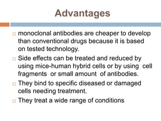
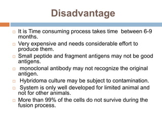
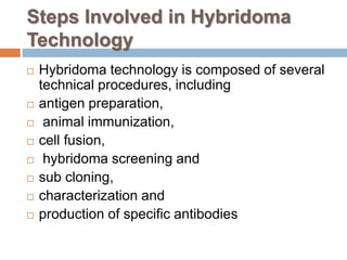
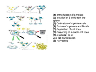
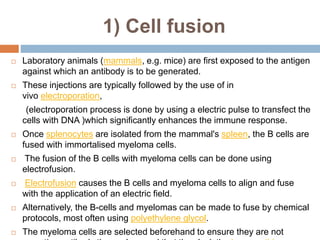
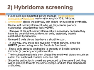
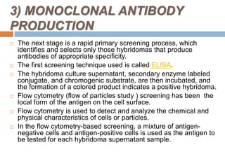
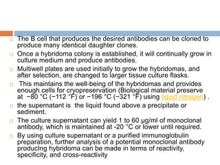
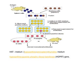
MONOCLONAL
ANTIBODIES
By
TEJASWINI L. ASAWE
ASSISTANT PROFESSOR
SIDHHIS INSTITUTE OF PHARMACY
THANE
Monoclonal antibodies
 History of Hybridoma Technology.
 Hybridoma technology is a well-established
method to produce monoclonal antibodies
(mAbs) specific to antigens.
 Hybridoma cell lines are formed via fusion
between a short-lived antibody-producing B
cell and an immortal or living myeloma cell.
 Myeloma is develops from cells in the bone
marrow blood cancer that called plasma cells.
HISTORY
 Hybridoma technology was discovered in 1975 by
two scientists, Georges Kohler and Cesar Milstein
 They wanted to create immortal hybrid cells by
fusing normal B cells from immunized mice with
their myeloma cells.
 By cloning individual hybrid cells, they established
the first hybridoma cell lines which can produce
single type of antibody specific to the specific
antigen.
 Their discovery is considered one of the greatest
breakthroughs in the field of biotechnology.
Advantages
 monoclonal antibodies are cheaper to develop
than conventional drugs because it is based
on tested technology.
 Side effects can be treated and reduced by
using mice-human hybrid cells or by using cell
fragments or small amount of antibodies.
 They bind to specific diseased or damaged
cells needing treatment.
 They treat a wide range of conditions
Disadvantage
 It is Time consuming process takes time between 6-9
months.
 Very expensive and needs considerable effort to
produce them.
 Small peptide and fragment antigens may not be good
antigens.
 monoclonal antibody may not recognize the original
antigen.
 Hybridoma culture may be subject to contamination.
 System is only well developed for limited animal and
not for other animals.
 More than 99% of the cells do not survive during the
fusion process.
Steps Involved in Hybridoma
Technology
 Hybridoma technology is composed of several
technical procedures, including
 antigen preparation,
 animal immunization,
 cell fusion,
 hybridoma screening and
 sub cloning,
 characterization and
 production of specific antibodies
(1) Immunization of a mouse
(2) Isolation of B cells from the
spleen
(3) Cultivation of myeloma cells
(4) Fusion of myeloma and B cells
(5) Separation of cell lines
(6) Screening of suitable cell lines
(7) in vitro (a) or in
vivo (b) multiplication
(8) Harvesting
1) Cell fusion
 Laboratory animals (mammals, e.g. mice) are first exposed to the antigen
against which an antibody is to be generated.
 These injections are typically followed by the use of in
vivo electroporation,
(electroporation process is done by using a electric pulse to transfect the
cells with DNA )which significantly enhances the immune response.
 Once splenocytes are isolated from the mammal's spleen, the B cells are
fused with immortalised myeloma cells.
 The fusion of the B cells with myeloma cells can be done using
electrofusion.
 Electrofusion causes the B cells and myeloma cells to align and fuse
with the application of an electric field.
 Alternatively, the B-cells and myelomas can be made to fuse by chemical
protocols, most often using polyethylene glycol.
 The myeloma cells are selected beforehand to ensure they are not
2) Hybridoma screening
 Fused cells are incubated in HAT medium (hypoxanthine-
aminopterin-thymidine medium) for roughly 10 to 14 days.
 Aminopterin blocks the pathway that allows for nucleotide synthesis.
 Hence, unfused myeloma cells die, as they cannot produce
nucleotides because they lack HGPRT.
 Removal of the unfused myeloma cells is necessary because they
have the potential to outgrow other cells, especially weakly
established hybridomas.
 Unfused B cells die as they have a short life span.
 In this way, only the B cell-myeloma hybrids survive, since the
HGPRT gene coming from the B cells is functional.
 These cells produce antibodies (a property of B cells) and are
immortal (a property of myeloma cells).
 The incubated medium is then diluted into multi-well plates to such an
extent that each well contains only one cell.
 Since the antibodies in a well are produced by the same B cell, they
will be directed towards the same epitope, and are thus monoclonal
antibodies.
3) MONOCLONAL ANTIBODY
PRODUCTION
 The next stage is a rapid primary screening process, which
identifies and selects only those hybridomas that produce
antibodies of appropriate specificity.
 The first screening technique used is called ELISA.
 The hybridoma culture supernatant, secondary enzyme labeled
conjugate, and chromogenic substrate, are then incubated, and
the formation of a colored product indicates a positive hybridoma.
 Flow cytometry (flow of particles study ) screening has been the
local form of the antigen on the cell surface.
 Flow cytometry is used to detect and analyze the chemical and
physical characteristics of cells or particles.
 In the flow cytometry-based screening, a mixture of antigen-
negative cells and antigen-positive cells is used as the antigen to
be tested for each hybridoma supernatant sample.
 The B cell that produces the desired antibodies can be cloned to
produce many identical daughter clones.
 Once a hybridoma colony is established, it will continually grow in
culture medium and produce antibodies.
 Multiwell plates are used initially to grow the hybridomas, and
after selection, are changed to larger tissue culture flasks.
 This maintains the well-being of the hybridomas and provides
enough cells for cryopreservation (Biological material preserve
at −80 °C (−112 °F) or −196 °C (−321 °F) using liquid nitrogen) .
 the supernatant is the liquid found above a precipitate or
sediment.
 The culture supernatant can yield 1 to 60 μg/ml of monoclonal
antibody, which is maintained at -20 °C or lower until required.
 By using culture supernatant or a purified immunoglobulin
preparation, further analysis of a potential monoclonal antibody
producIng hybridoma can be made in terms of reactivity,
specificity, and cross-reactivity
HAT - medium (hypoxanthine-aminopterin-thymidine medium
hypoxanthine-guanine phospho ribosyl transferase (HGPRT) gene,
Application
 Pregnancy Testing
 MAbs that have been developed to detect
human chorionic gonadotropin (HCG) are now
present in pregnancy test kits.
 Radioimmuno detection (RID) of Cancer
 An imaging technique used to detect the
presence of cancerous or cancer-specific cells
has been produced as mAbs.
 Treatment of Cancer through Drugs
 Many different drugs are being developed in
clinical trials used to treat various strains of
cancer.
 In fact, some of these are already on the
market.
 In 1997, a drug named Ritoxin was approved
by the FDA for commercial use which is based
on mAb technology.
Viral Disease Treatment
 Doctors hope that with further research into
mAbs and an increased knowledge of their
properties, treatments will become available for
diseases previously thought to be incurable,
such as AIDS.
 Identifying Pathogens
 MAbs can now be used to identify strains of a
single pathogen, for example neisseria
gonorrhoeae.
 Rhesus disease Immunization
 Rhesus disease is condition where antibodies
in a pregnant woman's blood destroy her
baby's blood cells.
 Anti-rhesus antiserum has been made to cure
rhesus disease.
References
 1. Zaroff, S., & Tan, G. (2019). Hybridoma technology:
the preferred method for monoclonal antibody
generation for in vivo applications.
 2. Zhang, C. (2012). Hybridoma technology for the
generation of monoclonal antibodies. In Antibody
methods and protocols (pp. 117-135). Humana Press,
Totowa, NJ.
 3. Liu, J. K. (2014). The history of monoclonal
antibody development–progress, remaining
challenges and future innovations. Annals of Medicine
and Surgery, 3(4), 113-116.
 4. Greenfield, E. A. (2018). Polyethylene Glycol
Fusion for Hybridoma Production. Cold Spring Harbor
Protocols, 2018(3), pdb-prot10317

hybridoma technology.pptx

  • 1.
    MONOCLONAL ANTIBODIES By TEJASWINI L. ASAWE ASSISTANTPROFESSOR SIDHHIS INSTITUTE OF PHARMACY THANE
  • 2.
    Monoclonal antibodies  Historyof Hybridoma Technology.  Hybridoma technology is a well-established method to produce monoclonal antibodies (mAbs) specific to antigens.  Hybridoma cell lines are formed via fusion between a short-lived antibody-producing B cell and an immortal or living myeloma cell.  Myeloma is develops from cells in the bone marrow blood cancer that called plasma cells.
  • 3.
    HISTORY  Hybridoma technologywas discovered in 1975 by two scientists, Georges Kohler and Cesar Milstein  They wanted to create immortal hybrid cells by fusing normal B cells from immunized mice with their myeloma cells.  By cloning individual hybrid cells, they established the first hybridoma cell lines which can produce single type of antibody specific to the specific antigen.  Their discovery is considered one of the greatest breakthroughs in the field of biotechnology.
  • 4.
    Advantages  monoclonal antibodiesare cheaper to develop than conventional drugs because it is based on tested technology.  Side effects can be treated and reduced by using mice-human hybrid cells or by using cell fragments or small amount of antibodies.  They bind to specific diseased or damaged cells needing treatment.  They treat a wide range of conditions
  • 5.
    Disadvantage  It isTime consuming process takes time between 6-9 months.  Very expensive and needs considerable effort to produce them.  Small peptide and fragment antigens may not be good antigens.  monoclonal antibody may not recognize the original antigen.  Hybridoma culture may be subject to contamination.  System is only well developed for limited animal and not for other animals.  More than 99% of the cells do not survive during the fusion process.
  • 6.
    Steps Involved inHybridoma Technology  Hybridoma technology is composed of several technical procedures, including  antigen preparation,  animal immunization,  cell fusion,  hybridoma screening and  sub cloning,  characterization and  production of specific antibodies
  • 7.
    (1) Immunization ofa mouse (2) Isolation of B cells from the spleen (3) Cultivation of myeloma cells (4) Fusion of myeloma and B cells (5) Separation of cell lines (6) Screening of suitable cell lines (7) in vitro (a) or in vivo (b) multiplication (8) Harvesting
  • 8.
    1) Cell fusion Laboratory animals (mammals, e.g. mice) are first exposed to the antigen against which an antibody is to be generated.  These injections are typically followed by the use of in vivo electroporation, (electroporation process is done by using a electric pulse to transfect the cells with DNA )which significantly enhances the immune response.  Once splenocytes are isolated from the mammal's spleen, the B cells are fused with immortalised myeloma cells.  The fusion of the B cells with myeloma cells can be done using electrofusion.  Electrofusion causes the B cells and myeloma cells to align and fuse with the application of an electric field.  Alternatively, the B-cells and myelomas can be made to fuse by chemical protocols, most often using polyethylene glycol.  The myeloma cells are selected beforehand to ensure they are not
  • 9.
    2) Hybridoma screening Fused cells are incubated in HAT medium (hypoxanthine- aminopterin-thymidine medium) for roughly 10 to 14 days.  Aminopterin blocks the pathway that allows for nucleotide synthesis.  Hence, unfused myeloma cells die, as they cannot produce nucleotides because they lack HGPRT.  Removal of the unfused myeloma cells is necessary because they have the potential to outgrow other cells, especially weakly established hybridomas.  Unfused B cells die as they have a short life span.  In this way, only the B cell-myeloma hybrids survive, since the HGPRT gene coming from the B cells is functional.  These cells produce antibodies (a property of B cells) and are immortal (a property of myeloma cells).  The incubated medium is then diluted into multi-well plates to such an extent that each well contains only one cell.  Since the antibodies in a well are produced by the same B cell, they will be directed towards the same epitope, and are thus monoclonal antibodies.
  • 10.
    3) MONOCLONAL ANTIBODY PRODUCTION The next stage is a rapid primary screening process, which identifies and selects only those hybridomas that produce antibodies of appropriate specificity.  The first screening technique used is called ELISA.  The hybridoma culture supernatant, secondary enzyme labeled conjugate, and chromogenic substrate, are then incubated, and the formation of a colored product indicates a positive hybridoma.  Flow cytometry (flow of particles study ) screening has been the local form of the antigen on the cell surface.  Flow cytometry is used to detect and analyze the chemical and physical characteristics of cells or particles.  In the flow cytometry-based screening, a mixture of antigen- negative cells and antigen-positive cells is used as the antigen to be tested for each hybridoma supernatant sample.
  • 11.
     The Bcell that produces the desired antibodies can be cloned to produce many identical daughter clones.  Once a hybridoma colony is established, it will continually grow in culture medium and produce antibodies.  Multiwell plates are used initially to grow the hybridomas, and after selection, are changed to larger tissue culture flasks.  This maintains the well-being of the hybridomas and provides enough cells for cryopreservation (Biological material preserve at −80 °C (−112 °F) or −196 °C (−321 °F) using liquid nitrogen) .  the supernatant is the liquid found above a precipitate or sediment.  The culture supernatant can yield 1 to 60 μg/ml of monoclonal antibody, which is maintained at -20 °C or lower until required.  By using culture supernatant or a purified immunoglobulin preparation, further analysis of a potential monoclonal antibody producIng hybridoma can be made in terms of reactivity, specificity, and cross-reactivity
  • 12.
    HAT - medium(hypoxanthine-aminopterin-thymidine medium hypoxanthine-guanine phospho ribosyl transferase (HGPRT) gene,
  • 13.
    Application  Pregnancy Testing MAbs that have been developed to detect human chorionic gonadotropin (HCG) are now present in pregnancy test kits.  Radioimmuno detection (RID) of Cancer  An imaging technique used to detect the presence of cancerous or cancer-specific cells has been produced as mAbs.
  • 14.
     Treatment ofCancer through Drugs  Many different drugs are being developed in clinical trials used to treat various strains of cancer.  In fact, some of these are already on the market.  In 1997, a drug named Ritoxin was approved by the FDA for commercial use which is based on mAb technology.
  • 15.
    Viral Disease Treatment Doctors hope that with further research into mAbs and an increased knowledge of their properties, treatments will become available for diseases previously thought to be incurable, such as AIDS.  Identifying Pathogens  MAbs can now be used to identify strains of a single pathogen, for example neisseria gonorrhoeae.
  • 16.
     Rhesus diseaseImmunization  Rhesus disease is condition where antibodies in a pregnant woman's blood destroy her baby's blood cells.  Anti-rhesus antiserum has been made to cure rhesus disease.
  • 17.
    References  1. Zaroff,S., & Tan, G. (2019). Hybridoma technology: the preferred method for monoclonal antibody generation for in vivo applications.  2. Zhang, C. (2012). Hybridoma technology for the generation of monoclonal antibodies. In Antibody methods and protocols (pp. 117-135). Humana Press, Totowa, NJ.  3. Liu, J. K. (2014). The history of monoclonal antibody development–progress, remaining challenges and future innovations. Annals of Medicine and Surgery, 3(4), 113-116.  4. Greenfield, E. A. (2018). Polyethylene Glycol Fusion for Hybridoma Production. Cold Spring Harbor Protocols, 2018(3), pdb-prot10317