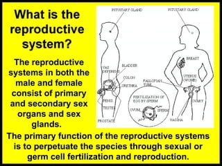
The reproductive
systems inboth the
male and female
consist of primary
and secondary sex
organs and sex
glands.
The primary function of the reproductive systems
is to perpetuate the species through sexual or
germ cell fertilization and reproduction.
3.
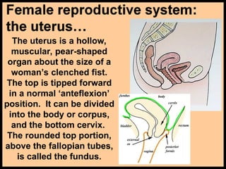
The uterus isa hollow,
muscular, pear-shaped
organ about the size of a
woman’s clenched fist.
The top is tipped forward
in a normal ‘anteflexion’
position. It can be divided
into the body or corpus,
and the bottom cervix.
The rounded top portion,
above the fallopian tubes,
is called the fundus.
4.
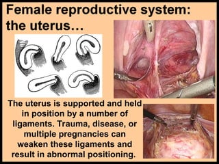
The uterus issupported and held
in position by a number of
ligaments. Trauma, disease, or
multiple pregnancies can
weaken these ligaments and
result in abnormal positioning.
5.
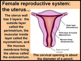
The uterus wall
has3 layers: the
outside layer
called the
perimetrium, the
muscular inside
layer called the
myometrium, and
the mucous
membrane lining
the uterus called
the endometrium.
Outer layer
(Perimetrium)
Thick layer of muscle
(Myometrium)
Mucous lining
(Endometrium)
The cervical opening is about
the diameter of a pencil.
6.
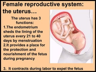
The uterus has3
functions:
1.The endometrium
sheds the lining of the
uterus every 21 to 40
days by menstruation
2.It provides a place for
the protection and
nourishment of the fetus
during pregnancy
3. It contracts during labor to expel the fetus
7.
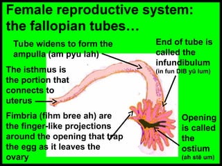
The isthmus is
theportion that
connects to
uterus
Tube widens to form the
ampulla (am pyu lah)
Fimbria (fihm bree ah) are
the finger-like projections
around the opening that trap
the egg as it leaves the
ovary
End of tube is
called the
infundibulum
(in fun DIB yū lum)
Opening
is called
the
ostium
(ah stē um)
8.
The fallopian tubeis 4-6 inches long. The egg, released
from the ovary, is captured by the fimbria and brought
into the fallopian tube. The egg is moved along inside
the tube by muscular contractions and the waving action
of cilia. It takes an egg about 3-4 days to travel the
length of the tube. If an egg is fertilized, it occurs here.
9.
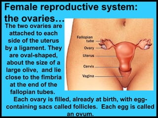
The two ovariesare
attached to each
side of the uterus
by a ligament. They
are oval-shaped,
about the size of a
large olive, and lie
close to the fimbria
at the end of the
fallopian tubes.
Each ovary is filled, already at birth, with egg-
containing sacs called follicles. Each egg is called
an ovum.
10.
Once every 21days,
one follicle in one
ovary ripens. This
mature follicle is a
graafian (GRAW fee un)
follicle. The follicle
ruptures in response
to hormones from the
pituitary gland,
releasing the
ovum/egg… a process
called ovulation.
After the follicle ruptures, it
becomes a mass of yellow
cells called the corpus
luteum. This is a temporary,
progesterone-producing
structure.
11.
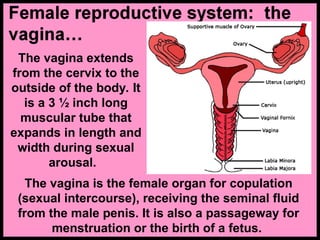
The vagina extends
fromthe cervix to the
outside of the body. It
is a 3 ½ inch long
muscular tube that
expands in length and
width during sexual
arousal.
The vagina is the female organ for copulation
(sexual intercourse), receiving the seminal fluid
from the male penis. It is also a passageway for
menstruation or the birth of a fetus.
12.
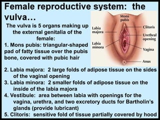
The vulva is5 organs making up
the external genitalia of the
female:
1. Mons pubis: triangular-shaped
pad of fatty tissue over the pubis
bone, covered with pubic hair
2. Labia majora: 2 large folds of adipose tissue on the sides
of the vaginal opening
3. Labia minora: 2 smaller folds of adipose tissue on the
inside of the labia majora
4. Vestibule: area between labia with openings for the
vagina, urethra, and two excretory ducts for Bartholin’s
glands (provide lubricant)
5. Clitoris: sensitive fold of tissue partially covered by hood
13.
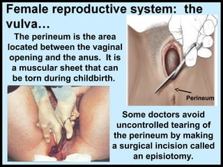
The perineum isthe area
located between the vaginal
opening and the anus. It is
a muscular sheet that can
be torn during childbirth.
Some doctors avoid
uncontrolled tearing of
the perineum by making
a surgical incision called
an episiotomy.
Perineum
14.
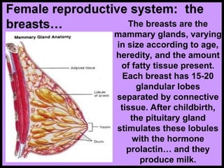
The breasts arethe
mammary glands, varying
in size according to age,
heredity, and the amount
of fatty tissue present.
Each breast has 15-20
glandular lobes
separated by connective
tissue. After childbirth,
the pituitary gland
stimulates these lobules
with the hormone
prolactin… and they
produce milk.
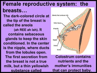
15.
The dark-colored circleat
the tip of the breast is
called the areola
(ah REE ah lah). It
contains sebaceous
glands to keep the skin
conditioned. In the center
is the nipple, where ducts
from the lobules open.
The first secretion from
the breast is not a true
milk, but a thin yellowish
substance called
Colostrum contains
nutrients and the
mother’s immunities
that can protect baby.
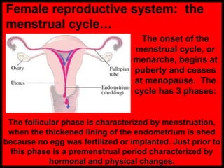
16.
The onset ofthe
menstrual cycle, or
menarche, begins at
puberty and ceases
at menopause. The
cycle has 3 phases:
The follicular phase is characterized by menstruation,
when the thickened lining of the endometrium is shed
because no egg was fertilized or implanted. Just prior to
this phase is a premenstrual period characterized by
hormonal and physical changes.
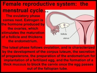
17.
The ovulatory phase
comesnext. Estrogen is
the hormone produced by
the ovaries, which
stimulates the maturation
of a follicle and thickens
the endometrium.
The luteal phase follows ovulation, and is characterized
by the development of the corpus luteum, the secretion
of progesterone, the preparation of the endometrium for
implantation of a fertilized egg, and the formation of a
thick mucous to block the cervix once the egg passes
out of the fallopian tube.
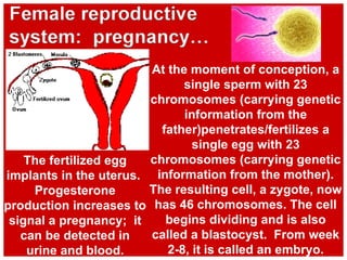
18.
At the momentof conception, a
single sperm with 23
chromosomes (carrying genetic
information from the
father)penetrates/fertilizes a
single egg with 23
chromosomes (carrying genetic
information from the mother).
The resulting cell, a zygote, now
has 46 chromosomes. The cell
begins dividing and is also
called a blastocyst. From week
2-8, it is called an embryo.
The fertilized egg
implants in the uterus.
Progesterone
production increases to
signal a pregnancy; it
can be detected in
urine and blood.
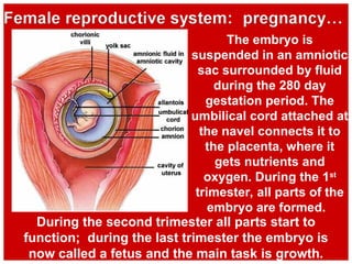
19.
The embryo is
suspendedin an amniotic
sac surrounded by fluid
during the 280 day
gestation period. The
umbilical cord attached at
the navel connects it to
the placenta, where it
gets nutrients and
oxygen. During the 1st
trimester, all parts of the
embryo are formed.
During the second trimester all parts start to
function; during the last trimester the embryo is
now called a fetus and the main task is growth.
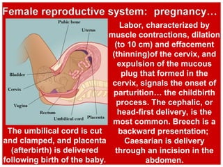
20.
Labor, characterized by
musclecontractions, dilation
(to 10 cm) and effacement
(thinning)of the cervix, and
expulsion of the mucous
plug that formed in the
cervix, signals the onset of
parturition… the childbirth
process. The cephalic, or
head-first delivery, is the
most common. Breech is a
backward presentation;
Caesarian is delivery
through an incision in the
abdomen.
The umbilical cord is cut
and clamped, and placenta
(afterbirth) is delivered
following birth of the baby.
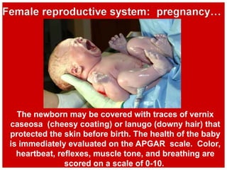
21.
The newborn maybe covered with traces of vernix
caseosa (cheesy coating) or lanugo (downy hair) that
protected the skin before birth. The health of the baby
is immediately evaluated on the APGAR scale. Color,
heartbeat, reflexes, muscle tone, and breathing are
scored on a scale of 0-10.
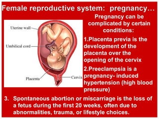
22.
3. Spontaneous abortionor miscarriage is the loss of
a fetus during the first 20 weeks, often due to
abnormalities, trauma, or lifestyle choices.
Pregnancy can be
complicated by certain
conditions:
1.Placenta previa is the
development of the
placenta over the
opening of the cervix
2.Preeclampsia is a
pregnancy- induced
hypertension (high blood
pressure)
23.
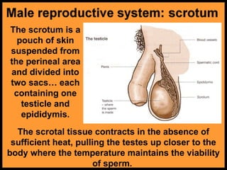
The scrotum isa
pouch of skin
suspended from
the perineal area
and divided into
two sacs… each
containing one
testicle and
epididymis.
The scrotal tissue contracts in the absence of
sufficient heat, pulling the testes up closer to the
body where the temperature maintains the viability
of sperm.
24.
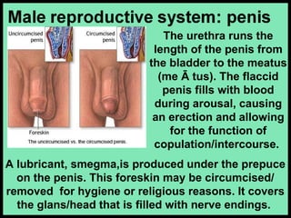
A lubricant, smegma,isproduced under the prepuce
on the penis. This foreskin may be circumcised/
removed for hygiene or religious reasons. It covers
the glans/head that is filled with nerve endings.
The urethra runs the
length of the penis from
the bladder to the meatus
(me Ā tus). The flaccid
penis fills with blood
during arousal, causing
an erection and allowing
for the function of
copulation/intercourse.
25.
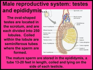
The oval-shaped
testes arelocated in
the scrotum, and are
each divided into 250
lobules. Coiled
within the lobule are
seminiferous tubes
where the sperm are
formed.
The mature sperm are stored in the epididymis, a
tube 13-20 feet in length, coiled and lying on the
side of each testicle.
26.
The acrosome
(AK rohzome)
covering the head
of the sperm
contains enzymes
that help it
penetrate the ova.
The head carries
the genetic
material. The
midpiece supplies
energy. The tail or
flagellum (flah JELL
um) provides
Sperm carry either an
X/female OR Y/male
chromosome. Since all ova
carry the X/female
chromosome, the male sperm
does influence the baby’s sex.
27.
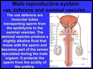
The vas deferensare
muscular tubes
transporting sperm from
the epididymis to the
seminal vesicles. The
seminal vesicles produce a
slightly alkaline fluid that
mixes with the sperm and
becomes part of the semen
ejaculated during the male
orgasm. It protects the
sperm from the acidity of
the urethra.
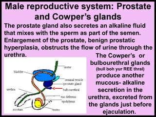
28.
The prostate glandalso secretes an alkaline fluid
that mixes with the sperm as part of the semen.
Enlargement of the prostate, benign prostatic
hyperplasia, obstructs the flow of urine through the
urethra. The Cowper’s or
bulbourethral glands
(bull boh yur REE thral)
produce another
mucous- alkaline
secretion in the
urethra, excreted from
the glands just before
ejaculation.