Final Project - Instrumentation and Signal Acquisition in Bioengineering.
As a result of ageing population and a progressively greedy will of better healthcare, new devices have arisen. Some focus on prevention, others in treating. The prototype developed here focus in the acquisition of the heart’s electrical activity with a simple electrical circuit embedded in an Arduino microcontroller, with the possibility of early heart diseases detection. An effective and cost contained solution was proposed and dozens of trials were performed with the device. These pointed to a high rate of detection of R, S and T peaks (~100%, 78,3% and 72,5%) with an accurate heart rate measure. Despite of the good results obtained, this device, for now, must not be used for other purposes than monitoring.
IST - 4th Year - 2nd Semestre - Biomedical Engineering.
1. X-rays were discovered in 1895 by Wilhelm Röntgen, a German physicist, while experimenting with cathode ray tubes. He noticed that materials near the tube would glow, even when shielded from known radiation sources, and concluded he had discovered a new type of radiation which he named X-rays.
2. X-rays are produced when high-energy electrons collide with a metal target, causing the electrons to lose energy which is released as X-ray photons. Modern X-ray tubes contain a tungsten target and operate by accelerating electrons toward the target with a high voltage.
3. X-rays have wavelengths between 10 picometers to 10 nanometers, shorter than visible light.
brief but informative knowledge about how CT works and what are its components ... easy to understand as well as presenting during lectures and in classes . share it
This document provides information about electroencephalography (EEG), electromyography (EMG), and patient monitoring. It discusses how EEG is used to measure brain activity through electrodes on the scalp. It describes the different frequency bands seen on EEG and how they relate to mental states. The document outlines the components of an EEG recording system and various EEG artifacts. It also discusses EMG and how it is used to measure muscle electrical activity. Finally, it covers patient monitoring systems, including bedside monitors, central monitoring stations, and the parameters that are measured like heart rate, blood pressure, respiration rate.
The document discusses the history and components of X-ray machines. It begins with a brief history of the discovery of X-rays by Wilhelm Roentgen in 1895 and important developments in dental radiology. It then describes the ideal requirements and main components of an X-ray tubehead, including the X-ray tube, position indicating device, and collimator. The document explains the circuitry and components within the X-ray tube, such as the cathode, anode, and line focus principle. It concludes with a discussion of advances in X-ray machines.
Dye lasers use an organic dye dissolved in a liquid as the active lasing medium and can produce a wide range of wavelengths. They work on the principle of population inversion using a pumping source like a flash lamp or other laser to excite the dye molecules. The major components are the active dye medium, pumping source, and resonator mirrors, with one mirror sometimes replaced by a diffraction grating to allow tuning of the output wavelength. Dye lasers offer tunability but have limitations in lifetime and output power.
The document discusses types of radiation detectors, including gas-filled detectors like ionization chambers and GM counters, and how they are used in medical applications. It covers concepts like detection methods, operating modes, dead time, and how detector performance is affected by high count rates. The goal is to classify detectors used in various departments like nuclear medicine.
X-rays are a form of electromagnetic radiation that can pass through soft tissue but are partially or fully blocked by dense tissues like bone. Wilhelm Roentgen discovered x-rays in 1895 while experimenting with cathode ray tubes. X-rays are used in medical imaging to generate diagnostic images of internal structures in the body by passing x-ray beams through the body onto photographic film or digital sensors. The denser tissues absorb more of the x-ray beam, resulting in contrast between different internal structures visible on the radiograph. Common medical uses of x-rays include detection of fractures, tumors, dental decay, and foreign objects in the body.
This document discusses various methods for detecting radiation. It outlines passive detectors like photographic film, electroscopes, dosimeters, and thermoluminescent dosimeters (TLDs) which do not require a power source. Active detectors mentioned include Geiger-Muller tubes and scintillation detectors, which need a constant energy supply. Both types detect radiation indirectly by ionizing matter and detecting the ions produced, though active detectors provide more information about the radiation type and energy.
Computed radiography (CR) uses reusable imaging plates and associated hardware and software to acquire and display digital x-ray images as an alternative to traditional film-based radiography. The document provides an overview of the key components of a CR system, including the imaging plate, reader/digitizer, and workstation. It describes how a latent image is captured and stored in the phosphor plate from x-ray exposure, then stimulated and converted to a digital image by the reader using a laser. The advantages of CR over conventional radiography are also summarized, such as reusability of plates and improved image manipulation, storage and sharing capabilities.
This document discusses different types of CT detectors. There are two main types: gas ionization detectors and scintillating crystal detectors. Gas ionization detectors use a gas mixture that produces electrons when struck by x-rays, while scintillating crystal detectors use crystals that produce light when struck by x-rays. Scintillating crystal detectors can be based on photomultiplier tubes or photodiodes, which convert the light into electrical signals. Detector features like quantum efficiency, response time, and cost must be considered when selecting a detector for a CT scanner.
This document discusses the cyclotron, a type of particle accelerator. It begins with an introduction and overview of key topics like principles, construction, diagrams, workings, calculations, applications, and limitations. Some key points made are:
- A cyclotron accelerates charged particles like protons and deuterons using electric and magnetic fields, generating energies from 1 MeV to over 100 MeV.
- It works on the principle that a charged particle moving perpendicular to a magnetic field experiences a force causing it to travel in a circular path, with increasing radius and velocity over time due to an oscillating electric field.
- Important applications of cyclotrons include production of beams for nuclear physics experiments and cancer particle therapy.
Fluoroscopy is a medical imaging technique that uses x-rays and an image intensifier to obtain real-time moving images of the internal structures of the body. It allows physicians to see the movement of internal body parts and is commonly used for procedures like barium swallow exams. The key components of a fluoroscope system include an x-ray generator, x-ray tube, image intensifier tube, focusing lenses, video camera, and CCD. The image intensifier tube converts x-rays into a visible light image using a photocathode, phosphor, and PMT to multiply electrons and allow real-time x-ray images to be captured by the video camera and displayed on a monitor.
This document discusses types of radiation, their interaction with matter, and radiation detectors. It covers the following types of radiation: photons (gamma rays and x-rays), neutrons, electrons, ions, protons, and alpha particles. It describes the processes of photoelectric effect, Compton scattering, and pair production for photon interaction, as well as scattering, capture and other interactions for neutrons. The document also discusses why radiation detection is important and gives examples of different types of radiation detectors like gas detectors, scintillation detectors, and semiconductor detectors.
This document provides an overview of biomedical instrumentation. It discusses how instrumentation is used to monitor and control process variables for measurement and control. Biomedical instrumentation specifically creates instruments to measure, record, and transmit data to and from the body. Some key types of biomedical instrumentation systems are direct/indirect, invasive/noninvasive, contact/remote for sensing and actuating in real-time or statically. Several important instruments are discussed in detail, including X-rays, electrocardiography, magnetic resonance imaging, ultrasound, and computed tomography. The document outlines the basic workings, advantages, and disadvantages of these key biomedical instruments.
This document provides information about x-ray generators. It discusses the key components of x-ray generators including transformers, rectifiers, and exposure timers. The transformers are used to increase or decrease voltage in the circuit. Rectifiers convert alternating current to direct current. Exposure timers control the length of x-ray exposures. The document also describes different types of x-ray generators such as three-phase generators, power storage generators, and automatic exposure control systems.
X-rays were discovered in 1895 by Wilhelm Conrad Roentgen, a German physicist. X-rays are a form of electromagnetic radiation that can penetrate opaque objects and are used in medical imaging and crystallography. They have wavelengths between 10 nanometers and 100 picometers. Common medical applications of x-rays include CT scanners, bone x-rays, linear accelerators, and backscatter x-rays. A CT scanner uses x-rays and a rotating tube to take multiple images from different angles and compiles them with a computer to produce cross-sectional images of the body.
This document provides an overview of medical x-ray equipment and radiological concepts. It discusses the nature and properties of x-rays, including their electromagnetic properties and interaction with matter. It describes the components of an x-ray tube, including the cathode, anode, window, and how x-rays are generated via the interaction of electrons with the anode. It also covers x-ray units and measurements, tissue contrast, the line focus principle, anode heel effect, x-ray spectra, tube ratings and heat load calculations to prevent thermal damage to tubes. Grids and collimators are discussed as methods to reduce dose by limiting the beam to the area of interest.
The document discusses the components and functioning of an X-ray tube. The key components are the glass envelope, cathode, and anode. Electrons are emitted from the cathode filament and accelerated toward the anode, where their impact produces X-rays. The rotating anode allows for greater heat dissipation to enable higher exposures. Factors like focal spot size and the anode heel effect determine the quality and characteristics of the emitted X-rays. Proper cooling and protective housing are also important for safe tube operation.
This document provides an overview of gamma cameras and their components. It discusses how gamma cameras work by detecting gamma rays emitted from radiotracers administered to patients. The key components of a gamma camera are the collimator, detector crystal, photomultiplier tubes, and position logic circuits. Different types of collimators, such as parallel hole, converging, and diverging collimators are described along with their effects on resolution and sensitivity. The document also provides background on the history and uses of nuclear medicine and gamma cameras.
Wilhelm Roentgen discovered x-rays in 1895 while studying cathode rays. He observed that a mysterious type of radiation was produced when electrons interacted with glass that could pass through objects and be detected outside the tube. X-rays are produced when high-energy electrons generated by an x-ray tube strike a metal target. They have properties such as being invisible, having no mass, and being able to pass through soft tissue but be absorbed by bone and metal. X-rays are used in medical imaging due to these properties allowing visualization of internal structures.
Ward radiography involves performing x-rays for patients who are too ill to be moved to the radiology department. It is done in various medical and surgical wards like ICUs, surgical wards, and emergency rooms. Common exams include chest, abdomen, spine and bone x-rays. Proper equipment handling and infection control measures must be followed. Radiation safety precautions like lead aprons and shields are also used to minimize exposure. Theater radiography refers to x-rays done in operating rooms for procedures like cholangiograms and orthopedic or urological interventions.
Wilhelm Conrad Röntgen discovered X-rays in 1895 while experimenting with cathode ray tubes. He found that a new type of radiation was being produced that could pass through objects and projected images onto photographic plates. Further investigation showed that X-rays were not deflected by magnetic fields and could penetrate materials. Röntgen took the first ever X-ray photo, an image of his wife's hand that revealed her bones and wedding ring. He named these new rays "X-rays" since the nature of the radiation was unknown at the time. X-rays have wavelengths shorter than visible light and can pass through objects, making them useful for medical and security imaging applications. However, exposure to X-rays also
X-rays are a form of electromagnetic radiation with wavelengths between 0.01 to 10 nanometers that can penetrate some materials like soft tissue. The three main components of an x-ray machine are the vacuum tube, high voltage power source, and operating console. X-rays are produced when electrons are accelerated toward a metal target in the vacuum tube. They are used medically for diagnostic imaging like radiography and mammograms due to their non-invasive nature, though overexposure can increase cancer risk.
The gantry assembly contains the x-ray tube and detectors that generate and detect x-rays, as well as the patient support and positioning equipment. There are two main types of detectors: scintillation detectors that use materials like sodium iodide and gas filled detectors that use gases like xenon and krypton. The data acquisition system converts the detected x-ray signals into digital images that are processed and reconstructed into scans by computer software. The operating console is used by the technician and physician to control the scan settings and movement of the patient.
This document discusses the components and functioning of an X-ray tube. It describes the main parts of an X-ray tube including the anode, cathode, glass envelope and housing. It focuses on the anode in detail, explaining the target material, types of anodes (stationary and rotating), and other anode components like the stem, bearings, rotor and motor system, and focal spot. The functions and properties of each part are provided to explain how an X-ray tube works to produce X-ray radiation for medical applications.
1. Surgical diathermy uses high frequency currents between 1-3 MHz for cutting and coagulation in surgery. This frequency range avoids muscle activity and electrocution hazards of lower frequencies.
2. The document discusses various surgical diathermy techniques like cutting, coagulation, fulguration, dessication, and haemostasis. It explains the principles and diagrams of surgical diathermy machines.
3. Safety is ensured by using high frequencies, monitoring power levels, and utilizing only the thermal effect for procedures. Modern solid-state machines have additional safety features and output monitoring.
Handling the emergencies in radiology and first aid in the x ray departmentAnupam Niraula
1) Emergency departments are designed to treat acute medical issues without appointments and are staffed by trauma physicians. They classify patients into non-urgent, urgent, and acute categories to prioritize care.
2) For trauma patients, MDCT is often the preferred imaging method and should be located near the emergency room along with radiography. Interventional radiology may perform procedures like embolization to stop hemorrhaging.
3) In reaction emergencies, treatments vary based on symptoms but may include oxygen, antihistamines, epinephrine, saline, and moving the patient to stabilize their condition. Staff are trained to recognize and respond to different types and severities of reactions.
vHealth Lab is a startup developing an AI-powered telemedicine platform to help cardiovascular patients better manage their health through remote ECG monitoring and diagnostics. The platform integrates with portable ECG devices and a mobile app. The team includes cardiologists and experts in computer science, business, and marketing. They have partnered with hospitals for data and trials, and are fundraising to expand their technology and operations.
Electrocardiogram signal processing algorithm on microcontroller using wavele...IJECEIAES
The electrocardiogram (ECG) is an important parameter for analyzing the cardiac system. It serves as the primary diagnostic tool for patients with suspected heart disease, guiding appropriate cardiac investigations according to the disease or condition suspected. However, ECG measurements may generate noise, leading to false diagnoses. The wavelet transform is an effective and widely-used technique for eliminating noise. Typically, analysis and generation algorithms are developed on computer and using software built in. This paper presents a noise elimination algorithm based on the wavelet transform method, designed to operate on resource-limited Node microcontroller unit (MCU). An efficiency study was conducted to determine the optimum mother wavelet implementation of the algorithm, and the results showed that, when considering synthetic ECG signals, db4 was the most suitable for eliminating interference by achieving the highest signal to noise ratio (SNR) and correlation coefficient. In addition, this algorithm prototype can analyze ECG signals using the wavelet transform method processed in a microcontroller and is accurate compared to reliable programs. It has the potential to be further developed into a low-cost portable ECG signal measurement tool for use in remote medicine, healthcare facilities in resource-limited areas, education and training, as well as home monitoring for chronic patients.
IRJET- Wireless Healthcare Monitoring using Android PhonesIRJET Journal
This document describes a wireless healthcare monitoring system using Android phones that measures heart rate through photoplethysmography and determines electrical resistance between acupuncture points. It uses an infrared LED and photodiode placed on the fingertip to detect changes in blood volume from the heartbeat. The signal is sent to an Arduino and processed to display the heart rate on an Android phone. Electrical resistance is also measured between acupuncture points P6 and P3 on the hands before and after pressure is applied. The results from 10 subjects show increased resistance values after pressure, indicating the importance of monitoring changes in acupuncture points.
1. Researchers developed a portable ECG device using dry capacitive electrodes and a driven right leg circuit to reject common-mode interference.
2. Simulation and testing of the electronic interface showed high input impedance, suitable common-mode rejection, and readable P, QRS, and T waves.
3. The developed device uses an ESP32 microcontroller to digitize and wirelessly transmit ECG signals, demonstrating the potential for multi-lead wearable ECG monitoring.
Measurement of GHT Glucose, Heart Rate, Temperature Using Non Invasive Methodijtsrd
"The medical field has been emerged in the various sectors but the current blood glucose monitoring BGM are invasive as they require a finger prick blood sample, a repetitively painful process that creates the risk of infection. Hundreds of millions of dollars have been invested in companies who have sought the solution to this long standing problem. Approaches that have been tried include near infrared spectroscopy measuring glucose through the skin using light of slightly longer wavelengths than the visible region , measuring the amount that polarized light is rotated by glucose in the front chamber of the eye containing the aqueous humor ,and many others. This device provide a solution by interfacing a electronic device that has the capability of monitoring the glucose level, heart rate and temperature using non invasive method which has less risk factors. This is a continuous monitoring device. So it helps to prevent the organ losses due to the low and high level of glucose, heart rate, temperature. Kailash. S | Karthick Raja. A | Mahesh. R | Murugappan. S | V. Mangaiyarkarasi M.E ""Measurement of GHT (Glucose, Heart Rate, Temperature) Using Non-Invasive Method"" Published in International Journal of Trend in Scientific Research and Development (ijtsrd), ISSN: 2456-6470, Volume-3 | Issue-3 , April 2019, URL: https://www.ijtsrd.com/papers/ijtsrd21670.pdf
Paper URL: https://www.ijtsrd.com/engineering/biological-and-bio-system-engineering/21670/measurement-of-ght-glucose-heart-rate-temperature-using-non-invasive-method/kailash-s"
Body Temperature & Blood Pressure Remote MonitoringIJMTST Journal
In this paper we present an electronic system to perform a non-invasive measurement of the blood pressure based on the oscillometric method, which does not suffer from the limitations of the well-known auscultatory one. With reference to other similar devices, a great improvement of our measurement system is achieved since it performs the transmission of the systolic and diastolic pressure values to a remote computer. This aspect is very important when the simultaneous monitoring of multi-patients is required. Blood pressure readings with help of developed algorithm has been calculated and transmitted via Bluetooth kit to the stationary computer. Numerical reading values of systolic and diastolic blood pressure remotely recorded and displayed with help of LCD as well stationary computer.
This document describes a portable wearable tele ECG monitoring system. The system uses textile electrodes that are comfortable for long-term wear and can reliably detect ECG signals. The signals are transmitted via Bluetooth to a smartphone, then to a server. This allows a patient's physician to monitor the ECG and heart rate in real-time from the web. If values exceed normal ranges or a help button is pressed, the physician is alerted. The system is intended to improve patient quality of life through remote psychological reassurance of their condition.
Implementation Of Real Time IoT Based Health monitoring systemkchakrireddy
The main aim of this project is to interconnect the available medical resources and offer smart, reliable, and effective healthcare service to elderly people. Health monitoring for active and assisted living is one of the paradigms that can use the IOT advantages to improve the elderly lifestyle in this project we present an IOT architecture customized for healthcare applications. The proposed architecture collects the data and relays it to the cloud where it is processed and analyzed. Feedback actions based on the analyzed data can be sent back to the user.
A cost minimisation analysis of an asynchronous teledermatology service in th...Josep Vidal-Alaball
This document summarizes a cost minimization analysis comparing an asynchronous teledermatology service to traditional dermatology outpatient consultations in Catalonia, Spain. During 2016, the teledermatology service evaluated 5,606 patients and saved 4,502 face-to-face visits. The estimated cost of the teledermatology service was €61,870 compared to an estimated cost of €113,034 for traditional consultations, representing savings of €51,164. The main cost savings came from reduced travel time for patients totalling €40,814 annually. The teledermatology service saved an estimated €11.40 per patient.
Real Time Health Monitoring System: A Reviewijtsrd
Generally in critical case patients are supposed to be monitored continuously for their heart rate, oxygen saturation level, blood pressure, body temperature, pulse-oximetry (SPO2) and ECG etc. In the previous methods, the doctors need to be present physically on sight, so that the real time health monitoring system is used every field such as hospital, home care unit, sports using wireless sensor network. This health monitoring system use for chronicle diseases patients who have daily check-up. So, researchers design a system as portable device. Researcher designed different health monitoring system based on requirement. Different platform like Microcontroller, ASIC, PIC microcontroller and embedded systems are used to design the system based on this performance and in the recent years cloud based e-healthcare systems have emerged. In future FPGA based or using IoT we can develop a system which will help to monitor different health parameters. Ajinkya Anant Bandegiri | Pradip Chandrakant Bhaskar"Real Time Health Monitoring System: A Review" Published in International Journal of Trend in Scientific Research and Development (ijtsrd), ISSN: 2456-6470, Volume-2 | Issue-1 , December 2017, URL: http://www.ijtsrd.com/papers/ijtsrd7092.pdf http://www.ijtsrd.com/engineering/electronics-and-communication-engineering/7092/real-time-health-monitoring-system-a-review/ajinkya-anant-bandegiri
A low-cost electro-cardiograph machine equipped with sensitivity and paper sp...TELKOMNIKA JOURNAL
This document summarizes a study that developed a low-cost electrocardiograph (ECG) machine with adjustable sensitivity and paper speed options. The ECG machine uses an ATmega16 microcontroller and includes a 12-channel ECG amplifier built with instrumentation amplifiers. Testing was conducted on 10 subjects and an ECG phantom to evaluate the machine's performance at different sensitivities (0.5 mV, 1 mV, 2 mV) and paper speeds (25 mm/s, 50 mm/s). Results showed the machine measured heart rates from the phantom within 2% error of the standard values and sensitivity measurements were also within specifications. The developed low-cost ECG machine achieved diagnostic-level performance with adjustable options, addressing
The ECG signals captured from the body of the patient using three electrode model is processed and
conditioned by the analog front end device is finally sent to the data acquisition unit. The data acquisition
unit used is the user pc/ laptop with MATLAB. Using very specific image processing techniques the critical
intelligence from the captured image is extracted. From this processed image any sort of abnormal
conditions is determined which is informed to the corresponding doctor via text message. Simultaneously
the processed image is sent to the doctor mail by using specific TCP/IP protocol.
Poincaré plots to analyze photoplethysmography signal between non-smokers and...IJECEIAES
An analysis of blood circulation was used to identify variations of heart rate and to create an early warning system of autonomic dysfunction. The Poincaré plot analyzed blood circulation using photoplethysmography (PPG) signals between non-smokers and smokers in three different indices: SD1, SD2, and SD1 SD2 ratio (SSR). There were twenty subjects separated into non-smoker and smoker groups with sample sizes of 10, respectively. An independent sample t-test to compare the continuous variables. Whereas, the comparison between two groups employed Fisher’s exact test for categorical variables. The result showed that SD1 was found to be considerably lower in the group of smokers (0.03±0.01) than that of the non-smokers (0.06±0.03). Similarly, SSR was recorded at 0.0012±0.0005 and 0.0023±0.0012 for smoking and non-smoking subjects, respectively. As a comparison, SD2 for non-smokers (25.7±0.5) was lower than smokers (27.3±0.4). In conclusion, we revealed that the parameters of Poincaré plots (SD1, SD2, and SSR) exert good performances to significantly differentiate the PPG signals of the group of non-smokers from those of smokers. We also supposed that the method promises to be a suitable method to distinguish the cardiovascular disease group. Therefore, this method can be applied as a part of early detection system of cardiovascular diseases.
BPL LifePhone Plus is a device that can attach to a smartphone and transform it into a personal health monitoring system. It can measure ECG, blood glucose, pulse rate, calories burned, and activity levels. The data is displayed on the smartphone and can be shared with doctors in real-time for remote monitoring and feedback. The device is validated for use by cardiac and diabetic patients to routinely monitor health metrics and receive guidance from physicians. It aims to make healthcare more convenient and accessible wherever patients are located.
BPL LifePhone Plus is a device that can attach to a smartphone and transform it into a personal health monitoring system. It can measure ECG, blood glucose, pulse rate, calories burned, and activity levels. The data is displayed on the smartphone and can be shared with doctors in real-time for remote monitoring and feedback. The device is validated for use by cardiac and diabetic patients to routinely monitor their health from home or on the go. It aims to improve wellness through self-management and reduce healthcare costs through remote care and prevention.
Noninvasive blood glucose monitoring system based on near-infrared method IJECEIAES
This document summarizes a study that developed a non-invasive blood glucose monitoring system using near-infrared spectroscopy. The system uses a finger sensor with an LED light source to collect photoplethysmography signals from the finger, which are preprocessed with an analog circuit and filtered with a Butterworth filter. A linear regression model is used to correlate the photoplethysmography peak data to blood glucose concentration measurements, developing individual calibration models for each of the 10 subjects. Experimental results found a root mean square error of 8.264-13.166 mg/dL between predicted and measured glucose values, with an R-squared value of 0.839, demonstrating clinically acceptable prediction in the standard error grid.
An IoT Based Patient Health Monitoring System Using Arduino UnoLeonard Goudy
This document summarizes a research paper that proposes an IoT-based patient health monitoring system using an Arduino Uno board. The system collects data on parameters like heart rate, body temperature, and blood pressure from sensors and sends it wirelessly to a IoT website. The data is analyzed to monitor patients' health and notify them or their doctors of any critical conditions. The proposed system was tested and able to accurately measure and transmit sensor data on the IoT site.
IRJET- Development of Portable Device for Measurement of Blood Glucose, T...IRJET Journal
This document describes the development of a portable device that can measure blood glucose, temperature, and pulse oximeter readings using an Arduino. It consists of four main parts: a near-infrared system to measure blood glucose levels non-invasively, an Arduino board to analyze sensor signals, an LCD display to show results, and wireless transmission of data to a laptop for storage. Sensors measure blood oxygen, temperature, and glucose levels. Arduino processes the signals and displays results on the LCD. Data is also transmitted to a laptop and stored in MATLAB for further analysis. The system aims to provide convenient, non-invasive monitoring of important health metrics for diabetic patients.
IoT Based Intelligent Medicine Box with AssistanceIRJET Journal
This document describes an intelligent medicine box that stores medicines and alerts patients to take their medicines on time. It measures various health parameters like pulse rate, blood pressure, temperature, and ECG and sends this data to the cloud for doctors to monitor. The system has two units - a GSM timer unit that sends medicine reminders to patients, and a sensor unit that measures health parameters and uploads the data to the cloud using Firebase IoT so doctors can monitor patients' health online. The system is designed to improve medication adherence and allow remote patient monitoring.
E-HEALTH BIOSENSOR PLATFORM FOR NONINVASIVE HEALTH MONITORING FOR THE ELDERLY...ijbesjournal
New technologies in the field of tele-health using biosensor systems for non-invasive vital signs monitoring of patients, especially elderly people who need long-term care, and marginalized areas with hard to reach health care services are emerging. A study involving a self-care approach within the cardiac domain, where late detection increases the likelihood of patient disability or of premature death is proposed. In the
study the application of e-health biosensors platform in medical services is experimented. The study resulted into the synthesis of vital signs from various body positions with biosensors that does not require a full coupled system. A model for the prevention of cardiovascular disease management based on noninvasive personal health monitoring systems with easy access for everybody, at any time or location is designed. A personal vital sign system such as ECG sensor which contain the functionality, allows recording anywhere and at any time a diagnostic quality ECG and analyzing it “on-board” by comparing it to a reference ECG, is modelled. The model called Mobile Health for the Elderly Persons (MOHELP)
which relies on with application in estimation and control of boolean processes based on noisy and incomplete measurements is designed. This enabled a reliable recommendation from a digital artificial intelligence-based diagnosis, which can support an elderly person to take timely and correct decisions upon his (her) health status. In a case of urgency, the assistant puts the elderly person in a contact with
healthcare providers. The signal pattern sensitivity related to sensors placement is one of the issues this study addressed using e-sensor platform. Sensors displacement errors have a direct impact on the medical diagnosis, especially if the diagnostic procedure is automated. The study resulted into the formulation of a methodology for e-Health Sensor Platform, in software architecture terms, that permits use of system
biosensors to adapt to the user-specific context for self-healthcare
Using Deep Learning to Identify Cyclists' Risk Factors in London | PresentationLuís Rita
The aim of this project was to use object detection and image segmentation models to extract cyclists’ road risk factors from GSV images of London. This involved compiling road safety indicators and risk factors; analysing a GSV dataset, before using two state-of-the-art tools, YOLOv5 and PSPNet101, to detect objects and segment images, respectively, and further analysing their results; determining the limitations of YOLOv5, PSPNet101 and suggesting ways of making cyclists’ safety assessment more accurate.
Machine Learning for Building a Food Recommendation SystemLuís Rita
Many factors influence individual’s health, such as physical exercise, sleep, nutrition, heredity and pollution. Being nutrition one of the biggest modifiable factors in our lives, small changes can have a big impact. With the exponential increase in the number of available food options, it is not possible to take them all into account anymore. The only way to consider user taste preferences, maximize the number of healthy compounds and minimize the unhealthy ones in food, is using (3D) recommendation systems.
The goal of this project was to use the largest publicly available collection of recipe data (Recipe1M+) to build a recommendation system for ingredients and recipes. Train, evaluate and test a model able to predict cuisines from sets of ingredients. Estimate the probability of negative recipe-drug interactions based on the predicted cuisine. Finally, to build a web application as a step forward in building a 3D recommendation system.
A vectorial representation for every ingredient and recipe was generated using Word2Vec. An SVC model was trained to return recipes’ cuisines from their set of ingredients. South Asian, East Asian and North American cuisines were predicted with more than 73% accuracy. African, Southern European and Middle East cuisines contain the highest number of cancer-beating molecules. Finally, it was developed a web application able to predict the ingredients from an image, suggest new combinations and retrieve the cuisine the recipe belongs, along with a score for the expected number of negative interactions with antineoplastic drugs (github.com/warcraft12321/HyperFoods).
Machine Learning | Food Recommendation | Web Application
INSaFLU | Innovation and Entrepreneurship ReportLuís Rita
Along with my master thesis Community Finding with Applications on Phylogenetic Networks, in which a set of visualization and analysis tools were developed, I was enrolled in an internship in Instituto Nacional de Saúde Doutor Ricardo Jorge. Some of the tools implemented during the thesis will be soon introduced in INSaFLU, a web application developed in this institution. Below, the app and the developed modules are detailed.
BIG Smart Cities is one of the biggest entrepreneurship competitions organized in Portugal. This year, it counted with more than 200 projects from all over the world.
Smarty is the name of the project I decided to submit. Mine was the winner in the category of "5G University Challenge".
Besides having received mentoring from Vodafone, Ericsson and Municipality of Cascais, it was given me the opportunity to have an internship in one of the 2 first companies. Plus, a monetary award to accelerate the project.
Website: https://warcraft12321.github.io/Smarty/index.html
App: https://play.google.com/store/apps/details?id=appinventor.ai_luis20dr.Smarty2&hl=pt
A remote controlled car and the respective app were developed under the supervision of Prof. Luís Sousa, from Instituto Superior Técnico (University of Lisbon).
[App]
Using MIT App Inventor 2, we developed an Android app that is able to control many features of a RC car, using a Bluetooth connection. Specifically, the angular velocity of the back wheels, the rotation angle of a front servo, a horn, music, light and GPS components.
[Car]
All parts of the RC car were modelled using the CAD software SolidWorks. The complete model was divided in several components (which were meant to be 3D printed separately): wheels, chassis, L-shape axles, connecting bar and the body of the car.
https://warcraft12321.github.io/RCar/
Community Finding with Applications on Phylogenetic Networks [Thesis]Luís Rita
The document is a thesis submitted by Luís Artur Domingues Rita to obtain a Master of Science degree in Biomedical Engineering. The thesis explores implementing community finding algorithms on phylogenetic networks, benchmarking them on synthetic networks, and applying them to real Staphylococcus aureus strain data. It compares the Louvain, Infomap, and Layered Label Propagation algorithms on Girvan-Newman and Lancichinetti-Fortunato-Radicchi benchmark networks and a S. aureus MLST dataset. It also compares the Cytoscape.js and D3.js visualization frameworks and makes the methods and results available through a web application.
Community Finding with Applications on Phylogenetic Networks [Extended Abstract]Luís Rita
[Master Thesis Extended Abstract]
With the advent of high-throughput sequencing methods, new ways of visualizing and analyzing increasingly amounts of data are needed. Although some software already exist, they do not scale well or require advanced skills to be useful in phylogenetics.
The aim of this thesis was to implement three community finding algorithms – Louvain, Infomap and Layered Label Propagation (LLP); to benchmark them using two synthetic networks – Girvan-Newman (GN) and Lancichinetti-Fortunato-Radicchi (LFR); to test them in real networks, particularly, in one derived from a Staphylococcus aureus MLST dataset; to compare visualization frameworks – Cytoscape.js and D3.js, and, finally, to make it all available online (mscthesis.herokuapp.com).
Louvain, Infomap and LLP were implemented in JavaScript. Unless otherwise stated, next conclusions are valid for GN and LFR. In terms of speed, Louvain outperformed all others. Considering accuracy, in networks with well-defined communities, Louvain was the most accurate. For higher mixing, LLP was the best. Contrarily to weakly mixed, it is advantageous to increase the resolution parameter in highly mixed GN. In LFR, higher resolution decreases the accuracy of detection, independently of the mixing parameter. The increase of the average node degree enhanced partitioning accuracy and suggested detection by chance was minimized. It is computationally more intensive to generate GN with higher mixing or average degree, using the algorithm developed in the thesis or the LFR implementation. In S. aureus network, Louvain was the fastest and the most accurate in detecting the clusters of seven groups of strains directly evolved from the common ancestor.
Community Finding with Applications on Phylogenetic Networks [Presentation]Luís Rita
The document describes algorithms and frameworks for community detection in networks. It introduces Louvain, Infomap, label propagation (LP) and layered label propagation (LLP) algorithms for detecting communities. It also discusses benchmark networks, evaluation metrics, visualization frameworks like Cytoscape.js and D3.js, and a web application for community detection. The results section compares the algorithms on benchmark and real-world networks and analyzes their time complexity.
Espetros de Absorção Eletrónica de CianinasLuís Rita
Relatório - Princípios de Química-Física.
Este trabalho laboratorial consiste no estudo dos espetros de absorção eletrónica de duas famílias de cianinas no VIS-UV próximo, mais precisamente as famílias 2Cn e 4Cn, recorrendo-se para tal a um espetrofotómetro de UV-VIS.
IST - 3º Ano - 2º Semestre - Engenharia Biomédica.
1) The document analyzes the absorption of 10 keV x-rays and 100 MeV gamma rays in soft tissue. It finds that x-rays are almost totally absorbed after 5 cm, while gamma rays lose only 8.3% of their energy.
2) It then calculates that while gamma rays lose a smaller fraction of energy, their overall energy loss is much greater due to their higher initial energy.
3) Different interaction mechanisms are discussed for each type of radiation, with photoelectric effect dominating for x-rays and pair production occurring for gamma rays.
2º Relatório - Física da Radiação.
1. Configuração e ajuste de parâmetros; O multicanal; Calibração em energia do sistema;
2. Estudo do espetro das fontes: 137Cs e 60Co;
3. Estudo da atenuação de gamas na matéria
IST - 4º Ano - 1º Semestre - Engenharia Biomédica.
1. O documento descreve estudos realizados com um detector Geiger-Müller para caracterizar sua resposta à radiação. Foram medidas taxas de contagem em função da tensão aplicada e da distância à fonte radioativa.
2. A zona de operação ótima do detector foi determinada entre 550V-950V, onde a taxa de contagens se mantém constante. Medições com diferentes fontes permitiram estimar a eficiência do detector para radiações β e γ.
3. Os resultados sugerem que a taxa de contagem varia inversamente com o quadrado da dist
1st Project - Health Systems.
Day after day, health is becoming an increasingly hot issue in our daily life. Particularly, ageing can be thought as one of the primary causes for such an increasing demand and expense in health services. Therefore, it’s not surprising a larger fraction of the countries’ domestic gross product is being allocated to improve care, provided by health authorities, as well as public services, guaranteeing a pleasurable and safe coexistence among people.
One way of achieving such goals, without excessive expenditure, is using decision support models. In one hand, it’s true that forecasting [Request 3], linear programming [Request 4] or a mere construction of a decision tree [Request 2] entails some costs. But, at the end, countries or health services that better apply these mathematical techniques are achieving better results with the same or lower costs.
IST - 4th Year - 2nd Semester - Biomedical Engineering.
The Role of Internet-of-Things (IoT) in HealthcareLuís Rita
The document discusses the role of Internet-of-Things (IoT) technologies in healthcare. It describes a multi-tier IoT system architecture consisting of biosensors, personal devices, and servers. Wireless communication standards are used to transmit health data from wearable devices to medical centers. Case studies from TigerPlace and Washington State University demonstrate how IoT can be used to detect physical and mental impairments through sensors and analyze activities of daily living.
The Role of Internet-of-Things (IoT) in HealthcareLuís Rita
1st Project - Health Systems.
As a result of ageing population, increasing demand and evolving technology on healthcare systems, the progress in the Internet of Things (IoT) has a key role in suppressing all these needs, in particular, redesigning modern health care with promising technological, economic and social prospects. This paper attempts to comprehensively review the current research and development on the impact of IoT in Healthcare. Relying on a comprehensive literature review, this paper analyses the architecture of an IoT-based systems, focusing on the main components and their value to the overall system. In addition, a perspective on electronic health records and on privacy and security issues are overviewed, along with the review of clinical cases of IoT-based systems. Given IoT clear acceptability and affordability among youngers and elders, combined to a broad range of devices and machine learning techniques, it’s expected these devices will facilitate in many ways health providers’ job, as long as other topics like data protection keep side-by-side.
IST - 4th Year - 2nd Semester - Biomedical Engineering.
Homework X - Biomaterials Science.
An extracorporeal artificial organ is a man-made device that is integrated into a human — interfacing with living tissue — to replace a natural organ, for the purpose of duplicating or augmenting a specific function or a group of related functions so the patient may return to a normal life as soon as possible. The replaced function doesn't necessarily have to be related to life support, but it often is.
IST - 4th Year - 2nd Semester - Biomedical Engineering.
The document discusses two implantable medical devices used in the eye: phakic lenses and subconjunctival implants. Phakic lenses are implanted during surgery to correct vision without removing the natural lens. Subconjunctival implants made of biodegradable polymers like PLGA and PLC can provide sustained drug delivery to lower intraocular pressure for glaucoma treatment overcoming issues with eye drops. Animal studies found both implants to be biocompatible with minimal inflammation and no evidence of toxicity.
Homework VIII - Biomaterials Science.
All biomaterials introduced in the human body, inevitably, will generate a biological response. The size, shape, and chemical and physical properties of the biomaterial and the physical dimensions and properties of the prosthesis or device are responsible for variations in the intensity and time duration of the inflammatory and wound healing processes.
IST - 4th Year - 2nd Semester - Biomedical Engineering.
Homework VII - Biomaterials Science
Essentially, all organisms from bacteria to humans are mechanosensitive. Physical forces are known to regulate an enormous amount of processes which play an important role in homeostasis. Thus, the main questions around this topic evolved from its importance to how it is possible to transduce mechanical stimulus into biochemical responses.
IST - 4th Year - 2nd Semester - Biomedical Engineering.
Mechanisms in Aqueous Solution for Corrosion of Metal AlloyLuís Rita
Homework VI - Biomaterials Science.
Corrosion is a natural process which can be found, not only in metals, but also in ceramics and polymers (instead of corrosion, it is usually called “degradation”). 2 main concerns around this topic include economic and security issues. In fact, big accidents related to corrosion are present in the world’s history... Some involved crashed bridges and sunk ships. Processes to avoid events like this should be carefully chosen, accordingly to our monetary resources, as well as considering the severity of a hypothetical situation where the material can fail (e.g. - if there are any lives at risk).
IST - 4th Year - 2nd Semester - Biomedical Engineering.
Physiology and chemistry of skin and pigmentation, hairs, scalp, lips and nail, Cleansing cream, Lotions, Face powders, Face packs, Lipsticks, Bath products, soaps and baby product,
Preparation and standardization of the following : Tonic, Bleaches, Dentifrices and Mouth washes & Tooth Pastes, Cosmetics for Nails.
Executive Directors Chat Leveraging AI for Diversity, Equity, and InclusionTechSoup
Let’s explore the intersection of technology and equity in the final session of our DEI series. Discover how AI tools, like ChatGPT, can be used to support and enhance your nonprofit's DEI initiatives. Participants will gain insights into practical AI applications and get tips for leveraging technology to advance their DEI goals.
This presentation was provided by Steph Pollock of The American Psychological Association’s Journals Program, and Damita Snow, of The American Society of Civil Engineers (ASCE), for the initial session of NISO's 2024 Training Series "DEIA in the Scholarly Landscape." Session One: 'Setting Expectations: a DEIA Primer,' was held June 6, 2024.
Strategies for Effective Upskilling is a presentation by Chinwendu Peace in a Your Skill Boost Masterclass organisation by the Excellence Foundation for South Sudan on 08th and 09th June 2024 from 1 PM to 3 PM on each day.
This slide is special for master students (MIBS & MIFB) in UUM. Also useful for readers who are interested in the topic of contemporary Islamic banking.
Assessment and Planning in Educational technology.pptxKavitha Krishnan
In an education system, it is understood that assessment is only for the students, but on the other hand, the Assessment of teachers is also an important aspect of the education system that ensures teachers are providing high-quality instruction to students. The assessment process can be used to provide feedback and support for professional development, to inform decisions about teacher retention or promotion, or to evaluate teacher effectiveness for accountability purposes.
Introduction to AI for Nonprofits with Tapp NetworkTechSoup
Dive into the world of AI! Experts Jon Hill and Tareq Monaur will guide you through AI's role in enhancing nonprofit websites and basic marketing strategies, making it easy to understand and apply.
This presentation includes basic of PCOS their pathology and treatment and also Ayurveda correlation of PCOS and Ayurvedic line of treatment mentioned in classics.
Macroeconomics- Movie Location
This will be used as part of your Personal Professional Portfolio once graded.
Objective:
Prepare a presentation or a paper using research, basic comparative analysis, data organization and application of economic information. You will make an informed assessment of an economic climate outside of the United States to accomplish an entertainment industry objective.
2. INSTRUMENTATION AND SIGNAL ACQUISITION IN BIOENGINEERING
ECG
2
Physiological Basis
A. Cardiac cycle and electrical activity
The cardiac cycle is controlled by the Sinoatrial node (SA node) via electrical stimulation. The node
consists on a group of very peculiar cells with the ability to spontaneously produce an electrical
impulse that propagates through a network of specialized fibers (Atrioventricular node, His
bundle, Purkinje fibers), regulating heart muscle’s contraction. Because it is responsible for
setting the heart’s rhythm, the SA node is often called the natural pacemaker.
This electrical activity of the heart is the biological fundament behind the electrocardiogram
(ECG) register. In fact, each feature of the ECG is related to some phase of the cardiac cycle.
At the beginning of the cardiac cycle, both the atria and ventricles are relaxed (diastole), and the
blood is flowing into the right and left atria from venae cavae and the four pulmonary veins,
respectively. Since both the tricuspid and mitral valves are open, blood flows unimpeded from
the atria to the ventricles.
Following atrial depolarization, represented by the P wave of the ECG, the atrial muscles contract
from the superior portion of the atria toward the atrioventricular septum, pumping the blood
into the ventricles through the open atrioventricular valves. At the start of atrial systole, the
ventricles are usually filled with approximately 70–80 percent of their capacity, during the atrial
systole the ventricles are completely filled. Atrial systole lasts approximately 100 ms and ends
before the ventricular systole.
Ventricular systole follows the depolarization of the ventricles and is represented by the QRS
complex in the ECG. Initially, the pressure generated in the ventricles isn’t sufficient to eject the
blood from the heart, the blood flows
back and closes the atrioventricular
valves, it’s the isovolumetric contraction
phase since blood’s volume remains
constant. When the contraction of the
ventricular muscle counterbalances the
pressures in the pulmonary trunk and
aorta, the ejection phase of the
ventricular systole begins and the blood is
pumped from the heart.
Ventricular relaxation, or diastole, follows
repolarization of the ventricles and is
represented by the T wave of the ECG.
Figure 1 - Cardiac cycle events and corresponding ECG feature
4. INSTRUMENTATION AND SIGNAL ACQUISITION IN BIOENGINEERING
ECG
4
Arduino
Arduino is an open-source electronic platform based on flexible and simple hardware and
software. It was created in 2003 by a group of students from Interaction Design Institute Ivrea, in
Ivrea, Italy. The name Arduino comes from a bar’s name located in its native city where the
creators used to meet. Its purpose is to create objects or environments. Arduino can sense the
environment by receiving input from a variety of sensors and can affect its surroundings by
controlling lights, motors and other actuators. The microcontroller on the board is programmed
using the Arduino programming language, which is based on C++, and the Arduino integrated
development environment (IDE), based on Processing. Most boards consist of an Atmel 8-bit AVR
microcontroller with different number of pins, rom and ram memory (among others…). These
pins use rows of female headers, in order to easily connect and integrate it with external circuits.
The Arduino microcontroller also an integrated timer based on an oscillating crystal at 16 MHz.
Arduino projects can be stand-alone or they can communicate with software running on a
computer, such as MATLAB.
The relevance of this hardware is its ability to convert a physiological signal into a
mathematical signal, which can be saved, modified and analyzed in a computer, using specific
biomedical software. This data processing could be essential in a medical diagnostic center,
where the least variation in the physiological signal could be crucial for the correct diagnosis of
certain pathology.
Any program written in Arduino is commonly called a sketch and is saved under the
extension “.ino”. In order to be executed, it only requires 2 basic functions: setup and a loop. The
1st
one is only executed once (after powering up the or resetting the board). Information like
setting output/input pins; bit rate (baud); variables (…) can be given in this section of the program.
Libraries can also be initialized in this section. These play an important role in Arduino
programming, since a limited number of functions are available to be promptly used. On the other
side, the loop will be executed cyclically with/without interruptions depending on the existence
of interrupts or functions that deliberately pause the execution (delay, delayMicroseconds).
Many projects prototype can be performed, robots, automatized dispensers, games,
printers and, of course, ECGs, EMGs, EEGs, Pletismography devices are some examples.
Figure 4 – Arduino Mega
2560 microcontroller board
5. INSTRUMENTATION AND SIGNAL ACQUISITION IN BIOENGINEERING
ECG
5
Prototype Development & Design
The circuit used to measure heart’s electric activity can divided in several blocks. Signal
acquisition, amplification, filtration and offsetting were the 4 main procedures used to extract a
very weak signal from ones’ chest. All the connections were performed in a breadboard and then
an Arduino Mega 2560 was chose to achieve all the digital processing.
A. Acquisition
The circuit proposed in this paper was based on a standard circuit of ECG acquisition. The input
electrodes are connected directly to the ampop’s positive terminals. Therefore, it was possible to
amplify a very weak signal without disturbing the body voltage (due to high input impedances).
To do so, ECG specific electrodes (Tiga-Med Gold) were used to gather the signal and transmit it
through a set of 3 coaxial cables created to better insulate the circuit from external
electromagnetic interferences. Along with this, another big advantage of these cables is
the electromagnetic fields carrying the signal exist only in the space between the inner and
outer conductors. This allows coaxial cable runs to be installed next to other metallic objects (e.g.
other cables).
The position of each electrode greatly influences the polarity and the shape of the electric heart
activity registered. Different positions empower one to study different specific ECG curve
parameters. This is why in a clinical environment, it’s expected to perform these tests with more
than 3 electrodes.
B. Amplification
Taking into consideration that biosignals tend to have small and weak amplitudes, ranging from
micro to mili volts, it is essential to amplify the signal before processing and display it. In order to
achieve that, it was used an Instrumentation Amplifier INA126, which was supplied with ±9 V.
Since it is intended to get a signal ranging Volts (able to be read by the Arduino, after passing
through the analog filters), it is necessary to dimension the gain in the order of magnitude of
1000, to do so we used 2 amplification stages. At the end, the amplification in our targeted
frequencies (1 to 5 Hz), was 2010 ×, which we verified experimentally by amplifying a signal
generated with a known amplitude.
One way of improving our circuit would be to purchase the INA126 in a chip, in order to
get a better amplification with the lowest noise (the right ampops would allow a higher CMRR).
Due to cost containment criteria, this wasn’t done.
6. INSTRUMENTATION AND SIGNAL ACQUISITION IN BIOENGINEERING
ECG
6
The energy supplied to each ampop was provided by a set of two 9 V batteries (maximum
voltage at the output of each one). To amplify the difference between the signal from each
electrode (V3 and V4) we had to remove the DC component, which was done adding a capacitor
in series (C1 and C2).
C. Filtration
The human body is a good conductor acting as an antenna which picks up electromagnetic
radiation present in the environment. In the current project, the most common electromagnetic
radiation, that was necessary to deal with, was the one coming from the power line, which has a
fundamental frequency around 50Hz. To reduce its sources, both ampops and the Arduino were
supplied with batteries. Other possible interference sources are muscular activity (apart the one
from the heart), including respiration and movement. It is also important to refer the limitations
of breadboards and the enormous number of connections which has interfered in the circuit
stability and contributed to signal distortions. Moreover, the electrodes should also be as tighten
as possible to the skin. To improve conductivity in the interface between the skin and the
electrode, a conductor gel could be used (although we didn’t use it for the sake of model
simplicity and low cost). To remove all these distortions sources, we designed 2 analog low-pass
filters and 1 analog high-pass.
Figure 5 - Acquisition and 2 stage amplification circuitries
7. INSTRUMENTATION AND SIGNAL ACQUISITION IN BIOENGINEERING
ECG
7
Low-Pass Filters
2 low-pass filters with 2 different cutoff frequencies (24 Hz and 44 Hz) were
used to eliminate power line frequency at a higher rate than 20 db/dec (Fig.
6). Since, we got an acceptable frequency response with these 2 passive filters,
we avoid using active filtration (with ampops) to get a circuit as energetically
efficient as possible, considering energy limitations present in portable
devices.
High-Pass Filter
This high-pass passive (same reason as before) filter aimed to remove DC
component and slow undesirable oscillations in the signal (Fig. 7).
ECG signal got undeniably better, although it still had some noise due to the
presence of a transition band in each frequency response. To complement these 3
filters, another 2 (digital) were added. Using ‘iirnotch’ and ‘butter’ functions in
Matlab, we extracted the corresponding IIR coefficients and implemented the filters
through the Arduino interface. In order to calculate each coefficient, sampling
frequency had to be provided. We will explain in further detail, how we were
able to sample an analog signal from the circuit with a constant and well-defined time step.
D. Offsetting
Arduino’s analog input pins are only able to read frequencies between 0 and 5V.
Since our output signal varied between positive and negative voltages, a voltage
divider had to be added to ensure the total signal was detected. 1 resistance of
1.1 MΩ and another of 200 kΩ between Vcc and the ground, allowed us to sum
1.4 V (DC) to the body signal.
E. Arduino
To visualize the obtained signal, we connected the prototype's output to Arduino Mega 2560
microcontroller.
The output signal (amplified and filtered) from the circuit was then received by the analogic pin
A0 and the ground reference of the circuit connected to GRD pin of the hardware.
In Arduino IDE the signal was filtered by an IIR notch filter (from 40 to 60 Hz) and another IIR low-
pass Butterworth (cut-off frequency 100 Hz) of 3rd
and 4th
orders, respectively:
Fig. 5 – 2
nd
order low-pass.
filter.
Fig. 6 – High-pass filter.
Figure 6 - Low-pass filter circuitry
Figure 7 - High-pass filter circuitry
Figure 8 - DC offset addition circuitry
8. INSTRUMENTATION AND SIGNAL ACQUISITION IN BIOENGINEERING
ECG
8
Notch Filter
y(t) = 0.2012 x t − 0.3256 x t − 1 + 0.2012 x t − 2 + 0.3256 y t − 1 + 0.5975 y(t − 2)
Low-Pass Filter
z t = 0.0495 y t + 0.1486 y t − 1 + 0.1486 y t − 2 + 0.0495 y t − 3 + 1.1619 z t − 1
− 0.6959 z(t − 2) + 0.1378 z(t − 3);
These coefficients were obtained using the Matlab function iirnotch and butter, respectively (see
Matlab script attached to this report to know exactly which parameters do these functions
receive and the frequency response of each one).
An important criterion for the filters’ parameter definition is the sampling frequency. To
monitor this, an interrupt was set. Every time the interrupt is ON, it interrupts the loop cycle and
some previously defined operations in the ISR function are performed, including the signal
acquisition from pin A0. We chose a sampling period of 2 ms, or equivalently, a sampling
frequency of 500 Hz. By the Nyquist theorem we should be able to acquire frequencies below
250 Hz without aliasing (we note the low-pass filters at 24 and 44 Hz plus the digital at 100 Hz).
We tested the sampling frequency acquiring a 50 Hz generated signal and confirming we had 10
samples per period.
After collecting signal from the circuit and filtering it using 2 digital filters, some peak
detection algorithms were applied to detect heart rate (4 digit 7 segment display showing it), QRS
and ST time interval in the ISR function. Finally, based on this value, a simple algorithm to detect
arrhythmias was proposed.
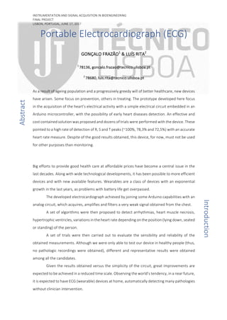
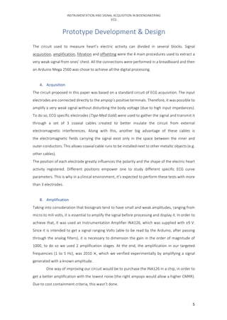
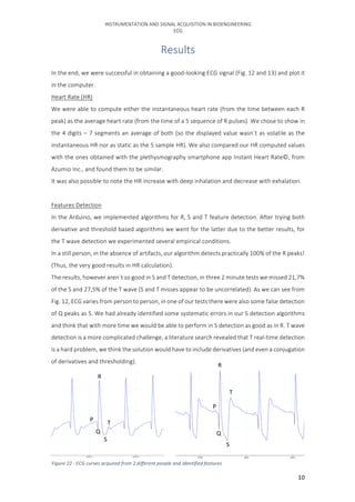
F. Complete Electric Circuit Scheme
After joining each block explained before, a final electric ECG circuit can be represented.
On the left side, the voltage source represents the expected signal from the body, 2
electrodes are connected to the chest of the person under clinical analysis. The 3rd
reference
electrode (left wrist) will be in contact with the circuit’s ground.
The circuit output signal is in the node V10 and is connected to an Arduino analog port.
Check PartSim reference [8], where all our ideas were brainstormed before implementation.
Figure 9 - Complete analogic circuitry
13. INSTRUMENTATION AND SIGNAL ACQUISITION IN BIOENGINEERING
ECG
13
Conclusion
Since the world's population is ageing and particular regions are becoming depopulated, the
healthcare assistance with continuous monitoring of physiological parameters (allowing
preventive care, which is more effective and less costly than treating) for patients who live in
remote locations can be a crucial device. Moreover, it would allow teleconsulting, patient
medication, diagnosis and the prevention of unnecessary specialist consultations or laboratory
examinations.
In this project, an electrocardiograph was designed and developed. A small prototype which
acquires, with three electrodes, one placed in the arm and 2 in the chest, the cardiac electrical
activity.
Despite the tremendous difficulty in recognizing the cardiac activity from the input signal
(acquired with the electrodes) we managed to greatly attenuate the noise and obtain a clean,
feature recognizable, final signal.
The signal acquisition and instrumentation part of the prototype is working properly, we obtain
a good-looking signal with a very simple circuit embedded in a highly portable system.
However, we fell short on the signal processing part, real-time feature detection algorithms
needed more time to be refined, and more controlled tests had to be performed to validate those
detections, this however was out of the scope of this course.
A future improvement, besides algorithm refinement, would be to attach a wireless emitter to
the system to allow signal monitoring without being connected to a computer. With the acquired
signal stored on a computer, harder algorithms for feature detection that can’t be performed on
real time, as Wavelet decomposition or Empirical Mode Detection, could also be used.