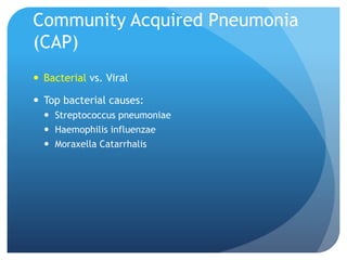
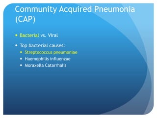
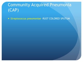
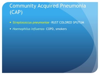
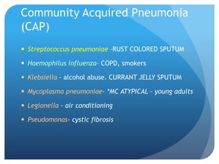
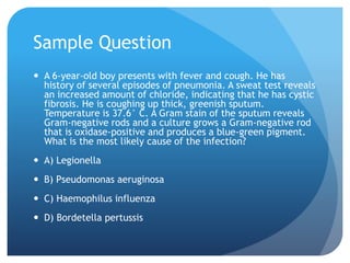
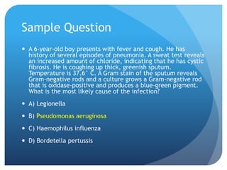
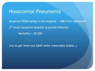
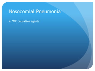
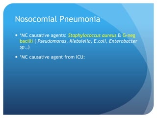
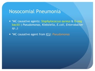
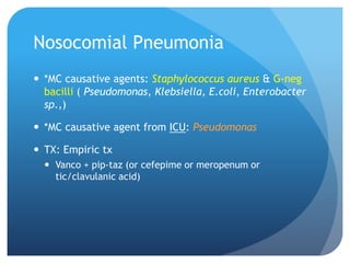
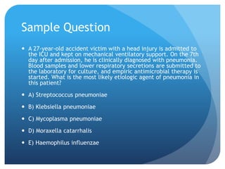
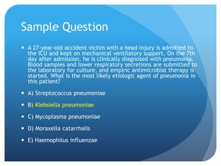
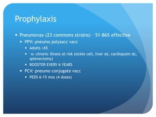
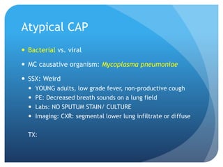
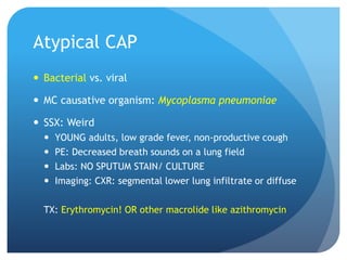
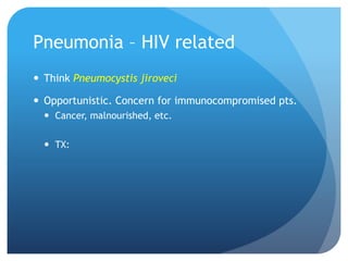
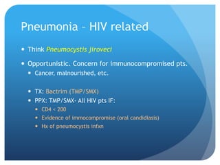
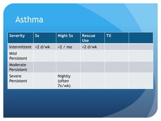
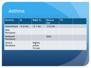
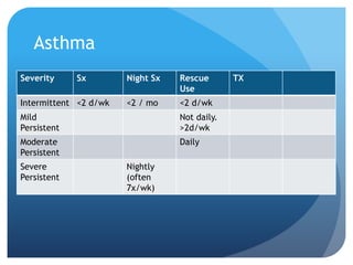
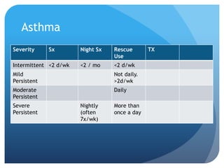
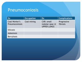
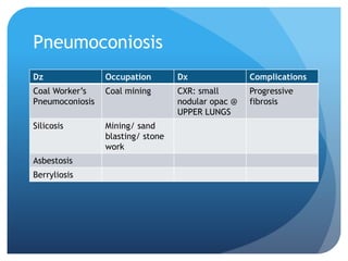
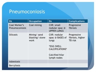
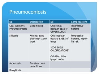
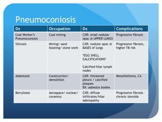
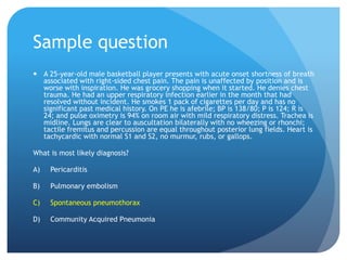
SlideShare a Scribd company logo
PULMONOLOGY REVIEW
Breathe. You’re going to be OK.
Community Acquired Pneumonia
(CAP)
 Bacterial vs. Viral
Community Acquired Pneumonia
(CAP)
 Bacterial vs. Viral
 Top bacterial causes:
Community Acquired Pneumonia
(CAP)
 Bacterial vs. Viral
 Top bacterial causes:
 Streptococcus pneumoniae
 Haemophilis influenzae
 Moraxella Catarrhalis
Community Acquired Pneumonia
(CAP)
 Bacterial vs. Viral
 Top bacterial causes:
 Streptococcus pneumoniae
 Haemophilis influenzae
 Moraxella Catarrhalis
Community Acquired Pneumonia
(CAP)
 Streptococcus pneumoniae –RUST COLORED SPUTUM
Community Acquired Pneumonia
(CAP)
 Streptococcus pneumoniae –RUST COLORED SPUTUM
 Haemophilus influenza- COPD, smokers
Community Acquired Pneumonia
(CAP)
 Streptococcus pneumoniae –RUST COLORED SPUTUM
 Haemophilus influenza- COPD, smokers
 Klebsiella – alcohol abuse. CURRANT JELLY SPUTUM
 Mycoplasma pneumoniae- *MC ATYPICAL – young adults
 Legionella – air conditioning
 Pseudomonas- cystic fibrosis
Community Acquired Pneumonia
(CAP)
 Viral cx:
 Influenza virus
 Respiratory syncytial virus (RSV)
 Adenovirus
 Parainfluenza virus
Sample question
 Some dude w/ hx of heavy alcohol use arrives in ER w/
10 day hx of increasing productive cough, fever, sweats.
On physical exam, patient coughs up currant jelly
sputum and crackles heard on left lower lung field. CXR
shows lobar infiltrates in lower left lobe. What is most
likely causative organism of the suspected diagnosis?
 A) Streptococcus pneumoniae
 B) Legionella sp.
 C) Parainfluenza virus
 D) Klebsiella
Sample Question
 A 47-year-old man presents with persistent chills following a
recent upper respiratory tract infection. The patient reports
cough with the production of yellowish sputum and myalgia.
The patient is febrile (37.9C), with a HR of 94 bmp and a
respiratory rate of 24/min. Knowing that the patient has had
a prolonged exposure to the air conditioned space in a large
office building, what may be the causative microbial agent in
this case?
 A) Klebsiella
 B) Mycoplasma
 C) Legionella
 D) Staphylococcus aureus
Sample Question
 A 6-year-old boy presents with fever and cough. He has
history of several episodes of pneumonia. A sweat test reveals
an increased amount of chloride, indicating that he has cystic
fibrosis. He is coughing up thick, greenish sputum.
Temperature is 37.6° C. A Gram stain of the sputum reveals
Gram-negative rods and a culture grows a Gram-negative rod
that is oxidase-positive and produces a blue-green pigment.
What is the most likely cause of the infection?
 A) Legionella
 B) Pseudomonas aeruginosa
 C) Haemophilus influenza
 D) Bordetella pertussis
To hospitalize or not?
 Hospitalize if…
 LOOKS BAD
 More than 1 lobe involved
 OLD + GOT ISSUES
 >50yo w/ comorbid or AMS
 WEAK
 Neutropenia, hemodynamically unstable, poor host resistance
CAP Treatment
 CAN be treated outpatient:
 Doxycycline/ Erythromycin/ Macrolides (Clarithromycin),
fluroquinolone
 IF >60yo, choose 2nd gen cephalosporin / levo + augmentin
 Should be hospitalized:
 Ceftriaxone / cefotaxime + azithromycin (or
fluroquinolone)
Nosocomial Pneumonia
Acquired FROM being in the hospital - >48h from admission
2nd most causative hospital acquired infection
Mortality = 20-50%
(try to get them out ASAP while reasonably stable…)
Nosocomial Pneumonia
 *MC causative agents:
Nosocomial Pneumonia
 *MC causative agents: Staphylococcus aureus & G-neg
bacilli ( Pseudomonas, Klebsiella, E.coli, Enterobacter
sp.,)
 *MC causative agent from ICU:
Nosocomial Pneumonia
 *MC causative agents: Staphylococcus aureus & G-neg
bacilli ( Pseudomonas, Klebsiella, E.coli, Enterobacter
sp.,)
 *MC causative agent from ICU: Pseudomonas
Nosocomial Pneumonia
 *MC causative agents: Staphylococcus aureus & G-neg
bacilli ( Pseudomonas, Klebsiella, E.coli, Enterobacter
sp.,)
 *MC causative agent from ICU: Pseudomonas
 TX: Empiric tx
 Vanco + pip-taz (or cefepime or meropenum or
tic/clavulanic acid)
Sample Question
 A 27-year-old accident victim with a head injury is admitted to
the ICU and kept on mechanical ventilatory support. On the 7th
day after admission, he is clinically diagnosed with pneumonia.
Blood samples and lower respiratory secretions are submitted to
the laboratory for culture, and empiric antimicrobial therapy is
started. What is the most likely etiologic agent of pneumonia in
this patient?
 A) Streptococcus pneumoniae
 B) Klebsiella pneumoniae
 C) Mycoplasma pneumoniae
 D) Moraxella catarrhalis
 E) Haemophilus influenzae
Prophylaxis
 Pneumovax (23 commons strains) – 51-86% effective
 PPV: pneumo polysacc vacc
 Adults >65
 w. chronic illness at risk (sickle cell, liver dz, cardiopulm dz,
splenectomy)
 BOOSTER EVERY 6 YEARS
 PCV: pneumo conjugate vacc
 PEDS 6-15 mos (4 doses)
Atypical CAP
 Bacterial vs. viral
Atypical CAP
 Bacterial vs. viral
 MC causative organism:
Atypical CAP
 Bacterial vs. viral
 MC causative organism: Mycoplasma pneumoniae
 SSX: Weird
 YOUNG adults, low grade fever, non-productive cough
 PE: Decreased breath sounds on a lung field
 Labs: NO SPUTUM STAIN/ CULTURE
 Imaging: CXR: segmental lower lung infiltrate or diffuse
TX:
Atypical CAP
 Bacterial vs. viral
 MC causative organism: Mycoplasma pneumoniae
 SSX: Weird
 YOUNG adults, low grade fever, non-productive cough
 PE: Decreased breath sounds on a lung field
 Labs: NO SPUTUM STAIN/ CULTURE
 Imaging: CXR: segmental lower lung infiltrate or diffuse
TX: Erythromycin! OR other macrolide like azithromycin
Atypical CAP
 Viral: influenza A/B, adenovirus, chlamydia
 TX: Supportive-
 Neuroaminidase inhibitor
 TAMIFLU (oseltamivir) – only good for 48hour window
Sample Question
A 22-year-old man presents with a 1-week history of fever, chills, dry or
mildly productive cough, and chest wall discomfort. He is on no medications,
but he has a history of mild asthma for which he does not need chronic
therapy. He states that he has been under a lot of pressure at work and has
not been sleeping very well. He decided to seek medical attention today
because he has not been improving and is concerned he might have
pneumonia. On examination, vitals include a temperature of 102.6ºF, BP:
126/76 mm Hg, P: 82 beats/min, R: 20/min. Lung exam reveals coarse
rhonchi throughout lung fields with rales in the right lower lobe with
egophony demonstrated over the right lower lobe. The patient is sent for PA
and lateral chest X-ray; he is found to have a right lower lobe infiltration.
Best empiric treatment for his illness?
 A) Amoxicillin 875 mg twice daily for 10 days
 B) Azithromycin 500 mg first dose, then 250mg daily for 4 days
 C) Ciprofloxacin 500 mg twice daily for 10 days
 D) Trimethoprim/sulfamethoxazole DS twice daily for 10 days
Pneumonia – HIV related
 Think what organism?
Pneumonia – HIV related
 Think Pneumocystis jiroveci
 Opportunistic. Concern for immunocompromised pts.
 Cancer, malnourished, etc.
 TX:
Pneumonia – HIV related
 Think Pneumocystis jiroveci
 Opportunistic. Concern for immunocompromised pts.
 Cancer, malnourished, etc.
 TX: Bactrim (TMP/SMX)
 PPX: TMP/SMX- All HIV pts IF:
 CD4 < 200
 Evidence of immocompromise (oral candidiasis)
 Hx of pneumocystis infxn
Sample Question
 A 56-year-old man presents for a routine follow-up regarding his
positive HIV status. He is compliant with his medications and has
been feeling well. In addition to his antivirals, he takes daily
trimethoprim/sulfamethoxazole for Pneumocystis jiroveci
pneumonia (PCP) prophylaxis.
 What detail in his history would warrant prophylaxis?
A) CD4 cell count < 400
B) History of previous PCP infection
C) CD4 cell count > 200
D) History of previous pneumonia infection
E) HIV viral load >100,000 copies/mL
Tuberculosis
 What kind of precautions?
Tuberculosis
 What kind of precautions? Airborne
 Organism:
Tuberculosis
 What kind of precautions? Airborne
 Organism: Mycobacterium tuberculosis
 Background:
Tuberculosis
 What kind of precautions? Airborne
 Organism: Mycobacterium tuberculosis
 Background: COUGH. FEVER. NIGHT SWEATS. ANOREXIA/
WEIGHT LOSS.
Tuberculosis
 What kind of precautions? Airborne
 Organism: Mycobacterium tuberculosis
 Background: COUGH. FEVER. NIGHT SWEATS. ANOREXIA/
WEIGHT LOSS.
 Review primary vs. latent/reactivated vs. healed
primary
 Highlight: latent/ reactivated/ secondary TB –
 loc: CXR: *APICES of lungs/ upper lobe cavitary dz; reactivated
at immunocompromise
 Highly tested: Tuberculin Skin Test
Sample Question
 A 45-year-old man presents with a fever accompanied
by a productive cough. He has had the symptoms for
several weeks. His temperature rises in the evenings,
and he has experienced weight loss. The chest X-ray
shows upper lobe cavitary lesions. What is the most
likely diagnosis?
 A) Secondary tuberculosis
 B) Asbestosis
 C) Pneumocystis carinii pneumonia
 D) Silicosis
PPD
 POSITIVE (induration diameter):
 >5mm = HIV+, recent active TB contact, new transplant
 >10mm = new immigrant, IVDA (HIV-), DM, CKD, close
quarters (college, military, prisons)
 >15mm = no risk factors
Sample Questions
 30yo male HIV+ with PPD test showed induration of
7mm. Positive or Negative PPD?
Sample Questions
 30yo male HIV+ with PPD test showed induration of
7mm. Positive or Negative PPD?
 70yo female recently immigrated from Cambodia had
PPD test showing induration of 8mm. Positive or
negative PPD?
Sample Questions
 30yo male HIV+ with PPD test showed induration of
7mm. Positive or Negative PPD?
 70yo female recently immigrated from Cambodia had
PPD test showing induration of 8mm. Positive or
negative PPD?
 47yo male with DM and CKD going to college for his
second bachelor’s and living at dorms. PPD induration
12mm. Positive or negative?
 28 yo female w/ asthma about to work at hospital. PPD
showed induration of 12mm. Positive or negative?
Tuberculosis
 Sputum stain:
Tuberculosis
 Sputum stain: acid fast bacilli
 (*supports dx but does not confirm!* )
 Ways to have definite diagnosis that identifies M.
tuberculosis:
Tuberculosis
 Sputum stain: acid fast bacilli
 (*supports dx but does not confirm!* )
 Ways to have definite diagnosis that identifies M.
tuberculosis:
 Sputum Culture
 DNA/RNA amplification (PCR)
 Lung Biopsy- histologic hallmark:
Tuberculosis
 Sputum stain: acid fast bacilli
 (*supports dx but does not confirm!* )
 Ways to have definite diagnosis that identifies M.
tuberculosis:
 Sputum Culture
 DNA/RNA amplification (PCR)
 Lung Biopsy- histologic hallmark: caseating (necrotizing)
granulomas
 Vs. Sarcoidosis?
Acute bronchiolitis
 Who:
Acute bronchiolitis
 Who: Infants
 Causative agent:
Acute bronchiolitis
 Who: Infants
 Causative agent: RSV
 TX:
Acute bronchiolitis
 Who: Infants
 Causative agent: RSV
 TX: premies- hospital & ribavirin
 Otherwise, supportive
Acute epiglottitis
(2-7 yo, 45-65yo)
 Which vaccination has decreased incidence?
Acute epiglottitis
 Which vaccination has decreased incidence?
 Haemophilus influenzae type B (Hib)
 SSX:
Acute epiglottitis
 Which vaccination has decreased incidence?
 Haemophilus influenzae type B (Hib)
 SSX: Tripoding*. Drooling. Sudden high fever, resp
distress.
 Tripoding: ?
What do you do FIRST?
Acute epiglottitis
 Which vaccination has decreased incidence?
 Haemophilus influenzae type B (Hib)
 SSX: Tripoding*. Drooling. Sudden high fever, resp
distress.
 Tripoding: sitting upright + neck extended
What do you do FIRST?
Acute epiglottitis
 Which vaccination has decreased incidence?
 Haemophilus influenzae type B (Hib)
 SSX: Tripoding*. Drooling. Sudden high fever, resp
distress.
 Tripoding: sitting upright + neck extended
What do you do FIRST? SECURE AIRWAY (intubate)
What do you see on lateral neck XR? :
Acute epiglottitis
 Which vaccination has decreased incidence?
 Haemophilus influenzae type B (Hib)
 SSX: Tripoding*. Drooling. Sudden high fever, resp
distress.
 Tripoding: sitting upright + neck extended
What do you do FIRST? SECURE AIRWAY (intubate)
What do you see on lateral neck XR? : THUMBPRINT SIGN
Thumbprint sign
Sample Question
 3 year old child presents to ER with sudden onset of
fever, difficulty swallowing, drooling, and dyspnea.
Exam reveals febrile child who is sitting, leaning
forward with his neck extended. Chest exam reveals
soft stridor with inspiratory retractions. What is the
next step in management of this patient?
A) Treatment with nebulized albuterol
B) Treatment with nebulized epinephrine
C) Inspection and intubation under controlled conditions
D) IV acetazolamide therapy
Croup
 Aka acute laryngotracheobronchitis
 Affects children 6mos to 5yo
 *MC causative agent:
Croup
 Aka acute laryngotracheobronchitis
 Affects children 6mos to 5yo
 *MC causative agent: parainfluenzae virus types 1 & 2
 (Note: CAN be caused by RSV, but parainfluenzae is MC)
 Buzz SSX:
Croup
 Aka acute laryngotracheobronchitis
 Affects children 6mos to 5yo
 *MC causative agent: parainfluenzae virus types 1 & 2
 (Note: CAN be caused by RSV, but parainfluenzae is MC)
 Buzz SSX: seal-like, barking cough
 Imaging:
Croup
 Aka acute laryngotracheobronchitis
 Affects children 6mos to 5yo
 *MC causative agent: parainfluenzae virus types 1 & 2
 (Note: CAN be caused by RSV, but parainfluenzae is MC)
 Buzz SSX: seal-like, barking cough
 Imaging: PA neck film- Steeple sign
Steeple sign
Croup
 Tx:
 Mild = NO stridor at rest = SUPPORTIVE
 Moderate to Severe = Stridor AT REST =
Croup
 Tx:
 Mild = NO stridor at rest = SUPPORTIVE
 Moderate to Severe = Stridor AT REST = Racemic EPI
(nebulized) & hospitalization
Sample Question
 A 3-year-old boy presents with difficulty in breathing and a
cough that sounds like a seal. On examination, the child has
fever, a harsh barking cough, a respiratory rate of 38/minute,
and minimal stridor on agitation. On lung auscultation, there
are no rales or wheezing. On cardiac auscultation, there is
tachycardia. Radiological examination reveals the so-called
'steeple sign'. What is the most likely diagnosis?
 A) Bronchiolitis
 B) Croup
 C) Epiglottitis
 D) Foreign body aspiration
Sample Question
 4 year old child presents to ER with low-grade fever,
barking cough, respiratory stridor with activity but not
at rest. On exam, you note cough and absence of
drooling. What is the most appropriate treatment for
this child?
 A) Dexamethasone IM
 B) Endotracheal intubation and IV antibiotics
 C) Nebulized racemic epinephrine
 D) Supportive therapy with oral hydration
Obstructive Lung Dz
 Asthma
 COPD
 Bronchiectasis
 Cystic Fibrosis
Obstructive Lung Dz: Asthma
 Classification! & Step Up therapy: What are classes?
Obstructive Lung Dz: Asthma
 Classification! & Step Up therapy:
 Intermittent
 Mild persistent
 Moderate persistent
 Severe persistent
Asthma
Severity Sx Night Sx Rescue
Use
TX
Intermittent <2 d/wk <2 / mo <2 d/wk
Mild
Persistent
Moderate
Persistent
Severe
Persistent
Asthma
Severity Sx Night Sx Rescue
Use
TX
Intermittent <2 d/wk <2 / mo <2 d/wk
Mild
Persistent
Moderate
Persistent
Severe
Persistent
Nightly
(often
7x/wk)
Asthma
Severity Sx Night Sx Rescue
Use
TX
Intermittent <2 d/wk <2 / mo <2 d/wk
Mild
Persistent
Moderate
Persistent
Daily
Severe
Persistent
Nightly
(often
7x/wk)
Asthma
Severity Sx Night Sx Rescue
Use
TX
Intermittent <2 d/wk <2 / mo <2 d/wk
Mild
Persistent
Not daily.
>2d/wk
Moderate
Persistent
Daily
Severe
Persistent
Nightly
(often
7x/wk)
Asthma
Severity Sx Night Sx Rescue
Use
TX
Intermittent <2 d/wk <2 / mo <2 d/wk
Mild
Persistent
Not daily.
>2d/wk
Moderate
Persistent
Daily
Severe
Persistent
Nightly
(often
7x/wk)
More than
once a day
Asthma
Severity Sx Night Sx Rescue
Use
TX
Intermittent <2 d/wk <2 / mo <2 d/wk
Mild
Persistent
>2d/wk 3-4 x/mo
(once a
wk)
Not daily.
>2d/wk
Moderate
Persistent
Some
limits ADL
>1 /wk but
not nightly
Daily
Severe
Persistent
Extremely
limited
phys act
Nightly
(often
7x/wk)
More than
once a day
Asthma
Severity Sx Night Sx Rescue
Use
TX Lung Fxn
Intermittent <2 d/wk <2 / mo <2 d/wk SABA
Mild
Persistent
>2d/wk 3-4 x/mo
(once a
wk)
Not daily.
>2d/wk
SABA + ICS
Moderate
Persistent
Some
limits ADL
>1 /wk but
not nightly
Daily SABA +ICS +
LABA
Severe
Persistent
Extremely
limited
phys act
Nightly
(often
7x/wk)
More than
once a day
PO
steroids,
hospital if
needed
Asthma
Severity Sx Night Sx Rescue
Use
TX Lung Fxn
Intermittent <2 d/wk <2 / mo <2 d/wk SABA FEV1>80%
predicted;
FEV1/FVC
normal
Mild
Persistent
>2d/wk 3-4 x/mo
(once a
wk)
Not daily.
>2d/wk
SABA + ICS “ “
Moderate
Persistent
Some
limits ADL
>1 /wk but
not nightly
Daily SABA +ICS +
LABA
FEV1>60%
but <80%
predict
FEV1/FVC
reduced 5%
Severe
Persistent
Extremely
limited
phys act
Nightly
(often
7x/wk)
More than
once a day
PO
steroids,
hospital if
needed
FEV1<60%
predicted
FEV1/FVC
reduced
Asthma
 Spirometry:
 Diagnostic if FEV1 decrease >20%
 Supportive of dx if
 FEV1/FVC = <75%
 FEV1 s/p bronchodilation = >10% decrease
Sample Question
 9 year old boy w/ hx of asthma presents with nocturnal
coughing occurring every night along with daily
exacerbations of wheezing and shortness of breath. How
would his asthma be classified?
 A) Intermittent
 B) Mild persistent
 C) Moderate persistent
 D) Severe persistent
Sample Question
 9 year old boy w/ hx of asthma presents with nocturnal
coughing occurring every night along with daily
exacerbations of wheezing and shortness of breath. How
would his asthma be classified?
 A) Intermittent
 B) Mild persistent
 C) Moderate persistent
 D) Severe persistent
COPD
 Stop smoking
 CXR: emphysema: ___________________?
COPD
 Stop smoking
 CXR: emphysema: parenchymal bullae/ subpleural blebs
 PATHOGNOMONIC !
 Vs. chronic bronchitis: nonspecific peribronchial & perivasc
markings
Key medication: anticholinergic: ?
COPD
 Stop smoking
 CXR: emphysema: parenchymal bullae/ subpleural blebs
 PATHOGNOMONIC !
 Vs. chronic bronchitis: nonspecific peribronchial & perivasc
markings
Key medication: anticholinergic: ipatropium / tiotropium
(Spiriva = tiotropium bromide)
PPX measures: ?
COPD
 Stop smoking
 CXR: emphysema: parenchymal bullae/ subpleural blebs
 PATHOGNOMONIC !
 Vs. chronic bronchitis: nonspecific peribronchial & perivasc
markings
Key medication: anticholinergic: ipatropium / tiotropium
(Spiriva = tiotropium bromide)
PPX measures: pneumococcal vaccine & annual flu
COPD
 What is the only therapy known to increase life
expectancy in COPD patients with chronic hypoxemia?
 A) Antibiotics
 B) Bronchodilator therapy
 C) Inhaled corticosteroids
 D) Home Oxygen
COPD
 What is the only therapy known to increase life
expectancy in COPD patients with chronic hypoxemia?
 A) Antibiotics
 B) Bronchodilator therapy
 C) Inhaled corticosteroids
 D) Home Oxygen
Bronchiectasis
 Think of what disease?
Bronchiectasis
 Think CYSTIC FIBROSIS
 50% of cases
SSX: ?
Bronchiectasis
 Think CYSTIC FIBROSIS
 50% of cases
SSX: Foul-smelling sputum/ hemoptysis/ recurrent pneumonia
Localized chest crackles/ clubbing
Imaging:
- High resolution CHEST CT: dilated tortuous airways
- CXR: crowded bronchial markings, basal cystic spaces
- *Tram-track lung markings, honeycombing, atelectasis
Tx: chest physio, bronchodilators, abx (10-14d for acute)
Cystic Fibrosis
 Autosomal recessive – CFTR gene
 Young, chronic lung dz, pancreatitis. Sputum excess,
steatorrhea, clubbing
 CXR: bronchiectasis
 Dx: sweat chloride test (pilocarpine ionotophoresis)
 Definitive: DNA testing
 TX: multidisciplinary.
 Acetylcysteine = mucolytic. (Mucomyst)
Sample Question
 17 year old girl with hx of CF presents with chronic
cough productive of copious, foul smelling, purulent
sputum. Patient is afebrile and lung exam reveals
crackles at lung bases bilaterally. What is the most
likely diagnosis?
 A) Asthma
 B) Bronchiectasis
 C) Bronchiolitis
 D) Croup
Restrictive Lung Dz
 Pneumoconiosis
 Sarcoidosis
Pneumoconiosis
Dz Occupation Dx Complications
Coal Worker’s
Pneumoconiosis
Silicosis
Asbestosis
Berryliosis
Pneumoconiosis
Dz Occupation Dx Complications
Coal Worker’s
Pneumoconiosis
Coal mining CXR:
Silicosis
Asbestosis
Berryliosis
Pneumoconiosis
Dz Occupation Dx Complications
Coal Worker’s
Pneumoconiosis
Coal mining CXR: small
nodular opac @
UPPER LUNGS
Progressive
fibrosis
Silicosis
Asbestosis
Berryliosis
Pneumoconiosis
Dz Occupation Dx Complications
Coal Worker’s
Pneumoconiosis
Coal mining CXR: small
nodular opac @
UPPER LUNGS
Progressive
fibrosis
Silicosis Mining/ sand
blasting/ stone
work
Asbestosis
Berryliosis
Pneumoconiosis
Dz Occupation Dx Complications
Coal Worker’s
Pneumoconiosis
Coal mining CXR: small
nodular opac @
UPPER LUNGS
Progressive
fibrosis
Silicosis Mining/ sand
blasting/ stone
work
CXR: nodular
opac @ BASES of
lungs
*EGG SHELL
CALCIFICATIONS*
Calcified hilar
lymph nodes
Progressive
fibrosis, higher
TB risk
Asbestosis Construction/
demolition
Berryliosis
Pneumoconiosis
Dz Occupation Dx Complications
Coal Worker’s
Pneumoconiosis
Coal mining CXR: small nodular
opac @ UPPER LUNGS
Progressive fibrosis
Silicosis Mining/ sand
blasting/ stone work
CXR: nodular opac @
BASES of lungs
*EGG SHELL
CALCIFICATIONS*
Calcified hilar lymph
nodes
Progressive fibrosis,
higher TB risk
Asbestosis Construction/
demolition
CXR: thickened
pleura / calcified
plaques
BX: asbestos bodies
Mesothelioma, CA
Berryliosis Aerospace/ nuclear/
ceramics
CXR: diffuse
infiltrates/hilar
adenopathy
Progressive fibrosis –
chronic steroids
Pneumoconiosis
 Tx:. Corticosteroids. Supportive
 Stop smoking
Sample Question
 A 59-year-old man presents with a 3-month history of progressive
exertional dyspnea. He has been experiencing dry cough, but
denies any history of fever, chest pain, or weight loss. On further
questioning, you discover that he works in the ceramic industry
at the outskirts of town. He is a non-smoker and drinks alcohol
very occasionally. Examination reveals bibasilar crackles, and
pulmonary function test indicates FEV1 of 67%, FVC of 73%, and
TLC of 75% with DLCO of 65%. Chest X-ray shows "eggshell
calcification" of hilar lymph nodes.
A) Asbestosis
B) Silicosis
C) Coal Worker’s Pneumoconiosis
D) Berylliosis
Sarcoidosis
 GRUELING mnemonic
 G
 R
 U
 E
 L
 I
 N
 G
Sarcoidosis
 GRUELING mnemonic
 Granulomas (what kind? Vs tuberculosis…)
 ARthritis
 Uveitis
 Erythema nodosum
 Lymphadenopathy
 Interstitial fibrosis
 Negative TB
 Gammaglobulinemia
Sarcoidosis
 GRUELING mnemonic
 Granulomas (NON-CASEATING)
 ARthritis
 Uveitis
 Erythema nodosum
 Lymphadenopathy
 Interstitial fibrosis
 Negative TB
 Gammaglobulinemia
Sarcoidosis
 Confirmatory test for diagnosis:
Sarcoidosis
 Confirmatory test for diagnosis: Fine needle node biopsy
Sarcoidosis
 Confirmatory test for diagnosis: Fine needle node biopsy
 NONCASEATING GRANULOMAS
TX: 90% responsive to what? Think multi-organ inflammation.
Sarcoidosis
 Confirmatory test for diagnosis: Fine needle node biopsy
 NONCASEATING GRANULOMAS
TX: 90% responsive to what? Think multi-organ inflammation.
CORTICOSTEROIDS
Sample Question
 A 32-year-old African-American woman, with no significant past medical history, has
been referred to a pulmonologist; she presents with a 2-month history of progressive
dyspnea. She notes associated low-grade fever, malaise, joint pain, and swollen neck
glands. She denies a history of travel, cigarette smoking, drug use, or sexually-
transmitted diseases (she has not been sexually active in the past year). All other
reviews of systems are negative. Her physical exam reveals tender, nodular formations
on her anterior lower extremities, parotid enlargement, hepatosplenomegaly, and
cervical lymphadenopathy. Her vital signs, heart, and lungs are unremarkable.
Diagnostic testing reveals leukopenia, increased ESR, hypercalcemia, hypercalciuria,
elevations of serum ACE levels, and bilateral hilar adenopathy with diffuse reticular
infiltrates. ANCA, ANA, and rheumatoid factor tests are negative. Histological
assessment confirms the presence of noncaseating granulomas.
What is the most likely diagnosis?
 A) Sarcoidosis
 B) Tuberculosis
 C) Pnemocystis jiroveci pneumonia
 D) Idiopathic pulmonary fibrosis
Pleural Dz:
 Pleural effusion
 Pneumothorax
 Pulmonary embolism
Pleural Effusion
 *MC cause:
Pleural Effusion
 *MC cause: CHF
 Transudate vs. Exudate
Pleural Effusion
 *MC cause: CHF
 Transudate vs. Exudate
Pleural Effusion
 *MC cause: CHF
 Transudate > Exudate
 Transudate: “intact capillaries” – hold protein in, so
pleural fluid is low protein
 CHF. Atelectasis. Renal/Liver dz (Cirrhosis).
 Exudate: “leaky capillaries” – pleural fluid high protein
 Infection. Malignancy. Trauma. Pulmonary Embolism.
Pleural Effusion
 What is the GOLD STANDARD for diagnosis (also
therapeutic) ?
Pleural Effusion
 What is the GOLD STANDARD for diagnosis (also
therapeutic) ? Thoracentesis
Pleural Effusion
 What is the GOLD STANDARD for diagnosis (also
therapeutic) ? Thoracentesis
 Light’s Criteria !
Pleural Effusion: Light’s
Transudate if pleural fluid: Exudate if pleural fluid:
Protein < 3 g/dL
Glucose >60 mg/dl
WBC’s <1,000
LDH <200 IU/L
> 3g/dL
< 60mg/dL
> 1,000
> 200 IU/L
OR
-Fluid protein: serum protein =
<0.5
- Fluid LDH: Serum LDH = <0.6
- Fluid LDH >2/3 of upper limit of
normal serum LDH
= <200 iU/mL
>0.5
>0.6
> 200 iu/mL
Sample Question
60 yr old man w/ hx of hypertension and left ventricular
hypertrophy presents with shortness of breath. Exam
reveals dullness to percussion bilaterally with decreased
breath sounds. Pleural fluid is aspirated and analyzed.
Which of the following results is consistent with his most
likely diagnosis?
A) Glucose 40mg/dL
B) LDH 300 iU/L
C) Protein 2.5 mg/dL
D) WBC 2,000
Pneumothorax
 Pt background:
Pneumothorax
 Pt background: TALL, THIN MALE (10-30yo)
 Increased or decreased fremitus?
Pneumothorax
 Pt background: TALL, THIN MALE (10-30yo)
 Increased or decreased fremitus?
Pneumothorax
 Pt background: TALL, THIN MALE (10-30yo)
 Increased or decreased fremitus?
 What happens to mediastinum?
Pneumothorax
 Pt background: TALL, THIN MALE (10-30yo)
 Increased or decreased fremitus?
 What happens to mediastinum? Shifts contralaterally
 What happens to the trachea? (late sign)
Pneumothorax
 Pt background: TALL, THIN MALE (10-30yo)
 Increased or decreased fremitus?
 What happens to mediastinum? Shifts contralaterally
 What happens to the trachea? (late sign) Deviates
contralaterally
 What do you do if high suspicion of tension ptx? (this is a
medical emergency!)
Pneumothorax
 Pt background: TALL, THIN MALE (10-30yo)
 Increased or decreased fremitus?
 What happens to mediastinum? Shifts contralaterally
 What happens to the trachea? (late sign) Deviates
contralaterally
 What do you do if high suspicion of tension ptx? (this is a
medical emergency!) Needle decompression+ chest tube
 *note: thoraCOSTomy vs thoraCOTomy
 What do you see on CXR?
Pneumothorax
 Pt background: TALL, THIN MALE (10-30yo)
 Increased or decreased fremitus?
 What happens to mediastinum? Shifts contralaterally
 What happens to the trachea? (late sign) Deviates
contralaterally
 What do you do if high suspicion of tension ptx? (this is a
medical emergency!) Needle decompression+ chest tube
 *note: thoraCOSTomy vs thoraCOTomy
 What do you see on CXR? Pleural line
Pneumothorax
Sample question
 A 25-year-old male basketball player presents with acute onset shortness of breath
associated with right-sided chest pain. The pain is unaffected by position and is
worse with inspiration. He was grocery shopping when it started. He denies chest
trauma. He had an upper respiratory infection earlier in the month that had
resolved without incident. He smokes 1 pack of cigarettes per day and has no
significant past medical history. On PE he is afebrile; BP is 138/80; P is 124; R is
24; and pulse oximetry is 94% on room air with mild respiratory distress. Trachea is
midline. Lungs are clear to auscultation bilaterally with no wheezing or rhonchi;
tactile fremitus and percussion are equal throughout posterior lung fields. Heart is
tachycardic with normal S1 and S2, no murmur, rubs, or gallops.
What is most likely diagnosis?
A) Pericarditis
B) Pulmonary embolism
C) Spontaneous pneumothorax
D) Community Acquired Pneumonia
Pulmonary Embolism
 Majority resulting from ?
Pulmonary Embolism
 Majority resulting from DVT
 Virchow’s Triad?
 Other causes:
 Central line  air emboli
 Long bone (femur) fracture  fat emboli
 Active labor  amniotic fluid emboli
What do you see on EKG ?
Pulmonary Embolism
 Majority resulting from DVT
 Virchow’s Triad: V (Venous stasis) , I (Increased
coagulability), R (recent injury, reduced activity)
 Other causes:
 Central line  air emboli
 Long bone (femur) fracture  fat emboli
 Active labor  amniotic fluid emboli
What do you see on EKG ? S1Q3T3
Normal EKG
Normal EKG vs Pulm Embolism
Pulmonary Embolism
 What lab can help rule out PE?
Pulmonary Embolism
 What lab can help rule out PE? D-Dimer
 Highly SENSITIVE. NOT specific.
 What is the INITIAL imaging test of choice for suspected
PE?
Pulmonary Embolism
 What lab can help rule out PE? D-Dimer
 Highly SENSITIVE. NOT specific.
 What is the INITIAL imaging test of choice for suspected
PE?
 SPIRAL CT
 What is DEFINITIVE imaging test for PE?
Pulmonary Embolism
 What lab can help rule out PE? D-Dimer
 Highly SENSITIVE. NOT specific.
 What is the INITIAL imaging test of choice for suspected
PE?
 SPIRAL CT
 What is DEFINITIVE imaging test for PE? Pulmonary
angiogram
Anticoagulation! – Heparin & warfarin!
If you can’t anticoagulate, what can you do?
Pulmonary Embolism
 What lab can help rule out PE? D-Dimer
 Highly SENSITIVE. NOT specific.
 What is the INITIAL imaging test of choice for suspected
PE?
 SPIRAL CT
 What is DEFINITIVE imaging test for PE? Pulmonary
angiogram
Anticoagulation! – Heparin & warfarin!
If you can’t anticoagulate, what can you do? Vena cava filter
Sample Question
A 45-year-old man presents with a 30-minute history of chest
pain; it began while he was on a long airline flight. He denies
any trauma to the chest. On exam, his pulse is 110 BPM;
respirations are 40/min. The chest radiograph reveals a wedge-
shaped opacity.
What is most likely cause of his chest pain?
A) Esophageal reflux
B) Pneumonia
C) Aortic dissection
D) Pulmonary embolism
Acute Resp Distress Syndrome
(ARDS)
 Pt background:
Acute Resp Distress Syndrome
(ARDS)
 Pt background:
 Severe multiple trauma
 Aspiration of gastric contents
 SEPSIS
 Shock/ toxic inhalation/ near-DROWNING
SX: tachypnea. FROTHY PINK/ RED SPUTUM.
CXR: Think fluid. Inflammation. Alveolar collapse.
*air bronchograms, patchy bilateral infiltrates that spare the
costophrenic angles.*
Normal CXR
ARDS
ARDS
 TX:
ARDS
 TX: Endotracheal intubation-
 POS PRESSURE VENT + low PEEP (Positive End- Expiratory
Pressure)
Sample Question
 A 25-year-old man presents at the hospital after a car
accident. He is intubated and placed on a ventilator. He
becomes progressively difficult to oxygenate despite
increasing the PEEP and the oxygen supply to 100%. Patient
remains afebrile. He dies several days later. At autopsy, the
lung shows diffuse hyaline membranes in the alveoli,
thickened alveolar walls, and many alveolar macrophages,
but few neutrophils. What condition did this patient have?
 A) Adult Respiratory Distress Syndrome
 B) Chronic bronchitis
 C) Bronchiectasis
 D) Viral pneumonia
Foreign Body Aspiration
 What is diagnostic and treatment of choice for removal?
Foreign Body Aspiration
 What is diagnostic and treatment of choice for removal?
 Bronchoscopy
Acute aspiration of gastric contents can lead to what?
Foreign Body Aspiration
 What is diagnostic and treatment of choice for removal?
 Bronchoscopy
Acute aspiration of gastric contents can lead to what?
ARDS !
Sample Question
 5 year old previously healthy presents with acute onset
of respiratory distress following ingestion of a piece of
candy. Which of the following signs is most ominous?
 A) Aphonia
 B) Cough
 C) Drooling
 D) Stridor
Sample Question
 5 year old previously healthy presents with acute onset
of respiratory distress following ingestion of a piece of
candy. Which of the following signs is most ominous?
 A) Aphonia
 B) Cough
 C) Drooling
 D) Stridor
Hyaline Membrane Dz
 *MC cause of respiratory dz in preterm infant!
 = deficiency of what?
Hyaline Membrane Dz
 *MC cause of respiratory dz in preterm infant!
 = deficiency of what? SURFACTANT
 PPX or rescue TX: exogenous surfactant in delivery room
 Synchronized intermittent ventilation
 For high risk premature-antenatal CS given to weeks 24-34
wks
Sample Question
 Premature infant born at 32 wks develops rapid shallow
respirations at 60/min, grunting retractions, and
duskiness of skin. CXR shows diffuse bilateral
atelectasis, ground glass appearance, and air
bronchograms. What is the most likely diagnosis?
A) Hyaline membrane disease
B) Acute laryngotracheobronchitis
C) Bronchiolitis
D) Pulmonary embolism
Neoplastic Dz
 Bronchogenic Carcinoma
General: #1 leading cause of CA death in men & women
(>colon, breast, & prostate combined)
**Smoking is #1 factor**
 SCLC vs NSCLC
 Pulmonary nodule
 Carcinoid Tumor
Small Cell (SCLC)
 Worst. “Small Cell Sucks” (worst prognosis)– use
alliteration to jog memory
 S-Spreads early, Super aggressive, Central bronchi
 Can’t even do Surgery
 TX: *COMBO Radiation & Chemo!*
Non small Cell (NSCLC)
 NOT small cell, so NOT as aggressive.
SLOWER growth. & more amenable to Surgery.
Tx: *Surgery
3 Types:
1. Adenocarcinoma (35-50%)
2. Squamous Cell Carcinoma (25-35%)
3. Large Cell Carcinoma
Non small Cell (NSCLC)
 NOT small cell, so NOT as aggressive.
SLOWER growth. & more amenable to Surgery.
TX: * Surgery
3 Types:
1. Adenocarcinoma  MC* CA type in NON-SMOKERS
 Appears in lung periphery (“Adeno” – “Away”)
2. Squamous Cell Carcinoma
3. Large Cell Carcinoma
Non small Cell (NSCLC)
 NOT small cell, so NOT as aggressive.
SLOWER growth. & more amenable to Surgery.
Tx: *Surgery
3 Types:
1. Adenocarcinoma  MC* CA type in NON-SMOKERS
 Appears in lung periphery (“Adeno” – “Away”)
2. Squamous Cell Carcinoma
 Bronchial & centrally located mass
 Likely present w/ hemoptysis
3. Large Cell Carcinoma
Non small Cell (NSCLC)
 NOT small cell, so NOT as aggressive.
SLOWER growth. & more amenable to Surgery.
TX: Surgery*
3 Types:
1. Adenocarcinoma  MC* CA type in NON-SMOKERS
 Appears in lung periphery (“Adeno” – “Away”)
2. Squamous Cell Carcinoma
 Bronchial & centrally located mass
 Likely present w/ hemoptysis
3. Large Cell Carcinoma
 Cytology: large cells
 Central OR peripheral mass
Drawing
Sample Question
 A 52-year-old man presents to discuss the results of his recent lung
biopsy. You saw him 3 weeks earlier due to his experiencing dyspnea. He
has no other significant past medical history. His chest X-ray reveals a 3-
centimeter (diameter) mass in the right upper lobe near the hilum. A CT
scan of his chest, abdomen, pelvis, and head reveals only the mass seen
on X-ray. His blood counts and blood chemistries are normal. He
underwent bronchoscopic biopsy of the lesion, which reveals small cell
lung cancer. What is the most appropriate course of treatment?
 A) Radiation only
 B) Radiation followed by chemotherapy
 C) Radiation followed by surgical resection
 D) Surgical resection only
 E) Comfort measures only
Sample Question
 A 60-year-old woman presents with a history of persistent cough. She is confined
to her bed; walking over 10 paces causes severe breathlessness. She has no energy
to carry out any of her regular activities. She has never smoked, and she drinks
the occasional glass of wine. On physical examination, she is found to have
decreased breath sounds and dullness to percussion over her right lower thorax.
Further evaluation reveals an irregular mass in the periphery of the right lung base
with a right sided pleural effusion. A needle is inserted into the pleural space and
divulges blood stained fluid.
 If results prove to be a malignancy, what is the most likely sub-type considering
she has never smoked?
 A) Small Cell Carcinoma
 B) Large Cell Carcinoma
 C) Squamous Cell Carcinoma
 D) Adenocarcinoma
Complications of Lung CA
 SPHERE mnemonic
 S
 P
 H
 E
 R
 E
Complications of Lung CA
 SPHERE mnemonic
 SVC Syndrome (compression of SVC excess fluid/blood, HA,
AMS
 Pancoast Tumor (lung apex tumor, horner’s syndrome +
shoulder pain, affects brachial plexus + cervical sympathetic
nerve)
 Horner’s Syndrome ( ?)
 Endocrine (Carcinoid syndrome ?)
 Recurrent laryngeal nerve (hoarseness)
 Effusion ( what kind?)
Complications of Lung CA
 SPHERE mnemonic
 SVC Syndrome (compression of SVC excess fluid/blood, HA,
AMS
 Pancoast Tumor (lung apex tumor, horner’s syndrome +
shoulder pain, affects brachial plexus + cervical sympathetic
nerve)
 Horner’s Syndrome (unilat facial ptosis, miosis, anhidrosis)
 Endocrine (Carcinoid syndrome?)
 Recurrent laryngeal nerve (hoarseness)
 Effusion ( what kind?)
Complications of Lung CA
 SPHERE mnemonic
 SVC Syndrome (compression of SVC excess fluid/blood, HA,
AMS
 Pancoast Tumor (lung apex tumor, horner’s syndrome + shoulder
pain, affects brachial plexus + cervical sympathetic nerve)
 Horner’s Syndrome (unilat facial ptosis, miosis, anhidrosis)
 Endocrine (Carcinoid syndrome- FDR: flushing/ diarrhea/ R-side
heart fail)
 Recurrent laryngeal nerve (hoarseness)
 Effusion ( exudative)
Pulmonary Nodule
Aka coin lesions. >3cm = Mass.
- Most benign solitary nodules are?
Pulmonary Nodule
Aka coin lesions. >3cm = Mass.
- Most benign solitary nodules are? Infectious granulomas
(from old/active TB, fungal infxn, FB reaction)
- Benign = No growth >2yrs
- Malignant-> carcinoma/ hamartoma/ bronchial adenoma
Malignant described as :
Pulmonary Nodule
Aka coin lesions. >3cm = Mass.
- Most benign solitary nodules are? Infectious granulomas
(from old/active TB, fungal infxn, FB reaction)
- Benign = No growth >2yrs
Malignant described as : INDISTINCT margins. Rapid
progression size. >2cm diameter. Rarely calcified.
Pulmonary Nodule
Management:
Low prob malignant: Observe- CT q3mos
Medium prob malig: Biopsy. High rest CT –delineat mass /
detect adenopathy/ presence of mulitple nodes
High prob malig: Resect ASAP. Interim bx not rec’d.
Carcinoid Tumor
 General: well differentiated neuroendocrine tumors
 Carcinoid syndrome !
 FDR
 ..also wheezing, hypotension
Labs: Bronchoscopy: pink/purple central lesion well
vascularied, may be pedunculated/sessile (immobile)
- CT/ octreotide scintigraphy  localize dz. CT monitors
growth.
- TX: Surgery! Good prognosis.
The End

More Related Content

What's hot

Pediatricpneumonia
PediatricpneumoniaPediatricpneumonia
Pediatricpneumonia
mousa elshamly
 
Pneumonia in children
Pneumonia in childrenPneumonia in children
Pneumonia in children
SourabhSharma296
 
Diagnosis and Management of Acute Community Acquired Pneumonia - Professor Iv...
Diagnosis and Management of Acute Community Acquired Pneumonia - Professor Iv...Diagnosis and Management of Acute Community Acquired Pneumonia - Professor Iv...
Diagnosis and Management of Acute Community Acquired Pneumonia - Professor Iv...
WAidid
 
Abpa aspergillosis -asthma day
Abpa aspergillosis -asthma dayAbpa aspergillosis -asthma day
Abpa aspergillosis -asthma day
Hiba Ashibany
 
MCQs & Case Discussion- 2
MCQs & Case Discussion- 2MCQs & Case Discussion- 2
MCQs & Case Discussion- 2
Suprakash Das
 
Hypersensitivity Pneumonitis
Hypersensitivity Pneumonitis  Hypersensitivity Pneumonitis
Hypersensitivity Pneumonitis
APCER Life Sciences
 
COMMUNITY ACQUIRED PNEUMONIA
COMMUNITY ACQUIRED PNEUMONIACOMMUNITY ACQUIRED PNEUMONIA
COMMUNITY ACQUIRED PNEUMONIA
mandar haval
 
G ferretti imaging of thoracic aspergillosis jfim hanoi 2015
G ferretti imaging of thoracic aspergillosis jfim hanoi 2015G ferretti imaging of thoracic aspergillosis jfim hanoi 2015
G ferretti imaging of thoracic aspergillosis jfim hanoi 2015
JFIM - Journées Francophones d'Imagerie Médicale
 
Community acquired pneumonia
Community acquired pneumoniaCommunity acquired pneumonia
Community acquired pneumonia
Adel Hamada
 
Community acquired pneumonia 2015 part 2
Community acquired pneumonia  2015  part 2Community acquired pneumonia  2015  part 2
Community acquired pneumonia 2015 part 2
samirelansary
 
Community acquired pneumonia by dr md abdullah saleem
Community acquired pneumonia by dr md abdullah saleemCommunity acquired pneumonia by dr md abdullah saleem
Community acquired pneumonia by dr md abdullah saleem
saleem051
 
Abpa
AbpaAbpa
MANAGEMENT OF PNEUMONIA
MANAGEMENT OF PNEUMONIAMANAGEMENT OF PNEUMONIA
MANAGEMENT OF PNEUMONIA
meducationdotnet
 
Abpa . a diagnostic dilemma
Abpa . a diagnostic dilemmaAbpa . a diagnostic dilemma
Abpa . a diagnostic dilemma
Veerendra Singh
 
Pneumonia management
Pneumonia managementPneumonia management
Pneumonia management
Askadockenya
 
Community acquired pneumonia
Community acquired pneumoniaCommunity acquired pneumonia
Community acquired pneumonia
Abhay Mange
 
community acquired pneumonia (CAP)
community acquired pneumonia (CAP) community acquired pneumonia (CAP)
community acquired pneumonia (CAP)
Hamdi Turkey
 
Dr.Vikas - Pulmonary manifestations of Aspergillosis
Dr.Vikas  -  Pulmonary manifestations of AspergillosisDr.Vikas  -  Pulmonary manifestations of Aspergillosis
Dr.Vikas - Pulmonary manifestations of Aspergillosis
Dr.MALCHETTY VIKAS
 
April 24th ppt
April 24th pptApril 24th ppt
April 24th ppt
Shashank Sridhara
 

What's hot (19)

Pediatricpneumonia
PediatricpneumoniaPediatricpneumonia
Pediatricpneumonia
 
Pneumonia in children
Pneumonia in childrenPneumonia in children
Pneumonia in children
 
Diagnosis and Management of Acute Community Acquired Pneumonia - Professor Iv...
Diagnosis and Management of Acute Community Acquired Pneumonia - Professor Iv...Diagnosis and Management of Acute Community Acquired Pneumonia - Professor Iv...
Diagnosis and Management of Acute Community Acquired Pneumonia - Professor Iv...
 
Abpa aspergillosis -asthma day
Abpa aspergillosis -asthma dayAbpa aspergillosis -asthma day
Abpa aspergillosis -asthma day
 
MCQs & Case Discussion- 2
MCQs & Case Discussion- 2MCQs & Case Discussion- 2
MCQs & Case Discussion- 2
 
Hypersensitivity Pneumonitis
Hypersensitivity Pneumonitis  Hypersensitivity Pneumonitis
Hypersensitivity Pneumonitis
 
COMMUNITY ACQUIRED PNEUMONIA
COMMUNITY ACQUIRED PNEUMONIACOMMUNITY ACQUIRED PNEUMONIA
COMMUNITY ACQUIRED PNEUMONIA
 
G ferretti imaging of thoracic aspergillosis jfim hanoi 2015
G ferretti imaging of thoracic aspergillosis jfim hanoi 2015G ferretti imaging of thoracic aspergillosis jfim hanoi 2015
G ferretti imaging of thoracic aspergillosis jfim hanoi 2015
 
Community acquired pneumonia
Community acquired pneumoniaCommunity acquired pneumonia
Community acquired pneumonia
 
Community acquired pneumonia 2015 part 2
Community acquired pneumonia  2015  part 2Community acquired pneumonia  2015  part 2
Community acquired pneumonia 2015 part 2
 
Community acquired pneumonia by dr md abdullah saleem
Community acquired pneumonia by dr md abdullah saleemCommunity acquired pneumonia by dr md abdullah saleem
Community acquired pneumonia by dr md abdullah saleem
 
Abpa
AbpaAbpa
Abpa
 
MANAGEMENT OF PNEUMONIA
MANAGEMENT OF PNEUMONIAMANAGEMENT OF PNEUMONIA
MANAGEMENT OF PNEUMONIA
 
Abpa . a diagnostic dilemma
Abpa . a diagnostic dilemmaAbpa . a diagnostic dilemma
Abpa . a diagnostic dilemma
 
Pneumonia management
Pneumonia managementPneumonia management
Pneumonia management
 
Community acquired pneumonia
Community acquired pneumoniaCommunity acquired pneumonia
Community acquired pneumonia
 
community acquired pneumonia (CAP)
community acquired pneumonia (CAP) community acquired pneumonia (CAP)
community acquired pneumonia (CAP)
 
Dr.Vikas - Pulmonary manifestations of Aspergillosis
Dr.Vikas  -  Pulmonary manifestations of AspergillosisDr.Vikas  -  Pulmonary manifestations of Aspergillosis
Dr.Vikas - Pulmonary manifestations of Aspergillosis
 
April 24th ppt
April 24th pptApril 24th ppt
April 24th ppt
 

Viewers also liked

Collection sputum afb
Collection sputum afbCollection sputum afb
Collection sputum afb
WahidahPuteriAbah
 
Internal Medicine Board Review
Internal Medicine  Board ReviewInternal Medicine  Board Review
Internal Medicine Board Review
jcm MD
 
TB-Sputum AFB Test
TB-Sputum AFB TestTB-Sputum AFB Test
TB-Sputum AFB Test
Sreejith T
 
Examination of sputum
Examination of sputumExamination of sputum
Examination of sputum
Atifa Ambreen
 
Use of Sputum sample for diagnosis of disease, interpretation, treatment & cl...
Use of Sputum sample for diagnosis of disease, interpretation, treatment & cl...Use of Sputum sample for diagnosis of disease, interpretation, treatment & cl...
Use of Sputum sample for diagnosis of disease, interpretation, treatment & cl...
narmeenarshad
 
Sputum Examination and Analysis
Sputum Examination and Analysis Sputum Examination and Analysis
Sputum Examination and Analysis
Alfred Martey
 
Sputum examination
Sputum examinationSputum examination
Sputum examination
Ashish Jawarkar
 
Pneumonia
Pneumonia Pneumonia
Pneumonia
Dr.Aslam calicut
 

Viewers also liked (8)

Collection sputum afb
Collection sputum afbCollection sputum afb
Collection sputum afb
 
Internal Medicine Board Review
Internal Medicine  Board ReviewInternal Medicine  Board Review
Internal Medicine Board Review
 
TB-Sputum AFB Test
TB-Sputum AFB TestTB-Sputum AFB Test
TB-Sputum AFB Test
 
Examination of sputum
Examination of sputumExamination of sputum
Examination of sputum
 
Use of Sputum sample for diagnosis of disease, interpretation, treatment & cl...
Use of Sputum sample for diagnosis of disease, interpretation, treatment & cl...Use of Sputum sample for diagnosis of disease, interpretation, treatment & cl...
Use of Sputum sample for diagnosis of disease, interpretation, treatment & cl...
 
Sputum Examination and Analysis
Sputum Examination and Analysis Sputum Examination and Analysis
Sputum Examination and Analysis
 
Sputum examination
Sputum examinationSputum examination
Sputum examination
 
Pneumonia
Pneumonia Pneumonia
Pneumonia
 

Similar to Hoang's pulm review

Diagnosis & Mangement of Community-Acquired Pneumonia, Hospital Acquired Pneu...
Diagnosis & Mangement of Community-Acquired Pneumonia, Hospital Acquired Pneu...Diagnosis & Mangement of Community-Acquired Pneumonia, Hospital Acquired Pneu...
Diagnosis & Mangement of Community-Acquired Pneumonia, Hospital Acquired Pneu...
Riaz Rahman
 
Pneumonia mksap 18
Pneumonia mksap 18Pneumonia mksap 18
Pneumonia, definition, symptoms, causes and cure
Pneumonia, definition, symptoms, causes and curePneumonia, definition, symptoms, causes and cure
Pneumonia, definition, symptoms, causes and cure
azadabubaker
 
Pathology of Pneumonia
Pathology of PneumoniaPathology of Pneumonia
Pathology of Pneumonia
Shashidhar Venkatesh Murthy
 
Pneumonia by Ussama
Pneumonia by UssamaPneumonia by Ussama
Pneumonia by Ussama
ussamamunir
 
Bela
BelaBela
Mmv sample-mc qs (1)
Mmv sample-mc qs (1)Mmv sample-mc qs (1)
Mmv sample-mc qs (1)
Arunadevi Subramani.B
 
April 24th ppt
April 24th pptApril 24th ppt
April 24th ppt
Shashank Sridhara
 
Pneumonia, causes, risk factors, treatment.pdf
Pneumonia, causes, risk factors, treatment.pdfPneumonia, causes, risk factors, treatment.pdf
Pneumonia, causes, risk factors, treatment.pdf
LankeSuneetha
 
Approach to uawo
Approach to uawoApproach to uawo
Approach to uawo
Raddaa
 
Radiology interactive session
Radiology interactive sessionRadiology interactive session
Radiology interactive session
Gamal Agmy
 
A Case Presentation on Pneumonia
A Case Presentation on PneumoniaA Case Presentation on Pneumonia
A Case Presentation on Pneumonia
DR. METI.BHARATH KUMAR
 
NEUMONIA PARA APRENDER SENCILLO Y FACILMENTE
NEUMONIA PARA APRENDER SENCILLO Y FACILMENTENEUMONIA PARA APRENDER SENCILLO Y FACILMENTE
NEUMONIA PARA APRENDER SENCILLO Y FACILMENTE
Oscar Augusto Quintana Blancas
 
Pneumonia
Pneumonia Pneumonia
Pneumonia
Vivian Barrera
 
Pneumonia
PneumoniaPneumonia
Pneumonia
Jack Frost
 
Pediatric pneumonia
Pediatric pneumoniaPediatric pneumonia
Pediatric pneumonia
Virendra Hindustani
 
Pneumonia
PneumoniaPneumonia
Pneumonia
DrMdMainUddin
 
Pneumonia.pptx
Pneumonia.pptxPneumonia.pptx
Pneumonia.pptx
KhalidAbdalaziz
 
Bronchiectasis
BronchiectasisBronchiectasis
Bronchiectasis
Yatheendra Vasanth
 
FETAL DECELERATION, GESTATIONAL HYPERTENSION
FETAL DECELERATION, GESTATIONAL HYPERTENSIONFETAL DECELERATION, GESTATIONAL HYPERTENSION
FETAL DECELERATION, GESTATIONAL HYPERTENSION
Jack Frost
 

Similar to Hoang's pulm review (20)

Diagnosis & Mangement of Community-Acquired Pneumonia, Hospital Acquired Pneu...
Diagnosis & Mangement of Community-Acquired Pneumonia, Hospital Acquired Pneu...Diagnosis & Mangement of Community-Acquired Pneumonia, Hospital Acquired Pneu...
Diagnosis & Mangement of Community-Acquired Pneumonia, Hospital Acquired Pneu...
 
Pneumonia mksap 18
Pneumonia mksap 18Pneumonia mksap 18
Pneumonia mksap 18
 
Pneumonia, definition, symptoms, causes and cure
Pneumonia, definition, symptoms, causes and curePneumonia, definition, symptoms, causes and cure
Pneumonia, definition, symptoms, causes and cure
 
Pathology of Pneumonia
Pathology of PneumoniaPathology of Pneumonia
Pathology of Pneumonia
 
Pneumonia by Ussama
Pneumonia by UssamaPneumonia by Ussama
Pneumonia by Ussama
 
Bela
BelaBela
Bela
 
Mmv sample-mc qs (1)
Mmv sample-mc qs (1)Mmv sample-mc qs (1)
Mmv sample-mc qs (1)
 
April 24th ppt
April 24th pptApril 24th ppt
April 24th ppt
 
Pneumonia, causes, risk factors, treatment.pdf
Pneumonia, causes, risk factors, treatment.pdfPneumonia, causes, risk factors, treatment.pdf
Pneumonia, causes, risk factors, treatment.pdf
 
Approach to uawo
Approach to uawoApproach to uawo
Approach to uawo
 
Radiology interactive session
Radiology interactive sessionRadiology interactive session
Radiology interactive session
 
A Case Presentation on Pneumonia
A Case Presentation on PneumoniaA Case Presentation on Pneumonia
A Case Presentation on Pneumonia
 
NEUMONIA PARA APRENDER SENCILLO Y FACILMENTE
NEUMONIA PARA APRENDER SENCILLO Y FACILMENTENEUMONIA PARA APRENDER SENCILLO Y FACILMENTE
NEUMONIA PARA APRENDER SENCILLO Y FACILMENTE
 
Pneumonia
Pneumonia Pneumonia
Pneumonia
 
Pneumonia
PneumoniaPneumonia
Pneumonia
 
Pediatric pneumonia
Pediatric pneumoniaPediatric pneumonia
Pediatric pneumonia
 
Pneumonia
PneumoniaPneumonia
Pneumonia
 
Pneumonia.pptx
Pneumonia.pptxPneumonia.pptx
Pneumonia.pptx
 
Bronchiectasis
BronchiectasisBronchiectasis
Bronchiectasis
 
FETAL DECELERATION, GESTATIONAL HYPERTENSION
FETAL DECELERATION, GESTATIONAL HYPERTENSIONFETAL DECELERATION, GESTATIONAL HYPERTENSION
FETAL DECELERATION, GESTATIONAL HYPERTENSION
 

More from Chelsea Elise

Pleural diseases (1)
Pleural diseases (1)Pleural diseases (1)
Pleural diseases (1)
Chelsea Elise
 
Cucchi acid base slides
Cucchi acid base slidesCucchi acid base slides
Cucchi acid base slides
Chelsea Elise
 
Bp asthma canvas 2015
Bp asthma canvas 2015Bp asthma canvas 2015
Bp asthma canvas 2015
Chelsea Elise
 
Respiratory lecture
Respiratory lectureRespiratory lecture
Respiratory lecture
Chelsea Elise
 
ARDS
ARDSARDS
Pneumonia and tuberculosis
Pneumonia and tuberculosis Pneumonia and tuberculosis
Pneumonia and tuberculosis
Chelsea Elise
 

More from Chelsea Elise (6)

Pleural diseases (1)
Pleural diseases (1)Pleural diseases (1)
Pleural diseases (1)
 
Cucchi acid base slides
Cucchi acid base slidesCucchi acid base slides
Cucchi acid base slides
 
Bp asthma canvas 2015
Bp asthma canvas 2015Bp asthma canvas 2015
Bp asthma canvas 2015
 
Respiratory lecture
Respiratory lectureRespiratory lecture
Respiratory lecture
 
ARDS
ARDSARDS
ARDS
 
Pneumonia and tuberculosis
Pneumonia and tuberculosis Pneumonia and tuberculosis
Pneumonia and tuberculosis
 

Recently uploaded

Michigan HealthTech Market Map 2024 with Policy Makers, Academic Innovation C...
Michigan HealthTech Market Map 2024 with Policy Makers, Academic Innovation C...Michigan HealthTech Market Map 2024 with Policy Makers, Academic Innovation C...
Michigan HealthTech Market Map 2024 with Policy Makers, Academic Innovation C...
Levi Shapiro
 
Champions of Health Spotlight On Leaders Shaping Germany's Healthcare.pdf
Champions of Health Spotlight On Leaders Shaping Germany's Healthcare.pdfChampions of Health Spotlight On Leaders Shaping Germany's Healthcare.pdf
Champions of Health Spotlight On Leaders Shaping Germany's Healthcare.pdf
eurohealthleaders
 
Unlocking the Secrets to Safe Patient Handling.pdf
Unlocking the Secrets to Safe Patient Handling.pdfUnlocking the Secrets to Safe Patient Handling.pdf
Unlocking the Secrets to Safe Patient Handling.pdf
Lift Ability
 
Luxurious Spa In Ajman Chandrima Massage Center
Luxurious Spa In Ajman Chandrima Massage CenterLuxurious Spa In Ajman Chandrima Massage Center
Luxurious Spa In Ajman Chandrima Massage Center
Chandrima Spa Ajman
 
Psychedelic Retreat Portugal - Escape to Lighthouse Retreats for an unforgett...
Psychedelic Retreat Portugal - Escape to Lighthouse Retreats for an unforgett...Psychedelic Retreat Portugal - Escape to Lighthouse Retreats for an unforgett...
Psychedelic Retreat Portugal - Escape to Lighthouse Retreats for an unforgett...
Lighthouse Retreat
 
LEAD Innovation Launch_WHO Innovation Initiative.pptx
LEAD Innovation Launch_WHO Innovation Initiative.pptxLEAD Innovation Launch_WHO Innovation Initiative.pptx
LEAD Innovation Launch_WHO Innovation Initiative.pptx
ChetanSharma78255
 
Hypertension and it's role of physiotherapy in it.
Hypertension and it's role of physiotherapy in it.Hypertension and it's role of physiotherapy in it.
Hypertension and it's role of physiotherapy in it.
Vishal kr Thakur
 
Can Allopathy and Homeopathy Be Used Together in India.pdf
Can Allopathy and Homeopathy Be Used Together in India.pdfCan Allopathy and Homeopathy Be Used Together in India.pdf
Can Allopathy and Homeopathy Be Used Together in India.pdf
Dharma Homoeopathy
 
FACIAL NERVE
FACIAL NERVEFACIAL NERVE
FACIAL NERVE
aditigupta1117
 
The Power of Superfoods and Exercise.pdf
The Power of Superfoods and Exercise.pdfThe Power of Superfoods and Exercise.pdf
The Power of Superfoods and Exercise.pdf
Dr Rachana Gujar
 
HUMAN BRAIN.pptx.PRIYA BHOJWANI@GAMIL.COM
HUMAN BRAIN.pptx.PRIYA BHOJWANI@GAMIL.COMHUMAN BRAIN.pptx.PRIYA BHOJWANI@GAMIL.COM
HUMAN BRAIN.pptx.PRIYA BHOJWANI@GAMIL.COM
priyabhojwani1200
 
Time line.ppQAWSDRFTGYUIOPÑLKIUYTREWASDFTGY
Time line.ppQAWSDRFTGYUIOPÑLKIUYTREWASDFTGYTime line.ppQAWSDRFTGYUIOPÑLKIUYTREWASDFTGY
Time line.ppQAWSDRFTGYUIOPÑLKIUYTREWASDFTGY
DianaRodriguez639773
 
PET CT beginners Guide covers some of the underrepresented topics in PET CT
PET CT  beginners Guide  covers some of the underrepresented topics  in PET CTPET CT  beginners Guide  covers some of the underrepresented topics  in PET CT
PET CT beginners Guide covers some of the underrepresented topics in PET CT
MiadAlsulami
 
Letter to MREC - application to conduct study
Letter to MREC - application to conduct studyLetter to MREC - application to conduct study
Letter to MREC - application to conduct study
Azreen Aj
 
DR SHAMIN EABENSON - JOURNAL CLUB - NEEDLE STICK INJURY
DR SHAMIN EABENSON - JOURNAL CLUB - NEEDLE STICK INJURYDR SHAMIN EABENSON - JOURNAL CLUB - NEEDLE STICK INJURY
DR SHAMIN EABENSON - JOURNAL CLUB - NEEDLE STICK INJURY
SHAMIN EABENSON
 
TEST BANK FOR Health Assessment in Nursing 7th Edition by Weber Chapters 1 - ...
TEST BANK FOR Health Assessment in Nursing 7th Edition by Weber Chapters 1 - ...TEST BANK FOR Health Assessment in Nursing 7th Edition by Weber Chapters 1 - ...
TEST BANK FOR Health Assessment in Nursing 7th Edition by Weber Chapters 1 - ...
rightmanforbloodline
 
Common Challenges in Dermatology Billing and How to Overcome.pptx
Common Challenges in Dermatology Billing and How to Overcome.pptxCommon Challenges in Dermatology Billing and How to Overcome.pptx
Common Challenges in Dermatology Billing and How to Overcome.pptx
patriciaava1998
 
Comprehensive Rainy Season Advisory: Safety and Preparedness Tips.pdf
Comprehensive Rainy Season Advisory: Safety and Preparedness Tips.pdfComprehensive Rainy Season Advisory: Safety and Preparedness Tips.pdf
Comprehensive Rainy Season Advisory: Safety and Preparedness Tips.pdf
Dr Rachana Gujar
 
PrudentRx: A Resource for Patient Education and Engagement
PrudentRx: A Resource for Patient Education and EngagementPrudentRx: A Resource for Patient Education and Engagement
PrudentRx: A Resource for Patient Education and Engagement
PrudentRx Program
 
MBC Support Group for Black Women – Insights in Genetic Testing.pdf
MBC Support Group for Black Women – Insights in Genetic Testing.pdfMBC Support Group for Black Women – Insights in Genetic Testing.pdf
MBC Support Group for Black Women – Insights in Genetic Testing.pdf
bkling
 

Recently uploaded (20)

Michigan HealthTech Market Map 2024 with Policy Makers, Academic Innovation C...
Michigan HealthTech Market Map 2024 with Policy Makers, Academic Innovation C...Michigan HealthTech Market Map 2024 with Policy Makers, Academic Innovation C...
Michigan HealthTech Market Map 2024 with Policy Makers, Academic Innovation C...
 
Champions of Health Spotlight On Leaders Shaping Germany's Healthcare.pdf
Champions of Health Spotlight On Leaders Shaping Germany's Healthcare.pdfChampions of Health Spotlight On Leaders Shaping Germany's Healthcare.pdf
Champions of Health Spotlight On Leaders Shaping Germany's Healthcare.pdf
 
Unlocking the Secrets to Safe Patient Handling.pdf
Unlocking the Secrets to Safe Patient Handling.pdfUnlocking the Secrets to Safe Patient Handling.pdf
Unlocking the Secrets to Safe Patient Handling.pdf
 
Luxurious Spa In Ajman Chandrima Massage Center
Luxurious Spa In Ajman Chandrima Massage CenterLuxurious Spa In Ajman Chandrima Massage Center
Luxurious Spa In Ajman Chandrima Massage Center
 
Psychedelic Retreat Portugal - Escape to Lighthouse Retreats for an unforgett...
Psychedelic Retreat Portugal - Escape to Lighthouse Retreats for an unforgett...Psychedelic Retreat Portugal - Escape to Lighthouse Retreats for an unforgett...
Psychedelic Retreat Portugal - Escape to Lighthouse Retreats for an unforgett...
 
LEAD Innovation Launch_WHO Innovation Initiative.pptx
LEAD Innovation Launch_WHO Innovation Initiative.pptxLEAD Innovation Launch_WHO Innovation Initiative.pptx
LEAD Innovation Launch_WHO Innovation Initiative.pptx
 
Hypertension and it's role of physiotherapy in it.
Hypertension and it's role of physiotherapy in it.Hypertension and it's role of physiotherapy in it.
Hypertension and it's role of physiotherapy in it.
 
Can Allopathy and Homeopathy Be Used Together in India.pdf
Can Allopathy and Homeopathy Be Used Together in India.pdfCan Allopathy and Homeopathy Be Used Together in India.pdf
Can Allopathy and Homeopathy Be Used Together in India.pdf
 
FACIAL NERVE
FACIAL NERVEFACIAL NERVE
FACIAL NERVE
 
The Power of Superfoods and Exercise.pdf
The Power of Superfoods and Exercise.pdfThe Power of Superfoods and Exercise.pdf
The Power of Superfoods and Exercise.pdf
 
HUMAN BRAIN.pptx.PRIYA BHOJWANI@GAMIL.COM
HUMAN BRAIN.pptx.PRIYA BHOJWANI@GAMIL.COMHUMAN BRAIN.pptx.PRIYA BHOJWANI@GAMIL.COM
HUMAN BRAIN.pptx.PRIYA BHOJWANI@GAMIL.COM
 
Time line.ppQAWSDRFTGYUIOPÑLKIUYTREWASDFTGY
Time line.ppQAWSDRFTGYUIOPÑLKIUYTREWASDFTGYTime line.ppQAWSDRFTGYUIOPÑLKIUYTREWASDFTGY
Time line.ppQAWSDRFTGYUIOPÑLKIUYTREWASDFTGY
 
PET CT beginners Guide covers some of the underrepresented topics in PET CT
PET CT  beginners Guide  covers some of the underrepresented topics  in PET CTPET CT  beginners Guide  covers some of the underrepresented topics  in PET CT
PET CT beginners Guide covers some of the underrepresented topics in PET CT
 
Letter to MREC - application to conduct study
Letter to MREC - application to conduct studyLetter to MREC - application to conduct study
Letter to MREC - application to conduct study
 
DR SHAMIN EABENSON - JOURNAL CLUB - NEEDLE STICK INJURY
DR SHAMIN EABENSON - JOURNAL CLUB - NEEDLE STICK INJURYDR SHAMIN EABENSON - JOURNAL CLUB - NEEDLE STICK INJURY
DR SHAMIN EABENSON - JOURNAL CLUB - NEEDLE STICK INJURY
 
TEST BANK FOR Health Assessment in Nursing 7th Edition by Weber Chapters 1 - ...
TEST BANK FOR Health Assessment in Nursing 7th Edition by Weber Chapters 1 - ...TEST BANK FOR Health Assessment in Nursing 7th Edition by Weber Chapters 1 - ...
TEST BANK FOR Health Assessment in Nursing 7th Edition by Weber Chapters 1 - ...
 
Common Challenges in Dermatology Billing and How to Overcome.pptx
Common Challenges in Dermatology Billing and How to Overcome.pptxCommon Challenges in Dermatology Billing and How to Overcome.pptx
Common Challenges in Dermatology Billing and How to Overcome.pptx
 
Comprehensive Rainy Season Advisory: Safety and Preparedness Tips.pdf
Comprehensive Rainy Season Advisory: Safety and Preparedness Tips.pdfComprehensive Rainy Season Advisory: Safety and Preparedness Tips.pdf
Comprehensive Rainy Season Advisory: Safety and Preparedness Tips.pdf
 
PrudentRx: A Resource for Patient Education and Engagement
PrudentRx: A Resource for Patient Education and EngagementPrudentRx: A Resource for Patient Education and Engagement
PrudentRx: A Resource for Patient Education and Engagement
 
MBC Support Group for Black Women – Insights in Genetic Testing.pdf
MBC Support Group for Black Women – Insights in Genetic Testing.pdfMBC Support Group for Black Women – Insights in Genetic Testing.pdf
MBC Support Group for Black Women – Insights in Genetic Testing.pdf
 

Hoang's pulm review

  • 3. Community Acquired Pneumonia (CAP)  Bacterial vs. Viral  Top bacterial causes:
  • 4. Community Acquired Pneumonia (CAP)  Bacterial vs. Viral  Top bacterial causes:  Streptococcus pneumoniae  Haemophilis influenzae  Moraxella Catarrhalis
  • 5. Community Acquired Pneumonia (CAP)  Bacterial vs. Viral  Top bacterial causes:  Streptococcus pneumoniae  Haemophilis influenzae  Moraxella Catarrhalis
  • 6. Community Acquired Pneumonia (CAP)  Streptococcus pneumoniae –RUST COLORED SPUTUM
  • 7. Community Acquired Pneumonia (CAP)  Streptococcus pneumoniae –RUST COLORED SPUTUM  Haemophilus influenza- COPD, smokers
  • 8. Community Acquired Pneumonia (CAP)  Streptococcus pneumoniae –RUST COLORED SPUTUM  Haemophilus influenza- COPD, smokers  Klebsiella – alcohol abuse. CURRANT JELLY SPUTUM  Mycoplasma pneumoniae- *MC ATYPICAL – young adults  Legionella – air conditioning  Pseudomonas- cystic fibrosis
  • 9. Community Acquired Pneumonia (CAP)  Viral cx:  Influenza virus  Respiratory syncytial virus (RSV)  Adenovirus  Parainfluenza virus
  • 10. Sample question  Some dude w/ hx of heavy alcohol use arrives in ER w/ 10 day hx of increasing productive cough, fever, sweats. On physical exam, patient coughs up currant jelly sputum and crackles heard on left lower lung field. CXR shows lobar infiltrates in lower left lobe. What is most likely causative organism of the suspected diagnosis?  A) Streptococcus pneumoniae  B) Legionella sp.  C) Parainfluenza virus  D) Klebsiella
  • 11. Sample question  Some dude w/ hx of heavy alcohol use arrives in ER w/ 10 day hx of increasing productive cough, fever, sweats. On physical exam, patient coughs up currant jelly sputum and crackles heard on left lower lung field. CXR shows lobar infiltrates in lower left lobe. What is most likely causative organism of the suspected diagnosis?  A) Streptococcus pneumoniae  B) Legionella sp.  C) Parainfluenza virus  D) Klebsiella
  • 12. Sample Question  A 47-year-old man presents with persistent chills following a recent upper respiratory tract infection. The patient reports cough with the production of yellowish sputum and myalgia. The patient is febrile (37.9C), with a HR of 94 bmp and a respiratory rate of 24/min. Knowing that the patient has had a prolonged exposure to the air conditioned space in a large office building, what may be the causative microbial agent in this case?  A) Klebsiella  B) Mycoplasma  C) Legionella  D) Staphylococcus aureus
  • 13. Sample Question  A 47-year-old man presents with persistent chills following a recent upper respiratory tract infection. The patient reports cough with the production of yellowish sputum and myalgia. The patient is febrile (37.9C), with a HR of 94 bmp and a respiratory rate of 24/min. Knowing that the patient has had a prolonged exposure to the air conditioned space in a large office building, what may be the causative microbial agent in this case?  A) Klebsiella  B) Mycoplasma  C) Legionella  D) Staphylococcus aureus
  • 14. Sample Question  A 6-year-old boy presents with fever and cough. He has history of several episodes of pneumonia. A sweat test reveals an increased amount of chloride, indicating that he has cystic fibrosis. He is coughing up thick, greenish sputum. Temperature is 37.6° C. A Gram stain of the sputum reveals Gram-negative rods and a culture grows a Gram-negative rod that is oxidase-positive and produces a blue-green pigment. What is the most likely cause of the infection?  A) Legionella  B) Pseudomonas aeruginosa  C) Haemophilus influenza  D) Bordetella pertussis
  • 15. Sample Question  A 6-year-old boy presents with fever and cough. He has history of several episodes of pneumonia. A sweat test reveals an increased amount of chloride, indicating that he has cystic fibrosis. He is coughing up thick, greenish sputum. Temperature is 37.6° C. A Gram stain of the sputum reveals Gram-negative rods and a culture grows a Gram-negative rod that is oxidase-positive and produces a blue-green pigment. What is the most likely cause of the infection?  A) Legionella  B) Pseudomonas aeruginosa  C) Haemophilus influenza  D) Bordetella pertussis
  • 16. To hospitalize or not?  Hospitalize if…  LOOKS BAD  More than 1 lobe involved  OLD + GOT ISSUES  >50yo w/ comorbid or AMS  WEAK  Neutropenia, hemodynamically unstable, poor host resistance
  • 17. CAP Treatment  CAN be treated outpatient:  Doxycycline/ Erythromycin/ Macrolides (Clarithromycin), fluroquinolone  IF >60yo, choose 2nd gen cephalosporin / levo + augmentin  Should be hospitalized:  Ceftriaxone / cefotaxime + azithromycin (or fluroquinolone)
  • 18. Nosocomial Pneumonia Acquired FROM being in the hospital - >48h from admission 2nd most causative hospital acquired infection Mortality = 20-50% (try to get them out ASAP while reasonably stable…)
  • 19. Nosocomial Pneumonia  *MC causative agents:
  • 20. Nosocomial Pneumonia  *MC causative agents: Staphylococcus aureus & G-neg bacilli ( Pseudomonas, Klebsiella, E.coli, Enterobacter sp.,)  *MC causative agent from ICU:
  • 21. Nosocomial Pneumonia  *MC causative agents: Staphylococcus aureus & G-neg bacilli ( Pseudomonas, Klebsiella, E.coli, Enterobacter sp.,)  *MC causative agent from ICU: Pseudomonas
  • 22. Nosocomial Pneumonia  *MC causative agents: Staphylococcus aureus & G-neg bacilli ( Pseudomonas, Klebsiella, E.coli, Enterobacter sp.,)  *MC causative agent from ICU: Pseudomonas  TX: Empiric tx  Vanco + pip-taz (or cefepime or meropenum or tic/clavulanic acid)
  • 23. Sample Question  A 27-year-old accident victim with a head injury is admitted to the ICU and kept on mechanical ventilatory support. On the 7th day after admission, he is clinically diagnosed with pneumonia. Blood samples and lower respiratory secretions are submitted to the laboratory for culture, and empiric antimicrobial therapy is started. What is the most likely etiologic agent of pneumonia in this patient?  A) Streptococcus pneumoniae  B) Klebsiella pneumoniae  C) Mycoplasma pneumoniae  D) Moraxella catarrhalis  E) Haemophilus influenzae
  • 24. Sample Question  A 27-year-old accident victim with a head injury is admitted to the ICU and kept on mechanical ventilatory support. On the 7th day after admission, he is clinically diagnosed with pneumonia. Blood samples and lower respiratory secretions are submitted to the laboratory for culture, and empiric antimicrobial therapy is started. What is the most likely etiologic agent of pneumonia in this patient?  A) Streptococcus pneumoniae  B) Klebsiella pneumoniae  C) Mycoplasma pneumoniae  D) Moraxella catarrhalis  E) Haemophilus influenzae
  • 25. Prophylaxis  Pneumovax (23 commons strains) – 51-86% effective  PPV: pneumo polysacc vacc  Adults >65  w. chronic illness at risk (sickle cell, liver dz, cardiopulm dz, splenectomy)  BOOSTER EVERY 6 YEARS  PCV: pneumo conjugate vacc  PEDS 6-15 mos (4 doses)
  • 27. Atypical CAP  Bacterial vs. viral  MC causative organism:
  • 28. Atypical CAP  Bacterial vs. viral  MC causative organism: Mycoplasma pneumoniae  SSX: Weird  YOUNG adults, low grade fever, non-productive cough  PE: Decreased breath sounds on a lung field  Labs: NO SPUTUM STAIN/ CULTURE  Imaging: CXR: segmental lower lung infiltrate or diffuse TX:
  • 29. Atypical CAP  Bacterial vs. viral  MC causative organism: Mycoplasma pneumoniae  SSX: Weird  YOUNG adults, low grade fever, non-productive cough  PE: Decreased breath sounds on a lung field  Labs: NO SPUTUM STAIN/ CULTURE  Imaging: CXR: segmental lower lung infiltrate or diffuse TX: Erythromycin! OR other macrolide like azithromycin
  • 30. Atypical CAP  Viral: influenza A/B, adenovirus, chlamydia  TX: Supportive-  Neuroaminidase inhibitor  TAMIFLU (oseltamivir) – only good for 48hour window
  • 31. Sample Question A 22-year-old man presents with a 1-week history of fever, chills, dry or mildly productive cough, and chest wall discomfort. He is on no medications, but he has a history of mild asthma for which he does not need chronic therapy. He states that he has been under a lot of pressure at work and has not been sleeping very well. He decided to seek medical attention today because he has not been improving and is concerned he might have pneumonia. On examination, vitals include a temperature of 102.6ºF, BP: 126/76 mm Hg, P: 82 beats/min, R: 20/min. Lung exam reveals coarse rhonchi throughout lung fields with rales in the right lower lobe with egophony demonstrated over the right lower lobe. The patient is sent for PA and lateral chest X-ray; he is found to have a right lower lobe infiltration. Best empiric treatment for his illness?  A) Amoxicillin 875 mg twice daily for 10 days  B) Azithromycin 500 mg first dose, then 250mg daily for 4 days  C) Ciprofloxacin 500 mg twice daily for 10 days  D) Trimethoprim/sulfamethoxazole DS twice daily for 10 days
  • 32. Sample Question A 22-year-old man presents with a 1-week history of fever, chills, dry or mildly productive cough, and chest wall discomfort. He is on no medications, but he has a history of mild asthma for which he does not need chronic therapy. He states that he has been under a lot of pressure at work and has not been sleeping very well. He decided to seek medical attention today because he has not been improving and is concerned he might have pneumonia. On examination, vitals include a temperature of 102.6ºF, BP: 126/76 mm Hg, P: 82 beats/min, R: 20/min. Lung exam reveals coarse rhonchi throughout lung fields with rales in the right lower lobe with egophony demonstrated over the right lower lobe. The patient is sent for PA and lateral chest X-ray; he is found to have a right lower lobe infiltration. Best empiric treatment for his illness?  A) Amoxicillin 875 mg twice daily for 10 days  B) Azithromycin 500 mg first dose, then 250mg daily for 4 days  C) Ciprofloxacin 500 mg twice daily for 10 days  D) Trimethoprim/sulfamethoxazole DS twice daily for 10 days
  • 33. Pneumonia – HIV related  Think what organism?
  • 34. Pneumonia – HIV related  Think Pneumocystis jiroveci  Opportunistic. Concern for immunocompromised pts.  Cancer, malnourished, etc.  TX:
  • 35. Pneumonia – HIV related  Think Pneumocystis jiroveci  Opportunistic. Concern for immunocompromised pts.  Cancer, malnourished, etc.  TX: Bactrim (TMP/SMX)  PPX: TMP/SMX- All HIV pts IF:  CD4 < 200  Evidence of immocompromise (oral candidiasis)  Hx of pneumocystis infxn
  • 36. Sample Question  A 56-year-old man presents for a routine follow-up regarding his positive HIV status. He is compliant with his medications and has been feeling well. In addition to his antivirals, he takes daily trimethoprim/sulfamethoxazole for Pneumocystis jiroveci pneumonia (PCP) prophylaxis.  What detail in his history would warrant prophylaxis? A) CD4 cell count < 400 B) History of previous PCP infection C) CD4 cell count > 200 D) History of previous pneumonia infection E) HIV viral load >100,000 copies/mL
  • 37. Sample Question  A 56-year-old man presents for a routine follow-up regarding his positive HIV status. He is compliant with his medications and has been feeling well. In addition to his antivirals, he takes daily trimethoprim/sulfamethoxazole for Pneumocystis jiroveci pneumonia (PCP) prophylaxis.  What detail in his history would warrant prophylaxis? A) CD4 cell count < 400 B) History of previous PCP infection C) CD4 cell count > 200 D) History of previous pneumonia infection E) HIV viral load >100,000 copies/mL
  • 38. Tuberculosis  What kind of precautions?
  • 39. Tuberculosis  What kind of precautions? Airborne  Organism:
  • 40. Tuberculosis  What kind of precautions? Airborne  Organism: Mycobacterium tuberculosis  Background:
  • 41. Tuberculosis  What kind of precautions? Airborne  Organism: Mycobacterium tuberculosis  Background: COUGH. FEVER. NIGHT SWEATS. ANOREXIA/ WEIGHT LOSS.
  • 42. Tuberculosis  What kind of precautions? Airborne  Organism: Mycobacterium tuberculosis  Background: COUGH. FEVER. NIGHT SWEATS. ANOREXIA/ WEIGHT LOSS.  Review primary vs. latent/reactivated vs. healed primary  Highlight: latent/ reactivated/ secondary TB –  loc: CXR: *APICES of lungs/ upper lobe cavitary dz; reactivated at immunocompromise  Highly tested: Tuberculin Skin Test
  • 43. Sample Question  A 45-year-old man presents with a fever accompanied by a productive cough. He has had the symptoms for several weeks. His temperature rises in the evenings, and he has experienced weight loss. The chest X-ray shows upper lobe cavitary lesions. What is the most likely diagnosis?  A) Secondary tuberculosis  B) Asbestosis  C) Pneumocystis carinii pneumonia  D) Silicosis
  • 44. Sample Question  A 45-year-old man presents with a fever accompanied by a productive cough. He has had the symptoms for several weeks. His temperature rises in the evenings, and he has experienced weight loss. The chest X-ray shows upper lobe cavitary lesions. What is the most likely diagnosis?  A) Secondary tuberculosis  B) Asbestosis  C) Pneumocystis carinii pneumonia  D) Silicosis
  • 45. PPD  POSITIVE (induration diameter):  >5mm = HIV+, recent active TB contact, new transplant  >10mm = new immigrant, IVDA (HIV-), DM, CKD, close quarters (college, military, prisons)  >15mm = no risk factors
  • 46. Sample Questions  30yo male HIV+ with PPD test showed induration of 7mm. Positive or Negative PPD?
  • 47. Sample Questions  30yo male HIV+ with PPD test showed induration of 7mm. Positive or Negative PPD?  70yo female recently immigrated from Cambodia had PPD test showing induration of 8mm. Positive or negative PPD?
  • 48. Sample Questions  30yo male HIV+ with PPD test showed induration of 7mm. Positive or Negative PPD?  70yo female recently immigrated from Cambodia had PPD test showing induration of 8mm. Positive or negative PPD?  47yo male with DM and CKD going to college for his second bachelor’s and living at dorms. PPD induration 12mm. Positive or negative?
  • 49. Sample Questions  30yo male HIV+ with PPD test showed induration of 7mm. Positive or Negative PPD?  70yo female recently immigrated from Cambodia had PPD test showing induration of 8mm. Positive or negative PPD?  47yo male with DM and CKD going to college for his second bachelor’s and living at dorms. PPD induration 12mm. Positive or negative?  28 yo female w/ asthma about to work at hospital. PPD showed induration of 12mm. Positive or negative?
  • 50. Sample Questions  30yo male HIV+ with PPD test showed induration of 7mm. Positive or Negative PPD?  70yo female recently immigrated from Cambodia had PPD test showing induration of 8mm. Positive or negative PPD?  47yo male with DM and CKD going to college for his second bachelor’s and living at dorms. PPD induration 12mm. Positive or negative?  28 yo female w/ asthma about to work at hospital. PPD showed induration of 12mm. Positive or negative?
  • 52. Tuberculosis  Sputum stain: acid fast bacilli  (*supports dx but does not confirm!* )  Ways to have definite diagnosis that identifies M. tuberculosis:
  • 53. Tuberculosis  Sputum stain: acid fast bacilli  (*supports dx but does not confirm!* )  Ways to have definite diagnosis that identifies M. tuberculosis:  Sputum Culture  DNA/RNA amplification (PCR)  Lung Biopsy- histologic hallmark:
  • 54. Tuberculosis  Sputum stain: acid fast bacilli  (*supports dx but does not confirm!* )  Ways to have definite diagnosis that identifies M. tuberculosis:  Sputum Culture  DNA/RNA amplification (PCR)  Lung Biopsy- histologic hallmark: caseating (necrotizing) granulomas  Vs. Sarcoidosis?
  • 56. Acute bronchiolitis  Who: Infants  Causative agent:
  • 57. Acute bronchiolitis  Who: Infants  Causative agent: RSV  TX:
  • 58. Acute bronchiolitis  Who: Infants  Causative agent: RSV  TX: premies- hospital & ribavirin  Otherwise, supportive
  • 59. Acute epiglottitis (2-7 yo, 45-65yo)  Which vaccination has decreased incidence?
  • 60. Acute epiglottitis  Which vaccination has decreased incidence?  Haemophilus influenzae type B (Hib)  SSX:
  • 61. Acute epiglottitis  Which vaccination has decreased incidence?  Haemophilus influenzae type B (Hib)  SSX: Tripoding*. Drooling. Sudden high fever, resp distress.  Tripoding: ? What do you do FIRST?
  • 62. Acute epiglottitis  Which vaccination has decreased incidence?  Haemophilus influenzae type B (Hib)  SSX: Tripoding*. Drooling. Sudden high fever, resp distress.  Tripoding: sitting upright + neck extended What do you do FIRST?
  • 63. Acute epiglottitis  Which vaccination has decreased incidence?  Haemophilus influenzae type B (Hib)  SSX: Tripoding*. Drooling. Sudden high fever, resp distress.  Tripoding: sitting upright + neck extended What do you do FIRST? SECURE AIRWAY (intubate) What do you see on lateral neck XR? :
  • 64. Acute epiglottitis  Which vaccination has decreased incidence?  Haemophilus influenzae type B (Hib)  SSX: Tripoding*. Drooling. Sudden high fever, resp distress.  Tripoding: sitting upright + neck extended What do you do FIRST? SECURE AIRWAY (intubate) What do you see on lateral neck XR? : THUMBPRINT SIGN
  • 66. Sample Question  3 year old child presents to ER with sudden onset of fever, difficulty swallowing, drooling, and dyspnea. Exam reveals febrile child who is sitting, leaning forward with his neck extended. Chest exam reveals soft stridor with inspiratory retractions. What is the next step in management of this patient? A) Treatment with nebulized albuterol B) Treatment with nebulized epinephrine C) Inspection and intubation under controlled conditions D) IV acetazolamide therapy
  • 67. Sample Question  3 year old child presents to ER with sudden onset of fever, difficulty swallowing, drooling, and dyspnea. Exam reveals febrile child who is sitting, leaning forward with his neck extended. Chest exam reveals soft stridor with inspiratory retractions. What is the next step in management of this patient? A) Treatment with nebulized albuterol B) Treatment with nebulized epinephrine C) Inspection and intubation under controlled conditions D) IV acetazolamide therapy
  • 68. Croup  Aka acute laryngotracheobronchitis  Affects children 6mos to 5yo  *MC causative agent:
  • 69. Croup  Aka acute laryngotracheobronchitis  Affects children 6mos to 5yo  *MC causative agent: parainfluenzae virus types 1 & 2  (Note: CAN be caused by RSV, but parainfluenzae is MC)  Buzz SSX:
  • 70. Croup  Aka acute laryngotracheobronchitis  Affects children 6mos to 5yo  *MC causative agent: parainfluenzae virus types 1 & 2  (Note: CAN be caused by RSV, but parainfluenzae is MC)  Buzz SSX: seal-like, barking cough  Imaging:
  • 71. Croup  Aka acute laryngotracheobronchitis  Affects children 6mos to 5yo  *MC causative agent: parainfluenzae virus types 1 & 2  (Note: CAN be caused by RSV, but parainfluenzae is MC)  Buzz SSX: seal-like, barking cough  Imaging: PA neck film- Steeple sign
  • 73. Croup  Tx:  Mild = NO stridor at rest = SUPPORTIVE  Moderate to Severe = Stridor AT REST =
  • 74. Croup  Tx:  Mild = NO stridor at rest = SUPPORTIVE  Moderate to Severe = Stridor AT REST = Racemic EPI (nebulized) & hospitalization
  • 75. Sample Question  A 3-year-old boy presents with difficulty in breathing and a cough that sounds like a seal. On examination, the child has fever, a harsh barking cough, a respiratory rate of 38/minute, and minimal stridor on agitation. On lung auscultation, there are no rales or wheezing. On cardiac auscultation, there is tachycardia. Radiological examination reveals the so-called 'steeple sign'. What is the most likely diagnosis?  A) Bronchiolitis  B) Croup  C) Epiglottitis  D) Foreign body aspiration
  • 76. Sample Question  A 3-year-old boy presents with difficulty in breathing and a cough that sounds like a seal. On examination, the child has fever, a harsh barking cough, a respiratory rate of 38/minute, and minimal stridor on agitation. On lung auscultation, there are no rales or wheezing. On cardiac auscultation, there is tachycardia. Radiological examination reveals the so-called 'steeple sign'. What is the most likely diagnosis?  A) Bronchiolitis  B) Croup  C) Epiglottitis  D) Foreign body aspiration
  • 77. Sample Question  4 year old child presents to ER with low-grade fever, barking cough, respiratory stridor with activity but not at rest. On exam, you note cough and absence of drooling. What is the most appropriate treatment for this child?  A) Dexamethasone IM  B) Endotracheal intubation and IV antibiotics  C) Nebulized racemic epinephrine  D) Supportive therapy with oral hydration
  • 78. Sample Question  4 year old child presents to ER with low-grade fever, barking cough, respiratory stridor with activity but not at rest. On exam, you note cough and absence of drooling. What is the most appropriate treatment for this child?  A) Dexamethasone IM  B) Endotracheal intubation and IV antibiotics  C) Nebulized racemic epinephrine  D) Supportive therapy with oral hydration
  • 79. Obstructive Lung Dz  Asthma  COPD  Bronchiectasis  Cystic Fibrosis
  • 80. Obstructive Lung Dz: Asthma  Classification! & Step Up therapy: What are classes?
  • 81. Obstructive Lung Dz: Asthma  Classification! & Step Up therapy:  Intermittent  Mild persistent  Moderate persistent  Severe persistent
  • 82. Asthma Severity Sx Night Sx Rescue Use TX Intermittent <2 d/wk <2 / mo <2 d/wk Mild Persistent Moderate Persistent Severe Persistent
  • 83. Asthma Severity Sx Night Sx Rescue Use TX Intermittent <2 d/wk <2 / mo <2 d/wk Mild Persistent Moderate Persistent Severe Persistent Nightly (often 7x/wk)
  • 84. Asthma Severity Sx Night Sx Rescue Use TX Intermittent <2 d/wk <2 / mo <2 d/wk Mild Persistent Moderate Persistent Daily Severe Persistent Nightly (often 7x/wk)
  • 85. Asthma Severity Sx Night Sx Rescue Use TX Intermittent <2 d/wk <2 / mo <2 d/wk Mild Persistent Not daily. >2d/wk Moderate Persistent Daily Severe Persistent Nightly (often 7x/wk)
  • 86. Asthma Severity Sx Night Sx Rescue Use TX Intermittent <2 d/wk <2 / mo <2 d/wk Mild Persistent Not daily. >2d/wk Moderate Persistent Daily Severe Persistent Nightly (often 7x/wk) More than once a day
  • 87. Asthma Severity Sx Night Sx Rescue Use TX Intermittent <2 d/wk <2 / mo <2 d/wk Mild Persistent >2d/wk 3-4 x/mo (once a wk) Not daily. >2d/wk Moderate Persistent Some limits ADL >1 /wk but not nightly Daily Severe Persistent Extremely limited phys act Nightly (often 7x/wk) More than once a day
  • 88. Asthma Severity Sx Night Sx Rescue Use TX Lung Fxn Intermittent <2 d/wk <2 / mo <2 d/wk SABA Mild Persistent >2d/wk 3-4 x/mo (once a wk) Not daily. >2d/wk SABA + ICS Moderate Persistent Some limits ADL >1 /wk but not nightly Daily SABA +ICS + LABA Severe Persistent Extremely limited phys act Nightly (often 7x/wk) More than once a day PO steroids, hospital if needed
  • 89. Asthma Severity Sx Night Sx Rescue Use TX Lung Fxn Intermittent <2 d/wk <2 / mo <2 d/wk SABA FEV1>80% predicted; FEV1/FVC normal Mild Persistent >2d/wk 3-4 x/mo (once a wk) Not daily. >2d/wk SABA + ICS “ “ Moderate Persistent Some limits ADL >1 /wk but not nightly Daily SABA +ICS + LABA FEV1>60% but <80% predict FEV1/FVC reduced 5% Severe Persistent Extremely limited phys act Nightly (often 7x/wk) More than once a day PO steroids, hospital if needed FEV1<60% predicted FEV1/FVC reduced
  • 90. Asthma  Spirometry:  Diagnostic if FEV1 decrease >20%  Supportive of dx if  FEV1/FVC = <75%  FEV1 s/p bronchodilation = >10% decrease
  • 91. Sample Question  9 year old boy w/ hx of asthma presents with nocturnal coughing occurring every night along with daily exacerbations of wheezing and shortness of breath. How would his asthma be classified?  A) Intermittent  B) Mild persistent  C) Moderate persistent  D) Severe persistent
  • 92. Sample Question  9 year old boy w/ hx of asthma presents with nocturnal coughing occurring every night along with daily exacerbations of wheezing and shortness of breath. How would his asthma be classified?  A) Intermittent  B) Mild persistent  C) Moderate persistent  D) Severe persistent
  • 93. COPD  Stop smoking  CXR: emphysema: ___________________?
  • 94. COPD  Stop smoking  CXR: emphysema: parenchymal bullae/ subpleural blebs  PATHOGNOMONIC !  Vs. chronic bronchitis: nonspecific peribronchial & perivasc markings Key medication: anticholinergic: ?
  • 95. COPD  Stop smoking  CXR: emphysema: parenchymal bullae/ subpleural blebs  PATHOGNOMONIC !  Vs. chronic bronchitis: nonspecific peribronchial & perivasc markings Key medication: anticholinergic: ipatropium / tiotropium (Spiriva = tiotropium bromide) PPX measures: ?
  • 96. COPD  Stop smoking  CXR: emphysema: parenchymal bullae/ subpleural blebs  PATHOGNOMONIC !  Vs. chronic bronchitis: nonspecific peribronchial & perivasc markings Key medication: anticholinergic: ipatropium / tiotropium (Spiriva = tiotropium bromide) PPX measures: pneumococcal vaccine & annual flu
  • 97. COPD  What is the only therapy known to increase life expectancy in COPD patients with chronic hypoxemia?  A) Antibiotics  B) Bronchodilator therapy  C) Inhaled corticosteroids  D) Home Oxygen
  • 98. COPD  What is the only therapy known to increase life expectancy in COPD patients with chronic hypoxemia?  A) Antibiotics  B) Bronchodilator therapy  C) Inhaled corticosteroids  D) Home Oxygen
  • 100. Bronchiectasis  Think CYSTIC FIBROSIS  50% of cases SSX: ?
  • 101. Bronchiectasis  Think CYSTIC FIBROSIS  50% of cases SSX: Foul-smelling sputum/ hemoptysis/ recurrent pneumonia Localized chest crackles/ clubbing Imaging: - High resolution CHEST CT: dilated tortuous airways - CXR: crowded bronchial markings, basal cystic spaces - *Tram-track lung markings, honeycombing, atelectasis Tx: chest physio, bronchodilators, abx (10-14d for acute)
  • 102. Cystic Fibrosis  Autosomal recessive – CFTR gene  Young, chronic lung dz, pancreatitis. Sputum excess, steatorrhea, clubbing  CXR: bronchiectasis  Dx: sweat chloride test (pilocarpine ionotophoresis)  Definitive: DNA testing  TX: multidisciplinary.  Acetylcysteine = mucolytic. (Mucomyst)
  • 103. Sample Question  17 year old girl with hx of CF presents with chronic cough productive of copious, foul smelling, purulent sputum. Patient is afebrile and lung exam reveals crackles at lung bases bilaterally. What is the most likely diagnosis?  A) Asthma  B) Bronchiectasis  C) Bronchiolitis  D) Croup
  • 104. Sample Question  17 year old girl with hx of CF presents with chronic cough productive of copious, foul smelling, purulent sputum. Patient is afebrile and lung exam reveals crackles at lung bases bilaterally. What is the most likely diagnosis?  A) Asthma  B) Bronchiectasis  C) Bronchiolitis  D) Croup
  • 105. Restrictive Lung Dz  Pneumoconiosis  Sarcoidosis
  • 106. Pneumoconiosis Dz Occupation Dx Complications Coal Worker’s Pneumoconiosis Silicosis Asbestosis Berryliosis
  • 107. Pneumoconiosis Dz Occupation Dx Complications Coal Worker’s Pneumoconiosis Coal mining CXR: Silicosis Asbestosis Berryliosis
  • 108. Pneumoconiosis Dz Occupation Dx Complications Coal Worker’s Pneumoconiosis Coal mining CXR: small nodular opac @ UPPER LUNGS Progressive fibrosis Silicosis Asbestosis Berryliosis
  • 109. Pneumoconiosis Dz Occupation Dx Complications Coal Worker’s Pneumoconiosis Coal mining CXR: small nodular opac @ UPPER LUNGS Progressive fibrosis Silicosis Mining/ sand blasting/ stone work Asbestosis Berryliosis
  • 110. Pneumoconiosis Dz Occupation Dx Complications Coal Worker’s Pneumoconiosis Coal mining CXR: small nodular opac @ UPPER LUNGS Progressive fibrosis Silicosis Mining/ sand blasting/ stone work CXR: nodular opac @ BASES of lungs *EGG SHELL CALCIFICATIONS* Calcified hilar lymph nodes Progressive fibrosis, higher TB risk Asbestosis Berryliosis
  • 111. Pneumoconiosis Dz Occupation Dx Complications Coal Worker’s Pneumoconiosis Coal mining CXR: small nodular opac @ UPPER LUNGS Progressive fibrosis Silicosis Mining/ sand blasting/ stone work CXR: nodular opac @ BASES of lungs *EGG SHELL CALCIFICATIONS* Calcified hilar lymph nodes Progressive fibrosis, higher TB risk Asbestosis Construction/ demolition Berryliosis
  • 112. Pneumoconiosis Dz Occupation Dx Complications Coal Worker’s Pneumoconiosis Coal mining CXR: small nodular opac @ UPPER LUNGS Progressive fibrosis Silicosis Mining/ sand blasting/ stone work CXR: nodular opac @ BASES of lungs *EGG SHELL CALCIFICATIONS* Calcified hilar lymph nodes Progressive fibrosis, higher TB risk Asbestosis Construction/ demolition CXR: thickened pleura / calcified plaques BX: asbestos bodies Mesothelioma, CA Berryliosis Aerospace/ nuclear/ ceramics CXR: diffuse infiltrates/hilar adenopathy Progressive fibrosis – chronic steroids
  • 113. Pneumoconiosis  Tx:. Corticosteroids. Supportive  Stop smoking
  • 114. Sample Question  A 59-year-old man presents with a 3-month history of progressive exertional dyspnea. He has been experiencing dry cough, but denies any history of fever, chest pain, or weight loss. On further questioning, you discover that he works in the ceramic industry at the outskirts of town. He is a non-smoker and drinks alcohol very occasionally. Examination reveals bibasilar crackles, and pulmonary function test indicates FEV1 of 67%, FVC of 73%, and TLC of 75% with DLCO of 65%. Chest X-ray shows "eggshell calcification" of hilar lymph nodes. A) Asbestosis B) Silicosis C) Coal Worker’s Pneumoconiosis D) Berylliosis
  • 115. Sample Question  A 59-year-old man presents with a 3-month history of progressive exertional dyspnea. He has been experiencing dry cough, but denies any history of fever, chest pain, or weight loss. On further questioning, you discover that he works in the ceramic industry at the outskirts of town. He is a non-smoker and drinks alcohol very occasionally. Examination reveals bibasilar crackles, and pulmonary function test indicates FEV1 of 67%, FVC of 73%, and TLC of 75% with DLCO of 65%. Chest X-ray shows "eggshell calcification" of hilar lymph nodes. A) Asbestosis B) Silicosis C) Coal Worker’s Pneumoconiosis D) Berylliosis
  • 116. Sarcoidosis  GRUELING mnemonic  G  R  U  E  L  I  N  G
  • 117. Sarcoidosis  GRUELING mnemonic  Granulomas (what kind? Vs tuberculosis…)  ARthritis  Uveitis  Erythema nodosum  Lymphadenopathy  Interstitial fibrosis  Negative TB  Gammaglobulinemia
  • 118. Sarcoidosis  GRUELING mnemonic  Granulomas (NON-CASEATING)  ARthritis  Uveitis  Erythema nodosum  Lymphadenopathy  Interstitial fibrosis  Negative TB  Gammaglobulinemia
  • 120. Sarcoidosis  Confirmatory test for diagnosis: Fine needle node biopsy
  • 121. Sarcoidosis  Confirmatory test for diagnosis: Fine needle node biopsy  NONCASEATING GRANULOMAS TX: 90% responsive to what? Think multi-organ inflammation.
  • 122. Sarcoidosis  Confirmatory test for diagnosis: Fine needle node biopsy  NONCASEATING GRANULOMAS TX: 90% responsive to what? Think multi-organ inflammation. CORTICOSTEROIDS
  • 123. Sample Question  A 32-year-old African-American woman, with no significant past medical history, has been referred to a pulmonologist; she presents with a 2-month history of progressive dyspnea. She notes associated low-grade fever, malaise, joint pain, and swollen neck glands. She denies a history of travel, cigarette smoking, drug use, or sexually- transmitted diseases (she has not been sexually active in the past year). All other reviews of systems are negative. Her physical exam reveals tender, nodular formations on her anterior lower extremities, parotid enlargement, hepatosplenomegaly, and cervical lymphadenopathy. Her vital signs, heart, and lungs are unremarkable. Diagnostic testing reveals leukopenia, increased ESR, hypercalcemia, hypercalciuria, elevations of serum ACE levels, and bilateral hilar adenopathy with diffuse reticular infiltrates. ANCA, ANA, and rheumatoid factor tests are negative. Histological assessment confirms the presence of noncaseating granulomas. What is the most likely diagnosis?  A) Sarcoidosis  B) Tuberculosis  C) Pnemocystis jiroveci pneumonia  D) Idiopathic pulmonary fibrosis
  • 124. Sample Question  A 32-year-old African-American woman, with no significant past medical history, has been referred to a pulmonologist; she presents with a 2-month history of progressive dyspnea. She notes associated low-grade fever, malaise, joint pain, and swollen neck glands. She denies a history of travel, cigarette smoking, drug use, or sexually- transmitted diseases (she has not been sexually active in the past year). All other reviews of systems are negative. Her physical exam reveals tender, nodular formations on her anterior lower extremities, parotid enlargement, hepatosplenomegaly, and cervical lymphadenopathy. Her vital signs, heart, and lungs are unremarkable. Diagnostic testing reveals leukopenia, increased ESR, hypercalcemia, hypercalciuria, elevations of serum ACE levels, and bilateral hilar adenopathy with diffuse reticular infiltrates. ANCA, ANA, and rheumatoid factor tests are negative. Histological assessment confirms the presence of noncaseating granulomas. What is the most likely diagnosis?  A) Sarcoidosis  B) Tuberculosis  C) Pnemocystis jiroveci pneumonia  D) Idiopathic pulmonary fibrosis
  • 125. Pleural Dz:  Pleural effusion  Pneumothorax  Pulmonary embolism
  • 127. Pleural Effusion  *MC cause: CHF  Transudate vs. Exudate
  • 128. Pleural Effusion  *MC cause: CHF  Transudate vs. Exudate
  • 129. Pleural Effusion  *MC cause: CHF  Transudate > Exudate  Transudate: “intact capillaries” – hold protein in, so pleural fluid is low protein  CHF. Atelectasis. Renal/Liver dz (Cirrhosis).  Exudate: “leaky capillaries” – pleural fluid high protein  Infection. Malignancy. Trauma. Pulmonary Embolism.
  • 130. Pleural Effusion  What is the GOLD STANDARD for diagnosis (also therapeutic) ?
  • 131. Pleural Effusion  What is the GOLD STANDARD for diagnosis (also therapeutic) ? Thoracentesis
  • 132. Pleural Effusion  What is the GOLD STANDARD for diagnosis (also therapeutic) ? Thoracentesis  Light’s Criteria !
  • 133. Pleural Effusion: Light’s Transudate if pleural fluid: Exudate if pleural fluid: Protein < 3 g/dL Glucose >60 mg/dl WBC’s <1,000 LDH <200 IU/L > 3g/dL < 60mg/dL > 1,000 > 200 IU/L OR -Fluid protein: serum protein = <0.5 - Fluid LDH: Serum LDH = <0.6 - Fluid LDH >2/3 of upper limit of normal serum LDH = <200 iU/mL >0.5 >0.6 > 200 iu/mL
  • 134. Sample Question 60 yr old man w/ hx of hypertension and left ventricular hypertrophy presents with shortness of breath. Exam reveals dullness to percussion bilaterally with decreased breath sounds. Pleural fluid is aspirated and analyzed. Which of the following results is consistent with his most likely diagnosis? A) Glucose 40mg/dL B) LDH 300 iU/L C) Protein 2.5 mg/dL D) WBC 2,000
  • 135. Sample Question 60 yr old man w/ hx of hypertension and left ventricular hypertrophy presents with shortness of breath. Exam reveals dullness to percussion bilaterally with decreased breath sounds. Pleural fluid is aspirated and analyzed. Which of the following results is consistent with his most likely diagnosis? A) Glucose 40mg/dL B) LDH 300 iU/L C) Protein 2.5 mg/dL D) WBC 2,000
  • 137. Pneumothorax  Pt background: TALL, THIN MALE (10-30yo)  Increased or decreased fremitus?
  • 138. Pneumothorax  Pt background: TALL, THIN MALE (10-30yo)  Increased or decreased fremitus?
  • 139. Pneumothorax  Pt background: TALL, THIN MALE (10-30yo)  Increased or decreased fremitus?  What happens to mediastinum?
  • 140. Pneumothorax  Pt background: TALL, THIN MALE (10-30yo)  Increased or decreased fremitus?  What happens to mediastinum? Shifts contralaterally  What happens to the trachea? (late sign)
  • 141. Pneumothorax  Pt background: TALL, THIN MALE (10-30yo)  Increased or decreased fremitus?  What happens to mediastinum? Shifts contralaterally  What happens to the trachea? (late sign) Deviates contralaterally  What do you do if high suspicion of tension ptx? (this is a medical emergency!)
  • 142. Pneumothorax  Pt background: TALL, THIN MALE (10-30yo)  Increased or decreased fremitus?  What happens to mediastinum? Shifts contralaterally  What happens to the trachea? (late sign) Deviates contralaterally  What do you do if high suspicion of tension ptx? (this is a medical emergency!) Needle decompression+ chest tube  *note: thoraCOSTomy vs thoraCOTomy  What do you see on CXR?
  • 143. Pneumothorax  Pt background: TALL, THIN MALE (10-30yo)  Increased or decreased fremitus?  What happens to mediastinum? Shifts contralaterally  What happens to the trachea? (late sign) Deviates contralaterally  What do you do if high suspicion of tension ptx? (this is a medical emergency!) Needle decompression+ chest tube  *note: thoraCOSTomy vs thoraCOTomy  What do you see on CXR? Pleural line
  • 145. Sample question  A 25-year-old male basketball player presents with acute onset shortness of breath associated with right-sided chest pain. The pain is unaffected by position and is worse with inspiration. He was grocery shopping when it started. He denies chest trauma. He had an upper respiratory infection earlier in the month that had resolved without incident. He smokes 1 pack of cigarettes per day and has no significant past medical history. On PE he is afebrile; BP is 138/80; P is 124; R is 24; and pulse oximetry is 94% on room air with mild respiratory distress. Trachea is midline. Lungs are clear to auscultation bilaterally with no wheezing or rhonchi; tactile fremitus and percussion are equal throughout posterior lung fields. Heart is tachycardic with normal S1 and S2, no murmur, rubs, or gallops. What is most likely diagnosis? A) Pericarditis B) Pulmonary embolism C) Spontaneous pneumothorax D) Community Acquired Pneumonia
  • 146. Sample question  A 25-year-old male basketball player presents with acute onset shortness of breath associated with right-sided chest pain. The pain is unaffected by position and is worse with inspiration. He was grocery shopping when it started. He denies chest trauma. He had an upper respiratory infection earlier in the month that had resolved without incident. He smokes 1 pack of cigarettes per day and has no significant past medical history. On PE he is afebrile; BP is 138/80; P is 124; R is 24; and pulse oximetry is 94% on room air with mild respiratory distress. Trachea is midline. Lungs are clear to auscultation bilaterally with no wheezing or rhonchi; tactile fremitus and percussion are equal throughout posterior lung fields. Heart is tachycardic with normal S1 and S2, no murmur, rubs, or gallops. What is most likely diagnosis? A) Pericarditis B) Pulmonary embolism C) Spontaneous pneumothorax D) Community Acquired Pneumonia
  • 147. Pulmonary Embolism  Majority resulting from ?
  • 148. Pulmonary Embolism  Majority resulting from DVT  Virchow’s Triad?  Other causes:  Central line  air emboli  Long bone (femur) fracture  fat emboli  Active labor  amniotic fluid emboli What do you see on EKG ?
  • 149. Pulmonary Embolism  Majority resulting from DVT  Virchow’s Triad: V (Venous stasis) , I (Increased coagulability), R (recent injury, reduced activity)  Other causes:  Central line  air emboli  Long bone (femur) fracture  fat emboli  Active labor  amniotic fluid emboli What do you see on EKG ?
  • 150. Pulmonary Embolism  Majority resulting from DVT  Virchow’s Triad: V (Venous stasis) , I (Increased coagulability), R (recent injury, reduced activity)  Other causes:  Central line  air emboli  Long bone (femur) fracture  fat emboli  Active labor  amniotic fluid emboli What do you see on EKG ? S1Q3T3
  • 152. Normal EKG vs Pulm Embolism
  • 153. Pulmonary Embolism  What lab can help rule out PE?
  • 154. Pulmonary Embolism  What lab can help rule out PE? D-Dimer  Highly SENSITIVE. NOT specific.  What is the INITIAL imaging test of choice for suspected PE?
  • 155. Pulmonary Embolism  What lab can help rule out PE? D-Dimer  Highly SENSITIVE. NOT specific.  What is the INITIAL imaging test of choice for suspected PE?  SPIRAL CT  What is DEFINITIVE imaging test for PE?
  • 156. Pulmonary Embolism  What lab can help rule out PE? D-Dimer  Highly SENSITIVE. NOT specific.  What is the INITIAL imaging test of choice for suspected PE?  SPIRAL CT  What is DEFINITIVE imaging test for PE? Pulmonary angiogram Anticoagulation! – Heparin & warfarin! If you can’t anticoagulate, what can you do?
  • 157. Pulmonary Embolism  What lab can help rule out PE? D-Dimer  Highly SENSITIVE. NOT specific.  What is the INITIAL imaging test of choice for suspected PE?  SPIRAL CT  What is DEFINITIVE imaging test for PE? Pulmonary angiogram Anticoagulation! – Heparin & warfarin! If you can’t anticoagulate, what can you do? Vena cava filter
  • 158. Sample Question A 45-year-old man presents with a 30-minute history of chest pain; it began while he was on a long airline flight. He denies any trauma to the chest. On exam, his pulse is 110 BPM; respirations are 40/min. The chest radiograph reveals a wedge- shaped opacity. What is most likely cause of his chest pain? A) Esophageal reflux B) Pneumonia C) Aortic dissection D) Pulmonary embolism
  • 159. Sample Question A 45-year-old man presents with a 30-minute history of chest pain; it began while he was on a long airline flight. He denies any trauma to the chest. On exam, his pulse is 110 BPM; respirations are 40/min. The chest radiograph reveals a wedge- shaped opacity. What is most likely cause of his chest pain? A) Esophageal reflux B) Pneumonia C) Aortic dissection D) Pulmonary embolism
  • 160. Acute Resp Distress Syndrome (ARDS)  Pt background:
  • 161. Acute Resp Distress Syndrome (ARDS)  Pt background:  Severe multiple trauma  Aspiration of gastric contents  SEPSIS  Shock/ toxic inhalation/ near-DROWNING SX: tachypnea. FROTHY PINK/ RED SPUTUM. CXR: Think fluid. Inflammation. Alveolar collapse. *air bronchograms, patchy bilateral infiltrates that spare the costophrenic angles.*
  • 163. ARDS
  • 165. ARDS  TX: Endotracheal intubation-  POS PRESSURE VENT + low PEEP (Positive End- Expiratory Pressure)
  • 166. Sample Question  A 25-year-old man presents at the hospital after a car accident. He is intubated and placed on a ventilator. He becomes progressively difficult to oxygenate despite increasing the PEEP and the oxygen supply to 100%. Patient remains afebrile. He dies several days later. At autopsy, the lung shows diffuse hyaline membranes in the alveoli, thickened alveolar walls, and many alveolar macrophages, but few neutrophils. What condition did this patient have?  A) Adult Respiratory Distress Syndrome  B) Chronic bronchitis  C) Bronchiectasis  D) Viral pneumonia
  • 167. Sample Question  A 25-year-old man presents at the hospital after a car accident. He is intubated and placed on a ventilator. He becomes progressively difficult to oxygenate despite increasing the PEEP and the oxygen supply to 100%. Patient remains afebrile. He dies several days later. At autopsy, the lung shows diffuse hyaline membranes in the alveoli, thickened alveolar walls, and many alveolar macrophages, but few neutrophils. What condition did this patient have?  A) Adult Respiratory Distress Syndrome  B) Chronic bronchitis  C) Bronchiectasis  D) Viral pneumonia
  • 168. Foreign Body Aspiration  What is diagnostic and treatment of choice for removal?
  • 169. Foreign Body Aspiration  What is diagnostic and treatment of choice for removal?  Bronchoscopy Acute aspiration of gastric contents can lead to what?
  • 170. Foreign Body Aspiration  What is diagnostic and treatment of choice for removal?  Bronchoscopy Acute aspiration of gastric contents can lead to what? ARDS !
  • 171. Sample Question  5 year old previously healthy presents with acute onset of respiratory distress following ingestion of a piece of candy. Which of the following signs is most ominous?  A) Aphonia  B) Cough  C) Drooling  D) Stridor
  • 172. Sample Question  5 year old previously healthy presents with acute onset of respiratory distress following ingestion of a piece of candy. Which of the following signs is most ominous?  A) Aphonia  B) Cough  C) Drooling  D) Stridor
  • 173. Hyaline Membrane Dz  *MC cause of respiratory dz in preterm infant!  = deficiency of what?
  • 174. Hyaline Membrane Dz  *MC cause of respiratory dz in preterm infant!  = deficiency of what? SURFACTANT  PPX or rescue TX: exogenous surfactant in delivery room  Synchronized intermittent ventilation  For high risk premature-antenatal CS given to weeks 24-34 wks
  • 175. Sample Question  Premature infant born at 32 wks develops rapid shallow respirations at 60/min, grunting retractions, and duskiness of skin. CXR shows diffuse bilateral atelectasis, ground glass appearance, and air bronchograms. What is the most likely diagnosis? A) Hyaline membrane disease B) Acute laryngotracheobronchitis C) Bronchiolitis D) Pulmonary embolism
  • 176. Sample Question  Premature infant born at 32 wks develops rapid shallow respirations at 60/min, grunting retractions, and duskiness of skin. CXR shows diffuse bilateral atelectasis, ground glass appearance, and air bronchograms. What is the most likely diagnosis? A) Hyaline membrane disease B) Acute laryngotracheobronchitis C) Bronchiolitis D) Pulmonary embolism
  • 177. Neoplastic Dz  Bronchogenic Carcinoma General: #1 leading cause of CA death in men & women (>colon, breast, & prostate combined) **Smoking is #1 factor**  SCLC vs NSCLC  Pulmonary nodule  Carcinoid Tumor
  • 178. Small Cell (SCLC)  Worst. “Small Cell Sucks” (worst prognosis)– use alliteration to jog memory  S-Spreads early, Super aggressive, Central bronchi  Can’t even do Surgery  TX: *COMBO Radiation & Chemo!*
  • 179. Non small Cell (NSCLC)  NOT small cell, so NOT as aggressive. SLOWER growth. & more amenable to Surgery. Tx: *Surgery 3 Types: 1. Adenocarcinoma (35-50%) 2. Squamous Cell Carcinoma (25-35%) 3. Large Cell Carcinoma
  • 180. Non small Cell (NSCLC)  NOT small cell, so NOT as aggressive. SLOWER growth. & more amenable to Surgery. TX: * Surgery 3 Types: 1. Adenocarcinoma  MC* CA type in NON-SMOKERS  Appears in lung periphery (“Adeno” – “Away”) 2. Squamous Cell Carcinoma 3. Large Cell Carcinoma
  • 181. Non small Cell (NSCLC)  NOT small cell, so NOT as aggressive. SLOWER growth. & more amenable to Surgery. Tx: *Surgery 3 Types: 1. Adenocarcinoma  MC* CA type in NON-SMOKERS  Appears in lung periphery (“Adeno” – “Away”) 2. Squamous Cell Carcinoma  Bronchial & centrally located mass  Likely present w/ hemoptysis 3. Large Cell Carcinoma
  • 182. Non small Cell (NSCLC)  NOT small cell, so NOT as aggressive. SLOWER growth. & more amenable to Surgery. TX: Surgery* 3 Types: 1. Adenocarcinoma  MC* CA type in NON-SMOKERS  Appears in lung periphery (“Adeno” – “Away”) 2. Squamous Cell Carcinoma  Bronchial & centrally located mass  Likely present w/ hemoptysis 3. Large Cell Carcinoma  Cytology: large cells  Central OR peripheral mass
  • 184. Sample Question  A 52-year-old man presents to discuss the results of his recent lung biopsy. You saw him 3 weeks earlier due to his experiencing dyspnea. He has no other significant past medical history. His chest X-ray reveals a 3- centimeter (diameter) mass in the right upper lobe near the hilum. A CT scan of his chest, abdomen, pelvis, and head reveals only the mass seen on X-ray. His blood counts and blood chemistries are normal. He underwent bronchoscopic biopsy of the lesion, which reveals small cell lung cancer. What is the most appropriate course of treatment?  A) Radiation only  B) Radiation followed by chemotherapy  C) Radiation followed by surgical resection  D) Surgical resection only  E) Comfort measures only
  • 185. Sample Question  A 52-year-old man presents to discuss the results of his recent lung biopsy. You saw him 3 weeks earlier due to his experiencing dyspnea. He has no other significant past medical history. His chest X-ray reveals a 3- centimeter (diameter) mass in the right upper lobe near the hilum. A CT scan of his chest, abdomen, pelvis, and head reveals only the mass seen on X-ray. His blood counts and blood chemistries are normal. He underwent bronchoscopic biopsy of the lesion, which reveals small cell lung cancer. What is the most appropriate course of treatment?  A) Radiation only  B) Radiation followed by chemotherapy  C) Radiation followed by surgical resection  D) Surgical resection only  E) Comfort measures only
  • 186. Sample Question  A 60-year-old woman presents with a history of persistent cough. She is confined to her bed; walking over 10 paces causes severe breathlessness. She has no energy to carry out any of her regular activities. She has never smoked, and she drinks the occasional glass of wine. On physical examination, she is found to have decreased breath sounds and dullness to percussion over her right lower thorax. Further evaluation reveals an irregular mass in the periphery of the right lung base with a right sided pleural effusion. A needle is inserted into the pleural space and divulges blood stained fluid.  If results prove to be a malignancy, what is the most likely sub-type considering she has never smoked?  A) Small Cell Carcinoma  B) Large Cell Carcinoma  C) Squamous Cell Carcinoma  D) Adenocarcinoma
  • 187. Sample Question  A 60-year-old woman presents with a history of persistent cough. She is confined to her bed; walking over 10 paces causes severe breathlessness. She has no energy to carry out any of her regular activities. She has never smoked, and she drinks the occasional glass of wine. On physical examination, she is found to have decreased breath sounds and dullness to percussion over her right lower thorax. Further evaluation reveals an irregular mass in the periphery of the right lung base with a right sided pleural effusion. A needle is inserted into the pleural space and divulges blood stained fluid.  If results prove to be a malignancy, what is the most likely sub-type considering she has never smoked?  A) Small Cell Carcinoma  B) Large Cell Carcinoma  C) Squamous Cell Carcinoma  D) Adenocarcinoma
  • 188. Complications of Lung CA  SPHERE mnemonic  S  P  H  E  R  E
  • 189. Complications of Lung CA  SPHERE mnemonic  SVC Syndrome (compression of SVC excess fluid/blood, HA, AMS  Pancoast Tumor (lung apex tumor, horner’s syndrome + shoulder pain, affects brachial plexus + cervical sympathetic nerve)  Horner’s Syndrome ( ?)  Endocrine (Carcinoid syndrome ?)  Recurrent laryngeal nerve (hoarseness)  Effusion ( what kind?)
  • 190. Complications of Lung CA  SPHERE mnemonic  SVC Syndrome (compression of SVC excess fluid/blood, HA, AMS  Pancoast Tumor (lung apex tumor, horner’s syndrome + shoulder pain, affects brachial plexus + cervical sympathetic nerve)  Horner’s Syndrome (unilat facial ptosis, miosis, anhidrosis)  Endocrine (Carcinoid syndrome?)  Recurrent laryngeal nerve (hoarseness)  Effusion ( what kind?)
  • 191. Complications of Lung CA  SPHERE mnemonic  SVC Syndrome (compression of SVC excess fluid/blood, HA, AMS  Pancoast Tumor (lung apex tumor, horner’s syndrome + shoulder pain, affects brachial plexus + cervical sympathetic nerve)  Horner’s Syndrome (unilat facial ptosis, miosis, anhidrosis)  Endocrine (Carcinoid syndrome- FDR: flushing/ diarrhea/ R-side heart fail)  Recurrent laryngeal nerve (hoarseness)  Effusion ( what kind?)
  • 192. Complications of Lung CA  SPHERE mnemonic  SVC Syndrome (compression of SVC excess fluid/blood, HA, AMS  Pancoast Tumor (lung apex tumor, horner’s syndrome + shoulder pain, affects brachial plexus + cervical sympathetic nerve)  Horner’s Syndrome (unilat facial ptosis, miosis, anhidrosis)  Endocrine (Carcinoid syndrome- FDR: flushing/ diarrhea/ R-side heart fail)  Recurrent laryngeal nerve (hoarseness)  Effusion ( exudative)
  • 193. Pulmonary Nodule Aka coin lesions. >3cm = Mass. - Most benign solitary nodules are?
  • 194. Pulmonary Nodule Aka coin lesions. >3cm = Mass. - Most benign solitary nodules are? Infectious granulomas (from old/active TB, fungal infxn, FB reaction) - Benign = No growth >2yrs - Malignant-> carcinoma/ hamartoma/ bronchial adenoma Malignant described as :
  • 195. Pulmonary Nodule Aka coin lesions. >3cm = Mass. - Most benign solitary nodules are? Infectious granulomas (from old/active TB, fungal infxn, FB reaction) - Benign = No growth >2yrs Malignant described as : INDISTINCT margins. Rapid progression size. >2cm diameter. Rarely calcified.
  • 196. Pulmonary Nodule Management: Low prob malignant: Observe- CT q3mos Medium prob malig: Biopsy. High rest CT –delineat mass / detect adenopathy/ presence of mulitple nodes High prob malig: Resect ASAP. Interim bx not rec’d.
  • 197. Carcinoid Tumor  General: well differentiated neuroendocrine tumors  Carcinoid syndrome !  FDR  ..also wheezing, hypotension Labs: Bronchoscopy: pink/purple central lesion well vascularied, may be pedunculated/sessile (immobile) - CT/ octreotide scintigraphy  localize dz. CT monitors growth. - TX: Surgery! Good prognosis.

Editor's Notes

  1. What is more common?
  2. What tells you the difference between bacterial & viral on CXR?
  3. Think of mnemonics to help you remember!
  4. Everyone understand hospitalization from CAP vs. nosocomial pneumonia?
  5. Paracroupers
  6. Paracroupers