The liver, bile ducts, and gallbladder play vital roles in the digestive process. When these organs are not functioning properly, it can lead to various symptoms and conditions. One imaging procedure commonly used to diagnose and evaluate issues with these organs is a HIDA scan. Let's explore more: https://www.southlakegeneralsurgery.com/hida-scan-understanding-the-procedure/
2. Overview
The liver, bile ducts, and gallbladder play vital roles in the digestive process. When these organs are not
functioning properly, it can lead to various symptoms and conditions. One imaging procedure commonly used to
diagnose and evaluate issues with these organs is a HIDA scan.
A HIDA scan, also called hepatobiliary scintigraphy or cholescintigraphy, is a non-invasive imaging procedure that
monitors the movement of bile from the liver to the small intestine. It helps healthcare providers assess the
function of the liver, bile ducts, and gallbladder to diagnose conditions such as acute cholecystitis, chronic
cholecystitis, sphincter of Oddi dysfunction, biliary atresia, and biliary leak.
A radioactive tracer is injected into the bloodstream during a HIDA scan. The liver absorbs the tracer and then
releases it into the gallbladder and small intestine. A gamma camera detects the energy emitted by the tracer
and creates detailed images that show the flow of bile through the biliary system.
HIDA scans are performed in the Department of Nuclear Medicine in Radiology. They are commonly used
alongside other imaging tests, such as X-rays and ultrasounds, to provide a comprehensive evaluation of the liver,
bile ducts, and gallbladder.
In this comprehensive guide, we will provide a detailed understanding of HIDA scans, their importance in
diagnosing gallbladder issues, how they work from a surgeon’s perspective, preparation for the scan, what to
expect during and after the procedure, and potential findings and next steps. We will also address common
concerns, risks, and considerations associated with HIDA scans.
3. Key Highlights
• A HIDA scan is an imaging procedure that tracks the flow of bile from the liver to the small intestine,
helping to diagnose gallbladder issues.
• It is used to evaluate conditions such as acute cholecystitis, chronic cholecystitis, sphincter of Oddi
dysfunction, biliary atresia, and biliary leak.
• The scan involves injecting a radioactive tracer into the bloodstream, where it is absorbed by the liver
and later released into the gallbladder and small intestine.
• Preparation for a HIDA scan requires fasting for a specific period and informing your healthcare provider
about any medications you are taking.
• During the scan, a gamma camera captures images of the tracer as it moves through the biliary system,
allowing healthcare providers to assess its function.
• After the scan, you can go about your day as usual, and the remaining radioactive tracer will be
eliminated from your body within a day or two.
4. Understanding HIDA Scans
A HIDA scan, also referred to as hepatobiliary scintigraphy, is a medical imaging technique that monitors the
movement of bile from the liver to the small intestine. It uses a radioactive tracer, usually hepatobiliary
iminodiacetic acid (HIDA), which is injected into the bloodstream.
The tracer is taken up by the liver cells and released into the bile ducts. It then flows through the gallbladder and
into the small intestine. During the scan, a gamma camera detects the radioactive energy emitted by the tracer
and creates images that show the function of the liver, bile ducts, and gallbladder.
Significance of HIDA Scans in Identifying Gallbladder Problems
HIDA scans play a crucial role in diagnosing and evaluating gallbladder issues. They are particularly important in
diagnosing conditions such as acute cholecystitis, which is a sudden inflammation of the gallbladder that can be
caused by gallstones. Acute cholecystitis often requires gallbladder surgery to alleviate symptoms and prevent
complications.
By tracking the flow of bile through the biliary system, HIDA scans can help healthcare providers identify any
obstructions or abnormalities in the gallbladder or bile ducts. This information is vital in determining the
appropriate treatment plan, whether it involves surgical intervention, medication, or monitoring.
HIDA scans provide valuable insights into the function and health of the gallbladder, allowing healthcare
providers to make accurate diagnoses and provide timely treatment for patients with gallbladder issues.
5. How HIDA Scans Work: A Surgeon’s Perspective
From a surgeon’s perspective, a HIDA scan provides valuable information about the function of the
gallbladder and the flow of bile. One essential parameter that can be measured during a HIDA scan is
the gallbladder ejection fraction. This refers to the percentage of bile that is released from the
gallbladder when it contracts.
During the scan, a radioactive tracer is injected into the bloodstream, and as it moves through the
liver, bile ducts, and gallbladder, a series of images are taken using a gamma camera. These images
help surgeons assess the flow of bile from the liver to the gallbladder and ultimately to the small
intestine.
By analyzing these images, surgeons can determine if there are any abnormalities or blockages in the
bile ducts or gallbladder. This information is crucial in guiding treatment decisions and determining
whether surgery or other interventions are necessary to address the underlying issue.
HIDA scans provide valuable insights into the function of the biliary system, allowing surgeons to
make informed decisions and provide optimal care for their patients.
6. Preparing for Your HIDA Scan
Before undergoing a HIDA scan, there are certain preparations you need
to follow to ensure accurate results. Your healthcare provider will provide
you with specific instructions, but here are some general guidelines to
keep in mind:
• Inform your healthcare provider about any medications you are taking,
as some medications may interfere with the accuracy of the scan.
• Fasting for a minimum of four hours before the scan is necessary, which
involves refraining from consuming any food or beverages except water.
• Remove any jewelry or accessories that may interfere with the scan,
and wear comfortable clothing.
By following these preparations, you can help ensure a smooth and
successful HIDA scan procedure. It is essential to communicate openly
with your healthcare provider and ask any questions you may have before
the scan.
7. Steps to Prepare for a HIDA Scan: Tips from Dr. Valeria
Simone MD
Preparing for a HIDA scan is relatively straightforward, but there are a few important steps to keep in mind.
Dr. Valeria Simone MD, shares some tips to help you navigate the preparation process:
1. Follow the instructions provided by your healthcare provider regarding fasting. Usually, you will need to
avoid food and drink for at least four hours before the scan, although specific requirements may vary.
2. Inform your healthcare provider about any medications you are taking, as they may need to be adjusted
or temporarily discontinued.
3. Dress comfortably and remove any jewelry or accessories that might interfere with the scan.
4. During the scan, you may experience slight discomfort from the injection of the radioactive tracer while
lying still on the scanning table. Nevertheless, this discomfort is usually slight and short-lived.
5. Rest assured that the amount of radiation exposure during a HIDA scan is considered safe and within
acceptable limits.
By following these steps and staying informed, you can prepare for your HIDA scan with confidence and
ensure accurate results.
8. Eating and Drinking: What You Need to Know Before
Your Scan
One of the key preparations for a HIDA scan is fasting. Fasting is typically required for at least
four hours before the scan to ensure accurate results. It is important to follow these fasting
guidelines to avoid interference with the scan.
During the fasting period, you should refrain from consuming any food or drink, except water. It
is essential to drink enough water to stay hydrated, but avoid consuming anything else, including
juice, coffee, or tea.
Fasting helps ensure that the radioactive tracer used during the scan is not influenced by the
digestion process, allowing for clearer and more accurate imaging of the biliary system.
If you have any concerns or questions about fasting before your HIDA scan, it is important to
discuss them with your healthcare provider. They can provide specific instructions based on your
circumstances and help alleviate any concerns you may have.
9. During the HIDA Scan
During a HIDA scan, you will be positioned on a scanning table, and a gamma camera will be
used to capture images of the radioactive tracer as it moves through your biliary system. It is
essential to stay still during the scan to achieve clear and accurate images.
The scan usually takes about an hour, during which you may be asked to change positions or
hold your breath briefly. The healthcare team will provide instructions and guide you through
the process to ensure optimal imaging.
If you experience any discomfort or have any questions or concerns during the scan, do not
hesitate to communicate with the healthcare team. They are there to support you and ensure
your comfort throughout the procedure.
10. What to Expect During the Procedure
During a HIDA scan, you can expect a non-invasive imaging test that tracks the flow of bile through your
biliary system. The process generally includes these steps:
1. You will be positioned on a scanning table.
2. A gamma camera, which is a specialized imaging device, will be placed over your abdomen.
3. The healthcare team will inject a radioactive tracer into your bloodstream. This tracer is taken up by the
liver and released into the gallbladder and small intestine.
4. The gamma camera will capture a series of images as the radioactive tracer moves through your biliary
system.
5. It is crucial to remain still during the procedure to ensure clear and accurate images.
The healthcare team will guide you throughout the procedure and provide any necessary instructions. The
entire process usually takes about an hour, after which you can resume your regular activities.
11. Understanding the Role of the Radiologist and Surgeon
During Your Scan
During a HIDA scan, both the radiologist and surgeon play important roles in interpreting the images and
assessing the function of the biliary system.
The radiologist is responsible for analyzing the images captured by the gamma camera. They will interpret
the flow of the radioactive tracer and assess how well bile is flowing from the liver to the gallbladder and
small intestine. Based on their findings, they can identify any abnormalities or obstructions in the biliary
system.
The surgeon, on the other hand, relies on the information provided by the radiologist to guide treatment
decisions. The images from the HIDA scan help the surgeon determine the appropriate course of action,
whether it involves surgical intervention or other treatment options.
By working together, the radiologist and surgeon ensure that patients receive accurate diagnoses and the
most appropriate care for their biliary system issues.
12. After the HIDA Scan
After a HIDA scan, there are a few important steps to follow to ensure optimal recovery and to
allow the remaining radioactive tracer to be eliminated from your body.
Drink plenty of fluids, particularly water, to help flush the tracer out of your system.
Use the restroom frequently to eliminate any radioactive tracer that may have been excreted in
your urine or stool.
Remember to wash your hands properly after using the bathroom.
Resume your normal activities and diet unless otherwise instructed by your healthcare provider.
It is important to note that the amount of radiation exposure during a HIDA scan is minimal and
poses no significant risk to your health or the health of those around you.
13. Immediate Steps Post-Scan: A Guide
After your HIDA scan, you can take some immediate steps to ensure comfort and well-being. Here is a guide
to help you through the post-scan period:
• Slowly get up from the scanning table to avoid dizziness or light-headedness.
• Drink plenty of fluids, particularly water, to help flush the remaining radioactive tracer out of your
system.
• Be aware of any signs of redness, swelling, or pain at the injection site. If you notice any of these signs,
inform your healthcare provider.
• Resume your regular activities and diet, unless otherwise instructed by your healthcare provider.
• Wash your hands thoroughly after using the restroom to eliminate any traces of the tracer.
By following these steps, you can ensure a smooth recovery after your HIDA scan and minimize any
potential discomfort or concern.
14. Reading Your HIDA Scan Results: Dr. Simone’s
Insights
Interpreting the results of a HIDA scan requires expertise and
experience, and provides valuable insights into understanding
the scan results:
• The scan image will show the flow of the radioactive tracer
through your biliary system.
• The overall quality of the scan, including the clarity and
consistency of the images, is crucial in assessing the
function of the biliary system.
• Any abnormalities or obstructions in the flow of bile can
indicate issues such as gallbladder inflammation or other
conditions affecting the biliary system.
Your healthcare provider will provide you with a
comprehensive interpretation of the images and explain the
implications for your health. By understanding the scan
results, you can make informed decisions about your
treatment and care.
15. Potential Findings and Next Steps
During a HIDA scan, various findings can be identified, providing valuable information about the
function of the biliary system. Some potential findings include:
• Normal flow of bile, indicating healthy biliary function.
• Slow movement of the radioactive tracer, suggesting a possible blockage or obstruction.
• No tracer was seen in the gallbladder, indicating acute inflammation of the gallbladder.
• Abnormally low gallbladder ejection fraction, which may indicate chronic inflammation.
Based on the scan findings, your healthcare provider will determine the next steps in your
treatment plan. This may involve further imaging, additional tests, or consultation with a
specialist.
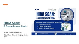
16. Common Gallbladder Issues Identified by HIDA Scans
HIDA scans are particularly useful in identifying and evaluating common gallbladder issues. Some
of these issues include:
• Gallbladder disease: This term encompasses a range of conditions that affect the gallbladder,
such as gallstones, inflammation, or infection.
• Chronic cholecystitis: This refers to repeated episodes of gallbladder inflammation, often
caused by gallstones blocking the cystic duct intermittently.
• Biliary tree abnormalities: HIDA scans can detect obstructions or abnormalities in the bile
ducts, which make up the biliary tree.
By identifying these issues through HIDA scans, healthcare providers can develop appropriate
treatment plans tailored to each patient’s specific condition and needs.
17. When Surgery Is Recommended: Navigating Your
Options in Texas
In some cases, surgery may be recommended based on the findings of a HIDA scan. Gallbladder
surgery, also known as cholecystectomy, is a common procedure used to remove the
gallbladder in cases of severe gallbladder disease or chronic cholecystitis.
In more complex cases, a liver transplant may be necessary to address issues with the biliary
system. This procedure involves replacing a diseased liver with a healthy liver from a donor.
The decision to undergo surgery depends on various factors, including the severity of the
condition, the patient’s overall health, and the gallbladder ejection fraction measured during the
HIDA scan.
It is important to consult with a healthcare provider who specializes in hepatobiliary surgery to
discuss your options and make an informed decision.
18. Risks and Considerations
As with any medical procedure, there are some risks and considerations associated with HIDA scans. It is
essential to be aware of these potential risks and discuss them with your healthcare provider. Some of the
key considerations include:
• Allergic reactions to medications containing radioactive tracers used during the scan, although they are
rare.
• Bruising at the injection site of the radioactive tracer.
• Minimal radiation exposure, which is within safe limits.
• Precautions for pregnant individuals, as nuclear medicine tests are generally not performed during
pregnancy due to potential harm to the developing fetus.
By understanding these risks and addressing any concerns with your healthcare provider, you can make
informed decisions regarding your medical care.
19. Understanding the Risks Associated with HIDA Scans
HIDA scans are generally considered safe with minimal risks. However, it is important to be aware of potential risks
associated with the procedure. These risks may include:
• Allergic reactions: Although rare, some individuals may experience allergic reactions to the radioactive tracer. It is
important to inform your healthcare provider of any known allergies before the scan.
• Bruising: Bruising at the injection site of the tracer is possible, but it is typically minimal.
• Radiation exposure: HIDA scans involve the use of a small amount of radiation, but the exposure is within safe limits
and not considered a significant risk.
Your healthcare provider will assess the potential risks and benefits of a HIDA scan based on your situation. They will
provide guidance and address any concerns you may have before proceeding with the procedure.
20. Addressing Patients’ Concerns: Safety
Measures in Place
Patient safety is a top priority during HIDA scans, and
various safety measures are in place to minimize
risks. These measures include:
Let’s explore more: HIDA Scan: Understanding the
Procedure - Southlake General Surgery
21. Make An Appointment
If you are experiencing symptoms related to your gallbladder or have concerns about your biliary
system, it is important to seek medical advice and schedule an appointment with our healthcare
expert at +1 (817) 748-0200 and click the link to book an online appointment with us. They can
evaluate your symptoms, discuss your medical history, and recommend appropriate diagnostic tests,
including HIDA scans.
Remember, early diagnosis and timely intervention are key to effective treatment and better
outcomes for gallbladder issues.
Medically Reviewed By: Dr. Valeria Simone MD
Board-certified General Surgeon at Southlake General Surgery, Texas, USA.
Follow us on Facebook and YouTube.
Source: HIDA Scan: Understanding the Procedure - Southlake General Surgery
22. THANK YOU!
SOUTHLAKE GENERAL SURGERY
1545 E. Southlake Blvd, Suite 270 Southlake, TX 76092
EMAIL: info@southlakegeneralsurgery.com
VISIT US AT: www.southlakegeneralsurgery.com