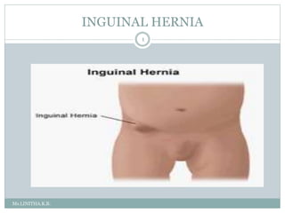
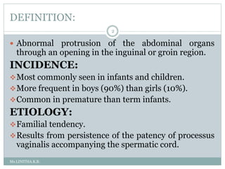
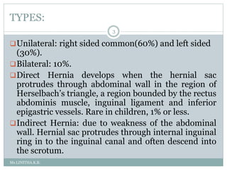
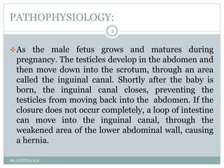
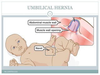
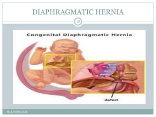
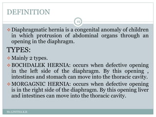
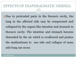
INGUINAL HERNIA
1
Ms.LINITHA.K.B.
DEFINITION:
Ms.LINITHA.K.B.
2
 Abnormal protrusion of the abdominal organs
through an opening in the inguinal or groin region.
INCIDENCE:
Most commonly seen in infants and children.
More frequent in boys (90%) than girls (10%).
Common in premature than term infants.
ETIOLOGY:
Familial tendency.
Results from persistence of the patency of processus
vaginalis accompanying the spermatic cord.
TYPES:
Ms.LINITHA.K.B.
3
Unilateral: right sided common(60%) and left sided
(30%).
Bilateral: 10%.
Direct Hernia develops when the hernial sac
protrudes through abdominal wall in the region of
Herselbach’s triangle, a region bounded by the rectus
abdominis muscle, inguinal ligament and inferior
epigastric vessels. Rare in children, 1% or less.
Indirect Hernia: due to weakness of the abdominal
wall. Hernial sac protrudes through internal inguinal
ring in to the inguinal canal and often descend into
the scrotum.
PATHOPHYSIOLOGY:
Ms.LINITHA.K.B.
4
As the male fetus grows and matures during
pregnancy. The testicles develop in the abdomen and
then move down into the scrotum, through an area
called the inguinal canal. Shortly after the baby is
born, the inguinal canal closes, preventing the
testicles from moving back into the abdomen. If the
closure does not occur completely, a loop of intestine
can move into the inguinal canal, through the
weakened area of the lower abdominal wall, causing
a hernia.
CLINICAL MANIFESTATIONS:
Ms.LINITHA.K.B.
5
May not be noticeable for several weeks or months
after birth.
When infant cries or strains, as a result of increased
intra abdominal pressure, the abdominal contents
come in the sac and a painless inguinal swelling of
varying size may be seen. The swelling disappears
when child is calm and relaxed. When the physician
pushes gently on the bulge. When the child is calm
and lying down, it will usually get smaller or go back
into the abdomen.
Ms.LINITHA.K.B.
6
Hernia that can’t be reduced is termed as irreducible
or incarcerated. When loop of intestine is caught in
the sac, generally at external inguinal ring, features
of intestinal obstruction occurs. Symptoms that arise
are vomiting contains bile, cramping pain,
abdominal distension, fever, irritable and restless.
Obstruction to venous return from the incarcerated
intestine and resulting edema leads to strangulation
which causes gangrene.
DIAGNOSTIC EVALUATIONS:
Ms.LINITHA.K.B.
7
Physical examination of the child.
The child is examined to determine if the hernia is
reducible or not.
Abdominal x-ray or ultrasound may be done to
examine the intestine closely.
MANAGEMENT:
Specific treatment for hernia depends on the child’s
age, overall health and type of hernia: whether it is
reducible or irreducible.
SURGICAL MANAGEMENT:
Ms.LINITHA.K.B.
8
 Two types of surgeries are done to repair hernia.
Open inguinal hernia repair (Herniorrhaphy,
Hernioplasty).
Laparoscopic Repair.
In case of uncomplicated hernia, an elective inguinal
Herniorrhaphy is done soon after the condition is
diagnosed so as to reduce the risk of incarceration. If
repair of reducible, uncomplicated hernia is
postponed, then parents are taught the method
reducing the hernia
Ms.LINITHA.K.B.
9
 In case of incarcerated hernia, reasonable attempts are
made to reduce it. Reduction may be successful if the infant
can be helped to relax by giving warm bath or allowing to
suck a pacifier. Compression is the exerted on the hernia.
 If non-operative reduction is unsuccessful, elective surgery
can be done in 2-3 days, after the inflammation has
subsided. If non-operative reduction is unsuccessful, an
irreducible incarcerated hernia requires immediately
surgery.
 Hernia repair is done using a small incision directly over
the weakened area. The intestine is then returned to the
abdominal cavity, hernia sac is excised and muscles are
closed tightly over the area.
PROGNOSIS:
Ms.LINITHA.K.B.
10
Overall good prognosis with early diagnosis and
treatment.
UMBILICAL HERNIA
Ms.LINITHA.K.B.
11
DEFINITION:
Ms.LINITHA.K.B.
12
Weakness or incomplete closure of umbilical ring,
through which blood vessels had passed during the
fetal life results in a portion of omentum and small
intestine passing through the ring and appearing as
soft protrusion covered with skin.
INCIDENCE:
Occurs in about 10% of all children.
More often in girls than boys.
Frequently occur in premature infants.
PATHOPHYSIOLOGY:
Ms.LINITHA.K.B.
13
When the fetus is growing and developing during
pregnancy, there is a small opening in the abdominal
muscles so that the umbilical cord can pass through,
connecting the mother to the baby.
After birth, the opening in the abdominal muscles
closes as the baby matures. Sometimes, these
muscles do not meet together completely, and a
small opening remains.
A loop of intestine can move into the opening
between the abdominal muscles and cause hernia.
CLINICAL MANIFESTATIONS:
Ms.LINITHA.K.B.
14
Bulge or swelling in the belly-button area. Swelling may
be more noticeable, when the baby cries and may get
smaller or disappear when the baby relaxes.
If the physician pushes gently on the bulge when the
child is calm and lying down it will usually get smaller or
go back into the abdomen.
DIAGNOSTIC EVALUATIONS:
Physical Examination determines the hernia is reducible
or not.
Abdominal X-Ray/Ultrasound: to examine intestine
closely.
MANAGEMENT:
Ms.LINITHA.K.B.
15
By 1 year of age, many umbilical hernias will have closed
on their own without needing surgery.
Nearly all umbilical hernia will have closed without
surgery by 5 years of age. Placing a coin or strap over the
hernia will not fix it.
In general, if the hernia becomes bigger with age, is not
reducible or is still present after 3 years of age, it needs to
be repaired surgically.
A small incision is made in the umbilicus and the loop of
intestine is placed back into the abdominal cavity. The
muscles are then sutured together. Sometimes a piece of
meshed material is used to help strengthen the area
where the muscles are repaired.
NURSING MANAGEMENT:PREOPERATIVE
Ms.LINITHA.K.B.
16
Preoperatively the infant is fed until a few hours
before surgery, in order to prevent dehydration.
Preoperative routine care and anesthesia
medications are administered and the child is sent
for surgery.
POST OPERATIVE NURSING CARE:
Ms.LINITHA.K.B.
17
Monitor vital signs.
Continue giving intravenous fluid till bowel movements
return.
Observe the incision site for bleeding.
Child is discharged from the hospital after starting taking
and retaining oral feeds.
Parents must be instructed to keep the incision site clean
and dry, until it heals completely.
Sponge bath can be given daily to maintain the personal
hygiene of the child.
Older children should be instructed not to engage in
sternous activities for at least 1 month after surgery.
DIAPHRAGMATIC HERNIA
Ms.LINITHA.K.B.
18
DEFINITION
Ms.LINITHA.K.B.
19
Diaphragmatic hernia is a congenital anomaly of children
in which protrusion of abdominal organs through an
opening in the diaphragm.
TYPES:
Mainly 2 types.
BOCHDALEK HERNIA: occurs when defective opening
in the left side of the diaphragm. By this opening ,
intestines and stomach can move into the thoracic cavity.
MORGAGNIC HERNIA: occurs when defective opening
is in the right side of the diaphragm. By this opening liver
and intestines can move into the thoracic cavity.
EFFECTS OF DIAPHRAGMATIC HERNIA:
Ms.LINITHA.K.B.
20
Due to protruded parts in the thoracic cavity, the
lung in the affected side may be compressed and
collapsed by the organs like intestine and stomach in
thoracic cavity. The intestine and stomach become
distended by the air which is swallowed and pushes
the mediastinum to one side and collapse of same
side lung can occur.
CLINICAL MANIFESTATIONS:
Ms.LINITHA.K.B.
21
Respiratory distress.
Dyspnea.
Tachypnea.
Cyanosis.
Scaphoid chest size.
On the affected side decreased chest movements and
breath sounds.
In left sided defect heart sounds are better heard than the
right sided effects.
Peristalsis may be heard in the chest on the affected side.
Shock and hypoxia.
Severe acidosis.
DIAGNOSTIC EVALUATIONS:
Ms.LINITHA.K.B.
22
History Collection.
Physical Examination.
Early Diagnosis may be done during the antenatal
period in cases with hydramnios , with the
aminography studies.
Chest X-Ray shows opaque, hemithorax, mediastein
shift, air filled intestinal loops, compressed lungs.
Abdominal X-Ray shows empty abdomen with gas
shadows.
Echocardiogram and ABG studies.
MANAGEMENT: MEDICAL MANAGEMENT
Ms.LINITHA.K.B.
23
 Supportive management of RD and acidosis.
 Endotracheal intubation.
 Maintain 02 level by o2 administration.
 Prophylatic use of antibiotics after culture and
sensitivity.
 Place baby in position with head raised at 45 degree on
the affected side.
 Regular monitoring of blood o2 and blood glucose level.
 Acidosis corrected by sodium bicarbonate.
 For nutrition IV glucose administration.
SURGICAL MANAGEMENT:
Ms.LINITHA.K.B.
24
 Herniotomy: surgical management by the abdominal
approach, abdominal organs are replaced and
diaphragmatic defects is corrected. Thoracic
drainage may be continued to prevent infection.
HOME MANAGEMENT:
 Maintain body temperature by avoiding exposure to
the environment.
Provide nutritious diet.
Follow hygienic measures and regular follow ups.
Ms.LINITHA.K.B.
25

HERNIA.pptx

  • 1.
  • 2.
    DEFINITION: Ms.LINITHA.K.B. 2  Abnormal protrusionof the abdominal organs through an opening in the inguinal or groin region. INCIDENCE: Most commonly seen in infants and children. More frequent in boys (90%) than girls (10%). Common in premature than term infants. ETIOLOGY: Familial tendency. Results from persistence of the patency of processus vaginalis accompanying the spermatic cord.
  • 3.
    TYPES: Ms.LINITHA.K.B. 3 Unilateral: right sidedcommon(60%) and left sided (30%). Bilateral: 10%. Direct Hernia develops when the hernial sac protrudes through abdominal wall in the region of Herselbach’s triangle, a region bounded by the rectus abdominis muscle, inguinal ligament and inferior epigastric vessels. Rare in children, 1% or less. Indirect Hernia: due to weakness of the abdominal wall. Hernial sac protrudes through internal inguinal ring in to the inguinal canal and often descend into the scrotum.
  • 4.
    PATHOPHYSIOLOGY: Ms.LINITHA.K.B. 4 As the malefetus grows and matures during pregnancy. The testicles develop in the abdomen and then move down into the scrotum, through an area called the inguinal canal. Shortly after the baby is born, the inguinal canal closes, preventing the testicles from moving back into the abdomen. If the closure does not occur completely, a loop of intestine can move into the inguinal canal, through the weakened area of the lower abdominal wall, causing a hernia.
  • 5.
    CLINICAL MANIFESTATIONS: Ms.LINITHA.K.B. 5 May notbe noticeable for several weeks or months after birth. When infant cries or strains, as a result of increased intra abdominal pressure, the abdominal contents come in the sac and a painless inguinal swelling of varying size may be seen. The swelling disappears when child is calm and relaxed. When the physician pushes gently on the bulge. When the child is calm and lying down, it will usually get smaller or go back into the abdomen.
  • 6.
    Ms.LINITHA.K.B. 6 Hernia that can’tbe reduced is termed as irreducible or incarcerated. When loop of intestine is caught in the sac, generally at external inguinal ring, features of intestinal obstruction occurs. Symptoms that arise are vomiting contains bile, cramping pain, abdominal distension, fever, irritable and restless. Obstruction to venous return from the incarcerated intestine and resulting edema leads to strangulation which causes gangrene.
  • 7.
    DIAGNOSTIC EVALUATIONS: Ms.LINITHA.K.B. 7 Physical examinationof the child. The child is examined to determine if the hernia is reducible or not. Abdominal x-ray or ultrasound may be done to examine the intestine closely. MANAGEMENT: Specific treatment for hernia depends on the child’s age, overall health and type of hernia: whether it is reducible or irreducible.
  • 8.
    SURGICAL MANAGEMENT: Ms.LINITHA.K.B. 8  Twotypes of surgeries are done to repair hernia. Open inguinal hernia repair (Herniorrhaphy, Hernioplasty). Laparoscopic Repair. In case of uncomplicated hernia, an elective inguinal Herniorrhaphy is done soon after the condition is diagnosed so as to reduce the risk of incarceration. If repair of reducible, uncomplicated hernia is postponed, then parents are taught the method reducing the hernia
  • 9.
    Ms.LINITHA.K.B. 9  In caseof incarcerated hernia, reasonable attempts are made to reduce it. Reduction may be successful if the infant can be helped to relax by giving warm bath or allowing to suck a pacifier. Compression is the exerted on the hernia.  If non-operative reduction is unsuccessful, elective surgery can be done in 2-3 days, after the inflammation has subsided. If non-operative reduction is unsuccessful, an irreducible incarcerated hernia requires immediately surgery.  Hernia repair is done using a small incision directly over the weakened area. The intestine is then returned to the abdominal cavity, hernia sac is excised and muscles are closed tightly over the area.
  • 10.
  • 11.
  • 12.
    DEFINITION: Ms.LINITHA.K.B. 12 Weakness or incompleteclosure of umbilical ring, through which blood vessels had passed during the fetal life results in a portion of omentum and small intestine passing through the ring and appearing as soft protrusion covered with skin. INCIDENCE: Occurs in about 10% of all children. More often in girls than boys. Frequently occur in premature infants.
  • 13.
    PATHOPHYSIOLOGY: Ms.LINITHA.K.B. 13 When the fetusis growing and developing during pregnancy, there is a small opening in the abdominal muscles so that the umbilical cord can pass through, connecting the mother to the baby. After birth, the opening in the abdominal muscles closes as the baby matures. Sometimes, these muscles do not meet together completely, and a small opening remains. A loop of intestine can move into the opening between the abdominal muscles and cause hernia.
  • 14.
    CLINICAL MANIFESTATIONS: Ms.LINITHA.K.B. 14 Bulge orswelling in the belly-button area. Swelling may be more noticeable, when the baby cries and may get smaller or disappear when the baby relaxes. If the physician pushes gently on the bulge when the child is calm and lying down it will usually get smaller or go back into the abdomen. DIAGNOSTIC EVALUATIONS: Physical Examination determines the hernia is reducible or not. Abdominal X-Ray/Ultrasound: to examine intestine closely.
  • 15.
    MANAGEMENT: Ms.LINITHA.K.B. 15 By 1 yearof age, many umbilical hernias will have closed on their own without needing surgery. Nearly all umbilical hernia will have closed without surgery by 5 years of age. Placing a coin or strap over the hernia will not fix it. In general, if the hernia becomes bigger with age, is not reducible or is still present after 3 years of age, it needs to be repaired surgically. A small incision is made in the umbilicus and the loop of intestine is placed back into the abdominal cavity. The muscles are then sutured together. Sometimes a piece of meshed material is used to help strengthen the area where the muscles are repaired.
  • 16.
    NURSING MANAGEMENT:PREOPERATIVE Ms.LINITHA.K.B. 16 Preoperatively theinfant is fed until a few hours before surgery, in order to prevent dehydration. Preoperative routine care and anesthesia medications are administered and the child is sent for surgery.
  • 17.
    POST OPERATIVE NURSINGCARE: Ms.LINITHA.K.B. 17 Monitor vital signs. Continue giving intravenous fluid till bowel movements return. Observe the incision site for bleeding. Child is discharged from the hospital after starting taking and retaining oral feeds. Parents must be instructed to keep the incision site clean and dry, until it heals completely. Sponge bath can be given daily to maintain the personal hygiene of the child. Older children should be instructed not to engage in sternous activities for at least 1 month after surgery.
  • 18.
  • 19.
    DEFINITION Ms.LINITHA.K.B. 19 Diaphragmatic hernia isa congenital anomaly of children in which protrusion of abdominal organs through an opening in the diaphragm. TYPES: Mainly 2 types. BOCHDALEK HERNIA: occurs when defective opening in the left side of the diaphragm. By this opening , intestines and stomach can move into the thoracic cavity. MORGAGNIC HERNIA: occurs when defective opening is in the right side of the diaphragm. By this opening liver and intestines can move into the thoracic cavity.
  • 20.
    EFFECTS OF DIAPHRAGMATICHERNIA: Ms.LINITHA.K.B. 20 Due to protruded parts in the thoracic cavity, the lung in the affected side may be compressed and collapsed by the organs like intestine and stomach in thoracic cavity. The intestine and stomach become distended by the air which is swallowed and pushes the mediastinum to one side and collapse of same side lung can occur.
  • 21.
    CLINICAL MANIFESTATIONS: Ms.LINITHA.K.B. 21 Respiratory distress. Dyspnea. Tachypnea. Cyanosis. Scaphoidchest size. On the affected side decreased chest movements and breath sounds. In left sided defect heart sounds are better heard than the right sided effects. Peristalsis may be heard in the chest on the affected side. Shock and hypoxia. Severe acidosis.
  • 22.
    DIAGNOSTIC EVALUATIONS: Ms.LINITHA.K.B. 22 History Collection. PhysicalExamination. Early Diagnosis may be done during the antenatal period in cases with hydramnios , with the aminography studies. Chest X-Ray shows opaque, hemithorax, mediastein shift, air filled intestinal loops, compressed lungs. Abdominal X-Ray shows empty abdomen with gas shadows. Echocardiogram and ABG studies.
  • 23.
    MANAGEMENT: MEDICAL MANAGEMENT Ms.LINITHA.K.B. 23 Supportive management of RD and acidosis.  Endotracheal intubation.  Maintain 02 level by o2 administration.  Prophylatic use of antibiotics after culture and sensitivity.  Place baby in position with head raised at 45 degree on the affected side.  Regular monitoring of blood o2 and blood glucose level.  Acidosis corrected by sodium bicarbonate.  For nutrition IV glucose administration.
  • 24.
    SURGICAL MANAGEMENT: Ms.LINITHA.K.B. 24  Herniotomy:surgical management by the abdominal approach, abdominal organs are replaced and diaphragmatic defects is corrected. Thoracic drainage may be continued to prevent infection. HOME MANAGEMENT:  Maintain body temperature by avoiding exposure to the environment. Provide nutritious diet. Follow hygienic measures and regular follow ups.
  • 25.