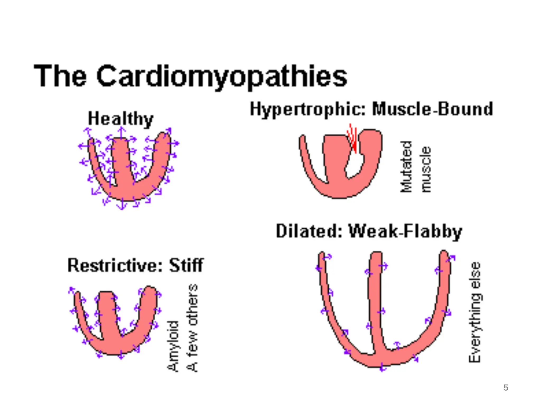
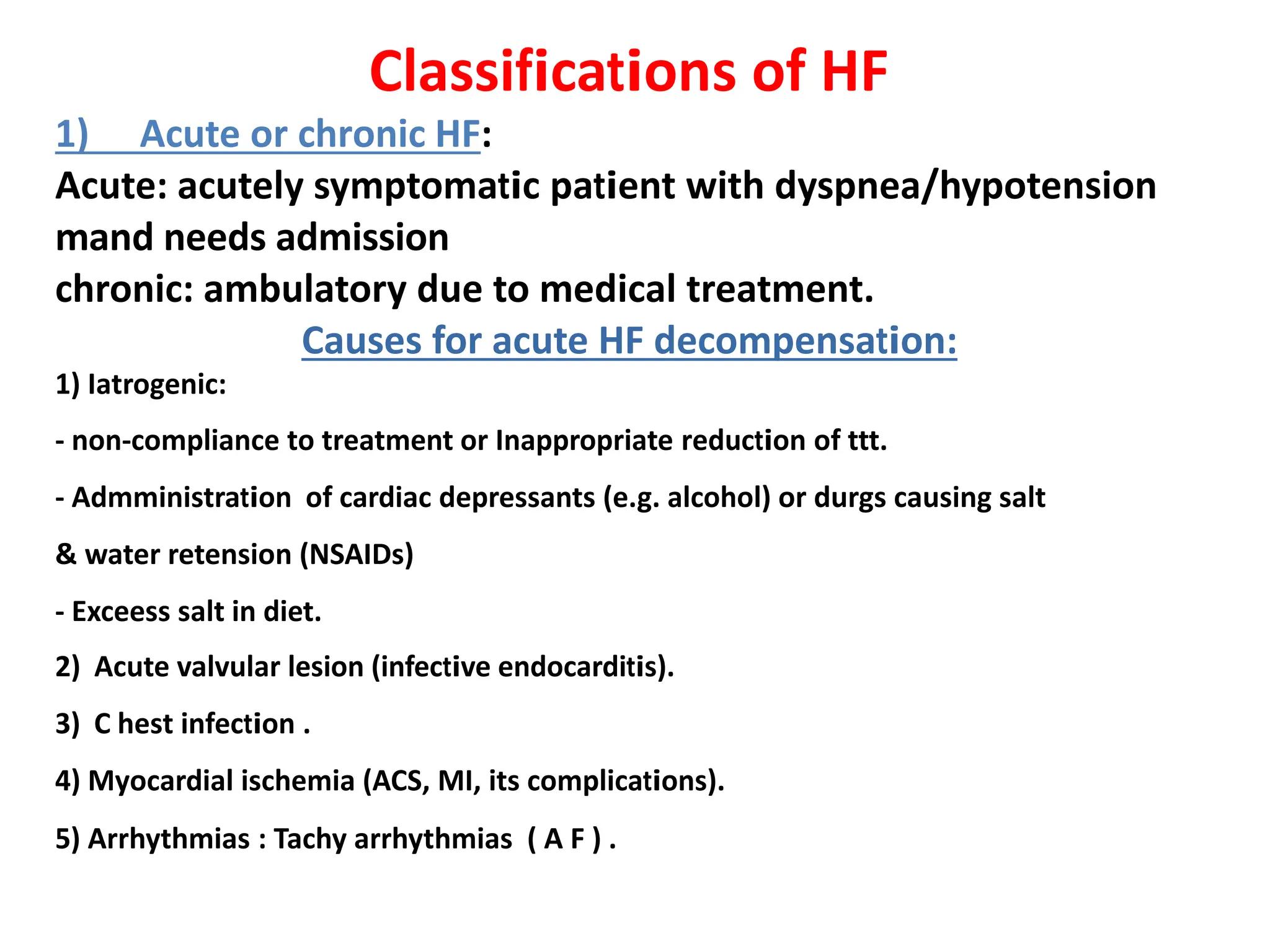
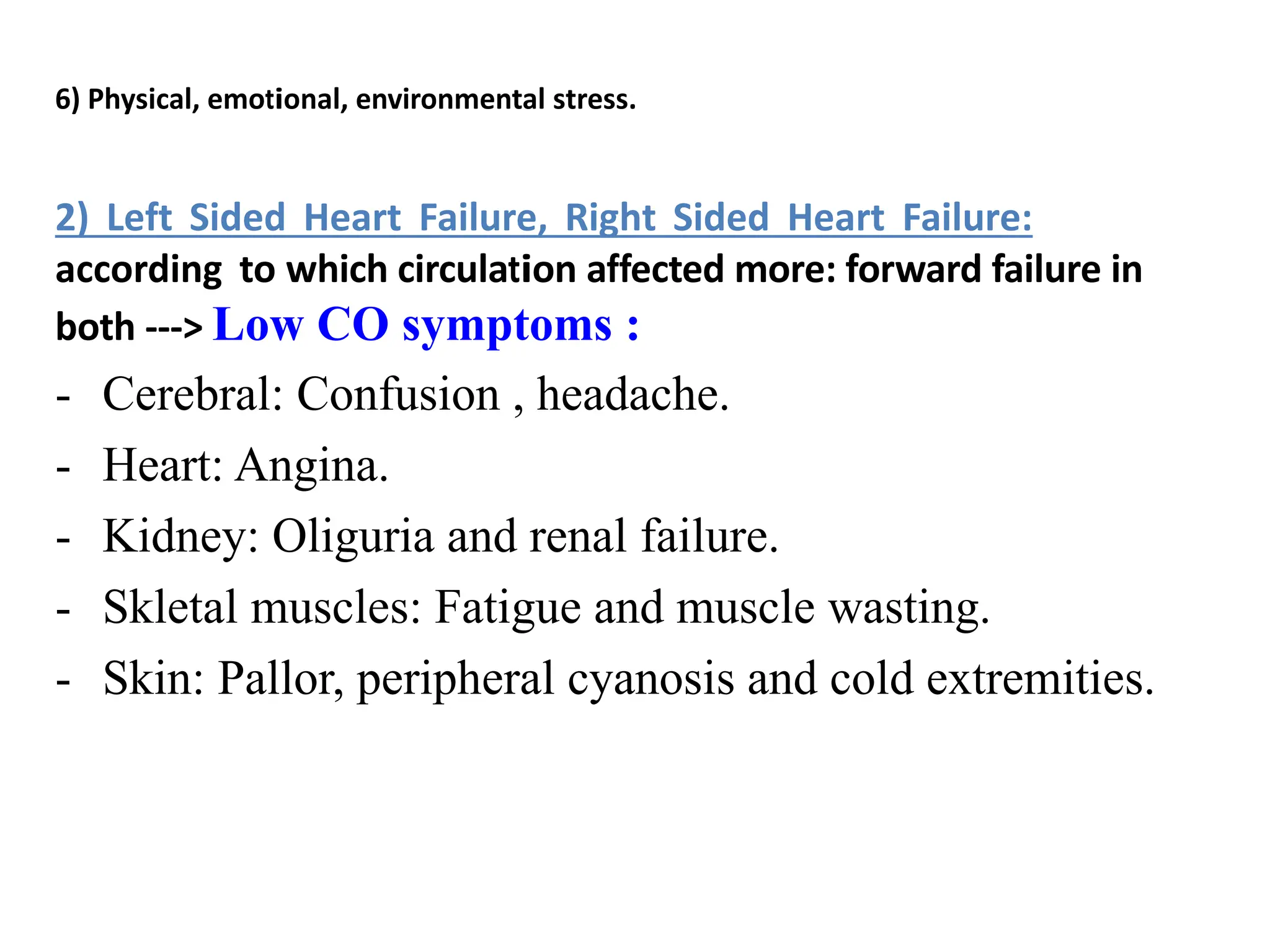
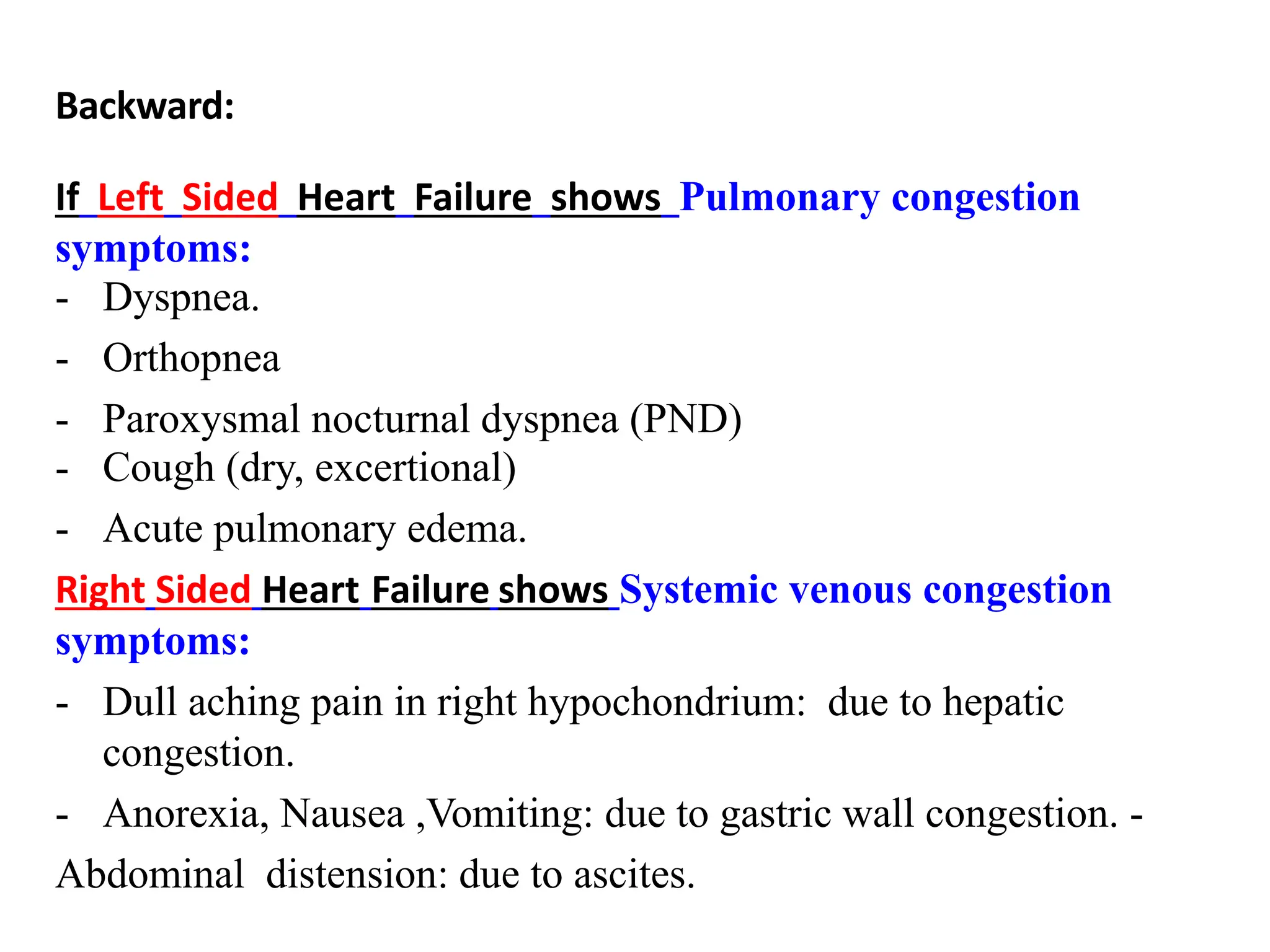
Heart Failure
Mohammad Shafiq, MD, FESC, FSCAI
Associate professor of cardiology
Defenition
• Heart failure is a clinical syndrome not an investigation disease.
• Clinically defined as the inability of the heart at the normal filling
pressures to maintain an output adequate to meet the metabolic
demands of the body.
Pathophysiology
• Nearly all compensatory mechanisms are harmful:
COP = SV x HR
- Heart dilatation (Cardiomegaly)
-Renin angiotensin system actication (↑ Na retension)
-Increased HR (Sympathetic actication)
Epidemiology:
• 1-2% of the general population
• U S A : 20% of hospital admissions among persons > 65
• 45% annual mortality in severe symptomatic heart failure
• More dollars are spent for diagnosis and treatment of heart
failure than for any other single diagnosis.
2
Aetiology:
1. Dilated Cardiomyopathy (about 50% of cases are familial)
2. Hypertrophic Cardiomyopathy (1:500)
3. Restrictive Cardiomyopathy (myocardial infiltrating diseases)
4. Arrhythmogenic Right Ventricular Cardiomyopathy
5. Unclassifiable Cardiomyopathies
6. Specific (Terminal fate) Cardiomyopathies (ischemic, hypertensive, valvular
obstruction/insufficiency, myocarditis).
7. Metabolic (thyrotoxicosis, hypothyroidism, pheochromocytoma)
8. General system disease (alcohol, anthracyclines, radiation, SLE, PAN, scleroderma,
sarcoidosis, muscular dystrophies, peripartum cardiomyopathy, etc.)
3
The most common causes of CHF
• Ischemic heart disease ~ 40 % .
• Dilated cardiomyopathy ~ 30 % .
• Primary valvular heart disease ~ 15 % .
• Hypertensive heart disease ~ 10 % .
• Other ~ 5 %.
4
5
Classifications of HF
1) Acute or chronic HF:
Acute: acutely symptomatic patient with dyspnea/hypotension
mand needs admission
chronic: ambulatory due to medical treatment.
Causes for acute HF decompensation:
1) Iatrogenic:
- non-compliance to treatment or Inappropriate reduction of ttt.
- Admministration of cardiac depressants (e.g. alcohol) or durgs causing salt
& water retension (NSAIDs)
- Exceess salt in diet.
2) Acute valvular lesion (infective endocarditis).
3) C hest infection .
4) Myocardial ischemia (ACS, MI, its complications).
5) Arrhythmias : Tachy arrhythmias ( A F ) .
6) Physical, emotional, environmental stress.
2) Left Sided Heart Failure, Right Sided Heart Failure:
according to which circulation affected more: forward failure in
both ---> Low CO symptoms :
- Cerebral: Confusion , headache.
- Heart: Angina.
- Kidney: Oliguria and renal failure.
- Skletal muscles: Fatigue and muscle wasting.
- Skin: Pallor, peripheral cyanosis and cold extremities.
Backward:
If Left Sided Heart Failure shows Pulmonary congestion
symptoms:
- Dyspnea.
- Orthopnea
- Paroxysmal nocturnal dyspnea (PND)
- Cough (dry, excertional)
- Acute pulmonary edema.
Right Sided Heart Failure shows Systemic venous congestion
symptoms:
- Dull aching pain in right hypochondrium: due to hepatic
congestion.
- Anorexia, Nausea ,Vomiting: due to gastric wall congestion. -
Abdominal distension: due to ascites.
- Bilateral lower Limb swelling (edema) : due to subcutaneous
edema.
NB: left & right side HF almost coexcist and left
sided can be complicated with right side heart
failure by time.
3) Systolic or diastolic HF: not all cases has weak myocardium
Ejection fraction: % of ejected blood from LV cavity (Normally
>50%)
According to contractility (Ejection fraction: assessed by
Echocardiography) of the left ventricle. It is either:
• Systolic HF if the EF% is < 40% also called Heart Failure with
reduced EF, HFrEF).
• Diastolic heart failure or heart failure with preserved EF
if EF% is > 50% (HFpEF) .
10
New York heart association classification
(NYHA) for dyspnea
• Class 1:
• Class II:
Dyspnea on more than ordinary effort.
Dyspnea on ordinary effort.
• Class III:
• Class 1V:
asthma.
Dyspnea on less than ordinary effort.
Dyspnea at rest including orthopnea, PND, cardiac
14
15
• Chest X-Ray: Cardiomegaly (Heart shadow >50% of
chest width) & pulmonary congestion.
• Echocardiography: Gold standard for diagnosis,
detect cause and degree of Left Ventricular systolic
fucntion (Ejection Fraction %, EF%).
https://youtube.com/clip/UgkxP7lJQk48jTelNlSD0M2dX
d0ChEdR7I3K?si=Z9-frkdQ7TDDRpqL
• Cardiac catheterization:
left heart catheter for coronary angiography to detect
Coronary Artery Disease (CAD) or right heart catheter to
take myocardial biopsy.
16
Laboratory work up
• Electrolytes: Na, K, Ca, Mg
• Kidney functions: urea, creatinin and creatinin clearence
• CBC to exclude anemia
• For specific causes: eg: serum Iron and ferritin if suspected
haemochrmatosis or abdominal fat biopsy if suspected
cardiac amyloidosis
17
Treatment of heart failure
1) General measures:
• Treatment of reversible causes (e.g. IHD or arrhytmias) & avoid preciptating factors
(e.g. chest infection and anemia)
• Salt restriction ( < 5 g/day) & bed rest.
• Oxygen therapy for acutely distressed patient.
2) Pharmacological ttt:
• Symptomatic ttt by Diuretics : Loop diuretics e.g: Furosemide (Lasix) or thiazide
diuretics e.g: Hydrochlorothaizides.
• Disease Modifying drugs:
1- Mineralocorticoids antagonists (MRAs) e.g: Spironolactone
2- Beta blockers (BB, e.g:bisoprolol)
3- Angiotensin Converting Enzyme (ACE) Inhibitors e.g: enalapril or Angiotensin
Receptor Blockers (ARBs) e.g: Valsartan.
• Other drugs: Ivabradine or Digoxin.
• Positive inotropic support for shocked patient : Dopamine , dobutamine or
norepinephrine.
3) Non-Pharmachological ttt:
• Mechanical ventilation for acute respiartory distress or acute pulmonary edema.
• Ultrafilteration by dialysis to releive volume overload in resistant cases.
• Device therapy by:
1- CRT (cardiac resynchronzation therapy if QRS complex in ECG is > 150msec) correct
delay between lateral wall and septum
2- ICD (Implantable cardiovertor defibrillator) to give automatic DC
shock to restore contractions after cardiac arrest due to ventricular
arrhythmia
• Surgical ttt : by Heart or heart-lung transplantation.
Thanks for your attention
and good luck

5-HF PTh basics of 1st semestwr diploma.pdf

  • 1.
    Heart Failure Mohammad Shafiq,MD, FESC, FSCAI Associate professor of cardiology Defenition • Heart failure is a clinical syndrome not an investigation disease. • Clinically defined as the inability of the heart at the normal filling pressures to maintain an output adequate to meet the metabolic demands of the body.
  • 2.
    Pathophysiology • Nearly allcompensatory mechanisms are harmful: COP = SV x HR - Heart dilatation (Cardiomegaly) -Renin angiotensin system actication (↑ Na retension) -Increased HR (Sympathetic actication) Epidemiology: • 1-2% of the general population • U S A : 20% of hospital admissions among persons > 65 • 45% annual mortality in severe symptomatic heart failure • More dollars are spent for diagnosis and treatment of heart failure than for any other single diagnosis. 2
  • 3.
    Aetiology: 1. Dilated Cardiomyopathy(about 50% of cases are familial) 2. Hypertrophic Cardiomyopathy (1:500) 3. Restrictive Cardiomyopathy (myocardial infiltrating diseases) 4. Arrhythmogenic Right Ventricular Cardiomyopathy 5. Unclassifiable Cardiomyopathies 6. Specific (Terminal fate) Cardiomyopathies (ischemic, hypertensive, valvular obstruction/insufficiency, myocarditis). 7. Metabolic (thyrotoxicosis, hypothyroidism, pheochromocytoma) 8. General system disease (alcohol, anthracyclines, radiation, SLE, PAN, scleroderma, sarcoidosis, muscular dystrophies, peripartum cardiomyopathy, etc.) 3
  • 4.
    The most commoncauses of CHF • Ischemic heart disease ~ 40 % . • Dilated cardiomyopathy ~ 30 % . • Primary valvular heart disease ~ 15 % . • Hypertensive heart disease ~ 10 % . • Other ~ 5 %. 4
  • 5.
  • 6.
    Classifications of HF 1)Acute or chronic HF: Acute: acutely symptomatic patient with dyspnea/hypotension mand needs admission chronic: ambulatory due to medical treatment. Causes for acute HF decompensation: 1) Iatrogenic: - non-compliance to treatment or Inappropriate reduction of ttt. - Admministration of cardiac depressants (e.g. alcohol) or durgs causing salt & water retension (NSAIDs) - Exceess salt in diet. 2) Acute valvular lesion (infective endocarditis). 3) C hest infection . 4) Myocardial ischemia (ACS, MI, its complications). 5) Arrhythmias : Tachy arrhythmias ( A F ) .
  • 7.
    6) Physical, emotional,environmental stress. 2) Left Sided Heart Failure, Right Sided Heart Failure: according to which circulation affected more: forward failure in both ---> Low CO symptoms : - Cerebral: Confusion , headache. - Heart: Angina. - Kidney: Oliguria and renal failure. - Skletal muscles: Fatigue and muscle wasting. - Skin: Pallor, peripheral cyanosis and cold extremities.
  • 8.
    Backward: If Left SidedHeart Failure shows Pulmonary congestion symptoms: - Dyspnea. - Orthopnea - Paroxysmal nocturnal dyspnea (PND) - Cough (dry, excertional) - Acute pulmonary edema. Right Sided Heart Failure shows Systemic venous congestion symptoms: - Dull aching pain in right hypochondrium: due to hepatic congestion. - Anorexia, Nausea ,Vomiting: due to gastric wall congestion. - Abdominal distension: due to ascites.
  • 9.
    - Bilateral lowerLimb swelling (edema) : due to subcutaneous edema. NB: left & right side HF almost coexcist and left sided can be complicated with right side heart failure by time.
  • 10.
    3) Systolic ordiastolic HF: not all cases has weak myocardium Ejection fraction: % of ejected blood from LV cavity (Normally >50%) According to contractility (Ejection fraction: assessed by Echocardiography) of the left ventricle. It is either: • Systolic HF if the EF% is < 40% also called Heart Failure with reduced EF, HFrEF). • Diastolic heart failure or heart failure with preserved EF if EF% is > 50% (HFpEF) . 10
  • 11.
    New York heartassociation classification (NYHA) for dyspnea • Class 1: • Class II: Dyspnea on more than ordinary effort. Dyspnea on ordinary effort. • Class III: • Class 1V: asthma. Dyspnea on less than ordinary effort. Dyspnea at rest including orthopnea, PND, cardiac 14
  • 12.
  • 13.
    • Chest X-Ray:Cardiomegaly (Heart shadow >50% of chest width) & pulmonary congestion.
  • 14.
    • Echocardiography: Goldstandard for diagnosis, detect cause and degree of Left Ventricular systolic fucntion (Ejection Fraction %, EF%). https://youtube.com/clip/UgkxP7lJQk48jTelNlSD0M2dX d0ChEdR7I3K?si=Z9-frkdQ7TDDRpqL
  • 15.
    • Cardiac catheterization: leftheart catheter for coronary angiography to detect Coronary Artery Disease (CAD) or right heart catheter to take myocardial biopsy. 16
  • 16.
    Laboratory work up •Electrolytes: Na, K, Ca, Mg • Kidney functions: urea, creatinin and creatinin clearence • CBC to exclude anemia • For specific causes: eg: serum Iron and ferritin if suspected haemochrmatosis or abdominal fat biopsy if suspected cardiac amyloidosis 17
  • 17.
    Treatment of heartfailure 1) General measures: • Treatment of reversible causes (e.g. IHD or arrhytmias) & avoid preciptating factors (e.g. chest infection and anemia) • Salt restriction ( < 5 g/day) & bed rest. • Oxygen therapy for acutely distressed patient. 2) Pharmacological ttt: • Symptomatic ttt by Diuretics : Loop diuretics e.g: Furosemide (Lasix) or thiazide diuretics e.g: Hydrochlorothaizides. • Disease Modifying drugs: 1- Mineralocorticoids antagonists (MRAs) e.g: Spironolactone 2- Beta blockers (BB, e.g:bisoprolol) 3- Angiotensin Converting Enzyme (ACE) Inhibitors e.g: enalapril or Angiotensin
  • 18.
    Receptor Blockers (ARBs)e.g: Valsartan. • Other drugs: Ivabradine or Digoxin. • Positive inotropic support for shocked patient : Dopamine , dobutamine or norepinephrine. 3) Non-Pharmachological ttt: • Mechanical ventilation for acute respiartory distress or acute pulmonary edema. • Ultrafilteration by dialysis to releive volume overload in resistant cases. • Device therapy by: 1- CRT (cardiac resynchronzation therapy if QRS complex in ECG is > 150msec) correct delay between lateral wall and septum
  • 19.
    2- ICD (Implantablecardiovertor defibrillator) to give automatic DC shock to restore contractions after cardiac arrest due to ventricular arrhythmia • Surgical ttt : by Heart or heart-lung transplantation. Thanks for your attention and good luck