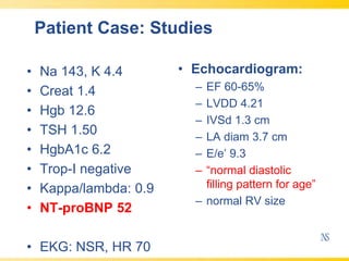
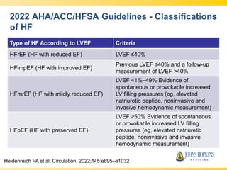
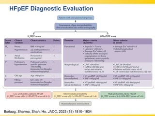
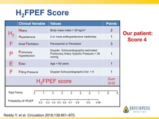
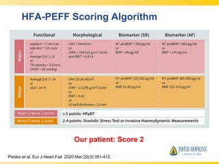
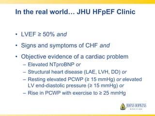
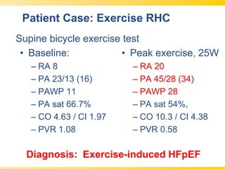
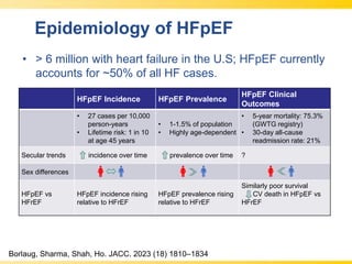
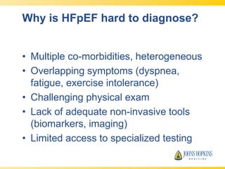
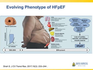
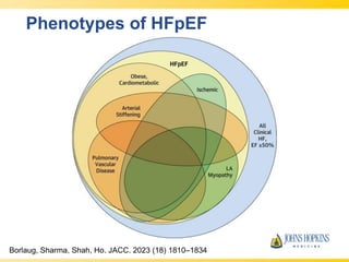
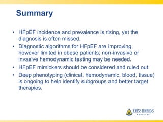
Acute Heart Failure: Current Standards and Evolution of Care.2015hivlifeinfo
Обсуждение последних данных, касающиеся диагностики и лечения острой сердечной недостаточности, в том числе использование биомаркеров для диагностики и оценке прогноза , преимущества и ограничения действующих стандартами медицинской помощи, и доказательств данных по современной терапии острой сердечной недостаточности.
Формат: Microsoft PowerPoint (.ppt)
Размер файла: 1.68 Мб
Дата публикации: 7/24/2015
Nick H. Kim, MD, Richard N. Channick, MD, and Vallerie V. McLaughlin, MD, prepared useful Practice Aids pertaining to pulmonary hypertension for this CME activity titled "Pulmonary Hypertension at the Crossroads of Current Clinical Challenges and Novel Therapeutic Strategies." For the full presentation, monograph, complete CME information, and to apply for credit, please visit us at http://bit.ly/2O9QbOh. CME credit will be available until July 30, 2019.
- Video recording of this lecture in English language: https://youtu.be/lK81BzxMqdo
- Video recording of this lecture in Arabic language: https://youtu.be/Ve4P0COk9OI
- Link to download the book free: https://nephrotube.blogspot.com/p/nephrotube-nephrology-books.html
- Link to NephroTube website: www.NephroTube.com
- Link to NephroTube social media accounts: https://nephrotube.blogspot.com/p/join-nephrotube-on-social-media.html
TEST BANK For Advanced Practice Nursing in the Care of Older Adults, 2nd Edit...kevinkariuki227
TEST BANK For Advanced Practice Nursing in the Care of Older Adults, 2nd Edition by Laurie Kennedy-Malone, Verified Chapters 1 - 19, Complete Newest Version.pdf
TEST BANK For Timby's Introductory Medical-Surgical Nursing, 13th American Ed...kevinkariuki227
TEST BANK For Timby's Introductory Medical-Surgical Nursing, 13th American Edition by Donnelly-Moreno, Verified Chapters 1 - 72, Complete Newest Version.pdf
New Directions in Targeted Therapeutic Approaches for Older Adults With Mantl...i3 Health
i3 Health is pleased to make the speaker slides from this activity available for use as a non-accredited self-study or teaching resource.
This slide deck presented by Dr. Kami Maddocks, Professor-Clinical in the Division of Hematology and
Associate Division Director for Ambulatory Operations
The Ohio State University Comprehensive Cancer Center, will provide insight into new directions in targeted therapeutic approaches for older adults with mantle cell lymphoma.
STATEMENT OF NEED
Mantle cell lymphoma (MCL) is a rare, aggressive B-cell non-Hodgkin lymphoma (NHL) accounting for 5% to 7% of all lymphomas. Its prognosis ranges from indolent disease that does not require treatment for years to very aggressive disease, which is associated with poor survival (Silkenstedt et al, 2021). Typically, MCL is diagnosed at advanced stage and in older patients who cannot tolerate intensive therapy (NCCN, 2022). Although recent advances have slightly increased remission rates, recurrence and relapse remain very common, leading to a median overall survival between 3 and 6 years (LLS, 2021). Though there are several effective options, progress is still needed towards establishing an accepted frontline approach for MCL (Castellino et al, 2022). Treatment selection and management of MCL are complicated by the heterogeneity of prognosis, advanced age and comorbidities of patients, and lack of an established standard approach for treatment, making it vital that clinicians be familiar with the latest research and advances in this area. In this activity chaired by Michael Wang, MD, Professor in the Department of Lymphoma & Myeloma at MD Anderson Cancer Center, expert faculty will discuss prognostic factors informing treatment, the promising results of recent trials in new therapeutic approaches, and the implications of treatment resistance in therapeutic selection for MCL.
Target Audience
Hematology/oncology fellows, attending faculty, and other health care professionals involved in the treatment of patients with mantle cell lymphoma (MCL).
Learning Objectives
1.) Identify clinical and biological prognostic factors that can guide treatment decision making for older adults with MCL
2.) Evaluate emerging data on targeted therapeutic approaches for treatment-naive and relapsed/refractory MCL and their applicability to older adults
3.) Assess mechanisms of resistance to targeted therapies for MCL and their implications for treatment selection
Heart Failure with Preserved Ejection Fraction: Diagnostic Updates
1. Heart Failure with Preserved Ejection
Fraction: Diagnostic Updates
Kavita Sharma, MD
Associate Professor of Medicine
Director, Heart Failure/Cardiac Transplantation
Director, JHU Heart Failure with Preserved Ejection Fraction Program
Johns Hopkins Univ. School of Medicine
Duke Heart Failure Symposium May 4, 2024
4. Patient F.S.
• 61 yo F referred to JHU HFpEF Clinic
• PMH: HTN, obesity, OSA, pre-DM
• HPI: 2 years of progressive dyspnea with
walking, 2 pillow orthopnea, bendopnea,
occasional PND
• Exam: BMI 40, BP 140/70, HR 75
Obese AA female, labored getting onto exam
table, JVP 12, + HJR, S1/S2 without gallop,
clear lungs, mild non-pitting edema.
5. Patient Case: Studies
• Na 143, K 4.4
• Creat 1.4
• Hgb 12.6
• TSH 1.50
• HgbA1c 6.2
• Trop-I negative
• Kappa/lambda: 0.9
• NT-proBNP 52
• EKG: NSR, HR 70
• Echocardiogram:
– EF 60-65%
– LVDD 4.21
– IVSd 1.3 cm
– LA diam 3.7 cm
– E/e’ 9.3
– “normal diastolic
filling pattern for age”
– normal RV size
6. Next Steps?
• Is this HFpEF?
• Empirically treat?
• Is more testing needed…?
7. 2022 AHA/ACC/HFSA Guidelines - Classifications
of HF
Type of HF According to LVEF Criteria
HFrEF (HF with reduced EF) LVEF ≤40%
HFimpEF (HF with improved EF)
Previous LVEF ≤40% and a follow-up
measurement of LVEF >40%
HFmrEF (HF with mildly reduced EF)
LVEF 41%–49% Evidence of
spontaneous or provokable increased
LV filling pressures (eg, elevated
natriuretic peptide, noninvasive and
invasive hemodynamic measurement)
HFpEF (HF with preserved EF)
LVEF ≥50% Evidence of spontaneous
or provokable increased LV filling
pressures (eg, elevated natriuretic
peptide, noninvasive and invasive
hemodynamic measurement)
May 7, 2024
Heidenreich PA et al. Circulation. 2022;145:e895–e1032
11. In the real world… JHU HFpEF Clinic
• LVEF ≥ 50% and
• Signs and symptoms of CHF and
• Objective evidence of a cardiac problem
– Elevated NTproBNP or
– Structural heart disease (LAE, LVH, DD) or
– Resting elevated PCWP (≥ 15 mmHg) or elevated
LV end-diastolic pressure (≥ 15 mmHg) or
– Rise in PCWP with exercise to ≥ 25 mmHg
12. Patient Case: Exercise RHC
• Baseline:
– RA 8
– PA 23/13 (16)
– PAWP 11
– PA sat 66.7%
– CO 4.63 / CI 1.97
– PVR 1.08
• Peak exercise, 25W
– RA 20
– PA 45/28 (34)
– PAWP 28
– PA sat 54%,
– CO 10.3 / CI 4.38
– PVR 0.58
Supine bicycle exercise test
Diagnosis: Exercise-induced HFpEF
13. Epidemiology of HFpEF
• > 6 million with heart failure in the U.S; HFpEF currently
accounts for ~50% of all HF cases.
HFpEF Incidence HFpEF Prevalence
HFpEF Clinical
Outcomes
• 27 cases per 10,000
person-years
• Lifetime risk: 1 in 10
at age 45 years
• 1-1.5% of population
• Highly age-dependent
• 5-year mortality: 75.3%
(GWTG registry)
• 30-day all-cause
readmission rate: 21%
Secular trends incidence over time prevalence over time ?
Sex differences
HFpEF vs
HFrEF
HFpEF incidence rising
relative to HFrEF
HFpEF prevalence rising
relative to HFrEF
Similarly poor survival
CV death in HFpEF vs
HFrEF
Borlaug, Sharma, Shah, Ho. JACC. 2023 (18) 1810–1834
14. Why is HFpEF hard to diagnose?
• Multiple co-morbidities, heterogeneous
• Overlapping symptoms (dyspnea,
fatigue, exercise intolerance)
• Challenging physical exam
• Lack of adequate non-invasive tools
(biomarkers, imaging)
• Limited access to specialized testing
20. Correlates of HFpEF-CA
Hahn V, Sharma K. JACC-HF 2020;8:712-24.
*Suspect cardiac amyloidosis in older patients, normal BMI,
normal BP, LVH, elevated NTproBNP + troponin
22. JHU HFpEF Precision Medicine Center of
Excellence
• Director: Kavita Sharma MD
• Over 900 patients seen; >400 in Redcap registry
• Clinical Team: Tiara Askew RN, Sarah Riley
CRNP
• Research Coordinators: Erin Ricketts, MJ Lee,
Antra Kumari
• Baseline assessments in Clinic:
– 6 minute walk test, Frailty , QOL assessment,
Echocardiogram, CAD assessment, labs
• Right heart catheterization with exercise and IRB protocol for
Endomyocardial biopsies (194 patients)
• Blood banked in over 250 patients
• Referral center for patient care, clinical trials, translational research
– Referral base: HF Bridge Clinic, Inpatient services, Pulmonary Clinics, JHU
Community Physicians, Regional and National referral center
23. Summary
• HFpEF incidence and prevalence is rising, yet the
diagnosis is often missed.
• Diagnostic algorithms for HFpEF are improving,
however limited in obese patients; non-invasive or
invasive hemodynamic testing may be needed.
• HFpEF mimickers should be considered and ruled out.
• Deep phenotyping (clinical, hemodynamic, blood, tissue)
is ongoing to help identify subgroups and better target
therapies.
It is important to note that the H2FPEF score was derived and validated in patients with unexplained dyspnea. In patients with overt signs and symptoms of HF (Figure 6) and elevated NPs, a low H2FPEF score does not exclude HFpEF, but may point to atypical causes of the HFpEF syndrome such as infiltrative cardiomyopathies.
An increase in pulmonary capillary wedge pressure (PCWP) at rest (≥15 mmHg) or exercise (≥25 mmHg) made at end expiration confirms the diagnosis during supine exercise.119 An increase in PCWP relative to the increase in cardiac output during exercise >2 mmHg/l/min has also been used to establish the diagnosis of HFpEF,121 but this metric may only apply to upright exercise as pressures are much higher in the supine position, and application to supine exercise may reclassify a significant minority of patients into the wrong diagnostic category.122
While invasive exercise testing provides definitive diagnosis, it is not universally available and carries greater cost and requirement for expertise compared to non-invasive imaging modalities. Alternative provocative maneuvers have been tested, including saline infusion and passive leg raise. An increase in PCWP ≥18 mmHg with saline or ≥19 mmHg with leg raise may also help to identify patients with HFpEF.123,124
Exercise stress echocardiography is also used as an alternative to invasive hemodynamic exercise testing and offers incremental value to resting echocardiography in isolation, but is often limited by the ability to acquire diagnostic quality imaging during stress, and false negative results as a number of patients with HFpEF may not display elevation in E/e’ or other surrogate markers with exertion.117,126
While the overall incidence of HF in the United States appears to be stable or even declining, the incidence of HFpEF specifically continues to rise (Figure 1).10,11 Across 4 community-based cohorts, the incidence rate of HFpEF is estimated at ~27 cases per 10,000 person-years.12 Over the span of two decades, secular trends in the Framingham Heart Study suggest a more than 50% higher incidence rate in 2000-2009 versus 1990-1999. Similarly, the prevalence of HFpEF is increasing and is expected to exceed that of HFrEF in the near future.13 Specifically, data from the national inpatient sample demonstrate that the number of HFpEF hospitalizations more than doubled from 189,260 in 2008 to 495,095 in 2018.14 Using pooled data from the Cardiovascular Health Study and Multi-Ethnic Study of Atherosclerosis, the estimated lifetime risk of HFpEF at age 45 years is more than 10% in both men and women.15 Taken together, these data suggest that the total burden of HFpEF is projected to become the dominant HF subtype in the future, affecting approximately 1 in 10 adults over their lifetime.
This paradigm shift in HFpEF has really nicely been depicted by Sanjiv Shah showing that HFpEF is increasingly being recognized to be far more complex, with multiple pathophysiologies, including hypertensive remodeling, obesity/metabolic stress, aging, and sedentary lifestyle, all leading to global loss of cardiac, vascular, and peripheral reserve, which are the hallmarks of HFpEF.
In particular, efforts to understand peripheral, skeletal muscle and microvasculature mechanisms really have been led at Penn through the work of Payman Zamani, Julio Chirinos and others in their group pursuing clinical and translational research in this area.
Figure 1. Assessment of Clinical Hemodynamic Profiles in HFpEF. We stratified our JHU HFpEF cohort that had undergone hemodynamic testing by congestion and perfusion status. We defined wet as PCWP > 15, and warm as CI > 2.2. To our surprise, over 34% of patients had a CI < 2.2, suggestion impaired perfusion in spite of the nomenclature “Preserved EF”
Figure 2. Kaplan-Meier Survival Curves According to Clinical Hemodynamic Profiles. When we evaluated for clinical outcomes by hemodynamic phenotype group, we found that those Congested (“wet) patients had worse outcomes, and within that group, cold profile with low CI fared worse.
Combined endpoint of all-cause mortality and heart failure hospitalization within 36 months of enrollment. The median follow-up time for all-cause mortality was 47 months. +P< 0.02; Wet-Warm vs. Dry-Cold. ‡P < 0.02, Dry-Warm vs. Dry-Cold; ∗P<0.01, Dry-Warm vs. Wet-Warm.
Largest myocardial tissue report in HFpEF to date.
N=108 patients referred to JHU HFpEF Clinic from 2014-2018 underwent RHC and EM Bx (IRB protocol), for assessment of histology, compared to HFrEF (n=20) and Controls (n=13)
Tissue assessed for fibrosis, inflammation (CD68+ cells), hypertrophy, and presence of amyloidosis