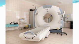
HEAD INJURY
Dr. Haydar Muneer Salih
Head Injury
Head injury is a frequent
cause of emergency
department attendance,
accounting for
approximately 3.4% of all
presentations.
Primary brian Injury
Primary brain injury occurs at
the time of impact and includes
injuries such as brainstem and
hemispheric contusions diffuse
axonal injury and cortical
lacerations.
Secondary Brian Injury
 Secondary brain injury occurs at some
time after the moment of impact and
is often preventable. The principle
causes of secondary brain injury are
hypoxia, hypotension, raised ICP,
reduced cerebral perfusion pressure
and pyrexia
Causes of secondary brain injury
■ Hypoxia: PO2 < 8 kPa
■ Hypotension: systolic blood pressure (SBP) <
90 mmHg
■ Raised intracranial pressure (ICP): ICP > 20
mmHg
■ Low cerebral perfusion pressure (CPP): CPP <
65 mmHg
■ Pyrexia
■ Seizures
■ Metabolic disturbance
Glasgow Coma scale
CLASSIFICATION OF HEAD INJURY
Glasgow Coma Score
Severity of head injury is classified according to the
Glasgow Coma Score (GCS)
• Minor head injury: GCS 15 with no loss of
consciousness (LOC);
• Mild head injury: GCS 14 or 15 with LOC;
• Moderate head injury: GCS 9–13;
• Severe head injury: GCS 3–8.
Taking a history in head injury
■ Mechanism of injury
■ Loss of consciousness or amnesia
■ Level of consciousness at scene and on transfer
■ Evidence of seizures
■ Probable hypoxia or hypotension
■ Pre-existing medical conditions
■ Medications (especially anticoagulants)
■ Illicit drugs and alcohol
MANAGEMENT OF MILD HEAD INJURY (GCS 14–15)
The following criteria must be met before discharge:
1. The patient must have accompanied by a responsible
adult
2. Should not be under the influence of alcohol or
other drugs;
3. verbal and written head injury advice must be given
to the patient and their accompanying adult
CT scan in a patient with mild head injury
■ Glasgow Coma Score (GCS) < 13 at any point
■ GCS 13 or 14 at 2 hours
■ Focal neurological deficit
■ Suspected open, depressed or basal skull fracture
■ Seizure
■ Vomiting > one episode
Urgent CT head scan if none of the above but:
■ Age > 65
■ Coagulopathy (e.g. on warfarin)
■ Dangerous mechanism of injury (CT within 8 hours)
■ Anterograde amnesia > 30 min (CT within 8 hours)
MANAGEMENT OF MODERATE TO SEVERE HEAD INJURY
 Begins with resuscitation and a primary survey
 The principle aim of treatment is the prevention of
secondary brain injury and this is best achieved by
the avoidance of hypoxia and hypotension.
 It follows that investigations such as a CT scan of
the head are of secondary importance to restoring
normal oxygenation and blood pressure
 The next appropriate step is a CT scan of the head
1. This investigation is aimed at identifying an
intracranial haematoma
2. The CT scan will also provide information about
scalp soft tissue injury
3. skull fracture, including base of skull fracture
4. lesions not requiring immediate surgery, such as
small intracerebral contusions
Extradural haematoma
 An extradural haematoma (EDH) is a
neurosurgical emergency. An EDH is nearly
always associated with a skull fracture and is
more common in young male patients. The
skull fracture is associated with tearing of a
meningeal artery and a haematoma
accumulates in the space between bone and
dura.
 The most common site is temporal, as the pterion
is not only the thinnest part of the skull but also
overlies the largest meningeal artery – the middle
meningeal
 when the patient complains of a headache but is
fully alert and orientated with no focal deficit.
After minutes or hours a rapid deterioration
occurs, with contralateral hemiparesis, reduced
conscious level and ipsilateral pupillary dilatation
as a result of brain compression and herniation
Extradural
Haematoma
The features of an
EDH on a CT scan
are a lentiform
(lensshaped or
biconvex)
hyperdense lesion
between the skull
and brain
Treatment
The treatment
of an EDH is
immediate
surgical
evacuation
Acute subdural haematoma
 Acute subdural haematoma (ASDH) differs
from EDH in terms of pathophysiology,
presentation and prognosis. An ASDH
accumulates in the space between the dura
and the arachnoid
 ASDH is nearly always associated with a
significant primary brain injury
Acute subdural haematoma
Patients with ASDH usually present
with an impaired conscious level from
the time of injury, but further
deterioration can occur as the
haematoma expands
The CT appearance
of an ASDH is also
hyperdense (acute
blood) but the
haematoma spreads
across the surface of
the brain giving it a
rather diffuse and
concave appearance.
Treatment
 The treatment of an ASDH is usually
evacuation via a craniotomy. Small
haematomas with little mass effect may be
managed conservatively in neurosurgical
centers.
Chronic subdural haematoma
 Chronic subdural haematomas (CSDH) usually occur
in the elderly and are more common in those on anti-
coagulant or antiplatelet agents. There is usually but
not always a history of minor head injury in the weeks
or months prior to presentation
 It is thought that small bridging veins tear and cause a
small ASDH which is clinically silent
Clinical features of CSDH include
headache, cognitive decline, focal
neurological deficits and seizures. It is
important to exclude hypoxic, metabolic and
endocrine disorders in this group of patients
chronic blood (> 2
weeks) is hypodense.
A CSDH will often
have areas of more
recent haemorrhage in
more dependent
(posterior) areas and is
then termed an acute-
on-chronic subdural
haematoma.
 Treatment of a CSDH and most acute-on-chronic
subdural haematomas is evacuation via burr hole(s)
rather than craniotomy.
Cerebral contusions
 Cerebral contusions are common in head
injury and result from the brain being
damaged by impacting against the skull
either at the point of impact (the ‘coup’) or
on the other side of the head (‘contre-coup’)
or as the brain slides forwards and
backwards over the ridged cranial fossa floor
CT appear
heterogeneous
with mixed
areas of high
and low density.
 Cerebral contusions rarely require immediate
surgical treatment. A head-injured patient
with cerebral contusions must be admitted
for observation as these lesions will tend to
mature and expand for 48–72 hours
following injury. A small proportion of
cerebral contusions will require delayed
surgical evacuation to reduce the mass effect
The Glasgow Outcome Score
(GOS)
Good recovery 5
Moderate disability 4
Severe disability 3
Persistent vegetative state 2
Dead 1
Head trauma

Head trauma

  • 1.
  • 2.
    Head Injury Head injuryis a frequent cause of emergency department attendance, accounting for approximately 3.4% of all presentations.
  • 3.
    Primary brian Injury Primarybrain injury occurs at the time of impact and includes injuries such as brainstem and hemispheric contusions diffuse axonal injury and cortical lacerations.
  • 5.
    Secondary Brian Injury Secondary brain injury occurs at some time after the moment of impact and is often preventable. The principle causes of secondary brain injury are hypoxia, hypotension, raised ICP, reduced cerebral perfusion pressure and pyrexia
  • 6.
    Causes of secondarybrain injury ■ Hypoxia: PO2 < 8 kPa ■ Hypotension: systolic blood pressure (SBP) < 90 mmHg ■ Raised intracranial pressure (ICP): ICP > 20 mmHg ■ Low cerebral perfusion pressure (CPP): CPP < 65 mmHg ■ Pyrexia ■ Seizures ■ Metabolic disturbance
  • 7.
  • 9.
    CLASSIFICATION OF HEADINJURY Glasgow Coma Score Severity of head injury is classified according to the Glasgow Coma Score (GCS) • Minor head injury: GCS 15 with no loss of consciousness (LOC); • Mild head injury: GCS 14 or 15 with LOC; • Moderate head injury: GCS 9–13; • Severe head injury: GCS 3–8.
  • 10.
    Taking a historyin head injury ■ Mechanism of injury ■ Loss of consciousness or amnesia ■ Level of consciousness at scene and on transfer ■ Evidence of seizures ■ Probable hypoxia or hypotension ■ Pre-existing medical conditions ■ Medications (especially anticoagulants) ■ Illicit drugs and alcohol
  • 11.
    MANAGEMENT OF MILDHEAD INJURY (GCS 14–15) The following criteria must be met before discharge: 1. The patient must have accompanied by a responsible adult 2. Should not be under the influence of alcohol or other drugs; 3. verbal and written head injury advice must be given to the patient and their accompanying adult
  • 13.
    CT scan ina patient with mild head injury ■ Glasgow Coma Score (GCS) < 13 at any point ■ GCS 13 or 14 at 2 hours ■ Focal neurological deficit ■ Suspected open, depressed or basal skull fracture ■ Seizure ■ Vomiting > one episode Urgent CT head scan if none of the above but: ■ Age > 65 ■ Coagulopathy (e.g. on warfarin) ■ Dangerous mechanism of injury (CT within 8 hours) ■ Anterograde amnesia > 30 min (CT within 8 hours)
  • 14.
    MANAGEMENT OF MODERATETO SEVERE HEAD INJURY  Begins with resuscitation and a primary survey  The principle aim of treatment is the prevention of secondary brain injury and this is best achieved by the avoidance of hypoxia and hypotension.  It follows that investigations such as a CT scan of the head are of secondary importance to restoring normal oxygenation and blood pressure
  • 16.
     The nextappropriate step is a CT scan of the head 1. This investigation is aimed at identifying an intracranial haematoma 2. The CT scan will also provide information about scalp soft tissue injury 3. skull fracture, including base of skull fracture 4. lesions not requiring immediate surgery, such as small intracerebral contusions
  • 17.
    Extradural haematoma  Anextradural haematoma (EDH) is a neurosurgical emergency. An EDH is nearly always associated with a skull fracture and is more common in young male patients. The skull fracture is associated with tearing of a meningeal artery and a haematoma accumulates in the space between bone and dura.
  • 19.
     The mostcommon site is temporal, as the pterion is not only the thinnest part of the skull but also overlies the largest meningeal artery – the middle meningeal  when the patient complains of a headache but is fully alert and orientated with no focal deficit. After minutes or hours a rapid deterioration occurs, with contralateral hemiparesis, reduced conscious level and ipsilateral pupillary dilatation as a result of brain compression and herniation
  • 20.
    Extradural Haematoma The features ofan EDH on a CT scan are a lentiform (lensshaped or biconvex) hyperdense lesion between the skull and brain
  • 21.
    Treatment The treatment of anEDH is immediate surgical evacuation
  • 22.
    Acute subdural haematoma Acute subdural haematoma (ASDH) differs from EDH in terms of pathophysiology, presentation and prognosis. An ASDH accumulates in the space between the dura and the arachnoid  ASDH is nearly always associated with a significant primary brain injury
  • 24.
    Acute subdural haematoma Patientswith ASDH usually present with an impaired conscious level from the time of injury, but further deterioration can occur as the haematoma expands
  • 25.
    The CT appearance ofan ASDH is also hyperdense (acute blood) but the haematoma spreads across the surface of the brain giving it a rather diffuse and concave appearance.
  • 26.
    Treatment  The treatmentof an ASDH is usually evacuation via a craniotomy. Small haematomas with little mass effect may be managed conservatively in neurosurgical centers.
  • 27.
    Chronic subdural haematoma Chronic subdural haematomas (CSDH) usually occur in the elderly and are more common in those on anti- coagulant or antiplatelet agents. There is usually but not always a history of minor head injury in the weeks or months prior to presentation  It is thought that small bridging veins tear and cause a small ASDH which is clinically silent
  • 28.
    Clinical features ofCSDH include headache, cognitive decline, focal neurological deficits and seizures. It is important to exclude hypoxic, metabolic and endocrine disorders in this group of patients
  • 29.
    chronic blood (>2 weeks) is hypodense. A CSDH will often have areas of more recent haemorrhage in more dependent (posterior) areas and is then termed an acute- on-chronic subdural haematoma.
  • 30.
     Treatment ofa CSDH and most acute-on-chronic subdural haematomas is evacuation via burr hole(s) rather than craniotomy.
  • 31.
    Cerebral contusions  Cerebralcontusions are common in head injury and result from the brain being damaged by impacting against the skull either at the point of impact (the ‘coup’) or on the other side of the head (‘contre-coup’) or as the brain slides forwards and backwards over the ridged cranial fossa floor
  • 32.
  • 33.
     Cerebral contusionsrarely require immediate surgical treatment. A head-injured patient with cerebral contusions must be admitted for observation as these lesions will tend to mature and expand for 48–72 hours following injury. A small proportion of cerebral contusions will require delayed surgical evacuation to reduce the mass effect
  • 34.
    The Glasgow OutcomeScore (GOS) Good recovery 5 Moderate disability 4 Severe disability 3 Persistent vegetative state 2 Dead 1