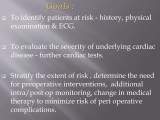
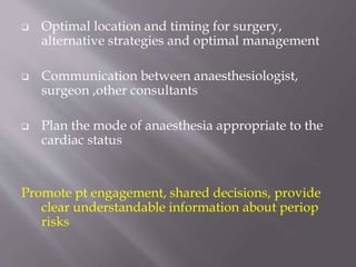
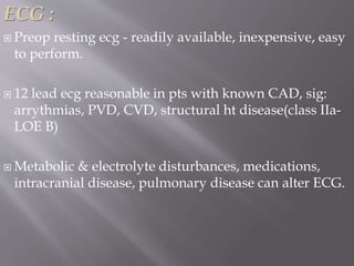
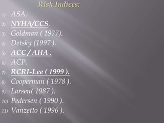
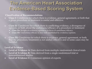
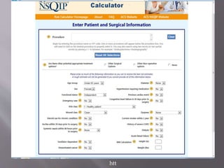
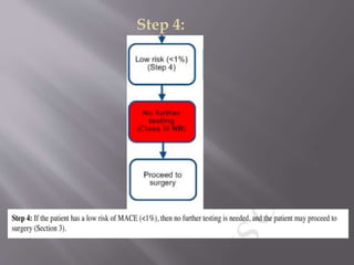
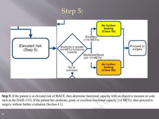
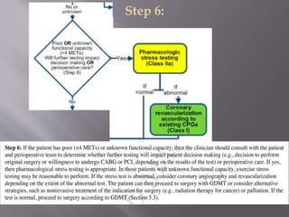
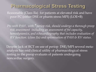
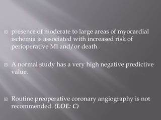
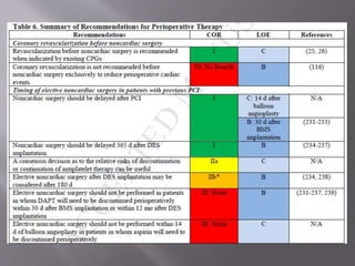
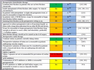
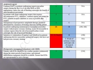
Dr Brijesh Savidhan
Department of Anaesthesiology
Travancore Medical College
Goals :
 To identify patients at risk - history, physical
examination & ECG.
 To evaluate the severity of underlying cardiac
disease - further cardiac tests.
 Stratify the extent of risk , determine the need
for preoperative interventions, additional
intra/post op monitoring, change in medical
therapy to minimize risk of peri operative
complications.
 Optimal location and timing for surgery,
alternative strategies and optimal management
 Communication between anaesthesiologist,
surgeon ,other consultants
 Plan the mode of anaesthesia appropriate to the
cardiac status
Promote pt engagement, shared decisions, provide
clear understandable information about periop
risks
 Major hemodynamic stress,
 Changes in cholinergic activity,
 Changes in catecholamine activity,
 Body temperature fluctuations, O2 consumption
 Pulmonary function altered,
 Fluid shifts, blood loss, electrolyte imbalance
 Pain.
 Effects of anaesthetic agents
 The initial history, physical examination, and
electrocardiogram assessment should focus on
identification of potentially serious cardiac
disorders.
 In addition it is essential to define disease
severity, stability, and prior treatment.
 GA & CNB Alters SVR,PVR,SV,CO
 Induction of GA  MAP by 20-30% ,tracheal
intubation  the BP by 20-30 mm Hg, and most
anesthetic agents  cardiac output by 15% by dose
dependent depression of contractility
 obtundation of baroreceptors, sympathetic
depression/stimulation
  arrythmogenic threshold for epinephrine
 May trigger bradycardia other dysrrythmias
 Cardiotoxicity, anaphylaxis, supply-demand
mismatch
 Pulmonary related issues
 Other factors that help determine cardiac risk-
functional capacity, age, co morbid conditions
(e.g., DM, PVD, renal dysfunction, and COPD).
 The type of surgery (vascular procedures and
prolonged, complicated thoracic, abdominal,
and head and neck procedures ) are considered
higher risk.
 The presence of anemia may also place a patient
at higher perioperative risk.
 Numerous risk indices have been developed
over the years on the basis of multivariate
analyses.
 In addition to CAD and HF, a history of CVD,
preoperative elevated creatinine greater than 2
mg/dl, insulin treatment for DM, and high-
risk surgery have all been associated with
increased periop cardiac morbidity.
( By Lee et al in 1999 in Revised Cardiac Risk
Index).
Evaluation of cardiac risk :
The cornerstone of preoperative cardiac
evaluation includes :-
- review of history ,
- physical examination,
- diagnostic tests,
- knowledge about the planned surgical
procedure.
History :
1) Risk factors : Age, HTN, DM, Dyslipidemias,
Smoking, Family history etc
2) Angina : Stable/ unstable, present medications.
3) Previous MI : details
4) CHF:
5) Dysrhythmias : Palpitations.
6) Associated CVS disease : Carotid, cerebral, aortic,
PVD.
7) Presence of pacemaker/ ICD.
Valvular Heart disease :
1) Dyspnea, Orthopnea, PND.
2) Embolic events.
3) Hemoptysis.
4) Heart failure, palpitations.
Prior cardiac evaluation :
1) Non invasive tests.
2) Angiography/ Stenting.
Medications :
1) Details.
2) Effectiveness.
Physical examination :
1) General examination :
Cyanosis, pallor, dyspnea during
conversation or with minimal activity, poor
nutritional status, obesity, skeletal deformities,
tremor & anxiety are just a few of the clues
of underlying disease or CAD.
2) Vitals : Pulse, BP , Respiration. Also pulse pressure
3) Cardiovascular examination :
JVP, pedal edema ,
Displaced apical impulse (cardiomegaly),
S3 gallop ( increased LVEDP ),
S4 ( decreased compliance),
Apical systolic murmur ( Papillary muscle
dysfunction ),
Presence of murmurs,
Pulmonary edema.
 The metabolic equivalent, or MET- the ratio of a
person's working metabolic rate relative to the
resting metabolic rate.
 One MET represents the oxygen consumption of a
resting adult-40 yr old 70kg (3.5 ml/kg/min).
 In the RCRI by Lee et al functional status was
not independently associated with risk.
 If pts reduce exertion because of cardiac
symptoms but still meet a 4-MET threshold,
clinicians will underestimate risk.
 Conversely, non cardiac functional limitations
(e.g., knee or back pain) may falsely
overestimate cardiac risk.
Functional capacity is classified as :
 poor (<4 METS),
 moderate (4–7METS),
 good (7–10METS) ,
 Excellent(>10 METS)
based on evaluation of the pts daily activity.
Measurements on a treadmill inducing ischemia at
low-level exercise (<5 MET or heart rate <100 /min)
identifies a high-risk group,
whereas the achievement of more than 7 MET (or
heart rate >130 / min) without ischemia identifies a
low-risk group.
ECG :
 Preop resting ecg - readily available, inexpensive, easy
to perform.
 12 lead ecg reasonable in pts with known CAD, sig:
arrythmias, PVD, CVD, structural ht disease(class IIa-
LOE B)
 Metabolic & electrolyte disturbances, medications,
intracranial disease, pulmonary disease can alter ECG.
 Prognostic significance of
numerous abnormalities has been identified in
studies, including arrhythmias, pathological Q-
waves, LVH, ST depressions , QTc interval
prolongn, and bundle-branch blocks
 Ecg taken within 1-3 mnths adeq for stable pts
Risk Indices:
1) ASA.
2) NYHA/CCS.
3) Goldman ( 1977).
4) Detsky (1997 ).
5) ACC / AHA .
6) ACP.
7) RCRI-Lee ( 1999 ).
8) Cooperman ( 1978 ).
9) Larsen( 1987 ).
10) Pedersen ( 1990 ).
11) Vanzetto ( 1996 ).
Classification of Recommendations
 Class I: Conditions for which there is evidence, general agreement, or both that
a given procedure or treatment is useful and effective.
 Class II: Conditions for which there is conflicting evidence, a divergence of
opinion, or both about the usefulness/efficacy of a procedure or treatment
 Class IIa: Weight of evidence/opinion is in favor of usefulness/efficacy.
 Class IIb: Usefulness/efficacy is less well established by evidence/opinion.
 Class III: Conditions for which there is evidence, general agreement, or both
that the procedure/treatment is not useful/effective and in some cases may be
harmful.
Level of Evidence
 Level of Evidence A: Data derived from multiple randomized clinical trials
 Level of Evidence B: Data derived from a single randomized trial or
nonrandomized studies
 Level of Evidence C: Consensus opinion of experts
2014…
ACC/AHA guidelines on
perioperative cardiovascular
evaluation and management
Life or limb is
threatened if
not in
operating room
within
24 hours
Delay of 1-6
weeks for
further
evaluation
would
negatively
affect outcome
Delay for up
to 1 year
Life or limb is
threatened if
not in operating
room within
6 hours
Emergent Urgent
Time-
Sensitive
Elective
 Low risk procedure – combined surgical and
patient characteristic predict a risk of major
adverse cardiac event (MACE) of death or MI
<1%- cataract, plastic surgery
 Elevated risk procedure- all procedures with
risk>= 1% of MACE. Risk by less invasive
techniques(endovascular) &  in case of
emergency procedures.
Previous classification of mild, intermediate, high
risk discarded
CAD
 Incidence of morbidity depends on- cardiac
markers to signs of ischaemia
 Isolated Trop I + 2% 30 day mortality
 Post op MI rates durn frm MI to surgery
 Modified by presence & type of coronary
revascularisn
 60 days after MI before non cardiac Sx- in the
absence of revascularisn
 Recent MI(<6months)- 8 fold in periop
mortality
 Age>65 overall risk for MACE in non cardiac
Sx
 Pts with clinical ht failure- peripheral edema,
jugular venous distension, rales, S3, CXR with
pulmonary vascular redistribution, pulmonary
edema- significant risk
 Original CRI- S3,JVD. modified CRI- h/o
HF,PE,PND O/E b/l rales, S3,CXR(PVR)
 30 day mortality 50 to 100% higher in pts with
CHF
Step 1:
/
Step 2:
/
GDMT-Guideline directed medical therapy
Step 3:
htt
Step 4:
Step 5:
Fli
Step 6:
Step 7:
/
 Assessment of LV function
 Exercise Stress Testing for Myocardial Ischemia and
Functional Capacity
 Pharmacological Stress Testing
 Noninvasive
 Radionuclide
 DSE
 Special Situations
LV fn
 reasonable for patients with dyspnea of unknown origin,
h/o HF to undergo preop evaluatn of LV fn.(class IIa-
LOE B)
 Sig:(<30%) LVEF- independent contributor to periop
outcome- survival significantly worse
 Though data related to DD ltd- asctd with sig:rate of
MACE, prolongd hospital stay, higher post op ht failure
 Prospctve cohort study(vascular surgery)- 2005 pts on
role of echo in preop pts- LV dysfn (EF<50%) in 50 % pts
of whom 80% asymptomatic- 30 day cardiovascular
event -23% in pts with asymptomatic LV dysfn
 pts with excellent (>10 METs) fnl capacity- reasonble
to forgo further exercise tstng with cardiac imaging
and proceed to Sx (classIIa-LOE B)
 pts with  risk and unknown fnl capacity/moderate-
good fnl capacity, reasonble to perform exercise testng
if it will change Mx(class IIb-LOE B)
Reasonable (Class IIa) for patients at elevated risk and have
poor FC (either DSE or pharm stress MPI) (LOE=B)
Pts with PAH , with periop risk, should undergo a thorough preop
risk assessment including an assessment of fnl capacity,
hemodynamics, and echocardiography that includes evaluation of
RV function. (class IIa) and optimisation of PAH & RV status.
Despite lack of RCT on use of periop DSE/MPI several meta-
analysis has estd clinical utility of pharmacological stress
testing in the preop evaluatn of patients undergoing
noncardiac surgery.
 presence of moderate to large areas of myocardial
ischemia is associated with increased risk of
perioperative MI and/or death.
 A normal study has a very high negative predictive
value.
 Routine preoperative coronary angiography is not
recommended. (LOE: C)
THANK YOU

Preop cardiovascular evaluation

  • 1.
    Dr Brijesh Savidhan Departmentof Anaesthesiology Travancore Medical College
  • 2.
    Goals :  Toidentify patients at risk - history, physical examination & ECG.  To evaluate the severity of underlying cardiac disease - further cardiac tests.  Stratify the extent of risk , determine the need for preoperative interventions, additional intra/post op monitoring, change in medical therapy to minimize risk of peri operative complications.
  • 3.
     Optimal locationand timing for surgery, alternative strategies and optimal management  Communication between anaesthesiologist, surgeon ,other consultants  Plan the mode of anaesthesia appropriate to the cardiac status Promote pt engagement, shared decisions, provide clear understandable information about periop risks
  • 4.
     Major hemodynamicstress,  Changes in cholinergic activity,  Changes in catecholamine activity,  Body temperature fluctuations, O2 consumption  Pulmonary function altered,  Fluid shifts, blood loss, electrolyte imbalance  Pain.  Effects of anaesthetic agents
  • 5.
     The initialhistory, physical examination, and electrocardiogram assessment should focus on identification of potentially serious cardiac disorders.  In addition it is essential to define disease severity, stability, and prior treatment.
  • 6.
     GA &CNB Alters SVR,PVR,SV,CO  Induction of GA  MAP by 20-30% ,tracheal intubation  the BP by 20-30 mm Hg, and most anesthetic agents  cardiac output by 15% by dose dependent depression of contractility  obtundation of baroreceptors, sympathetic depression/stimulation
  • 7.
      arrythmogenicthreshold for epinephrine  May trigger bradycardia other dysrrythmias  Cardiotoxicity, anaphylaxis, supply-demand mismatch  Pulmonary related issues
  • 8.
     Other factorsthat help determine cardiac risk- functional capacity, age, co morbid conditions (e.g., DM, PVD, renal dysfunction, and COPD).  The type of surgery (vascular procedures and prolonged, complicated thoracic, abdominal, and head and neck procedures ) are considered higher risk.  The presence of anemia may also place a patient at higher perioperative risk.
  • 9.
     Numerous riskindices have been developed over the years on the basis of multivariate analyses.  In addition to CAD and HF, a history of CVD, preoperative elevated creatinine greater than 2 mg/dl, insulin treatment for DM, and high- risk surgery have all been associated with increased periop cardiac morbidity. ( By Lee et al in 1999 in Revised Cardiac Risk Index).
  • 10.
    Evaluation of cardiacrisk : The cornerstone of preoperative cardiac evaluation includes :- - review of history , - physical examination, - diagnostic tests, - knowledge about the planned surgical procedure.
  • 11.
    History : 1) Riskfactors : Age, HTN, DM, Dyslipidemias, Smoking, Family history etc 2) Angina : Stable/ unstable, present medications. 3) Previous MI : details 4) CHF: 5) Dysrhythmias : Palpitations. 6) Associated CVS disease : Carotid, cerebral, aortic, PVD. 7) Presence of pacemaker/ ICD.
  • 12.
    Valvular Heart disease: 1) Dyspnea, Orthopnea, PND. 2) Embolic events. 3) Hemoptysis. 4) Heart failure, palpitations. Prior cardiac evaluation : 1) Non invasive tests. 2) Angiography/ Stenting. Medications : 1) Details. 2) Effectiveness.
  • 13.
    Physical examination : 1)General examination : Cyanosis, pallor, dyspnea during conversation or with minimal activity, poor nutritional status, obesity, skeletal deformities, tremor & anxiety are just a few of the clues of underlying disease or CAD. 2) Vitals : Pulse, BP , Respiration. Also pulse pressure
  • 14.
    3) Cardiovascular examination: JVP, pedal edema , Displaced apical impulse (cardiomegaly), S3 gallop ( increased LVEDP ), S4 ( decreased compliance), Apical systolic murmur ( Papillary muscle dysfunction ), Presence of murmurs, Pulmonary edema.
  • 15.
     The metabolicequivalent, or MET- the ratio of a person's working metabolic rate relative to the resting metabolic rate.  One MET represents the oxygen consumption of a resting adult-40 yr old 70kg (3.5 ml/kg/min).
  • 16.
     In theRCRI by Lee et al functional status was not independently associated with risk.  If pts reduce exertion because of cardiac symptoms but still meet a 4-MET threshold, clinicians will underestimate risk.  Conversely, non cardiac functional limitations (e.g., knee or back pain) may falsely overestimate cardiac risk.
  • 17.
    Functional capacity isclassified as :  poor (<4 METS),  moderate (4–7METS),  good (7–10METS) ,  Excellent(>10 METS) based on evaluation of the pts daily activity. Measurements on a treadmill inducing ischemia at low-level exercise (<5 MET or heart rate <100 /min) identifies a high-risk group, whereas the achievement of more than 7 MET (or heart rate >130 / min) without ischemia identifies a low-risk group.
  • 19.
    ECG :  Preopresting ecg - readily available, inexpensive, easy to perform.  12 lead ecg reasonable in pts with known CAD, sig: arrythmias, PVD, CVD, structural ht disease(class IIa- LOE B)  Metabolic & electrolyte disturbances, medications, intracranial disease, pulmonary disease can alter ECG.
  • 20.
     Prognostic significanceof numerous abnormalities has been identified in studies, including arrhythmias, pathological Q- waves, LVH, ST depressions , QTc interval prolongn, and bundle-branch blocks  Ecg taken within 1-3 mnths adeq for stable pts
  • 21.
    Risk Indices: 1) ASA. 2)NYHA/CCS. 3) Goldman ( 1977). 4) Detsky (1997 ). 5) ACC / AHA . 6) ACP. 7) RCRI-Lee ( 1999 ). 8) Cooperman ( 1978 ). 9) Larsen( 1987 ). 10) Pedersen ( 1990 ). 11) Vanzetto ( 1996 ).
  • 22.
    Classification of Recommendations Class I: Conditions for which there is evidence, general agreement, or both that a given procedure or treatment is useful and effective.  Class II: Conditions for which there is conflicting evidence, a divergence of opinion, or both about the usefulness/efficacy of a procedure or treatment  Class IIa: Weight of evidence/opinion is in favor of usefulness/efficacy.  Class IIb: Usefulness/efficacy is less well established by evidence/opinion.  Class III: Conditions for which there is evidence, general agreement, or both that the procedure/treatment is not useful/effective and in some cases may be harmful. Level of Evidence  Level of Evidence A: Data derived from multiple randomized clinical trials  Level of Evidence B: Data derived from a single randomized trial or nonrandomized studies  Level of Evidence C: Consensus opinion of experts
  • 23.
    2014… ACC/AHA guidelines on perioperativecardiovascular evaluation and management
  • 24.
    Life or limbis threatened if not in operating room within 24 hours Delay of 1-6 weeks for further evaluation would negatively affect outcome Delay for up to 1 year Life or limb is threatened if not in operating room within 6 hours Emergent Urgent Time- Sensitive Elective
  • 25.
     Low riskprocedure – combined surgical and patient characteristic predict a risk of major adverse cardiac event (MACE) of death or MI <1%- cataract, plastic surgery  Elevated risk procedure- all procedures with risk>= 1% of MACE. Risk by less invasive techniques(endovascular) &  in case of emergency procedures. Previous classification of mild, intermediate, high risk discarded
  • 26.
    CAD  Incidence ofmorbidity depends on- cardiac markers to signs of ischaemia  Isolated Trop I + 2% 30 day mortality  Post op MI rates durn frm MI to surgery  Modified by presence & type of coronary revascularisn
  • 27.
     60 daysafter MI before non cardiac Sx- in the absence of revascularisn  Recent MI(<6months)- 8 fold in periop mortality  Age>65 overall risk for MACE in non cardiac Sx
  • 28.
     Pts withclinical ht failure- peripheral edema, jugular venous distension, rales, S3, CXR with pulmonary vascular redistribution, pulmonary edema- significant risk  Original CRI- S3,JVD. modified CRI- h/o HF,PE,PND O/E b/l rales, S3,CXR(PVR)  30 day mortality 50 to 100% higher in pts with CHF
  • 29.
  • 30.
  • 31.
  • 32.
  • 33.
  • 34.
  • 35.
  • 36.
  • 37.
     Assessment ofLV function  Exercise Stress Testing for Myocardial Ischemia and Functional Capacity  Pharmacological Stress Testing  Noninvasive  Radionuclide  DSE  Special Situations
  • 38.
    LV fn  reasonablefor patients with dyspnea of unknown origin, h/o HF to undergo preop evaluatn of LV fn.(class IIa- LOE B)  Sig:(<30%) LVEF- independent contributor to periop outcome- survival significantly worse  Though data related to DD ltd- asctd with sig:rate of MACE, prolongd hospital stay, higher post op ht failure  Prospctve cohort study(vascular surgery)- 2005 pts on role of echo in preop pts- LV dysfn (EF<50%) in 50 % pts of whom 80% asymptomatic- 30 day cardiovascular event -23% in pts with asymptomatic LV dysfn
  • 39.
     pts withexcellent (>10 METs) fnl capacity- reasonble to forgo further exercise tstng with cardiac imaging and proceed to Sx (classIIa-LOE B)  pts with  risk and unknown fnl capacity/moderate- good fnl capacity, reasonble to perform exercise testng if it will change Mx(class IIb-LOE B)
  • 40.
    Reasonable (Class IIa)for patients at elevated risk and have poor FC (either DSE or pharm stress MPI) (LOE=B) Pts with PAH , with periop risk, should undergo a thorough preop risk assessment including an assessment of fnl capacity, hemodynamics, and echocardiography that includes evaluation of RV function. (class IIa) and optimisation of PAH & RV status. Despite lack of RCT on use of periop DSE/MPI several meta- analysis has estd clinical utility of pharmacological stress testing in the preop evaluatn of patients undergoing noncardiac surgery.
  • 41.
     presence ofmoderate to large areas of myocardial ischemia is associated with increased risk of perioperative MI and/or death.  A normal study has a very high negative predictive value.  Routine preoperative coronary angiography is not recommended. (LOE: C)
  • 45.