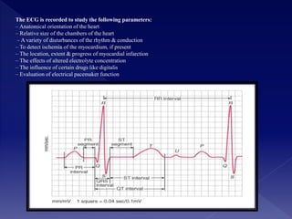
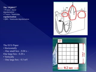
Electrocardiograph
Prepared by:
• Hawzhin Hussein
• First Stage
Supervised by:
L/soma
sulaimani polytechnic university
INTRODUCTION • Electrocardiograph is a device that records the electrical activity of the heart which will produce the
result in the graphic form which called electrocardiogram (ECG).
• Electrical waves cause the heart muscle to pump.
• These waves pass through the body and can be measured at electrodes attached to the skin.
Electrocardiography is the method of recording of an electrocardiogram (ECG)
• ECG or EKG is the graphic recording of the electrical activities of the heart
• Electrocardiograph is the machine that records the ECG
• It is an important diagnostic & prognostic tool for assessment of cardiovascular function
MEDICAL APPLICATION The ECG is useful for three basic parameters of clinically interest;
• check the rhythm and rate of the heart, whether it is regular and normal, faster (tachycardia) or slower.
• Check the axis of the heart, which can reveal any enlargement of the heart (hypertrophy)
. • Check the state of the myocardial muscle
NORMAL ECG
It will be clear from above that the first structure to be depolarized during normal sinus rhythm is the right atrium
Closely followed by the left atrium
So the first electrical signal on a normal ECG originates from the atria and is known as the P wave
Although there is usually only one p wave in most leads of an ECG
The P wave is in fact the sum of the electrical signals from the two atria
Which are usually superimposed
There is then short :a short period where no electrical activity is seen on the ECG
Depolarization of the ventricles results in usually the largest part of the ECG
Signal( because of the greater muscle mass in ventricles)and this is known as the
(ORS) cpmplex
The ECG is recorded to study the following parameters:
– Anatomical orientation of the heart
– Relative size of the chambers of the heart
– A variety of disturbances of the rhythm & conduction
– To detect ischemia of the myocardium, if present
– The location, extent & progress of myocardial infarction
– The effects of altered electrolyte concentration
– The influence of certain drugs like digitalis
– Evaluation of electrical pacemaker function
ECG leads • Direct leads – Leads applied directly to the surface of the heart – These leads are used to record
cardiac activities during cardiac surgery or during an experiment • Indirect leads – Leads applied away form the
heart to record the cardiac activities – Different indirect leads are limb leads, chest leads
Limb leads (bipolar & unipolar) • Bipolar limb leads
• Bipolar standard limb leads are original leads selected by Einthoven to record electrical potential on frontal
plane
• electrodes are attached to right arm, left arm, & left foot – another electrode is applied to the right leg, which
acts as a ground wire to prevent external disturbance during recording
Standard leads :
• Lead I: between right arm (negative electrode) & left arm (positive electrode)
Lead II: between right arm (negative electrode) & left leg (positive electrode)
Lead III: between left arm (negative electrode) & left leg (positive electrode)
Augmented unipolar leads
aVR : right arm
Avl:left arm
aVF:left leg
The “PQRST”
• P wave - Atrial
depolarization
• T wave - Ventricular
repolarization
• QRS - Ventricular depolarization
The ECG Paper
• Horizontally
– One small box - 0.04 s –
One large box - 0.20 s
• Vertically
– One large box - 0.5 mV
• Unipolar chest leads • six chest leads are used routinely; V1 to V6 (V7 -V9 ) • The reference electrode is
connected to the right arm, left arm & left leg through a high resistance. • V1 : in the right fourth intercostal
space at the right border of the sternum • V2 : in the left fourth intercostal space at the left border of the sternum
• V3 : at the midpoint between V2 & V4 • V4 : in the left fifth intercostal space on the midclavicular line • V5 :
in the left fifth intercostal space on the anterior axillary line • V6 : in the left fifth intercostal space on the mid-
axillary line • V7 : in the left fifth intercostal space on the posterior axillary line • V8 : in the left fifth intercostal
space on the posterior scapular line • V9 : in the left fifth intercostal space on the back just left to the spine
Abnormalities of rhythm
• Tachycardia
– fast heart rate (>100 bpm)
• Bradycardia –
slow heart rate (60bpm)
• Sinus arrhythmia
– ↑ heart rate during inspiration & ↓ during expiration
• First degree block
– when the P-R interval ↑ 0.20s
• Second degree block
– when the P-R interval ↑ 0.35 to 0.45s •
Third degree block
– complete block of the impulse from the atria into the
ventricles occurs
ADVANTAGES •.
• ECG also require to detect the chest pain or discomfort , shortness of breath , weakness
, anxiety, abdominal pain.
• ECG can use to detect any latent (silent) cardiac conditions that might worsen with the stresses of surgery and anesthesia
DISADVANTAGES
• A static picture and may not reflect severe underlying heart problems at a time when the patient is not having
any symptoms.
• It don’t always permit an accurate diagnosis. It can be normal despite a serious heart problem.
Pacemakers of the Heart
• SA Node - Dominant pacemaker with an intrinsic rate of 60 - 100 beats/minute.
• AV Node - Back-up pacemaker with an intrinsic rate of 40 - 60 beats/minute.
• Ventricular cells - Back-up pacemaker with an intrinsic rate of 20 - 45 bpm.
CONCLUSION
• Electrocardiograph is a device that can record the electrical activities of the heart and display the data in
graphic data called electrocardiogram (ECG).
• electrocardiographs share the same basic circuit in all ECG devices.
This circuit use to analysis the signal from patient heart and produce the result in graphical output.
• This is the most important device which can help the doctor to identify the problems in certain heart failure.
References • Ganong Review of Medical Physiology, 23/E
• Textbook of Medical Physiology, 12/E Guyton & Hall
• Understanding Medical Physiology, 4/E Bijlani & Manjunatha
• http://www.n3wt.nildram.co.uk/ECG/
S • Crawford M H, DiMarco J P, Paulus W J. Cardiology.3rd ed.Philadelphia:Elsevier
publication;2010.
• Fauci A S, Braunwald E, Kasper D L, Hauser S L, Longo D L, Jameson J L.Harrison’s
Principles of Internal Medicine.17th ed (vol I).USA:McGraw Hill;2008.
• Black M.J,Hawks H.K.Medical Surgical Nursing.7th
edn.Missouri:Saunders;2005
Hawzhin

Hawzhin

  • 1.
    Electrocardiograph Prepared by: • HawzhinHussein • First Stage Supervised by: L/soma sulaimani polytechnic university
  • 2.
    INTRODUCTION • Electrocardiographis a device that records the electrical activity of the heart which will produce the result in the graphic form which called electrocardiogram (ECG). • Electrical waves cause the heart muscle to pump. • These waves pass through the body and can be measured at electrodes attached to the skin. Electrocardiography is the method of recording of an electrocardiogram (ECG) • ECG or EKG is the graphic recording of the electrical activities of the heart • Electrocardiograph is the machine that records the ECG • It is an important diagnostic & prognostic tool for assessment of cardiovascular function MEDICAL APPLICATION The ECG is useful for three basic parameters of clinically interest; • check the rhythm and rate of the heart, whether it is regular and normal, faster (tachycardia) or slower. • Check the axis of the heart, which can reveal any enlargement of the heart (hypertrophy) . • Check the state of the myocardial muscle
  • 3.
    NORMAL ECG It willbe clear from above that the first structure to be depolarized during normal sinus rhythm is the right atrium Closely followed by the left atrium So the first electrical signal on a normal ECG originates from the atria and is known as the P wave Although there is usually only one p wave in most leads of an ECG The P wave is in fact the sum of the electrical signals from the two atria Which are usually superimposed There is then short :a short period where no electrical activity is seen on the ECG Depolarization of the ventricles results in usually the largest part of the ECG Signal( because of the greater muscle mass in ventricles)and this is known as the (ORS) cpmplex
  • 4.
    The ECG isrecorded to study the following parameters: – Anatomical orientation of the heart – Relative size of the chambers of the heart – A variety of disturbances of the rhythm & conduction – To detect ischemia of the myocardium, if present – The location, extent & progress of myocardial infarction – The effects of altered electrolyte concentration – The influence of certain drugs like digitalis – Evaluation of electrical pacemaker function
  • 5.
    ECG leads •Direct leads – Leads applied directly to the surface of the heart – These leads are used to record cardiac activities during cardiac surgery or during an experiment • Indirect leads – Leads applied away form the heart to record the cardiac activities – Different indirect leads are limb leads, chest leads Limb leads (bipolar & unipolar) • Bipolar limb leads • Bipolar standard limb leads are original leads selected by Einthoven to record electrical potential on frontal plane • electrodes are attached to right arm, left arm, & left foot – another electrode is applied to the right leg, which acts as a ground wire to prevent external disturbance during recording Standard leads : • Lead I: between right arm (negative electrode) & left arm (positive electrode) Lead II: between right arm (negative electrode) & left leg (positive electrode) Lead III: between left arm (negative electrode) & left leg (positive electrode) Augmented unipolar leads aVR : right arm Avl:left arm aVF:left leg
  • 7.
    The “PQRST” • Pwave - Atrial depolarization • T wave - Ventricular repolarization • QRS - Ventricular depolarization The ECG Paper • Horizontally – One small box - 0.04 s – One large box - 0.20 s • Vertically – One large box - 0.5 mV
  • 8.
    • Unipolar chestleads • six chest leads are used routinely; V1 to V6 (V7 -V9 ) • The reference electrode is connected to the right arm, left arm & left leg through a high resistance. • V1 : in the right fourth intercostal space at the right border of the sternum • V2 : in the left fourth intercostal space at the left border of the sternum • V3 : at the midpoint between V2 & V4 • V4 : in the left fifth intercostal space on the midclavicular line • V5 : in the left fifth intercostal space on the anterior axillary line • V6 : in the left fifth intercostal space on the mid- axillary line • V7 : in the left fifth intercostal space on the posterior axillary line • V8 : in the left fifth intercostal space on the posterior scapular line • V9 : in the left fifth intercostal space on the back just left to the spine
  • 9.
    Abnormalities of rhythm •Tachycardia – fast heart rate (>100 bpm) • Bradycardia – slow heart rate (60bpm) • Sinus arrhythmia – ↑ heart rate during inspiration & ↓ during expiration • First degree block – when the P-R interval ↑ 0.20s • Second degree block – when the P-R interval ↑ 0.35 to 0.45s • Third degree block – complete block of the impulse from the atria into the ventricles occurs
  • 10.
    ADVANTAGES •. • ECGalso require to detect the chest pain or discomfort , shortness of breath , weakness , anxiety, abdominal pain. • ECG can use to detect any latent (silent) cardiac conditions that might worsen with the stresses of surgery and anesthesia DISADVANTAGES • A static picture and may not reflect severe underlying heart problems at a time when the patient is not having any symptoms. • It don’t always permit an accurate diagnosis. It can be normal despite a serious heart problem. Pacemakers of the Heart • SA Node - Dominant pacemaker with an intrinsic rate of 60 - 100 beats/minute. • AV Node - Back-up pacemaker with an intrinsic rate of 40 - 60 beats/minute. • Ventricular cells - Back-up pacemaker with an intrinsic rate of 20 - 45 bpm.
  • 11.
    CONCLUSION • Electrocardiograph isa device that can record the electrical activities of the heart and display the data in graphic data called electrocardiogram (ECG). • electrocardiographs share the same basic circuit in all ECG devices. This circuit use to analysis the signal from patient heart and produce the result in graphical output. • This is the most important device which can help the doctor to identify the problems in certain heart failure.
  • 12.
    References • GanongReview of Medical Physiology, 23/E • Textbook of Medical Physiology, 12/E Guyton & Hall • Understanding Medical Physiology, 4/E Bijlani & Manjunatha • http://www.n3wt.nildram.co.uk/ECG/ S • Crawford M H, DiMarco J P, Paulus W J. Cardiology.3rd ed.Philadelphia:Elsevier publication;2010. • Fauci A S, Braunwald E, Kasper D L, Hauser S L, Longo D L, Jameson J L.Harrison’s Principles of Internal Medicine.17th ed (vol I).USA:McGraw Hill;2008. • Black M.J,Hawks H.K.Medical Surgical Nursing.7th edn.Missouri:Saunders;2005